DCCC Ventral Body Cavity Organs #8 - Ventral Body Cavity...
16
Bio 151 – Laboratory Manual Human Anatomy & Physiology II DCCC Ventral Body Cavity Organs Last updated 08/2019 Page1 #8 - Ventral Body Cavity Organs Objectives: Use a cat dissection to study the organs of the ventral body cavity; Use virtual human dissection software and a human model to observe the organs of the Respiratory, Digestive, Urinary, and Reproductive systems. Equipment: Remember to bring gloves, goggles, dissecting kit, appropriate shoes, and photographic atlas. I. Virtual Human Dissection and Human Model Observation A. Preparation 1. Start the Anatomy & Physiology Revealed® software by double clicking its icon. 2. Select the appropriate System (as indicated in the tables below) from the Module (top center) drop down menu. 3. On the home screen, click the dissection icon (top center, leftmost icon). 4. Using the directions on the following pages, find all of the structures on your lists. Use the human models to locate the structures you are viewing with the human dissection software. Within each system, there will be instructions that guide you in the removal of the organs from the human model. Remove organs only when instructed. Your instructor will sign off on the model before you leave the lab. After you have completed the lab, answer the questions in the laboratory report pages.
Transcript of DCCC Ventral Body Cavity Organs #8 - Ventral Body Cavity...
Bio 151 – Laboratory Manual Human Anatomy & Physiology II DCCC Ventral Body Cavity Organs
Last updated 08/2019
Pag
e1
#8 - Ventral Body Cavity Organs
Objectives:
Use a cat dissection to study the organs of the ventral body cavity;
Use virtual human dissection software and a human model to observe the organs of the
Respiratory, Digestive, Urinary, and Reproductive systems.
Equipment: Remember to bring gloves, goggles, dissecting kit, appropriate shoes, and
photographic atlas.
I. Virtual Human Dissection and Human Model Observation
A. Preparation
1. Start the Anatomy & Physiology
Revealed® software by double clicking its icon.
2. Select the appropriate System (as indicated in the
tables below) from the Module (top center) drop
down menu.
3. On the home screen, click the dissection icon
(top center, leftmost icon).
4. Using the directions on the following pages, find
all of the structures on your lists. Use the human
models to locate the structures you are viewing with
the human dissection software. Within each system, there will be instructions that guide you in
the removal of the organs from the human model. Remove organs only when instructed. Your
instructor will sign off on the model before you leave the lab. After you have completed the lab,
answer the questions in the laboratory report pages.
Bio 151 – Laboratory Manual Human Anatomy & Physiology II DCCC Ventral Body Cavity Organs
Last updated 08/2019
Pag
e2
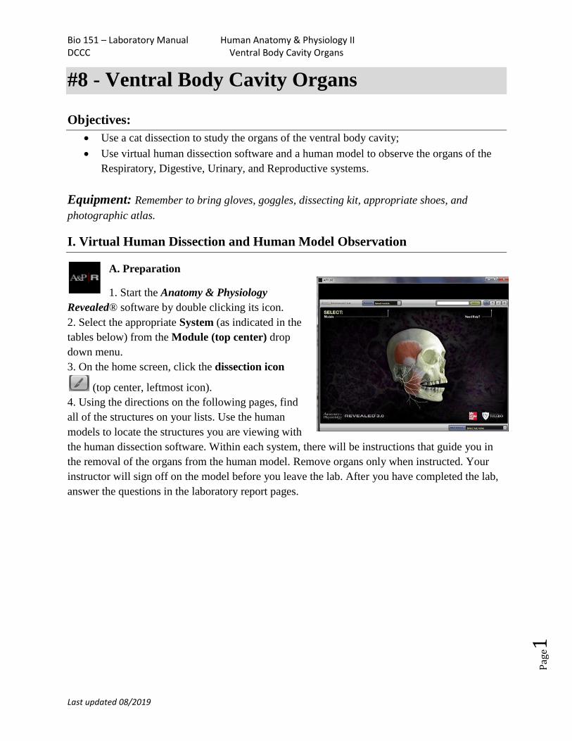
Respiratory System – Virtual Dissection
1. lungs
2. trachea
3. diaphragm
4. larynx
4a. epiglottis
1. From the Module dropdown menu, chose the 11.Respiratory system.
3. Click the dissection icon (top center, leftmost icon).
2. Choose Lower respiratory from the Topic menu.
3. Using the sliders (bottom left), move to Layer 4 (slide Layers 1, 2, and 3
down).
4. From the Select structures type dropdown menu (below View, top left),
choose Respiratory tract.
5. From the Select structures group dropdown menu, click on lung, to view
the structures. Remember that the pleural membranes cover the lung and line
the thoracic cavity.
6. Change the Select structures group to Trachea and bronchi to highlight the trachea (2)
(APR Fig.1).
7. Observe the diaphgram (3) (APR Fig.1), a muscle used for breathing.
8. In the Topic menu, choose larynx, View, anterior. Identify the trachea (2), larynx (4) and
epiglottis (4a) (RA Fig.2).
9. View the epiglottis (part of the larynx) in posterior, by using the sliders (bottom left), to move
to Layer 4 (slide Layers 1, 2, and 3 down) and choose the posterior View (APR Fig.3).
APR Fig.1 APR Fig. 2 APR Fig. 3
Bio 151 – Laboratory Manual Human Anatomy & Physiology II DCCC Ventral Body Cavity Organs
Last updated 08/2019
Pag
e3
Respiratory System – Human Model
Organ Instructions
☐ Larynx ☐ Left lung ☐ Right lung ☐
Diaphragm
Can be viewed without removing
organs
☐ Trachea Remove the lungs first.
Then, remove the thymus and heart.
Note: Epiglottis is not visible on the human model.
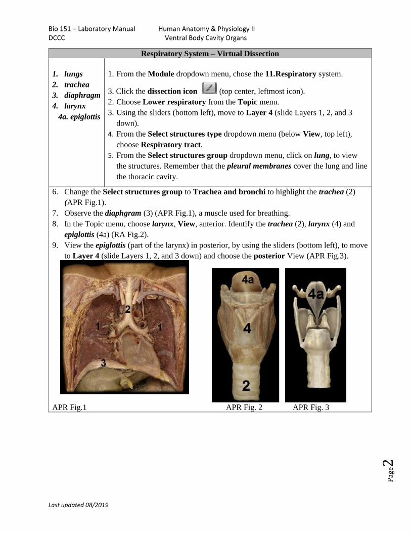
Digestive System – Virtual Dissection
5. greater
omentum
(part of the
peritoneum)
6. liver
7. gallbladder
1. From the Module dropdown menu, chose
the 12. Digestive system.
2. Click the dissection icon .
3. Choose Abdominal cavity from the
Topic menu. Using the sliders (bottom
left), move to Layer 2 (slide Layer 1
down).
4. From the Select structures type
dropdown menu (below View, top left),
choose Peritoneum.
5. Observe the greater omentum (5), liver
(6), and gallbladder (7) (APR Fig.4).
APR Fig.4 (Layer 2)
Bio 151 – Laboratory Manual Human Anatomy & Physiology II DCCC Ventral Body Cavity Organs
Last updated 08/2019
Pag
e4
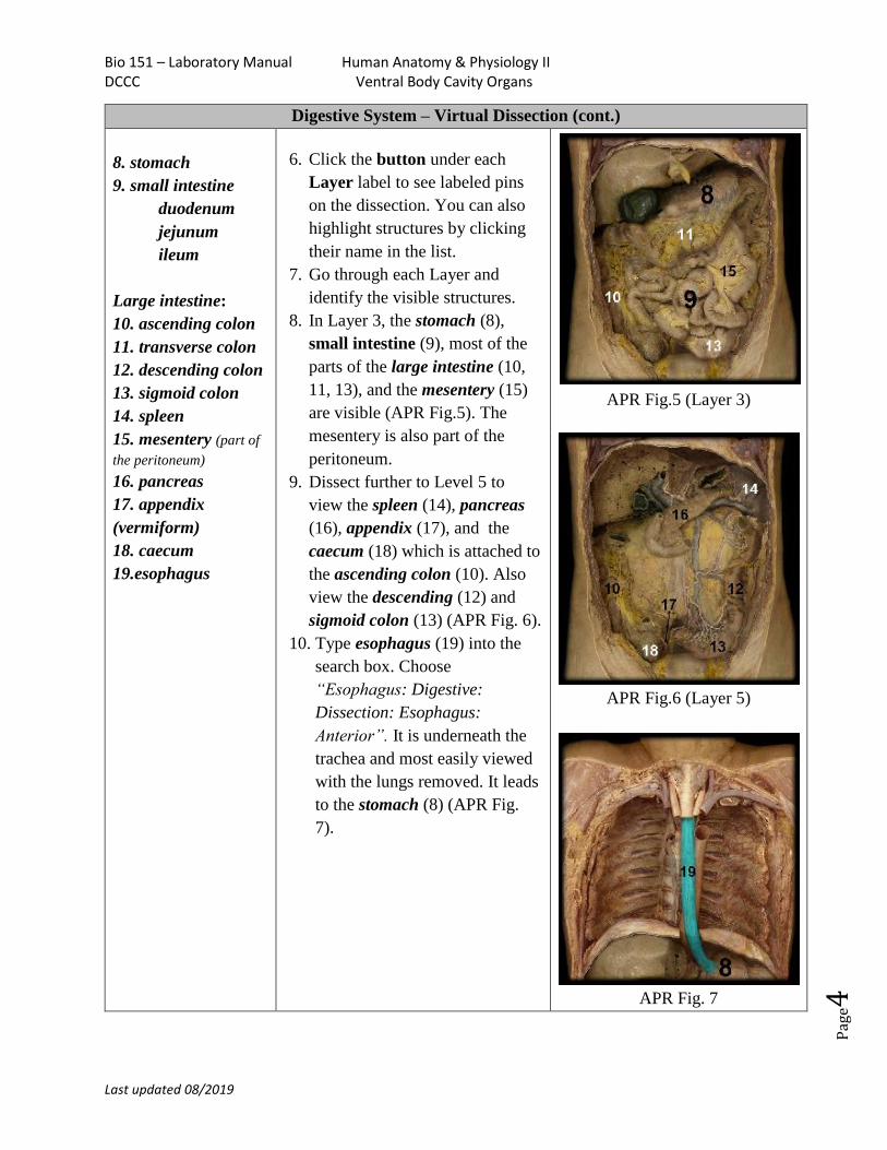
Digestive System – Virtual Dissection (cont.)
8. stomach
9. small intestine
duodenum
jejunum
ileum
Large intestine:
10. ascending colon
11. transverse colon
12. descending colon
13. sigmoid colon
14. spleen
15. mesentery (part of
the peritoneum)
16. pancreas
17. appendix
(vermiform)
18. caecum
19.esophagus
6. Click the button under each
Layer label to see labeled pins
on the dissection. You can also
highlight structures by clicking
their name in the list.
7. Go through each Layer and
identify the visible structures.
8. In Layer 3, the stomach (8),
small intestine (9), most of the
parts of the large intestine (10,
11, 13), and the mesentery (15)
are visible (APR Fig.5). The
mesentery is also part of the
peritoneum.
9. Dissect further to Level 5 to
view the spleen (14), pancreas
(16), appendix (17), and the
caecum (18) which is attached to
the ascending colon (10). Also
view the descending (12) and
sigmoid colon (13) (APR Fig. 6).
10. Type esophagus (19) into the
search box. Choose
“Esophagus: Digestive:
Dissection: Esophagus:
Anterior”. It is underneath the
trachea and most easily viewed
with the lungs removed. It leads
to the stomach (8) (APR Fig.
7).
APR Fig.5 (Layer 3)
APR Fig.6 (Layer 5)
APR Fig. 7
Bio 151 – Laboratory Manual Human Anatomy & Physiology II DCCC Ventral Body Cavity Organs
Last updated 08/2019
Pag
e5
Digestive System – Human Model
Organ Instructions
☐ Liver ☐ Esophagus ☐ Stomach Can be viewed without removing organs
☐ Gallbladder ☐ Spleen ☐ Pancreas Remove the diaphragm, liver and stomach
☐ Duodenum ☐ Jejunum ☐ Ileum
☐ Ascending colon ☐ Transverse colon
☐ Descending colon ☐ Caecum
Can be viewed without removing organs
☐ Sigmoid Colon ☐ Appendix (vermiform) Remove the small and large intestines
Notes: The mesentery cannot be seen on the human model.
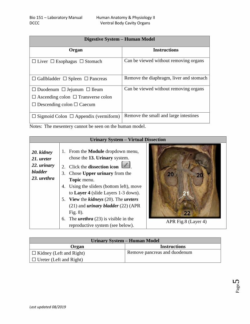
Urinary System – Virtual Dissection
20. kidney
21. ureter
22. urinary
bladder
23. urethra
1. From the Module dropdown menu,
chose the 13. Urinary system.
2. Click the dissection icon .
3. Chose Upper urinary from the
Topic menu.
4. Using the sliders (bottom left), move
to Layer 4 (slide Layers 1-3 down).
5. View the kidneys (20). The ureters
(21) and urinary bladder (22) (APR
Fig. 8).
6. The urethra (23) is visible in the
reproductive system (see below). APR Fig.8 (Layer 4)
Urinary System – Human Model
Organ Instructions
☐ Kidney (Left and Right)
☐ Ureter (Left and Right)
Remove pancreas and duodenum
Bio 151 – Laboratory Manual Human Anatomy & Physiology II DCCC Ventral Body Cavity Organs
Last updated 08/2019
Pag
e6
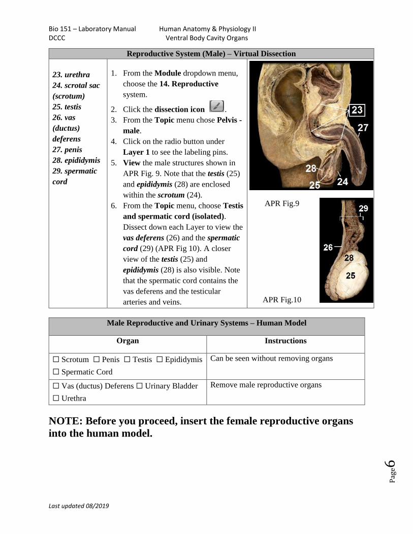
Reproductive System (Male) – Virtual Dissection
23. urethra
24. scrotal sac
(scrotum)
25. testis
26. vas
(ductus)
deferens
27. penis
28. epididymis
29. spermatic
cord
1. From the Module dropdown menu,
choose the 14. Reproductive
system.
2. Click the dissection icon .
3. From the Topic menu chose Pelvis -
male.
4. Click on the radio button under
Layer 1 to see the labeling pins.
5. View the male structures shown in
APR Fig. 9. Note that the testis (25)
and epididymis (28) are enclosed
within the scrotum (24).
6. From the Topic menu, choose Testis
and spermatic cord (isolated).
Dissect down each Layer to view the
vas deferens (26) and the spermatic
cord (29) (APR Fig 10). A closer
view of the testis (25) and
epididymis (28) is also visible. Note
that the spermatic cord contains the
vas deferens and the testicular
arteries and veins.
APR Fig.9
APR Fig.10
Male Reproductive and Urinary Systems – Human Model
Organ Instructions
☐ Scrotum ☐ Penis ☐ Testis ☐ Epididymis
☐ Spermatic Cord
Can be seen without removing organs
☐ Vas (ductus) Deferens ☐ Urinary Bladder
☐ Urethra
Remove male reproductive organs
NOTE: Before you proceed, insert the female reproductive organs
into the human model.
Bio 151 – Laboratory Manual Human Anatomy & Physiology II DCCC Ventral Body Cavity Organs
Last updated 08/2019
Pag
e7
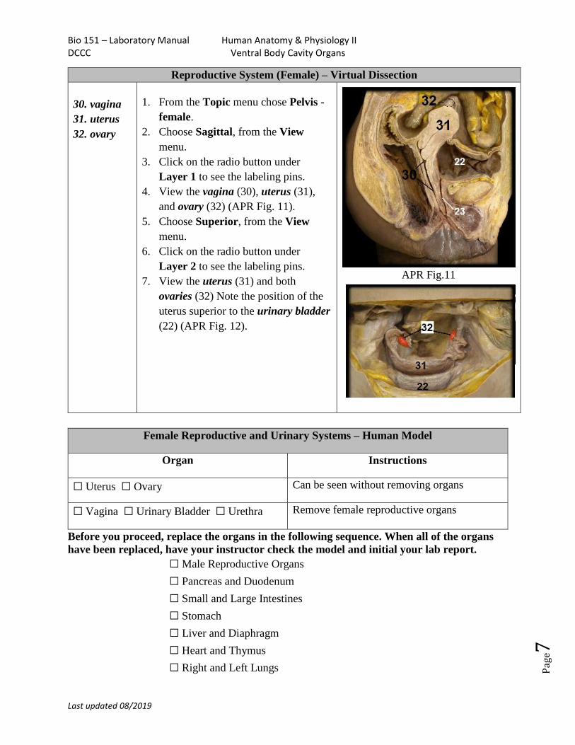
Reproductive System (Female) – Virtual Dissection
30. vagina
31. uterus
32. ovary
1. From the Topic menu chose Pelvis -
female.
2. Choose Sagittal, from the View
menu.
3. Click on the radio button under
Layer 1 to see the labeling pins.
4. View the vagina (30), uterus (31),
and ovary (32) (APR Fig. 11).
5. Choose Superior, from the View
menu.
6. Click on the radio button under
Layer 2 to see the labeling pins.
7. View the uterus (31) and both
ovaries (32) Note the position of the
uterus superior to the urinary bladder
(22) (APR Fig. 12).
APR Fig.11
APR Fig.12
Female Reproductive and Urinary Systems – Human Model
Organ Instructions
☐ Uterus ☐ Ovary Can be seen without removing organs
☐ Vagina ☐ Urinary Bladder ☐ Urethra Remove female reproductive organs
Before you proceed, replace the organs in the following sequence. When all of the organs
have been replaced, have your instructor check the model and initial your lab report.
☐ Male Reproductive Organs
☐ Pancreas and Duodenum
☐ Small and Large Intestines
☐ Stomach
☐ Liver and Diaphragm
☐ Heart and Thymus
☐ Right and Left Lungs
22
Bio 151 – Laboratory Manual Human Anatomy & Physiology II DCCC Ventral Body Cavity Organs
Last updated 08/2019
Pag
e8
II. Cat Dissection
A. Preparation
1. Wear goggles, gloves, and an apron for the entire time that you are working with
preserved specimens.
2. Use scissors to cut open the bag and remove the cat, but do not let the preservative
spill out of the bag.
3. Carefully dump the preservative into a special bin provided at certain lab sinks.
DO NOT DUMP THE PRESERVATIVE DOWN THE SINK.
4. The plastic bag should then be thrown away in the regular trash containers.
5. Do not use a scalpel for dissecting.
6. For most of your work, use only your scissors and blunt probe. It is the safest and most
efficient dissecting tool for muscles.
7. Use scissors only when specifically directed to do so in the dissection directions.
B. The Respiratory System - CAT
1. Lungs
2. Trachea
3. Diaphragm
4. Larynx
4a. Epiglottis
Lungs - Observe the lungs on either side of the heart. Note that like in humans, the left lung is
smaller and has fewer lobes than the right lung.
Trachea – The trachea can be seen as a white tube at the midline of the neck leading to the
lungs. Feel the hard cartilage rings in the trachea with your finger. Note that the trachea is still
wide open because of the support provided by the cartilage rings.
Diaphragm – Observe the diaphragm at the base of the lungs. This large dome shaped muscle
flattens to expand the thoracic cavity during inhalation. Note that it is skeletal muscle.
Larynx – Follow the trachea towards the head. The larynx can be observed as a widening of the
cartilage in the neck and will appear shiny white. The largest cartilage visible is the thyroid
cartilage. The larynx contains the vocal folds and epiglottis.
Epiglottis – Very carefully use your scissors to cut open the connective tissue at the very
superior end of the larynx above the cartilage in an upside-down “V” shape. Use your
finger or your blunt probe to lift up the flap-like epiglottis. This structure closes off the
larynx during swallowing to prevent food from entering the airways.
Bio 151 – Laboratory Manual Human Anatomy & Physiology II DCCC Ventral Body Cavity Organs
Last updated 08/2019
Pag
e9
C. Digestive System – CAT
5. Greater omentum
6. Liver
7. Gall bladder
8. Stomach
9. Small intestine (duodenum, jejunum, ileum)
Large intestine:
10. Ascending colon
11. Transverse colon
12. Descending colon
13. Rectum
14. Spleen
15. Mesentery
16. Pancreas
17. Caecum (no appendix in the cat)
19. Esophagus
Greater omentum – The fatty lace-like structure covering the abdominal organs is the greater
omentum. It is part of the peritoneum – the serous membrane of the abdominopelvic cavity. Lift
the greater omentum up and place it to the side to view the other organs. Do not detach it.
Liver – Observe the large red/brown liver in the right upper quadrant. Note that there are several
lobes similar to humans.
Gallbladder – Between the lobes of the liver find the gallbladder. Sometimes a dark greenish
stain is visible on the surface of the liver. The gallbladder itself is a sac-like structure with a
green tint. In some animals you will have to spread the lobes of the liver apart to find the
gallbladder.
Stomach – In the left upper quadrant, locate the stomach. You may need to move the greater
omentum. It is a pinkish sac that varies in size. Find where the esophagus attaches to it close to
the diaphragm. Use your scissors to cut open the stomach and observe the interior folds called
rugae.
Small Intestine –The small intestine leads from the stomach. It coils around and is connected
together by the mesentery. The first part of the small intestine is short called the duodenum. In
the cat it continues to the first bend (flexure) and then it becomes the jejunum. This portion is
the longest and leads to the ileum which empties into the caecum of the large intestine.
Bio 151 – Laboratory Manual Human Anatomy & Physiology II DCCC Ventral Body Cavity Organs
Last updated 08/2019
Pag
e10
Mesentery – This thin membrane connects the small intestine coils. Note the many blood vessels
running through it. It is part of the peritoneum.
Large intestine (colon) – The large intestine has a diameter of about twice the small intestine.
Find where the ileum of the small intestine meets the caecum, a pouch at the beginning of the
colon. Unlike the human, the cat has no appendix attaching to the caecum. Follow the colon up
(the ascending colon) to the very short transverse colon (basically a turn) and down the
descending colon to the rectum. Most of the cat’s rectum is not visible without further
dissection. The anal opening is visible from the exterior.
Spleen – The spleen is a reddish-brown structure visible behind the left side of the stomach.
Pancreas – The pancreas is best visible under the stomach by lifting the greater omentum and
placing it up over the stomach. At the juncture where the omentum attaches to the stomach you
can see a very delicate lobular material sometimes pinkish-white embedded in the connective
tissue. The pancreas runs horizontally across the abdomen and may continue down the
duodenum of the small intestine.
Esophagus – In the thoracic cavity, find the esophagus by gently moving the trachea aside to the
cat’s right. It is a soft brown/pink flattened tube directly underneath the trachea. Follow it down
to the stomach.
D. Urinary System – CAT
20. kidney
21. ureter
22. urinary bladder
23. urethra
Kidney – The kidneys are located on either side of the abdomen behind the other organs.
They are behind the peritoneum and so are called, retroperitoneal. You will need to remove this
layer to see the kidneys. Be careful not to damage the ureters as you do so. Remove one of the
kidneys and use your scalpel to slice it open using a frontal section. Observe the outer cortex and
inner medulla – note differences in color and texture.
Ureter – From the medial surface of each kidney a ureter attaches. These delicate flattened tubes
carry urine produced in the kidneys down to the urinary bladder for storage.
Urinary bladder – The urinary bladder is found in the pelvic cavity as a rounded sac. The
ureters attach to the posterior wall of the bladder and the urethra lies inferior to it.
Bio 151 – Laboratory Manual Human Anatomy & Physiology II DCCC Ventral Body Cavity Organs
Last updated 08/2019
Pag
e11
Urethra – The urethra is visible by lifting the bladder slightly and cleaning away the connective
tissue at the base of the bladder. Observe the beginning of the tubular urethra coming out of the
bladder which carries urine from the bladder to the outside of the body. The rest of the urethra
goes through the pelvis and its opening can be observed near the anal opening (see description of
the vagina below).
E. Male Reproductive System - CAT
24. scrotal sac (scrotum)
25. testis (testes pl.)
26. vas (ductus) deferens
27. penis
28. epididymis
29. spermatic cord
Scrotal sac (scrotum) – The scrotum is the skin covering the testicles (testes). The skin with fur
will need to be removed with scissors and forceps to view the testes. If your male cat has been
castrated, it will have no scrotum or testes.
Testis (testes pl.) – Two testes can be seen once the fur is removed. To view the testis better, as
well as the epididymis, remove the capsule around one testis using scissors and forceps.
Epididymis – The epididymis is a coiled tube located along the posterior surface of each testis
more easily visible when the testis capsule has been removed. It collects and helps to mature
sperm produced in the testes and passes them to the vas deferens.
Vas (ductus) deferens – The vas deferens is a tube leading from the epididymis through the
spermatic cord and loops behind the urinary bladder to fuse with the urethra at the prostate. In
the cat with the pelvis intact, it is most easily viewed within the spermatic cord near the testes.
Spermatic cord – This structure encompasses the vas deferens, testicular arteries, veins and
nerves from each testis. It passes through the inguinal canal to the abdominal cavity.
Penis – The penis of the cat is visible externally, located between and slightly above the testes.
To view the structures more closely you can remove the fur covered skin using scissors.
F. Female Reproductive System -CAT
30. Vagina (viewed as the urogenital sinus)
31. Uterus
32. Ovary
Bio 151 – Laboratory Manual Human Anatomy & Physiology II DCCC Ventral Body Cavity Organs
Last updated 08/2019
Pag
e12
Vagina – In the cat the urethra and the vagina open into a common orifice called the urogenital
sinus. It is located ventral to the anal opening.
Uterus – The cat uterus has two “horns” which are extensions going up the lateral abdominal
wall each attaching to an ovary. Each long uterine horn allows space for many offspring to grow.
The cat uterus lacks the long uterine (Fallopian) tubes seen in the human. The thickness and
color of the uterine horns varies according to whether the cat has had / does have a pregnancy.
The uterine horns can vary from very thin and pinkish, similar in appearance to the ureters, or
very thick. A pregnant cat will have an enlarged uterus with large blood vessels.
Ovary – At the end of each uterine horn, a small flattened lobular ovary is attached. It may
appear whitish, yellowish, or pinkish in color and often resembles a very small bean.
G. Directions for Putting Your Cat Away and Cleaning Up
1. All loose cat parts should be thrown away in the appropriate container as indicated by
your instructor. Please do NOT allow cat parts to end up in the sink. They will clog the
drain and cause extra work for lab assistants.
2. Place the cat into the container provided for disposal as directed by your instructor.
3. Thoroughly wash your dissecting tray with soap and water, dry, and return it to the lab
bench where you found it.
4. Use the spray disinfectant cleaner provided to thoroughly wipe down your lab bench
area so that it will be ready for the next group of students.
5. Dispose of your gloves, paper towels and old plastic bags in the regular trash
containers as directed by your instructor.
6. Make sure that you take your dissecting kit and goggles with you when you leave the lab.
Bio 151 – Laboratory Manual Human Anatomy & Physiology II
DCCC Ventral Body Cavity Organs Name:__________________________
Last updated 08/2019
Pag
e13
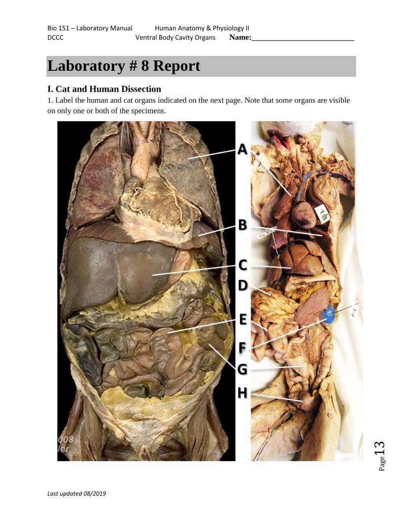
Laboratory # 8 Report
I. Cat and Human Dissection
1. Label the human and cat organs indicated on the next page. Note that some organs are visible
on only one or both of the specimens.
Bio 151 – Laboratory Manual Human Anatomy & Physiology II
DCCC Ventral Body Cavity Organs Name:__________________________
Last updated 08/2019
Pag
e14
1. Diagram labels: A. ______________________________________
B.______________________________________
C. ______________________________________
D. ______________________________________
E. ______________________________________
F. ______________________________________
G. ______________________________________
H. ______________________________________
2. Name the two parts of the peritoneum that a.) __________________________
we observed in the human and cat: b.) _________________________
3. What part of the respiratory system contains the vocal folds? _______________________
4. Name the three parts of the small intestine: a)__________________________
b)__________________________
c)__________________________
5. Name the tubes which take urine from the kidneys to the urinary bladder. ________________
6. What tube eliminates urine from the body? _____________________
7. What four structures are in the spermatic cord? a)_________________________
b)_________________________
c)_________________________
d)_________________________
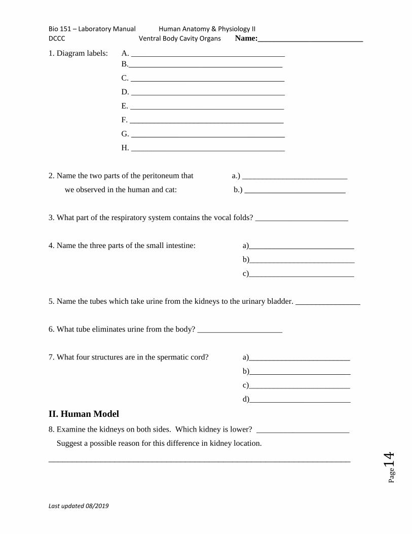
II. Human Model
8. Examine the kidneys on both sides. Which kidney is lower? _______________________
Suggest a possible reason for this difference in kidney location.
________________________________________________________________
Bio 151 – Laboratory Manual Human Anatomy & Physiology II
DCCC Ventral Body Cavity Organs Name:__________________________
Last updated 08/2019
Pag
e15
9. Place the following in order from superior to inferior: transverse colon, liver, diaphragm.
A. ______________________________________
B._______________________________________
C. ______________________________________
10. Since most of this organ lies behind the stomach, the ____________________ can only be
observed clearly after the stomach is removed from the human model.
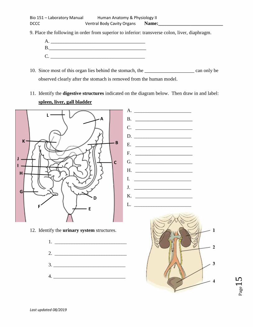
11. Identify the digestive structures indicated on the diagram below. Then draw in and label:
spleen, liver, gall bladder
A. _______________________
B. _______________________
C. _______________________
D. _______________________
E. _______________________
F. _______________________
G. _______________________
H. _______________________
I. _______________________
J. _______________________
K. _______________________
L. _______________________
12. Identify the urinary system structures.
1. _____________________________
2. _____________________________
3. _____________________________
4. _____________________________
A
D
E
L
B
C
F
G
H
I
J
K
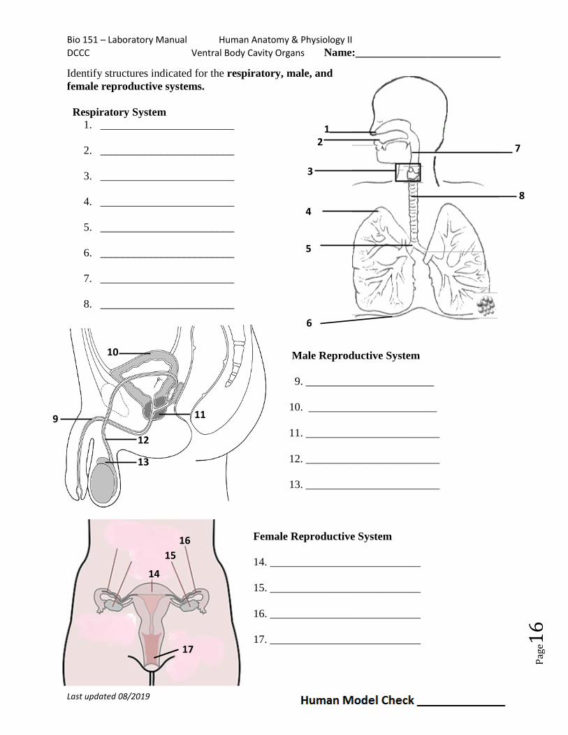
Bio 151 – Laboratory Manual Human Anatomy & Physiology II
DCCC Ventral Body Cavity Organs Name:__________________________
Last updated 08/2019
Pag
e16
Identify structures indicated for the respiratory, male, and
female reproductive systems.
Respiratory System
1. ________________________
2. ________________________
3. ________________________
4. ________________________
5. ________________________
6. ________________________
7. ________________________
8. ________________________
Male Reproductive System
9. _______________________
10. _______________________
11. ________________________
12. ________________________
13. ________________________
Female Reproductive System
14. ___________________________
15. ___________________________
16. ___________________________
17. ___________________________
10
14
15
16
17
7
8
1 2
3
4
5
6
9 11
12
13