Cytotoxic T Cells Mediate Pathology and Metastasis in ...
11
Cytotoxic T Cells Mediate Pathology and Metastasis in Cutaneous Leishmaniasis Fernanda O. Novais 1 , Lucas P. Carvalho 2 , Joel W. Graff 3 , Daniel P. Beiting 4 , Gordon Ruthel 1 , David S. Roos 4 , Michael R. Betts 5 , Michael H. Goldschmidt 1 , Mary E. Wilson 3 , Camila I. de Oliveira 6 , Phillip Scott 1 * 1 Department of Pathobiology, School of Veterinary Medicine, University of Pennsylvania, Philadelphia, Pennsylvania, United States of America, 2 Instituto Nacional de Cie ˆ ncia e Tecnologia de Doenc ¸as Tropicais-INCT-DT(CNPq/MCT), Servic ¸o de Imunologia, Hospital Universitario Prof. Edgard Santos, Universidade Federal da Bahia Salvador, Bahia, Brazil, 3 Iowa City VA Medical Center, Iowa City, Iowa, United States of America, 4 Department of Biology, University of Pennsylvania, Philadelphia, Pennsylvania, United States of America, 5 Department of Microbiology, Perelman School of Medicine, University of Pennsylvania, Philadelphia, Pennsylvania, United States of America, 6 Centro de Pesquisas Gonc ¸alo Moniz, Fundac ¸ao Oswaldo Cruz, Salvador, Brazil Abstract Disease progression in response to infection can be strongly influenced by both pathogen burden and infection-induced immunopathology. While current therapeutics focus on augmenting protective immune responses, identifying therapeutics that reduce infection-induced immunopathology are clearly warranted. Despite the apparent protective role for murine CD8+ T cells following infection with the intracellular parasite Leishmania, CD8+ T cells have been paradoxically linked to immunopathological responses in human cutaneous leishmaniasis. Transcriptome analysis of lesions from Leishmania braziliensis patients revealed that genes associated with the cytolytic pathway are highly expressed and CD8+ T cells from lesions exhibited a cytolytic phenotype. To determine if CD8+ T cells play a causal role in disease, we turned to a murine model. These studies revealed that disease progression and metastasis in L. braziliensis infected mice was independent of parasite burden and was instead directly associated with the presence of CD8+ T cells. In mice with severe pathology, we visualized CD8+ T cell degranulation and lysis of L. braziliensis infected cells. Finally, in contrast to wild-type CD8+ T cells, perforin-deficient cells failed to induce disease. Thus, we show for the first time that cytolytic CD8+ T cells mediate immunopathology and drive the development of metastatic lesions in cutaneous leishmaniasis. Citation: Novais FO, Carvalho LP, Graff JW, Beiting DP, Ruthel G, et al. (2013) Cytotoxic T Cells Mediate Pathology and Metastasis in Cutaneous Leishmaniasis. PLoS Pathog 9(7): e1003504. doi:10.1371/journal.ppat.1003504 Editor: Peter C. Melby, University of Texas Medical Branch, United States of America Received January 30, 2013; Accepted June 2, 2013; Published July 18, 2013 This is an open-access article, free of all copyright, and may be freely reproduced, distributed, transmitted, modified, built upon, or otherwise used by anyone for any lawful purpose. The work is made available under the Creative Commons CC0 public domain dedication. Funding: These studies were funded in part by the National Institutes of Health: (RO176257, http://grants.nih.gov/grants/oer.htm); International Centers for Infectious Disease Research grant (U01- AUI 088650), Conselho Nacional de Desenvolvimento Cientı ´fico e Tecnolo ´ gico (CNPq, http://www.cnpq.br/), Brazil. The funders had no role in study design, data collection and analysis, decision to publish, or preparation of the manuscript. Competing Interests: The authors have declared that no competing interests exist. * E-mail: [email protected] Introduction CD8+ T cells contribute to the control of pathogens by cytokine production, cytolytic activity or both. In the case of intracellular parasites, the production of IFN-c by CD8+ T cells is protective, while in viral infections CD8+ T cells provide protection by inducing cytokine production and killing virally infected cells [1]. Nevertheless, these same CD8+ T cell effector functions can also promote increased pathology, and the presence of CD8+ T cells has been associated with increased pathology in several infectious and autoimmune diseases [2,3,4,5,6,7,8]. In some cases the pathology is believed to be associated with IFN-c or IL-17 production, while in other situations cytolytic activity is linked with disease. Still, the mechanistic basis by which CD8+ T cells could potentially contribute to increased pathology is difficult to determine in humans. Cutaneous leishmaniasis is one of many diseases where the outcome of the infection depends on both the extent of parasite elimination and the relative induction of potentially immunopath- ologic responses. A great deal is known about how leishmania parasites are eliminated. Thus, control of these intracellular parasites requires a CD4+ Th1 cell response, which leads to IFN-c production that enhances the killing capacity of infected macro- phages and dendritic cells [9,10]. CD8+ T cells respond during infection and contribute to the control of Leishmania by producing IFN-c, which not only activates macrophages to kill the parasites, but also promotes the differentiation of naı ¨ve T cells into Th1 cells [11,12]. On the other hand, few studies have addressed how immunopathology develops in cutaneous leishmaniasis. Correla- tions with enhanced immunopathology and lower levels of IL-10 or IL-10 receptor expression have been observed in patients, but the unregulated responses that promote pathology are not defined [13,14]. In patients infected with L. braziliensis, the number of CD8+ T cells increases in lesions as the disease worsens, and patients with mucosal disease–where metastatic lesions develop in the nasopha- ryngeal mucosa due to a destructive inflammatory response–have elevated numbers of cytotoxic cells in the blood [15,16,17]. Interestingly, it has also been reported that L. major-infected Rag12/2 mice reconstituted with CD8+ T cells develop much larger lesions than unreconstituted Rag12/2 mice [11]. Together, these observations implicate CD8+ T cells as inducers of PLOS Pathogens | www.plospathogens.org 1 July 2013 | Volume 9 | Issue 7 | e1003504
Transcript of Cytotoxic T Cells Mediate Pathology and Metastasis in ...

Cytotoxic T Cells Mediate Pathology and Metastasis inCutaneous LeishmaniasisFernanda O. Novais1, Lucas P. Carvalho2, Joel W. Graff3, Daniel P. Beiting4, Gordon Ruthel1,
David S. Roos4, Michael R. Betts5, Michael H. Goldschmidt1, Mary E. Wilson3, Camila I. de Oliveira6,
Phillip Scott1*
1 Department of Pathobiology, School of Veterinary Medicine, University of Pennsylvania, Philadelphia, Pennsylvania, United States of America, 2 Instituto Nacional de
Ciencia e Tecnologia de Doencas Tropicais-INCT-DT(CNPq/MCT), Servico de Imunologia, Hospital Universitario Prof. Edgard Santos, Universidade Federal da Bahia
Salvador, Bahia, Brazil, 3 Iowa City VA Medical Center, Iowa City, Iowa, United States of America, 4 Department of Biology, University of Pennsylvania, Philadelphia,
Pennsylvania, United States of America, 5 Department of Microbiology, Perelman School of Medicine, University of Pennsylvania, Philadelphia, Pennsylvania, United States
of America, 6 Centro de Pesquisas Goncalo Moniz, Fundacao Oswaldo Cruz, Salvador, Brazil
Abstract
Disease progression in response to infection can be strongly influenced by both pathogen burden and infection-inducedimmunopathology. While current therapeutics focus on augmenting protective immune responses, identifying therapeuticsthat reduce infection-induced immunopathology are clearly warranted. Despite the apparent protective role for murineCD8+ T cells following infection with the intracellular parasite Leishmania, CD8+ T cells have been paradoxically linked toimmunopathological responses in human cutaneous leishmaniasis. Transcriptome analysis of lesions from Leishmaniabraziliensis patients revealed that genes associated with the cytolytic pathway are highly expressed and CD8+ T cells fromlesions exhibited a cytolytic phenotype. To determine if CD8+ T cells play a causal role in disease, we turned to a murinemodel. These studies revealed that disease progression and metastasis in L. braziliensis infected mice was independent ofparasite burden and was instead directly associated with the presence of CD8+ T cells. In mice with severe pathology, wevisualized CD8+ T cell degranulation and lysis of L. braziliensis infected cells. Finally, in contrast to wild-type CD8+ T cells,perforin-deficient cells failed to induce disease. Thus, we show for the first time that cytolytic CD8+ T cells mediateimmunopathology and drive the development of metastatic lesions in cutaneous leishmaniasis.
Citation: Novais FO, Carvalho LP, Graff JW, Beiting DP, Ruthel G, et al. (2013) Cytotoxic T Cells Mediate Pathology and Metastasis in Cutaneous Leishmaniasis. PLoSPathog 9(7): e1003504. doi:10.1371/journal.ppat.1003504
Editor: Peter C. Melby, University of Texas Medical Branch, United States of America
Received January 30, 2013; Accepted June 2, 2013; Published July 18, 2013
This is an open-access article, free of all copyright, and may be freely reproduced, distributed, transmitted, modified, built upon, or otherwise used by anyone forany lawful purpose. The work is made available under the Creative Commons CC0 public domain dedication.
Funding: These studies were funded in part by the National Institutes of Health: (RO176257, http://grants.nih.gov/grants/oer.htm); International Centers forInfectious Disease Research grant (U01- AUI 088650), Conselho Nacional de Desenvolvimento Cientıfico e Tecnologico (CNPq, http://www.cnpq.br/), Brazil. Thefunders had no role in study design, data collection and analysis, decision to publish, or preparation of the manuscript.
Competing Interests: The authors have declared that no competing interests exist.
* E-mail: [email protected]
Introduction
CD8+ T cells contribute to the control of pathogens by cytokine
production, cytolytic activity or both. In the case of intracellular
parasites, the production of IFN-c by CD8+ T cells is protective,
while in viral infections CD8+ T cells provide protection by
inducing cytokine production and killing virally infected cells [1].
Nevertheless, these same CD8+ T cell effector functions can also
promote increased pathology, and the presence of CD8+ T cells
has been associated with increased pathology in several infectious
and autoimmune diseases [2,3,4,5,6,7,8]. In some cases the
pathology is believed to be associated with IFN-c or IL-17
production, while in other situations cytolytic activity is linked with
disease. Still, the mechanistic basis by which CD8+ T cells could
potentially contribute to increased pathology is difficult to
determine in humans.
Cutaneous leishmaniasis is one of many diseases where the
outcome of the infection depends on both the extent of parasite
elimination and the relative induction of potentially immunopath-
ologic responses. A great deal is known about how leishmania
parasites are eliminated. Thus, control of these intracellular
parasites requires a CD4+ Th1 cell response, which leads to IFN-cproduction that enhances the killing capacity of infected macro-
phages and dendritic cells [9,10]. CD8+ T cells respond during
infection and contribute to the control of Leishmania by producing
IFN-c, which not only activates macrophages to kill the parasites,
but also promotes the differentiation of naıve T cells into Th1 cells
[11,12]. On the other hand, few studies have addressed how
immunopathology develops in cutaneous leishmaniasis. Correla-
tions with enhanced immunopathology and lower levels of IL-10
or IL-10 receptor expression have been observed in patients, but
the unregulated responses that promote pathology are not defined
[13,14].
In patients infected with L. braziliensis, the number of CD8+ T
cells increases in lesions as the disease worsens, and patients with
mucosal disease–where metastatic lesions develop in the nasopha-
ryngeal mucosa due to a destructive inflammatory response–have
elevated numbers of cytotoxic cells in the blood [15,16,17].
Interestingly, it has also been reported that L. major-infected
Rag12/2 mice reconstituted with CD8+ T cells develop much
larger lesions than unreconstituted Rag12/2 mice [11]. Together,
these observations implicate CD8+ T cells as inducers of
PLOS Pathogens | www.plospathogens.org 1 July 2013 | Volume 9 | Issue 7 | e1003504

pathology. As CD8+ T cells can produce IFN-c in leishmaniasis, it
is possible that an overproduction of IFN-c promotes increased
pathology. On the other hand, the severity of disease in patients
infected with L. braziliensis is directly associated with increased
numbers of granzyme expressing CD8+ T cells [15]. Thus, it
remains to be determined whether CD8+ T cells are indeed
pathogenic, and if so, whether they increase disease severity by
cytokine production and/or enhanced cytolytic activity.
Defining the mechanisms that promote the immunopathology
observed in cutaneous leishmaniasis is a critical first step in
developing an approach to control the disease. Here, we define the
pathologic role that CD8+ T cells play in L. braziliensis infections.
We found that the most highly expressed genes in leishmanial
lesions are associated with the lytic pathway and that CD8+ T cells
within the lesions of L. braziliensis patients are functionally cytolytic.
Using a murine model we found that CD8+ T cells contribute to
increased lesion size following infection with L. braziliensis parasites.
Strikingly, we found that the development of metastatic lesions was
also promoted by the presence of CD8+ T cells. Mechanistically,
we demonstrated that the pathology associated with unregulated
CD8 function is not due to enhanced IFN-c or IL-17 production,
but rather is due to excessive perforin-dependent cytolytic activity
by CD8+ T cells. Thus, our findings show for the first time that
cytolytic CD8+ T cells are not only present during infection, but
that they promote increased immunopathology and metastatic
lesions in cutaneous leishmaniasis.
Results
CD8+ T cells from L. braziliensis patients exhibit increasedcytolytic activity
To better understand the local immune environment during
human leishmaniasis and the extent to which a cytolytic program
is associated with the infection, we carried out whole genome
expression profiling of lesions from patients infected with L.
braziliensis. Over 500 genes were expressed $3-fold (p#0.05) in
lesions compared to normal skin from uninfected donors (not
shown). Strikingly, many known components of cytolytic granules
were found amongst the most strongly expressed genes in lesions
[18] (Fig. 1A). For example, granzyme B, granzyme A and
granulysin, a pore-forming molecule found in the granules of
human CTLs and NK cells, were the first, seventh and tenth genes
most strongly expressed genes overall in lesions, respectively.
Moreover, gene set enrichment analysis of the entire expression
Figure 1. Cytolytic profile of CD8+ T cells in lesions from L.braziliensis infected patients. (A) Heatmap showing induction of keymediators of cytolysis from microarray profiling of ten human lesionbiopsies and two normal skin biopsies. Average fold change (FC) foreach gene in lesion samples, relative to normal skin controls, is shown,as is the rank for this fold change within the entire expression data set.(B) PBMCs and cells isolated from lesions obtained from L. braziliensispatients were incubated with anti-CD107a. Depicted are representative(A) contour plots and scatter plots of (C) GZMB, (D) PRF and (E) CD107aexpression by CD8+ T cells (pregated on Singlets/CD3+/CD8+). Datafrom patients [8 (blood) and 12 (biopsy)] obtained in two independentexperiments are shown. **p,0.01; ***p,0.001.doi:10.1371/journal.ppat.1003504.g001
Author Summary
Leishmaniasis is a parasitic disease where the host immuneresponse plays an essential role in pathogenesis. However,the mechanisms promoting immunopathology in patientsare still unclear. We performed gene expression profiling ofskin lesions from cutaneous leishmaniasis patients andnormal skin and the results demonstrated that the mostexpressed genes in leishmanial lesions were associatedwith the cytolytic pathway. Using both human samplesand mouse models we showed that CD8+ T cells arecytolytic within leishmanial lesions and kill Leishmaniainfected target cells. We found that the CD8+ T cellcytolytic response was not protective, but rather promotedincreased immunopathology, associated with enhancedrecruitment of neutrophils to the site of infection. CD8+ Tcells also promoted the development of metastatic lesionsat distant skin sites. Together, our results clearly demon-strate that activation of CD8+ T cell cytolytic responses isdetrimental to the host and that targeting this pathwaycould be a new approach to treat patients with leishman-iasis.
Pathogenic CD8+ T Cells in Cutaneous Leishmanaisis
PLOS Pathogens | www.plospathogens.org 2 July 2013 | Volume 9 | Issue 7 | e1003504

data set revealed a significant enrichment for KEGG pathways
involved in NK cell mediated cytotoxicity, graft-versus-host
diseases and allograft rejection (not shown), all of which involve
cytolysis [19].
Next we measured the protein levels of granzyme B and
perforin in CD8+ T cells recovered from the peripheral blood or
from lesions of L. braziliensis infected patients (Fig. 1B). More
CD8+ T cells obtained from lesions expressed granzyme B
(Fig. 1C) in comparison to cells from the blood, and both
populations contained cells expressing perforin (Fig. 1D). These
data suggest that both skin and peripheral CD8+ T cells have the
capacity to degranulate. To determine if in fact these cells were
degranulating, we assessed their surface expression of CD107a
(Fig. 1B, lower panels). CD107a is a lysosomal membrane
glycoprotein (also known as Lamp1) present in CD8+ T cell
granules. During degranulation, this molecule is transiently
exposed on the cell surface and thus is a marker for the release
of cytotoxic granules by CD8+ T cells [20]. We found a significant
increase in CD8+ T cells expressing surface CD107a from lesions,
while CD8+ T cells from the blood failed to express CD107a
(Fig. 1E). These results not only confirm previous studies showing
that CD8+ T cells express granzyme in leishmaniasis [15], but also
extend these findings to show that the CD8+ T cells are
cytolytically active within human leishmanial lesions.
CD8+ T cells induce pathology in L. braziliensis infectedmice
To determine if our results from L. braziliensis patients could be
mechanistically dissected using animal models we first asked if
depletion of CD8+ T cells would affect lesion development in
BALB/c mice. In contrast to L. major, L. braziliensis infections in
BALB/c mice results in the development of an ulcerated lesion
that eventually resolves [21]. Thus, BALB/c mice infected with
105 L. braziliensis developed a substantial lesion that healed
spontaneously when treated with control isotype antibody
(Fig. 2A, closed circles). In contrast, mice depleted of CD8+ T
cells with an anti-CD8-specific antibody developed substantially
smaller lesions (Fig. 2A, open circles), suggesting that CD8+ T cells
contribute to lesion size. The relative decrease in lesion size in
CD8 depleted mice was not due to an alteration in parasite
number (Fig. 2B), indicating that the change in lesion size was due
primarily to differences in the inflammatory response. Thus, these
data indicate that CD8+ T cells contribute to the inflammatory
response following L. braziliensis infection in BALB/c mice.
Two factors seem to be associated with immunopathology in L.
braziliensis patients: an increase in CD8+ T cells recruited to the
lesions and a decrease in immunoregulatory cytokines. To test if
CD8+ T cells could directly enhance disease, L. braziliensis infected
Rag12/2 mice were reconstituted with naive CD8+ T cells
(RAG+CD8) or CD8+ and CD4+ T cells (RAG+CD4+CD8), at a
1:1 ratio, and the course of infection was followed. Unrecon-
stituted Rag12/2 mice infected with L. braziliensis developed
minimal lesions. Thus, similar to L. major or L. amazonensis [11,22],
lesion development with L. braziliensis likely depends upon
generation of a T cell-dependent inflammatory response. As
expected, RAG+CD4+CD8 mice developed small nodules that
resolved within 7 weeks following infection (Fig. 2C). In contrast,
transfer of CD8+ T cells alone to Rag12/2 mice led to the
development of an uncontrolled lesion (Fig. 2C). To determine if
the increased pathology observed in RAG+CD8 mice was due to
uncontrolled parasite growth, parasite loads were assessed within
the lesions. We found that Rag12/2 mice and RAG+CD8 mice
had similar numbers of parasites at the infection site, in spite of the
disparity in lesion size observed in these animals, while transfer of
CD4+ and CD8+ T cells into Rag12/2 mice led to significantly
better control of the parasite in the infected ear (Fig. 2D). Thus,
the exacerbated lesion development in RAG+CD8 mice compared
with Rag12/2 mice is due to CD8+ T cell mediated pathology
rather than differences in the number of parasites in the ear.
Most notably, the enhanced lesion size in RAG+CD8 mice was
accompanied by a rampant immunopathologic response. By 5
weeks post infection we observed destruction of the infected ear in
RAG+CD8 mice, but minimal pathology in either Rag12/2 or
RAG+CD4+CD8 mice (Fig. 2E). This destruction was accompa-
nied by infiltration of many more CD8+ T cells than in Rag12/2
mice that received both CD4+ and CD8+ T cells (data not shown).
Histologically, we could observe at lower magnification the
substantial differences in lesion thickness in mice without T cells
and RAG+CD4+CD8 or RAG+CD8 mice. Higher magnification
showed that lesions from Rag12/2 mice were composed of
infected macrophages and granulocytes (Fig. 2E). At this time
point, lesions from RAG+CD4+CD8 mice were healing and
exhibited a mild dermal lymphocytic infiltrate accompanied by
epidermal hyperplasia and spongiosis with few leishmania
organisms (Fig. 2E). In contrast, the lesions from RAG+CD8
mice showed a dramatic cellular infiltration consisting of
lymphocytes, granulocytes and many highly infected cells
(Fig. 2E). Moreover, the epidermis in these lesions exhibited
substantial hyperplasia and areas of ulceration, and the severe
inflammatory response in the dermis led to alterations in the
cartilage. Using a pathology score that goes from 0 to 5, where 0 is
mild and 5 is severe disease, at 7 weeks post-infection RAG+CD8
mice had the most severe disease (5) followed by Rag12/2 mice
with moderate disease (2) and RAG+CD4+CD8 mice with no
disease (0). We also characterized the myeloid cell composition
present in lesions from RAG+CD8 mice, and found that a
majority of the myeloid cells within lesions were neutrophils. (Fig.
S1). Together, these observations illustrate the critical role of the
inflammatory response as the main factor driving lesion develop-
ment, further highlighting the importance of identifying the
mechanisms that control pathology in cutaneous leishmaniasis.
An additional unexpected result observed in RAG+CD8 mice
was the development of metastatic lesions. This was particularly
notable in the contralateral ear, which developed gross pathology
indistinguishable from the primary lesion (Fig. 2F). In addition to
the contralateral ear, we observed lesions at other skin sites,
including the nose, tail, and footpad. We were able to culture
parasites from these regions, confirming the spread of parasites to
these additional skin sites (data not shown). In contrast, we did not
observe metastatic lesions in Rag12/2 or RAG+CD4+CD8 mice
(Fig. 2F). Thus, the development of both primary and metastatic
lesions in Rag12/2 mice was dependent upon CD8+ T cells.
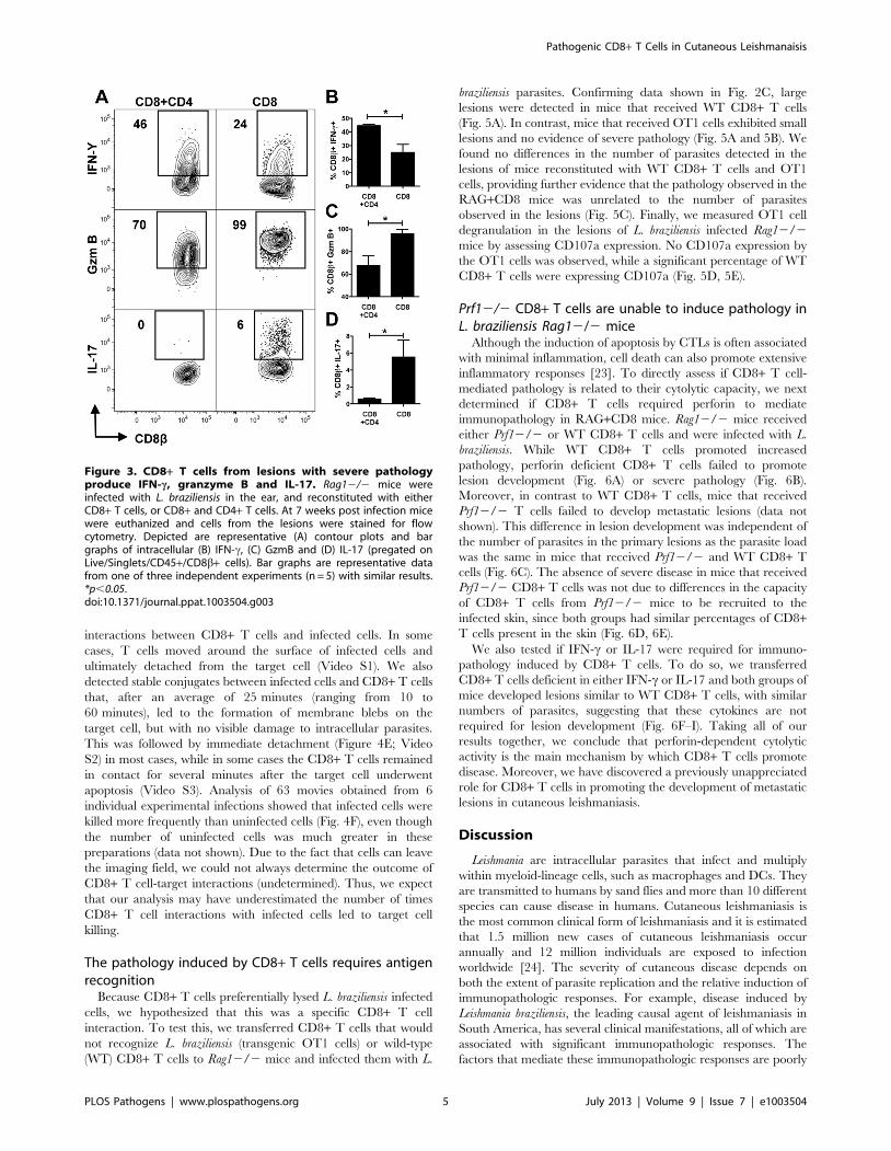
CD8+ T cells within lesions express IFN-c, IL-17 and highlevels of granzyme B
To characterize the functions of the CD8+ T cells transferred in
the presence and absence of CD4+ T cells, cells from lesions of L.
braziliensis infected reconstituted Rag12/2 mice were assessed for
IFN-c, IL-17 and granzyme B by flow cytometry (Fig. 3A). A
higher percentage of CD8+ T cells made IFN-c when these cells
were transferred together with CD4+ T cells, although CD8+ T
cells made IFN-c in the absence of CD4+ T cells (Fig. 3B). In
contrast, CD8+ T cells from RAG+CD8 mice produced
significantly more granzyme B in the absence of CD4+ T cells
(Fig. 3C). Finally, although only a small percentage of CD8+ T
cells from RAG+CD8 mice produced IL-17, IL-17 production was
completely abrogated by the presence of CD4+ T cells in
RAG+CD4+CD8 mice (Fig. 3D). Overall, these results suggest
Pathogenic CD8+ T Cells in Cutaneous Leishmanaisis
PLOS Pathogens | www.plospathogens.org 3 July 2013 | Volume 9 | Issue 7 | e1003504

that CD8+ T cells could be mediating increased pathology due to
cytolytic activity (indicated by high levels of granzyme B), IL-17, or
IFN-c, and indicate that CD4+ T cells may regulate these
responses.
CD8+ T cells degranulate and kill L. braziliensis infectedcells
As we observed expression of genes associated with cytolysis in
leishmanial lesions, we first assessed if the immunopathology
observed in RAG+CD8 mice was related to cytolytic activity. Cells
were obtained 5 weeks after infection of RAG+CD4+CD8 or
RAG+CD8 mice, and were stained for CD107a expression
directly ex vivo. In RAG+CD4+CD8 mice analysis of CD107a
expression showed a small percentage of degranulating CD8+ T
cells at the infection site (Fig. 4A, 4B). On the other hand, a high
percentage of CD8+ T cells from lesions of RAG+CD8 mice
expressed surface CD107a (Fig. 4A, 4B).
To confirm that CD107a expression was indicative of degran-
ulation, we sought to visualize CD107a at the interface between
CD8+ T cells and infected cells. For these experiments, eGFP+
CD8+ T cells were transferred into Rag12/2 mice that were
subsequently infected with L. braziliensis parasites expressing
mCherry. Cells from lesions taken 5 weeks after infection were
incubated for 1 hour in the presence of anti-CD107a monoclonal
antibody and then run on an ImageStream flow cytometer. We
observed the presence of CD107a (blue) at the synapse between
CD8+ T cells (green) and L. braziliensis infected target cells (red)
providing further support that CD8+ T cells from RAG+CD8
mice were degranulating (Fig. 4C). Analysis of the total doublets
that contained eGFP+ CD8+ T cells showed that surface
expression of CD107a on CD8+ T cells was more frequent when
CD8+ T cells were in contact with infected in comparison to
uninfected target cells (Fig. 4D).
Finally, to directly show that CD8+ T cells from RAG+CD8
mice induce infected cell death, we visualized the interactions
between CD8+ T cells and infected cells using a spinning disk
confocal microscope. As above, mice were infected with mCherry
L. braziliensis parasites and reconstituted with GFP+ CD8+ T cells,
and after 5 weeks cells from the lesions were isolated and
immediately visualized. We observed several different types of
Figure 2. CD8+ T cells induce pathology during L. braziliensis infection. (A) BALB/c mice were infected with L. braziliensis in the ear, and thecourse of infection monitored, and (B) parasite burden assessed in the lesions at 5 weeks. Rag12/2 mice were infected with L. braziliensis in the ear,and reconstituted with either CD8+ T cells, or CD8+ and CD4+ T cells, or did not receive cells and (C) ear thickness at the site of infection assessed. At7 weeks post infection mice were euthanized and shown are (D) the parasite burden in the lesions and (E) histological comparison of lesions by H&Estaining. Scale bar represents 200 mm for lower magnification (top row) and 20 mm for higher magnification (bottom row). (F) Representative frontimages and back images (showing metastasis) of leishmanial lesions at 6 weeks post infection. Representative data from one of three or moreindependent experiments (n = 5) with similar results are presented. ns, non-significant. *p,0.05.doi:10.1371/journal.ppat.1003504.g002
Pathogenic CD8+ T Cells in Cutaneous Leishmanaisis
PLOS Pathogens | www.plospathogens.org 4 July 2013 | Volume 9 | Issue 7 | e1003504
interactions between CD8+ T cells and infected cells. In some
cases, T cells moved around the surface of infected cells and
ultimately detached from the target cell (Video S1). We also
detected stable conjugates between infected cells and CD8+ T cells
that, after an average of 25 minutes (ranging from 10 to
60 minutes), led to the formation of membrane blebs on the
target cell, but with no visible damage to intracellular parasites.
This was followed by immediate detachment (Figure 4E; Video
S2) in most cases, while in some cases the CD8+ T cells remained
in contact for several minutes after the target cell underwent
apoptosis (Video S3). Analysis of 63 movies obtained from 6
individual experimental infections showed that infected cells were
killed more frequently than uninfected cells (Fig. 4F), even though
the number of uninfected cells was much greater in these
preparations (data not shown). Due to the fact that cells can leave
the imaging field, we could not always determine the outcome of
CD8+ T cell-target interactions (undetermined). Thus, we expect
that our analysis may have underestimated the number of times
CD8+ T cell interactions with infected cells led to target cell
killing.
The pathology induced by CD8+ T cells requires antigenrecognition
Because CD8+ T cells preferentially lysed L. braziliensis infected
cells, we hypothesized that this was a specific CD8+ T cell
interaction. To test this, we transferred CD8+ T cells that would
not recognize L. braziliensis (transgenic OT1 cells) or wild-type
(WT) CD8+ T cells to Rag12/2 mice and infected them with L.
braziliensis parasites. Confirming data shown in Fig. 2C, large
lesions were detected in mice that received WT CD8+ T cells
(Fig. 5A). In contrast, mice that received OT1 cells exhibited small
lesions and no evidence of severe pathology (Fig. 5A and 5B). We
found no differences in the number of parasites detected in the
lesions of mice reconstituted with WT CD8+ T cells and OT1
cells, providing further evidence that the pathology observed in the
RAG+CD8 mice was unrelated to the number of parasites
observed in the lesions (Fig. 5C). Finally, we measured OT1 cell
degranulation in the lesions of L. braziliensis infected Rag12/2
mice by assessing CD107a expression. No CD107a expression by
the OT1 cells was observed, while a significant percentage of WT
CD8+ T cells were expressing CD107a (Fig. 5D, 5E).
Prf12/2 CD8+ T cells are unable to induce pathology inL. braziliensis Rag12/2 mice
Although the induction of apoptosis by CTLs is often associated
with minimal inflammation, cell death can also promote extensive
inflammatory responses [23]. To directly assess if CD8+ T cell-
mediated pathology is related to their cytolytic capacity, we next
determined if CD8+ T cells required perforin to mediate
immunopathology in RAG+CD8 mice. Rag12/2 mice received
either Prf12/2 or WT CD8+ T cells and were infected with L.
braziliensis. While WT CD8+ T cells promoted increased
pathology, perforin deficient CD8+ T cells failed to promote
lesion development (Fig. 6A) or severe pathology (Fig. 6B).
Moreover, in contrast to WT CD8+ T cells, mice that received
Prf12/2 T cells failed to develop metastatic lesions (data not
shown). This difference in lesion development was independent of
the number of parasites in the primary lesions as the parasite load
was the same in mice that received Prf12/2 and WT CD8+ T
cells (Fig. 6C). The absence of severe disease in mice that received
Prf12/2 CD8+ T cells was not due to differences in the capacity
of CD8+ T cells from Prf12/2 mice to be recruited to the
infected skin, since both groups had similar percentages of CD8+T cells present in the skin (Fig. 6D, 6E).
We also tested if IFN-c or IL-17 were required for immuno-
pathology induced by CD8+ T cells. To do so, we transferred
CD8+ T cells deficient in either IFN-c or IL-17 and both groups of
mice developed lesions similar to WT CD8+ T cells, with similar
numbers of parasites, suggesting that these cytokines are not
required for lesion development (Fig. 6F–I). Taking all of our
results together, we conclude that perforin-dependent cytolytic
activity is the main mechanism by which CD8+ T cells promote
disease. Moreover, we have discovered a previously unappreciated
role for CD8+ T cells in promoting the development of metastatic
lesions in cutaneous leishmaniasis.
Discussion
Leishmania are intracellular parasites that infect and multiply
within myeloid-lineage cells, such as macrophages and DCs. They
are transmitted to humans by sand flies and more than 10 different
species can cause disease in humans. Cutaneous leishmaniasis is
the most common clinical form of leishmaniasis and it is estimated
that 1.5 million new cases of cutaneous leishmaniasis occur
annually and 12 million individuals are exposed to infection
worldwide [24]. The severity of cutaneous disease depends on
both the extent of parasite replication and the relative induction of
immunopathologic responses. For example, disease induced by
Leishmania braziliensis, the leading causal agent of leishmaniasis in
South America, has several clinical manifestations, all of which are
associated with significant immunopathologic responses. The
factors that mediate these immunopathologic responses are poorly
Figure 3. CD8+ T cells from lesions with severe pathologyproduce IFN-c, granzyme B and IL-17. Rag12/2 mice wereinfected with L. braziliensis in the ear, and reconstituted with eitherCD8+ T cells, or CD8+ and CD4+ T cells. At 7 weeks post infection micewere euthanized and cells from the lesions were stained for flowcytometry. Depicted are representative (A) contour plots and bargraphs of intracellular (B) IFN-c, (C) GzmB and (D) IL-17 (pregated onLive/Singlets/CD45+/CD8b+ cells). Bar graphs are representative datafrom one of three independent experiments (n = 5) with similar results.*p,0.05.doi:10.1371/journal.ppat.1003504.g003
Pathogenic CD8+ T Cells in Cutaneous Leishmanaisis
PLOS Pathogens | www.plospathogens.org 5 July 2013 | Volume 9 | Issue 7 | e1003504
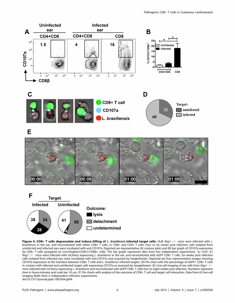
Figure 4. CD8+ T cells degranulate and induce killing of L. braziliensis infected target cells. (A,B) Rag12/2 mice were infected with L.braziliensis in the ear, and reconstituted with either CD8+ T cells, or CD8+ and CD4+ T cells. Four to six weeks post infection cells isolated fromuninfected and infected ears were incubated with anti-CD107a. Depicted are representative (A) contour plots and (B) bar graph of CD107a expressionby CD8+ T cells (pregated on Live/Singlets/CD45+/CD8b+ cells). The bar graph represents data from five independent experiments. *p,0.05. (C)Rag12/2 mice were infected with mCherry expressing L. braziliensis in the ear, and reconstituted with eGFP CD8+ T cells. Six weeks post infectioncells isolated from infected ears were incubated with anti-CD107a and acquired by ImageStream. Depicted are four representative images showingCD107a expression at the interface between CD8+ T cells and L. braziliensis infected targets. (D) Pie chart with the percentage of eGFP+ CD8+ T cellsin contact with infected and uninfected target cells expressing CD107a as assessed by ImageStream. (E) Live-cell imaging of ear cells from Rag12/2mice infected with mCherry expressing L. braziliensis and reconstituted with eGFP CD8+ T cells four to eight weeks post infection. Numbers representtime in hours:minutes and scale bar 10 mm. (F) Pie charts with analysis of the outcome of CD8+ T cell and target cell interaction. Data from 63 live-cellimaging fields from 6 independent infection experiments.doi:10.1371/journal.ppat.1003504.g004
Pathogenic CD8+ T Cells in Cutaneous Leishmanaisis
PLOS Pathogens | www.plospathogens.org 6 July 2013 | Volume 9 | Issue 7 | e1003504

defined. Here, by combining clinical data from patients with
results from our experimental L. braziliensis infection, we have
identified CD8+ T cell-mediated cytotoxicity as a major contrib-
utor to increased pathology in cutaneous leishmaniasis.
While CD4+ Th1 cells are critical for controlling Leishmania,
CD8+ T cells also play a protective role, since mice lacking CD8+T cells exhibit increased susceptibility to L. major [11,12,25].
Moreover, in several experimental vaccine models CD8+ T cells
contribute to immunity [26,27,28,29,30,31]. In contrast, we
found that depletion of CD8+ T cells in L. braziliensis infected
mice decreases inflammation. It is currently unclear why CD8+ T
cell depletion has different effects in L. major and L. braziliensis
infected mice. However, CD8+ T cell derived IFN-c blocks the
development of a Th2 response in L. major infected mice [12], and
the apparent lack of Th2 induction in L. braziliensis infection may
contribute to these differential effects [21,32]. Nevertheless, our
findings indicate that CD8+ T cells can promote increased
disease. Although this may seem paradoxical, our current results
suggest that a bifurcation in CD8+ T cell effector function
provides a potential explanation. Specifically, CD8+ T cells
provide protection by releasing IFN-c, thereby activating
macrophages to kill the parasites, and promoting a stronger
CD4+ Th1 response in response to L. major infections [12]. In
contrast, our current results with L. braziliensis suggest that
cytolytic function, rather than IFN-c production, promotes
increased pathology. These findings are consistent with our
own observations, as well as others, that in L. braziliensis patients
there is a direct correlation between granzyme expressing CD8+T cells in lesions and disease severity [15,33]. While we found
that perforin-expressing CD8+ T cells were required for this
pathology, we cannot exclude a role for NK cells. Indeed, NK
cells have been linked with pathology in mucocutaneous
leishmaniasis patients [17] and NK cells were present in the
lesions of L. braziliensis infected RAG+CD8 mice (data not
shown). Similarly, we cannot exclude a role for CD8 derived
chemokines in promoting infiltration of inflammatory cells into
these lesions [34]. Taken together, the human studies suggest that
CD8+ T cell cytolytic activity is pathologic, and using a mouse
model we are the first to show conclusively that indeed a
dysregulated CD8+ T cell response promotes perforin-dependent
immunopathology in cutaneous leishmaniasis. Therefore, defin-
ing mechanisms that control CD8+ T cell function, including
regulation by factors present within the cytokine milieu [35], will
be a crucial step in identifying therapeutic targets to treat
immunopathologic sequelae.
Figure 5. CD8+ T cell induced pathology requires specificity.Rag12/2 mice were infected with L. braziliensis in the ear, andreconstituted with either WT or OT1 CD8+ T cells. (A) Ear thickness wasevaluated at the site of infection. (B) Representative images ofleishmanial lesions at 6 weeks post infection. At 7 weeks post infectionmice were euthanized and shown are: (C) parasite burden in the lesions;(D) contour plots and (E) bar graph of CD107a expression by CD8+ Tcells (pregated on: Live/Singlets/CD45+/CD8b+ cells). Graphs arerepresentative data from one of two independent experiments (n = 3)with similar results. *p,0.05.doi:10.1371/journal.ppat.1003504.g005
Figure 6. Perforin is required for CD8+ T cell inducedpathology in L. braziliensis infection. Rag12/2 mice were infectedwith L. braziliensis in the ear, and reconstituted with either WT or Prf2/2 CD8+ T cells. (A) Ear thickness at the site of infection. (B)Representative images of leishmanial lesions at 5 weeks post infection.At 7 weeks post infection mice were euthanized and shown are: (C)parasite burden in the lesions; representative (D) contour plots and (E)bar graph of CD8+ T cell in the lesion (pregated on: Live/Singlets/CD45+/CD8b+ cells). Graphs are representative data from one of threeindependent experiments (n = 5). Rag12/2 mice were infected with L.braziliensis in the ear, and reconstituted with either WT, IFN-c2/2, or IL-172/2 CD8+ T cells. (F,H) Ear thickness at the site of infection; (G,I)Parasite burden in the lesions at 7 weeks post infection. Data from one(F,G) experiment or one representative experiment of two (H,I) (n = 5)are presented. *p,0.05.doi:10.1371/journal.ppat.1003504.g006
Pathogenic CD8+ T Cells in Cutaneous Leishmanaisis
PLOS Pathogens | www.plospathogens.org 7 July 2013 | Volume 9 | Issue 7 | e1003504

Surprisingly, the role of CTLs in cutaneous leishmaniasis has
not been extensively explored. Nevertheless, studies have shown
that CD8+ T cell lines or clones can be lytic for Leishmania infected
cells [17,34,36,37,38,39,40]. Here, we extend those studies and
show the ability of effector CD8+ T cells from infected mice to kill
naturally infected targets. We were able to visualize for the first
time CD8+ T cells lysing L. braziliensis infected targets and
characterize the interactions that preceded the lytic event. In this
regard, we found that CD8+ T cells bind target cells for an
average of 25 minutes before target cell lysis, similar to what is
seen for CD8+ T cell induced apoptosis of peptide pulsed B cells
[41]. This killing appeared to be specific, since OT1 T cells–which
cannot recognize leishmanial antigens–failed to degranulate or
induce pathology in L. braziliensis infected mice. Additionally, while
almost 40% of the interactions between CD8+ T cells and infected
cells resulted in target cell lysis, a much smaller fraction (,4%) of
interactions between CD8+ T cells and uninfected cells resulted in
apoptosis. Although the mechanisms promoting killing of unin-
fected targets was not explored, these targets may have internal-
ized leishmanial antigens or phagocytosed infected apoptotic cells,
resulting in cross presentation [42]. Alternatively, the killing could
be antigen-independent, as occurs when stressed cells express
ligands recognized by NKG2D on CD8+ T cells [43]. Finally, it
remains to be determined whether parasites are killed when the
target cell is killed. We were unable to determine the fate of the
parasites following killing of their host cell, primarily because
dying cells did not remain in the field of focus. However, since
parasite numbers were similar in Rag12/2 and RAG+CD8 mice,
it seems unlikely that the parasites are killed, although we cannot
disregard the possibility that in an immunocompetent mouse the
parasites might be killed. Consistent with this view, a previous
study with a CTL clone indicated that parasites survive after their
host cell is killed by CD8+ T cells [40].
CD8+ T cells are best known for their ability to protect against
viral infections by lysing virus-infected targets. However, a
pathologic role for CD8+ T cells has also been observed. For
example, virally induced myocarditis is prevented in mice deficient
in perforin [5]. Similarly, experimental cerebral malaria is
mediated by CD8 CTLs [6,7]. Perhaps most analogous to the
pathologic CTL responses that we observe in leishmaniasis are
data from experimental Trypanosoma cruzi infections in which
CD8+ T cell responses that have traditionally been considered
protective have more recently been linked to pathology [8,44].
Such a finding is again consistent with a bifurcation in CD8+ T
cell function in which IFN-c producing CD8+ T cells are
protective, while perforin expressing CD8+ T cells mediate
increased pathology in the heart [8]. Leishmania infection is a
good model to understand this duality of CD8+ T cell function in
vivo, since CD8+ T cells are protective in visceral leishmaniasis
[26,45,46,47], whereas we now understand that this is the opposite
for L. braziliensis. Understanding what mechanisms drive CD8+ T
cells to become pathogenic or protective is an important goal to
design new treatments and is now under investigation in our lab.
At present, it is unclear why CTLs induce pathology in cutaneous
leishmaniasis, since apoptosis is primarily thought to drive anti-
inflammatory responses [48]. However, apoptotic cells can
undergo secondary necrosis if not rapidly cleared by phagocytes.
In secondary necrosis the integrity of the plasma membrane is lost
and intracellular constituents of the cell are released, increasing the
inflammatory response [23], and providing positive feedback to
increase the cytolytic activity of CD8+ T cells [49,50].
In several clinical forms of leishmaniasis, parasites metastasize to
distant cutaneous sites. In mucosal leishmaniasis, metastatic lesions
develop in the nasopharyngeal region, which leads to substantial
morbidity [16], while in disseminated leishmaniasis individual
nodules develop at multiple sites in the skin [51,52]. It has been
suggested that an RNA virus present in some South American
strains of Leishmania enhances the immune response, potentially
promoting metastasis [53]. However, this is not a universal finding,
as parasite strains associated with metastatic disease do not all
contain this virus (the strain that we have used here does not
contain the virus). Nevertheless, the association of enhanced
immune responses with metastasis is consistent with our results,
and suggests that dysregulated immune responses play an essential
role in this process. Although the mechanistic basis for immune-
mediated metastasis is unclear, the immune system may simply be
responding to disseminated parasites, thereby inducing the
inflammatory response required for lesion development at distal
sites. Alternatively, the immune response itself may contribute to
parasite spread. One attractive possibility is that CTL killing of
infected cells enhances the release of parasites, allowing them to
metastasize more efficiently to distant skin sites.
The relative decrease in immunopathology in mice reconstitut-
ed with both CD4+ and CD8+ T cells suggests that CD4+ T cells
control the pathogenicity of CD8+ T cells. This may be, in part,
by controlling the number of CD8+ T cells or the number of
parasites within the lesion. However, we also found that CD8+ T
cells transferred in the absence of CD4+ T cells expressed
relatively higher levels of GrzB and IL-17, and less IFN-y, than did
CD8+ T cells co-transferred with CD4+ T cells. We predict that
this difference is due to the absence of regulatory T cells (Tregs)
since in tumor models Tregs control the cytotoxicity of CD8+ T
cells in a TGF-b dependent manner [41,54], and in leishmaniasis
both CD4+ Th1 cells and CD4+ T regulatory cells (Treg) dampen
the immune response [55,56,57]. While we have not yet
determined how CD4+ T cells control the pathologic CD8+ T
cell response, we believe that the ratio of CD4+ and CD8+ T cells
may be a critical determinant in disease outcome, since we found
that Rag12/2 mice developed disease even in the presence of
CD4+ T cells, as long as they are in relatively low numbers (data
not shown). This result is consistent with the observed change in
ratio of CD4+ and CD8+ T cells present in lesions as disease
progresses in human patients [15]. Thus, we suggest that an
analysis of the CD4:CD8 ratio in biopsies taken for diagnostic
purposes might be useful in predicting disease outcome.
In summary, our results define a new role for CD8+ T cells in
leishmaniasis. While CD8+ T cells have previously been thought
of as primarily protective, our results demonstrate that they can
mediate severe pathologic responses. This finding makes it
essential that more effort is directed at delineating the factors that
determine CD8+ T cell effector function during leishmaniasis,
since such information is critical in considering therapies or
vaccines that may impact CD8+ T cells. Moreover, our data
highlight the importance of evaluating CD8+ T cell effector
function in many infections where CD8+ T cells may be playing
dual protective and pathologic roles.
Materials and Methods
Ethics statementThis study was conducted according to the principles specified
in the Declaration of Helsinki and under local ethical guidelines
(Ethical Committee of the Maternidade Climerio de Oliveira,
Salvador, Bahia, Brazil; and the University of Pennsylvania
Institutional Review Board). This study was approved by the
Ethical Committee of the Federal University of Bahia (Salvador,
Bahia, Brazil)(010/10) and the University of Pennsylvania IRB
(Philadelphia, Pa)(813390). All patients provided written informed
Pathogenic CD8+ T Cells in Cutaneous Leishmanaisis
PLOS Pathogens | www.plospathogens.org 8 July 2013 | Volume 9 | Issue 7 | e1003504

consent for the collection of samples and subsequent analysis. This
study was carried out in strict accordance with the recommenda-
tions in the Guide for the Care and Use of Laboratory Animals of
the National Institutes of Health. The protocol was approved by
the Institutional Animal Care and Use Committee, University of
Pennsylvania Animal Welfare Assurance Number A3079-01.
PatientsAll cutaneous leishmaniasis patients were seen at the health post
in Corte de Pedra, Bahia, Brazil, which is a well-known area of L.
braziliensis transmission. The criteria for diagnosis were a clinical
picture characteristic of cutaneous leishmaniasis in conjunction
with parasite isolation or a positive delayed-type hypersensitivity
response to Leishmania antigen, plus histological features of
cutaneous leishmaniasis. In all cases, the immunological analysis
was performed before therapy. Biopsies were performed using a
4 mm punch, treated with Liberase (Roche) for 90 mins at 37uC/
5% CO2. Biopsies were dissociated and passed through a 50 mm
Medicon filter (BD phamingen). Peripheral blood mononuclear
cells were obtained from heparinized venous blood layered over a
Ficoll-Hypaque gradient (GE Healthcare), then washed and
resuspended in RPMI1640 and stained for flow cytometry as
described below.
Microarray-based expression profiling of human lesionsFor whole genome expression microarray, lesion biopsies
preserved in RNAlater (Qiagen) were homogenized using a
rotor-stator and RNA was isolated using the RNeasy Plus kit
(Qiagen). Biotin-labeled complementary RNA (cRNA) was gen-
erated using the Illumina TotalPrep RNA amplification kit
(Ambion). RNA and cRNA quality were assessed on a BioAna-
lyzer (Agilent). Illumina HumanHT-12 version 4 expression
beadchips were hybridized with cRNA from ten L. braziliensis
lesion biopsies and two biopses collected from uninfected donors.
Data was quantile normalized and differential expression analysis
was carried out using GenomeStudio v1.8 software (Illumina).
Genes were considered differentially regulated if expression
increased or decreased $3-fold with a diffscore of $13 or
#213 (equivalent to p#0.05). Data was deposited on the Gene
Expression Omnibus (GEO) database for public access (GSE#GSE43880). Heat map tools available on GenePattern [58] were
used to graphically display differentially regulated genes in
Figure 1.
MiceBALB/c and C57BL/6 mice (6 weeks old) were purchased from
NCI, and Rag12/2 (B6.12957-RAG1tm1Mom)(N14F12), Ifn-c2/2
(B6.129S7-Ifngtm1Ts)(N8+2F23), Prf12/2 (perforin)(C57BL/6-
Prf1tm1Sdz)(F?+52) were purchased from The Jackson Laboratory.
C57BL/6 IL17a2/2 mice (N9) were provided by Dr. Yoichiro
Iwakura (University of Tokyo, Japan) and OT1 (B6.129S7-
Rag1tm1MomTg(TcraTcrb)) mice were purchased from Taconic Farms
and mice expressing eGFP in all T cells were originally obtained
from Ulrich van Andrian (Harvard University). All mice were
maintained in a specific pathogen-free environment at the
University of Pennsylvania Animal Care Facilities.
ParasitesL. braziliensis parasites (strain MHOM/BR/01/BA788) (de-
Moura et al., 2005) were grown in Schneider’s insect medium
(GIBCO) supplemented with 20% heat-inactivated FBS, 2 mM
glutamine, 100 U/ml penicillin, and 100 mg/ml streptomycin.
Metacyclic enriched promastigotes were used for infection [59].
Transgenic parasites expressing both luciferase and mCherry were
generated by transfecting the parental L. braziliensis strain with
SwaI linearized pLucCherry, a modified version of pIR1SAT
encoding firefly luciferase and mCherry in the SmaI and BglII
sites, respectively. Transgenic parasites were selected by plating
transfected parasites on M199 agar supplemented with nourseo-
thricin (50 mg/ml; Sigma-Aldrich). Mice were infected with 105 L.
braziliensis in the right ear, and the course of lesion progression was
monitored weekly by measuring the diameter of ear induration
with digital calipers (Fisher Scientific).
AntibodiesMouse: anti-CD45.2 APC-AlexaFluor 750, anti-CD11b eF450,
anti-CD11c FITC, anti-F4/80 PE-Cy7, anti-CD3 eFluor 450,
anti-IFN-c PeCy7, anti-CD107a eFluor 660 (all from eBioscience).
Anti-CD4 APC-Cy7, anti-IL-17A PE and Ly6C PerCP-Cy5.5 (BD
Pharmingen), anti-CD8b PerCPCy5.5 (Biolegend) and anti-
granzyme B APC (Invitrogen). Anti-Ly6G APC (Biolegend).
Human: anti-CD3 APCCy7, anti-CD8a PeCy5.5 and anti-
perforin FITC (all from eBioscience). Anti-CD107a PE (BD
Pharmingen) and anti-granzyme B APC (Invitrogen). For in vivo
CD4 or CD8 depletion, mice received i.p. injections of 250 mg of
GK1.5 or 53-6.72 (BioXcell), respectively.
Cell purification and adoptive transferSplenocytes from C57BL/6, Prf12/2, Ifn-c2/2, Il17a2/2
and OT1 mice were collected, red blood cells lysed with ACK
lysing buffer (LONZA) and CD8+ T or CD4+ T cells were
purified using a magnetic bead separation kit (Miltenyi Biotec).
Three million CD8+ or CD4+ T cells were transferred alone or
together to Rag12/2 mice that were subsequently infected with L.
braziliensis. Mice reconstituted with CD8+ T cells alone received 4
injections of 250 mg of anti-CD4 within the first 2 weeks.
Ear preparationInfected and uninfected ears were harvested, the dorsal and
ventral layers of the ear separated, and the ears incubated in
RPMI (Gibco) with 250 mg/mL of Liberase (Roche) for 90 mins at
37uC/5% CO2. Following incubation, the enzyme reaction was
stopped using 1 mL of RPMI media containing 10% FBS. Ears
were dissociated using a cell strainer (40 mm, BD Pharmingen) and
an aliquot of the cell suspension was used for parasite titration.
Parasite titrationThe parasite burden in the ears was quantified as described
previously [12]. Briefly, the homogenate was serially diluted (1:10)
in 96-well plates and incubated at 26uC. The number of viable
parasites was calculated from the highest dilution at which
parasites were observed after 7 days. To determine if parasites
disseminate, footpad, opposite ear, and nose were cultured in
complete Schneider’s medium at 26uC and parasite growth was
evaluated after 7 days.
Flow cytometric analysisCell suspensions from mice were incubated with PMA (50 ng/
mL), ionomycin (500 ng/mL) and Brefeldin A (10 mg/mL) (all
from SIGMA) for cytokine and granzyme B intracellular staining.
For degranulation assays, cells were resuspended in 46106/mL
and incubated for 6 hours at 37uC/5% CO2 with anti-CD107a
and monensin (eBioscience). Before surface and intracellular
staining, cells were washed and stained for live/dead fixable aqua
dead cell stain kit (Molecular Probes), according to manufacturer
instructions. For human granzyme B, perforin and CD107a
Pathogenic CD8+ T Cells in Cutaneous Leishmanaisis
PLOS Pathogens | www.plospathogens.org 9 July 2013 | Volume 9 | Issue 7 | e1003504

expression, cells were incubated for 6 hours with anti-CD107a
antibody and Brefeldin A without stimulation followed by surface
and intracellular staining.
Image-stream flow cytometryRag12/2 mice were reconstituted with 36106 eGFP CD8+ T
cells and infected with 105 metacyclic enriched mCherry L.
braziliensis. Six weeks post infection ear tissue was dissociated and
cells were incubated with anti-CD107a fluorescent antibody for
1 hour. Total ear cell suspension was acquired on the Image-
Stream machine and analyzed using the IDEAS software (Amnis
Corporation).
Live cell imagingRag12/2 mice were reconstituted with 36106 eGFP CD8+ T
cells and infected with 105 metacyclic enriched mCherry L.
braziliensis. Five to eight weeks post infection ears were harvested
and 26106 cells used for imaging. Cells were maintained at 37uCat 5% CO2 on the stage of a fully enclosed microscope. Fields were
selected randomly where both mCherry+ cells and eGFP+ cells
could be detected. Images from multiple fields were acquired every
60 seconds on a Leica DMI 4000 inverted microscope equipped
with a Yokagawa spinning disk confocal head and a Hamamatsu
EMCCD 510 camera. eGFP+ and mCherry images were taken
sequentially with 488 and 561 nm laser excitation, respectively.
Image acquisition was controlled by MetaMorph Software.
Histological analysisAt seven weeks post infection ears were harvested and fixed in
10% formalin. Ears were embedded in paraffin and 5 mm sections
were cut and stained with hematoxylin and eosin. Histological
sections were blindly scored and the system was pre-defined as the
following: the lowest score (0) was defined by the absence of a
lesion; moderate disease was defined as a small lesion with a
localized cell infiltration ranging from minimal (1), moderate (2) to
intense (3); lesions showing ulceration and intense localized cell
infiltration were scored as 4; and the highest score (5) was defined
by severe ulceration, cell infiltration involving the whole ear as well
as cartilage destruction.
Statistical analysisStatistical analysis was performed with the Mann–Whitney test
(two-sided t-test) using Prism (GraphPad Software).
Supporting Information
Figure S1 CD8+ T cells induce a greater recruitment ofneutrophils in mice infected with L. braziliensis. C57BL/
6 and Rag12/2 mice were infected with L. braziliensis in the ear
and Rag12/2 mice were reconstituted with either CD8+ T cells
or CD8+ and CD4+ T cells or no T cells. At 7 weeks post infection
mice were euthanized and cell suspensions from infected ears and
contralateral ears (C57BL/6 and Rag12/2 only) were stained
directly ex vivo for inflammatory cell markers and shown are: (A)
total CD11b+ cells, (B) neutrophils, (C) macrophages, (D)
monocytes and (E) dendritic cells. Representative data from three
independent experiments (n = 5) with similar results are presented.
*p,0.05.
(TIF)
Video S1 CD8+ T cell moves on the surface of infectedtarget. Live-cell imaging of cells isolated from leishmanial lesions
in Rag12/2 mice infected with mCherry expressing L. braziliensis
and reconstituted with eGFP CD8+ T cells six weeks post
infection. Numbers represent time in hours:minutes:seconds.
(MOV)
Video S2 CD8+ T cell kills target cell and immediatelydetaches from target. Live-cell imaging of cells isolated from
leishmanial lesions in Rag12/2 mice infected with mCherry
expressing L. braziliensis and reconstituted with eGFP CD8+ T cells
four weeks post infection. Numbers represent time in hours:mi-
nutes:seconds.
(MOV)
Video S3 Infected cell is killed by CD8+ T cell and losesmembrane integrity. Live-cell imaging of cells isolated from
leishmanial lesions in Rag12/2 mice infected with mCherry
expressing L. braziliensis and reconstituted with eGFP CD8+ T cells
six weeks post infection. Numbers represent time in hours:minu-
tes:seconds.
(MOV)
Acknowledgments
The authors wish to thank Drs. Alan Sher, Dragana Jankovic, Dan Barber,
and Leslie King for critical comments on the manuscript; Ba Nguyen,
Ednaldo Lago, Carolina Pombo, and Drs. Adriano Queiroz, Luiz
Guimaraes, Sarah Passos and Morgan Reuter for assistance; and Drs.
David Artis, Yoichiro Iwakura, and Ulrich Von Andrian for reagents and
mice.
Author Contributions
Conceived and designed the experiments: FON LPC JWG DPB DSR
MRB MEW CIO PS. Performed the experiments: FON LPC JWG DPB
GR. Analyzed the data: FON DPB GR MHG MRB PS. Contributed
reagents/materials/analysis tools: DSR MRB MEW. Wrote the paper:
FON PS.
References
1. Jordan KA, Hunter CA (2010) Regulation of CD8+ T cell responses to infection
with parasitic protozoa. Experimental parasitology 126: 318–325.
2. Gunderson AJ, Mohammed J, Horvath FJ, Podolsky MA, Anderson CR, et al.
(2012) CD8(+) T Cells Mediate RAS-Induced Psoriasis-Like Skin Inflammation
through IFN-gamma. The Journal of investigative dermatology.
3. Ortega C, Fernandez AS, Carrillo JM, Romero P, Molina IJ, et al. (2009) IL-17-
producing CD8+ T lymphocytes from psoriasis skin plaques are cytotoxic
effector cells that secrete Th17-related cytokines. Journal of leukocyte biology 86:
435–443.
4. Res PC, Piskin G, de Boer OJ, van der Loos CM, Teeling P, et al. (2010)
Overrepresentation of IL-17A and IL-22 producing CD8 T cells in lesional
skin suggests their involvement in the pathogenesis of psoriasis. PloS one 5:
e14108.
5. Gebhard JR, Perry CM, Harkins S, Lane T, Mena I, et al. (1998) Coxsackievirus
B3-induced myocarditis: perforin exacerbates disease, but plays no detectable
role in virus clearance. The American journal of pathology 153: 417–428.
6. Haque A, Best SE, Unosson K, Amante FH, de Labastida F, et al. (2011)
Granzyme B expression by CD8+ T cells is required for the development of
experimental cerebral malaria. Journal of immunology 186: 6148–6156.
7. Nitcheu J, Bonduelle O, Combadiere C, Tefit M, Seilhean D, et al. (2003) Perforin-
dependent brain-infiltrating cytotoxic CD8+ T lymphocytes mediate experimental
cerebral malaria pathogenesis. Journal of immunology 170: 2221–2228.
8. Silverio JC, Pereira IR, Cipitelli Mda C, Vinagre NF, Rodrigues MM, et al.
(2012) CD8+ T-cells expressing interferon gamma or perforin play antagonistic
roles in heart injury in experimental Trypanosoma cruzi-elicited cardiomyop-
athy. PLoS pathogens 8: e1002645.
9. Kaye P, Scott P (2011) Leishmaniasis: complexity at the host-pathogen interface.
Nat Rev Microbiol 9: 604–615.
10. Sacks D, Noben-Trauth N (2002) The immunology of susceptibility and resistance
to Leishmania major in mice. Nature reviews Immunology 2: 845–858.
11. Belkaid Y, Von Stebut E, Mendez S, Lira R, Caler E, et al. (2002) CD8+ T cells
are required for primary immunity in C57BL/6 mice following low-dose,
Pathogenic CD8+ T Cells in Cutaneous Leishmanaisis
PLOS Pathogens | www.plospathogens.org 10 July 2013 | Volume 9 | Issue 7 | e1003504
intradermal challenge with Leishmania major. Journal of immunology 168:
3992–4000.
12. Uzonna JE, Joyce KL, Scott P (2004) Low dose Leishmania major promotes a
transient T helper cell type 2 response that is down-regulated by interferon
gamma-producing CD8+ T cells. The Journal of experimental medicine 199:
1559–1566.
13. Novoa R, Bacellar O, Nascimento M, Cardoso TM, Ramasawmy R, et al.
(2011) IL-17 and Regulatory Cytokines (IL-10 and IL-27) in L. braziliensis
Infection. Parasite immunology 33: 132–136.
14. Faria DR, Gollob KJ, Barbosa J, Jr., Schriefer A, Machado PR, et al. (2005)
Decreased in situ expression of interleukin-10 receptor is correlated with the
exacerbated inflammatory and cytotoxic responses observed in mucosal
leishmaniasis. Infection and immunity 73: 7853–7859.
15. Faria DR, Souza PE, Duraes FV, Carvalho EM, Gollob KJ, et al. (2009)
Recruitment of CD8(+) T cells expressing granzyme A is associated with lesion
progression in human cutaneous leishmaniasis. Parasite immunology 31: 432–
439.
16. de Oliveira CI, Brodskyn CI (2012) The immunobiology of Leishmania
braziliensis infection. Frontiers in immunology 3: 145.
17. Brodskyn CI, Barral A, Boaventura V, Carvalho E, Barral-Netto M (1997)
Parasite-driven in vitro human lymphocyte cytotoxicity against autologous
infected macrophages from mucosal leishmaniasis. Journal of immunology 159:
4467–4473.
18. Smyth MJ, Kelly JM, Sutton VR, Davis JE, Browne KA, et al. (2001) Unlocking
the secrets of cytotoxic granule proteins. Journal of leukocyte biology 70: 18–29.
19. Hung JH, Yang TH, Hu Z, Weng Z, DeLisi C (2012) Gene set enrichment
analysis: performance evaluation and usage guidelines. Briefings in bioinfor-
matics 13: 281–291.
20. Betts MR, Brenchley JM, Price DA, De Rosa SC, Douek DC, et al. (2003)
Sensitive and viable identification of antigen-specific CD8+ T cells by a flow
cytometric assay for degranulation. Journal of immunological methods 281: 65–
78.
21. de Moura TR, Novais FO, Oliveira F, Clarencio J, Noronha A, et al. (2005)
Toward a novel experimental model of infection to study American cutaneous
leishmaniasis caused by Leishmania braziliensis. Infection and immunity 73:
5827–5834.
22. Soong L, Chang CH, Sun J, Longley BJ, Jr., Ruddle NH, et al. (1997) Role of
CD4+ T cells in pathogenesis associated with Leishmania amazonensis infection.
J Immunol 158: 5374–5383.
23. Kono H, Rock KL (2008) How dying cells alert the immune system to danger.
Nature reviews Immunology 8: 279–289.
24. Desjeux P (2004) Leishmaniasis: current situation and new perspectives.
Comparative immunology, microbiology and infectious diseases 27: 305–318.
25. Muller I, Pedrazzini T, Kropf P, Louis J, Milon G (1991) Establishment of
resistance to Leishmania major infection in susceptible BALB/c mice requires
parasite-specific CD8+ T cells. International immunology 3: 587–597.
26. Stager S, Rafati S (2012) CD8(+) T cells in leishmania infections: friends or foes?
Front Immunol 3: 5.
27. Gurunathan S, Sacks DL, Brown DR, Reiner SL, Charest H, et al. (1997)
Vaccination with DNA encoding the immunodominant LACK parasite antigen
confers protective immunity to mice infected with Leishmania major. J Exp Med
186: 1137–1147.
28. Maroof A, Brown N, Smith B, Hodgkinson MR, Maxwell A, et al. (2012)
Therapeutic vaccination with recombinant adenovirus reduces splenic parasite
burden in experimental visceral leishmaniasis. The Journal of infectious diseases
205: 853–863.
29. Jayakumar A, Castilho TM, Park E, Goldsmith-Pestana K, Blackwell JM, et al.
(2011) TLR1/2 activation during heterologous prime-boost vaccination (DNA-
MVA) enhances CD8+ T Cell responses providing protection against
Leishmania (Viannia). PLoS neglected tropical diseases 5: e1204.
30. Kronenberg K, Brosch S, Butsch F, Tada Y, Shibagaki N, et al. (2010)
Vaccination with TAT-antigen fusion protein induces protective, CD8(+) T cell-
mediated immunity against Leishmania major. The Journal of investigative
dermatology 130: 2602–2610.
31. Sanchez-Sampedro L, Gomez CE, Mejias-Perez E, Sorzano CO, Esteban M
(2012) High quality long-term CD4+ and CD8+ effector memory populations
stimulated by DNA-LACK/MVA-LACK regimen in Leishmania major BALB/
c model of infection. PloS one 7: e38859.
32. Rocha FJ, Schleicher U, Mattner J, Alber G, Bogdan C (2007) Cytokines,
signaling pathways, and effector molecules required for the control of
Leishmania (Viannia) braziliensis in mice. Infection and immunity 75: 3823–
3832.
33. Silva C, Boaventura VS, Cardoso CR, Tavares NM, Lordelo MJ, et al. (2013)
CD8+ Granzyme B+ T cells-mediated tissue injury versus CD4+ IFN-g+ T cells-
mediated parasite killing in human cutaneous leishmaniasis. Journal of
Investigative Dermatology In press.
34. Tsagozis P, Karagouni E, Dotsika E (2003) CD8(+) T cells with parasite-specific
cytotoxic activity and a Tc1 profile of cytokine and chemokine secretion developin experimental visceral leishmaniasis. Parasite immunology 25: 569–579.
35. Soudja SM, Ruiz AL, Marie JC, Lauvau G (2012) Inflammatory Monocytes
Activate Memory CD8(+) T and Innate NK Lymphocytes Independent ofCognate Antigen during Microbial Pathogen Invasion. Immunity 37: 549–562.
36. Barral-Netto M, Barral A, Brodskyn C, Carvalho EM, Reed SG (1995)Cytotoxicity in human mucosal and cutaneous leishmaniasis. Parasite Immunol
17: 21–28.
37. Kima PE, Ruddle NH, McMahon-Pratt D (1997) Presentation via the class Ipathway by Leishmania amazonensis-infected macrophages of an endogenous
leishmanial antigen to CD8+ T cells. Journal of immunology 159: 1828–1834.38. Pinelli E, Gonzalo RM, Boog CJ, Rutten VP, Gebhard D, et al. (1995)
Leishmania infantum-specific T cell lines derived from asymptomatic dogs thatlyse infected macrophages in a major histocompatibility complex-restricted
manner. European journal of immunology 25: 1594–1600.
39. Russo DM, Chakrabarti P, Higgins AY (1999) Leishmania: naive human T cellssensitized with promastigote antigen and IL-12 develop into potent Th1 and
CD8(+) cytotoxic effectors. Experimental parasitology 93: 161–170.40. Smith LE, Rodrigues M, Russell DG (1991) The interaction between CD8+
cytotoxic T cells and Leishmania-infected macrophages. The Journal of
experimental medicine 174: 499–505.41. Mempel TR, Pittet MJ, Khazaie K, Weninger W, Weissleder R, et al. (2006)
Regulatory T cells reversibly suppress cytotoxic T cell function independent ofeffector differentiation. Immunity 25: 129–141.
42. Winau F, Weber S, Sad S, de Diego J, Hoops SL, et al. (2006) Apoptotic vesiclescrossprime CD8 T cells and protect against tuberculosis. Immunity 24: 105–117.
43. Gonzalez S, Lopez-Soto A, Suarez-Alvarez B, Lopez-Vazquez A, Lopez-Larrea
C (2008) NKG2D ligands: key targets of the immune response. Trends inimmunology 29: 397–403.
44. Tarleton RL, Koller BH, Latour A, Postan M (1992) Susceptibility of beta 2-microglobulin-deficient mice to Trypanosoma cruzi infection. Nature 356: 338–
340.
45. Polley R, Stager S, Prickett S, Maroof A, Zubairi S, et al. (2006) Adoptiveimmunotherapy against experimental visceral leishmaniasis with CD8+ T cells
requires the presence of cognate antigen. Infection and immunity 74: 773–776.46. Kaye PM, Cooke A, Lund T, Wattie M, Blackwell JM (1992) Altered course of
visceral leishmaniasis in mice expressing transgenic I-E molecules. Europeanjournal of immunology 22: 357–364.
47. Stern JJ, Oca MJ, Rubin BY, Anderson SL, Murray HW (1988) Role of L3T4+and LyT-2+ cells in experimental visceral leishmaniasis. Journal of immunology140: 3971–3977.
48. Savill J, Dransfield I, Gregory C, Haslett C (2002) A blast from the past:clearance of apoptotic cells regulates immune responses. Nature reviews
Immunology 2: 965–975.
49. Bonilla WV, Frohlich A, Senn K, Kallert S, Fernandez M, et al. (2012) Thealarmin interleukin-33 drives protective antiviral CD8(+) T cell responses.
Science 335: 984–989.50. Shi Y, Evans JE, Rock KL (2003) Molecular identification of a danger signal that
alerts the immune system to dying cells. Nature 425: 516–521.51. David CV, Craft N (2009) Cutaneous and mucocutaneous leishmaniasis.
Dermatologic therapy 22: 491–502.
52. Carvalho LP, Passos S, Bacellar O, Lessa M, Almeida RP, et al. (2007)Differential immune regulation of activated T cells between cutaneous and
mucosal leishmaniasis as a model for pathogenesis. Parasite immunology 29:251–258.
53. Ives A, Ronet C, Prevel F, Ruzzante G, Fuertes-Marraco S, et al. (2011)
Leishmania RNA virus controls the severity of mucocutaneous leishmaniasis.Science 331: 775–778.
54. Chen ML, Pittet MJ, Gorelik L, Flavell RA, Weissleder R, et al. (2005)Regulatory T cells suppress tumor-specific CD8 T cell cytotoxicity through
TGF-beta signals in vivo. Proceedings of the National Academy of Sciences of
the United States of America 102: 419–424.55. Belkaid Y, Piccirillo CA, Mendez S, Shevach EM, Sacks DL (2002)
CD4+CD25+ regulatory T cells control Leishmania major persistence andimmunity. Nature 420: 502–507.
5 6 . A n d e r s o n C F , O u k k a M , K u c h r o o V J , S a c k s D ( 2 0 0 7 )CD4(+)CD25(2)Foxp3(2) Th1 cells are the source of IL-10-mediated immune
suppression in chronic cutaneous leishmaniasis. The Journal of experimental
medicine 204: 285–297.57. Falcao SC, de Moura TR, Clarencio J, Brodskyn C, Barral A, et al. (2012) The
presence of Tregs does not preclude immunity to reinfection with Leishmaniabraziliensis. International journal for parasitology 42: 771–780.
58. Reich M, Liefeld T, Gould J, Lerner J, Tamayo P, et al. (2006) GenePattern 2.0.
Nature genetics 38: 500–501.59. Spath GF, Beverley SM (2001) A lipophosphoglycan-independent method for
isolation of infective Leishmania metacyclic promastigotes by density gradientcentrifugation. Experimental parasitology 99: 97–103.
Pathogenic CD8+ T Cells in Cutaneous Leishmanaisis
PLOS Pathogens | www.plospathogens.org 11 July 2013 | Volume 9 | Issue 7 | e1003504