Current Trends in Otolaryngology and Rhinology
9
1 Volume 2018; Issue 01 Current Trends in Otolaryngology and Rhinology Review Article Catalano P and Walker J. Curr Trends Otolaryngol Rhinol: CTOR-121. Understanding Nasal Breathing: The Key to Evaluating and Treating Sleep Disordered Breathing in Adults and Children Peter Catalano, MD 1* , John Walker, DMD 2 1 Department of Otolaryngology, St. Elizabeth’s Medical Center, Tufts University School of Medicine Medical, MA, USA 2 Department of Orthodontics, Boston University School of Medicine, Boston, MA, USA * Corresponding author: Peter Catalano, MD, Department of Otolaryngology, St. Elizabeth’s Medical Center, 736 Cambridge Street, SMC-8, Brighton, MA 02134, USA. Tel:+16177796440; Fax: +16177895088; Email: [email protected] Citation: Catalano P, Walker J (2018) Understanding Nasal Breathing: The Key to Evaluating and Treating Sleep Disordered Breath- ing in Adults and Children. Curr Trends Otolaryngol Rhinol: CTOR-121. DOI: 10.29011/ CTOR-121. 000021 Received Date: 11 May, 2018; Accepted Date: 22 June, 2018; Published Date: 29 June, 2018 Abstract Nasal breathing is a basic and critical function that we rarely think much about, and most would be quite surprised to learn how important and influential it is in growth and development, and a healthy productive life. Fundamentally speaking, nasal breathing provides us with a sense of smell, enhances oxygen absorption by our lungs (via nitric oxide production from the sinuses [1]), warms and humidifies the air we breathe before it reaches the lower airway, and helps filter impurities from the air. However, it is also responsible for proper craniofacial development [2], tempero-mandibular joint function [3], head posture [4] and more. When nasal breathing does not occur for any reason, the body is forced into a series of compromises that prioritize getting oxygen into our blood at the expense of the functions provided by the nasal breathing. Because oxygen is required for survival, we are forced to live with a host of acquired health issues as a consequence of chronic or intermittent mouth breathing. One such con- sequence, known as Sleep Disordered Breathing [5] (SDB), is becoming pandemic in children, and directly affects their growth, development, intellect, academic performance, behavior, and much more. Let’s begin with a quick review of the causes of nasal obstruction. DOI: 10.29011/ CTOR-121. 000021 The Common Etiologies of Nasal Obstruction Nasal breathing is an extremely important function, and we have come to appreciate that many health problems develop when nasal breathing is compromised. The etiology of reduced nasal airflow is limited, and includes the following 5 major categories. Anatomic deformities, such as a septal deviation, alar collapse, turbinate hypertrophy, or the presence of concha bullosa are relatively common. These problems are typically genetic, but the first two can also be the result of nasal trauma. Atopy can cause swelling of the nasal submucosa and mucosal glands, and thus limit airflow. The majority of glands are located in the nasal turbinates and septal swell body, and are a major cause of their hypertrophy [6]. Atopy can be treated with various medications, however, while topical nasal steroids are the preferred treatment, a paradox arises when nasal congestion actually minimizes the access of topical medications to the nose, this markedly decreasing their effectiveness. A nasal mass or nasal polyps can also cause nasal obstruction, with symptoms that can include congestion, bloody discharge, facial pain or pressure, headache, and hyposmia. These growths can occur in children and adults, be benign or malignant, and have the best prognosis when diagnosed and treated early. Adenoid hypertrophy is very common in children and can cause severe nasal obstruction. The adenoids typically shrink by age > 4. Adenoids, like tonsils, are part of our immune system and serve little purpose after age 2 [7]. In addition to nasal obstruction, enlarged adenoids are implicated in the development of chronic serous otitis media and chronic sinusitis in children [8]. Vasomotor Rhinitis (VMR) is perhaps the most challenging cause of nasal obstruction and is due to and imbalance in the autonomic nervous system which controls nasal blood flow. This leads to intermittent vascular engorgement on one side or in one part of the nose, and is occasionally non-remitting. Improved breathing may occur sporadically during the day, and symptoms are almost always worse after lying down, especially on one’s side. As with most conditions, there are varying degrees of VMR. These patients often describe alternating nasal congestion, especially when supine. It is also possible to have several of these factors present simultaneously (i.e. anatomic and atopic, or atopy and polyps).
Transcript of Current Trends in Otolaryngology and Rhinology
1 Volume 2018; Issue 01
Current Trends in Otolaryngology and Rhinology Review Article
Catalano P and Walker J. Curr Trends Otolaryngol Rhinol: CTOR-121.
Understanding Nasal Breathing: The Key to Evaluating and Treating Sleep Disordered Breathing in Adults and ChildrenPeter Catalano, MD1*, John Walker, DMD2
1Department of Otolaryngology, St. Elizabeth’s Medical Center, Tufts University School of Medicine Medical, MA, USA2Department of Orthodontics, Boston University School of Medicine, Boston, MA, USA
*Corresponding author: Peter Catalano, MD, Department of Otolaryngology, St. Elizabeth’s Medical Center, 736 Cambridge Street, SMC-8, Brighton, MA 02134, USA. Tel:+16177796440; Fax: +16177895088; Email: [email protected]
Citation: Catalano P, Walker J (2018) Understanding Nasal Breathing: The Key to Evaluating and Treating Sleep Disordered Breath-ing in Adults and Children. Curr Trends Otolaryngol Rhinol: CTOR-121. DOI: 10.29011/ CTOR-121. 000021
Received Date: 11 May, 2018; Accepted Date: 22 June, 2018; Published Date: 29 June, 2018
AbstractNasal breathing is a basic and critical function that we rarely think much about, and most would be quite surprised to learn
how important and influential it is in growth and development, and a healthy productive life. Fundamentally speaking, nasal breathing provides us with a sense of smell, enhances oxygen absorption by our lungs (via nitric oxide production from the sinuses [1]), warms and humidifies the air we breathe before it reaches the lower airway, and helps filter impurities from the air. However, it is also responsible for proper craniofacial development [2], tempero-mandibular joint function [3], head posture [4] and more. When nasal breathing does not occur for any reason, the body is forced into a series of compromises that prioritize getting oxygen into our blood at the expense of the functions provided by the nasal breathing. Because oxygen is required for survival, we are forced to live with a host of acquired health issues as a consequence of chronic or intermittent mouth breathing. One such con-sequence, known as Sleep Disordered Breathing [5] (SDB), is becoming pandemic in children, and directly affects their growth, development, intellect, academic performance, behavior, and much more. Let’s begin with a quick review of the causes of nasal obstruction.
DOI: 10.29011/ CTOR-121. 000021
The Common Etiologies of Nasal ObstructionNasal breathing is an extremely important function, and we
have come to appreciate that many health problems develop when nasal breathing is compromised. The etiology of reduced nasal airflow is limited, and includes the following 5 major categories. Anatomic deformities, such as a septal deviation, alar collapse, turbinate hypertrophy, or the presence of concha bullosa are relatively common. These problems are typically genetic, but the first two can also be the result of nasal trauma. Atopy can cause swelling of the nasal submucosa and mucosal glands, and thus limit airflow. The majority of glands are located in the nasal turbinates and septal swell body, and are a major cause of their hypertrophy [6]. Atopy can be treated with various medications, however, while topical nasal steroids are the preferred treatment, a paradox arises when nasal congestion actually minimizes the access of topical medications to the nose, this markedly decreasing their effectiveness. A nasal mass or nasal polyps can also cause nasal obstruction, with symptoms that can include congestion, bloody discharge, facial pain or pressure, headache, and hyposmia.
These growths can occur in children and adults, be benign or malignant, and have the best prognosis when diagnosed and treated early. Adenoid hypertrophy is very common in children and can cause severe nasal obstruction. The adenoids typically shrink by age > 4. Adenoids, like tonsils, are part of our immune system and serve little purpose after age 2 [7]. In addition to nasal obstruction, enlarged adenoids are implicated in the development of chronic serous otitis media and chronic sinusitis in children [8]. Vasomotor Rhinitis (VMR) is perhaps the most challenging cause of nasal obstruction and is due to and imbalance in the autonomic nervous system which controls nasal blood flow. This leads to intermittent vascular engorgement on one side or in one part of the nose, and is occasionally non-remitting. Improved breathing may occur sporadically during the day, and symptoms are almost always worse after lying down, especially on one’s side. As with most conditions, there are varying degrees of VMR. These patients often describe alternating nasal congestion, especially when supine. It is also possible to have several of these factors present simultaneously (i.e. anatomic and atopic, or atopy and polyps).

Citation: Catalano P, Walker J (2018) Understanding Nasal Breathing: The Key to Evaluating and Treating Sleep Disordered Breathing in Adults and Children. Curr Trends Otolaryngol Rhinol: CTOR-121. DOI: 10.29011/ CTOR-121. 000021
2 Volume 2018; Issue 01
Symptoms Differ in Adults Vs ChildrenThe most common etiology of nasal obstruction differs
between adults and children. For the former they are, in order of decreasing frequency, anatomic deformities, allergy, VMR, a nasal mass (adenoid remnants are extremely rare); whereas in the latter the order would be adenoid hypertrophy, anatomic deformities, allergy, VMR, and a nasal mass. Regardless of the cause(s), nasal obstruction in adults and children leads to major physiologic changes that can have serious health consequences. In adults, a common example of the consequence of nasal obstruction is snoring and Obstructive Sleep Apnea (OSA). The most common symptom of OSA in adults is daytime fatigue and cognitive impairment, followed by a significantly increased risk for heart disease, high blood pressure, stroke, diabetes, memory loss, and reduced libido [9]. Bruxism and TMJ dysfunction are now also linked to nasal obstruction in sleep [3]. In children, OSA and Sleep Disordered Breathing (SDB) leads to very different signs and symptoms that are very common, yet often overlooked or misunderstood. These include mouth breathing, snoring, restless sleep, frequent awakenings, enuresis, lack of focus, head-forward posturing, disinterest in eating, Attention Deficit Disorder (ADD), Hyperactivity (ADHD), poor school performance, anger, and frank aggression [10]. Headaches may also be a significant complaint. Teens can show more of the daytime fatigue seen in adults.
A key point to remember is that not all symptoms are present in each patient and the exact constellation of symptoms varies widely among patients. For patients with OSA and SDB, more sleep is not helpful because it is during sleep when their heart, brain and other organs are stressed due to oxygen debt. They are also in what is called “sympathetic dystrophy”, where their body is subject to continuous release of adrenaline during sleep. The repetitive adrenaline surge (often called arousals on a polysomnogram) has very different physiologic effects in adults versus children. More on this to follow. CPAP (continuous positive airway pressure) machines, worn on the face or nose, attempt to force oxygen past the nasal obstruction to provide adequate oxygen during sleep. (Figure 1) However, CPAP tolerance in generally quite poor, with large sample data showing few patients using it more than 4 hours a night [11].
Figure 1: CPAP devices in use.
Another very important point is that many patients with OSA or SDB are unaware of their nasal congestion. The two most common reasons for this are that in many patients, nasal obstruction is positional and only occurs once the patient is lying down for sleep. It takes anywhere from 30 to 90 minutes for the nasal congestion to develop when supine, and by then the patient is often already asleep and unaware of the problem. The second reason is the variability in subjective appreciation or self-awareness of nasal compromise. Many of these patients have had nasal obstruction since early childhood, thus they have no frame of reference for normal nasal breathing. It is for this very reason that we test children for hearing and visual loss in grade school. Children have no frame of reference for normal hearing or sight.
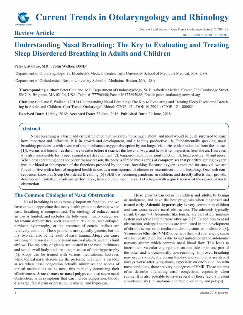
Likewise, they have no frame of reference for normal nasal breathing, and unfortunately, there is no standard screening test for them at this time. Another common compensation for nasal obstruction is forward head posturing [10]. In order to mouth breath better, the oral airway needs to be aligned with the trachea, this would produce a chin up, extended head position. (Figure 2) Many children will sleep in an extended head position for this
Citation: Catalano P, Walker J (2018) Understanding Nasal Breathing: The Key to Evaluating and Treating Sleep Disordered Breathing in Adults and Children. Curr Trends Otolaryngol Rhinol: CTOR-121. DOI: 10.29011/ CTOR-121. 000021
3 Volume 2018; Issue 01
reason. (Figure 3) However, during daytime, we cannot see where we are going in this position. Therefore, we must lean our extended neck forward. This forward head posturing places significant stress on the posterior neck muscles and leads to cervical spine alignment. Poor cervical spine alignment can lead to nerve root compression and mal-alignment of the lower spine. Thus, once the first domino falls, it is uncertain which will be the last domino to fall- i.e. will head forward posturing affect cervical spine position alone, or will it result in lower back problems, gait issues, neck pain, etc. The bottom line is that many children who suffer from SDB go undiagnosed. Awareness for the signs and symptoms as described herein should alert teachers, parents, and physicians to the possibility of SDB in children.
Figure 2: Physical changes in mouth breathing children.
Figure 3: Mouth breather with extended neck posture.
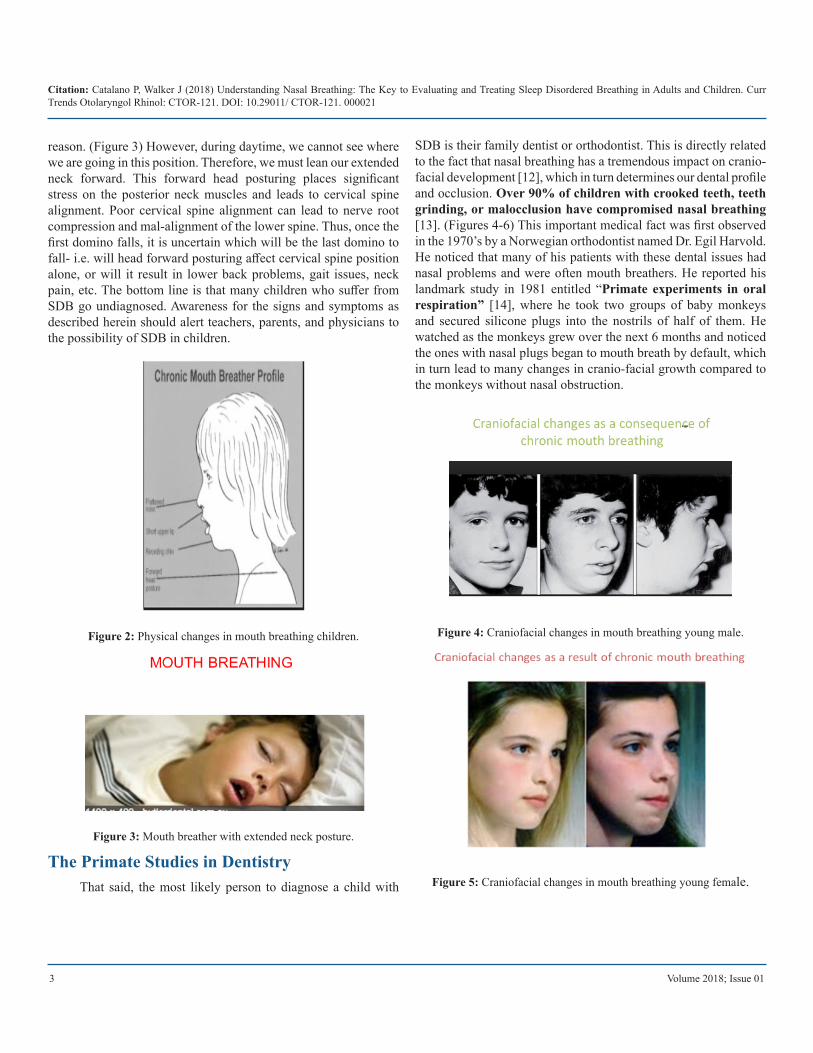
The Primate Studies in DentistryThat said, the most likely person to diagnose a child with
SDB is their family dentist or orthodontist. This is directly related to the fact that nasal breathing has a tremendous impact on cranio-facial development [12], which in turn determines our dental profile and occlusion. Over 90% of children with crooked teeth, teeth grinding, or malocclusion have compromised nasal breathing [13]. (Figures 4-6) This important medical fact was first observed in the 1970’s by a Norwegian orthodontist named Dr. Egil Harvold. He noticed that many of his patients with these dental issues had nasal problems and were often mouth breathers. He reported his landmark study in 1981 entitled “Primate experiments in oral respiration” [14], where he took two groups of baby monkeys and secured silicone plugs into the nostrils of half of them. He watched as the monkeys grew over the next 6 months and noticed the ones with nasal plugs began to mouth breath by default, which in turn lead to many changes in cranio-facial growth compared to the monkeys without nasal obstruction.
Figure 4: Craniofacial changes in mouth breathing young male.
Figure 5: Craniofacial changes in mouth breathing young female.

Citation: Catalano P, Walker J (2018) Understanding Nasal Breathing: The Key to Evaluating and Treating Sleep Disordered Breathing in Adults and Children. Curr Trends Otolaryngol Rhinol: CTOR-121. DOI: 10.29011/ CTOR-121. 000021
4 Volume 2018; Issue 01
Figure 6: Craniofacial and dental changes in young female.
He reported the narrow maxilla, high arched palate, elongated and downwards rotation of the mandible, and varying degrees of malocclusion were hallmarks of nasal obstruction in children. Not only were skeletal changes being observed, but compensatory changes by the masticator muscles were causing lower jaw malposition. The affected monkeys subsequently developed narrow faces with elongated mandibles, known today as “adenoid facies”. (Figure 7) This finding is the physical hallmark of nasal obstruction in young children, and unfortunately, is an end-stage situation. Interestingly, Harvold then removed the nasal plugs from the monkeys who had them, and noted that many of the occlusion and jaw issues resolved over the next 6 months, therefore proving that restoration of nasal breathing can lead to reversal of these changes.
Figure 7: Young rhesus monkey with “adenoid facies” after nasal blockage.
As a result of compromised nasal breathing, the narrow maxilla cannot accommodate the full complement of upper teeth and a crowded dental pattern develops. (Figure 8) It is important to note that if nasal obstruction is not corrected after the teeth have been repositioned, the once straightened teeth will likely shift again because the forces causing the shift has never been corrected. We frequently see adults and teens requiring a second, and sometimes third set of braces for this exact reason. In the past, orthodontists would extract teeth to “un-crowd” the maxilla and use braces to realign the remaining teeth. Historically, orthodontics has been the science of restorative aesthetic dentistry, whereas now it is far more focused on functional upper airway development. Thus, it is also important to diagnose and treat children at an early age because 60% of their facial growth occurs by age 4, 70% by age 5, and 90% by age 12 [15].
Figure 8: Narrow and high vs normal palatal arch.
The old philosophy of waiting until a child is a teen or pre-teen to address their crooked teeth is no longer appropriate. Dental extractions to un-crowd the maxilla are now largely abandoned in place of what is called palatal expansion [16]. By temporarily attaching an oral device onto the inner table of the alveolar ridge of the maxilla, the orthodontist can perform osteo-distraction, whereby they slowly move the palatal bones apart over a 6-12-month period. (Figure 9) There is no pain as the process is slow and gentle. Palatal expanders can actually bear the child’s initials or name and have become an oral fashion statement for kids. Palatal expansion ultimately causes widening of the constricted maxilla and will allow proper alignment of all teeth using braces without requiring extractions.

Citation: Catalano P, Walker J (2018) Understanding Nasal Breathing: The Key to Evaluating and Treating Sleep Disordered Breathing in Adults and Children. Curr Trends Otolaryngol Rhinol: CTOR-121. DOI: 10.29011/ CTOR-121. 000021
5 Volume 2018; Issue 01
Figure 9: Palatal expanders.
Palatal ExpansionIt was recently reported by Dr. Melih Motro, a researcher at
Boston University’s Goldman School of Dentistry, that for every 1 mm of lateral palatal expansion, the nasal airway increases by 2.4% [17]. This is an extremely important discovery because it shows us how a simple minimally invasive office treatment can begin to correct the craniofacial abnormalities that develop from reduced nasal breathing. Since the palatal bones in children are not yet fused in the midline, it is relatively easy to obtain expansion of 4-10 mm in a relatively short time. Unfortunately, this is not as easy in adults because the palatal suture has already fused. As a result of this study, palatal expansion in children with SDB and dental mal-alignment is now standard practice as the first step in modern orthodontic care [18]. Wearing head-frames or other dental devices designed to correct these orthodontic problems are actually counter-productive and place restrictive forces on a maxilla that is developing in an anterior and inferior direction [18].
ADD/ADHD and Behavioral Changes in Children
It has also been shown with increasing evidence, that approximately 40% of children who suffer from SDB develop ADD, ADHD, and/or a learning disability [19]. Statistically, if a child snores by the age of 8 and is untreated, there is an 80% chance the child will have a permanent 20% reduction in mental capacity [20]. Additionally, if a child is diagnosed with SDB in the first 5 years of life and untreated, they are 60% more likely
to require special needs education by age 8 [21]. However, the clinicians diagnosing these children and offering treatment for ADD/ADHD rarely consider nasal obstruction as a cause, and are also unaware that the condition is likely reversible without the need for medications or psychological therapy. ADD and ADHD are the direct result of the concept of sympathetic dystrophy I introduced earlier. Sleep deprived children develop hyperactive behavior, and this phenomenon is witnessed in most young children as their bedtime approaches. Exposing a child to hours of adrenaline over the course of a night leads to restless sleep at night and hours of hyperactive behavior the following day. It is also responsible for their enuresis, frequent awakenings, etc. [22]. In adults, sympathetic dystrophy leads to hypertension, heart disease, stroke, and diabetes. The fatigue and cognitive issues are due to hours of oxygen debt during poor sleep. Note the vastly different phenotypes of SDB and OSA in children versus adults.
Our group is currently investigating the reversibility of ADD/ADHD in these children. Early results show a significant ability to reverse the condition once normal nasal breathing is established. While the results of this study are not complete, we are extremely encouraged by the early data and the significant number of children whose behavior has been positively affected by reversing mouth breathing tendencies and providing quality sleep. We are currently evaluating the data with respect to the child’s age, gender, and severity of baseline ADD/ADHD to determine if there are any differences in outcomes with respect to these parameters. The ability to reverse or reduce the effects of ADD/ADHD in children is a major breakthrough with profound quality of life, social, and family implications. We have even seen children with mild autism, anxiety, or aggression become highly functioning students to the astonishment of their parents and teachers.
The Role of Tonsils & AdenoidsThus far, we have described the major causes of compromised
nasal breathing, the different phenotypes in children versus adults, and have introduced some treatment options. In the 1980’s it was discovered that hypertrophic tonsils and adenoids were responsible for SDB and OSA, and at that time, it was considered the primary etiology [23]. Hence, many children underwent T&A with the goal of correcting their SDB. Did we finally have a cure for SDB in children? (Figure 10) The answer is yes and no! Studies looking at large groups of children with SDB who underwent corrective T&A have shown that about 67% relapse with SDB > 2 years of surgery [24]. This is a very important point that was revealed after longitudinal study of affected children, and shows the importance of long-term follow-up.
Citation: Catalano P, Walker J (2018) Understanding Nasal Breathing: The Key to Evaluating and Treating Sleep Disordered Breathing in Adults and Children. Curr Trends Otolaryngol Rhinol: CTOR-121. DOI: 10.29011/ CTOR-121. 000021
6 Volume 2018; Issue 01
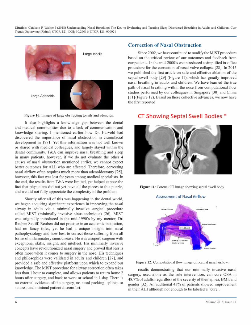
Figure 10: Images of large obstructing tonsils and adenoids.
It also highlights a knowledge gap between the dental and medical communities due to a lack of communication and knowledge sharing. I mentioned earlier how Dr. Harvold had discovered the importance of nasal obstruction in craniofacial development in 1981. Yet this information was not well known or shared with medical colleagues, and largely stayed within the dental community. T&A can improve nasal breathing and sleep in many patients, however, if we do not evaluate the other 4 causes of nasal obstruction mentioned earlier, we cannot expect better outcomes for ALL who are affected. Therefore, correcting nasal airflow often requires much more than adenoidectomy [25], however, this fact was lost for years among medical specialists. In the end, the results from T&A were limited, yet helped expose the fact that physicians did not yet have all the pieces to this puzzle, and we did not fully appreciate the complexity of the problem.
Shortly after all of this was happening in the dental world, we began acquiring significant experience in improving the nasal airway in adults via a minimally invasive surgical procedure called MIST (minimally invasive sinus technique) [26]. MIST was originally introduced in the mid-1990’s by my mentor, Dr. Reuben Setliff. Reuben did not practice in an academic institution, had no fancy titles, yet he had a unique insight into nasal pathophysiology and how best to correct those suffering from all forms of inflammatory sinus disease. He was a superb surgeon with exceptional skills, insight, and intellect. His minimally invasive concepts have revolutionized nasal surgery and proved that less is often more when it comes to surgery in the nose. His techniques and philosophies were validated in adults and children [27], and provided a safe and effective platform upon which to expand our knowledge. The MIST procedure for airway correction often takes less than 1 hour to complete, and allows patients to return home 2 hours after surgery, and back to work or school in 1 day. There is no external evidence of the surgery, no nasal packing, splints, or sutures, and minimal patient discomfort.
Correction of Nasal ObstructionSince 2002, we have continued to modify the MIST procedure
based on the critical review of our outcomes and feedback from our patients. In the mid-2000’s we introduced a simplified in-office procedure for the correction of nasal valve collapse [28]. In 2015 we published the first article on safe and effective ablation of the septal swell body [29] (Figure 11), which has greatly improved nasal breathing in adults and children. We have learned the true path of nasal breathing within the nose from computational flow studies performed by our colleagues in Singapore [30] and China [31] (Figure 12). Based on these collective advances, we now have the first reported
Figure 11: Coronal CT image showing septal swell body.
Figure 12: Computational flow image of normal nasal airflow.
results demonstrating that our minimally invasive nasal surgery, used alone as the sole intervention, can cure OSA in 48.7% of adults, regardless of the severity of their apnea, BMI, and gender [32]. An additional 43% of patients showed improvement in their AHI although not enough to be labeled a “cure”.

Citation: Catalano P, Walker J (2018) Understanding Nasal Breathing: The Key to Evaluating and Treating Sleep Disordered Breathing in Adults and Children. Curr Trends Otolaryngol Rhinol: CTOR-121. DOI: 10.29011/ CTOR-121. 000021
7 Volume 2018; Issue 01
Previously, nasal surgery alone had been shown ineffective in correcting OSA with cure rates < 17%, and was primarily used to improve one’s tolerance of CPAP [33]. This has led sleep surgeons to pursue more aggressive and morbid procedures such as palatoplasty (UPPP), jaw advancement surgery, hyoid suspension, and robotic excision of the base of the tongue (known as TORS). These options are far from the minimally invasive goals of contemporary surgical practice, and proving unnecessary. Even the hypoglossal nerve stimulator, an expensive implant designed to stimulate selective portions of the hypoglossal nerve during sleep to prevent hypopharyngeal obstruction, has success rates approaching 50% [34].
The latter surgery takes about 3-5 hours to complete, prevents the use of MR imaging in the future, and is far more morbid and costly than MIST. Almost all the patients who were not cured after MIST had absolute or relative macroglossia, due to either a large tongue or small oral cavity. Our solution to tongue base prolapse is a device called Linguaflex. Linguaflex is an elastic tongue implant that is easily placed into the tongue in about 3-5 minutes using a straight needle [35]. Patients are unaware it is present once placed and it does not interfere with their speech or swallowing. It is adjustable, and easily reversible should the patient want it removed. This device is currently before the FDA awaiting 510K approval, and our data from clinical trials done in SE Asia show OSA cure rates from Linguaflex alone of over 70%. Combined with MIST, we believe we will have the ultimate minimally invasive options to cure OSA, SDB, and snoring in the majority of adult patients.
New Data Supports the ConceptSince 2015, we have applied these minimally invasive
surgical principles to children with OSA and SDB, and continue to receive tremendous feedback from the children and their parents. With the use of a 3-dimensional CT imaging program (Figure 13), we have begun to measure the actual airway changes responsible for the subjective improvement in these children. We have found that both minimal throat area (2 dimensional measurement) and volume (3 dimensional space) have both increased an average of 42%, with every child studied showing improvement [36]. We have also just completed another study measuring the volume changes in the nasal airway that occur after our modified MIST procedure, and the average child saw a 46% improvement in nasal airway volume [37], which translates into a 3-4-fold increase in nasal airflow!
Figure 13: 3-D CT images can measure pharyngeal airway.
These findings were predictable based on the Starling Resistor Model of airway collapse and also validate the concept. (Figure 14) This model states that when one reduces nasal/upstream airway resistance, you will simultaneously reduce collapse of the pharyngeal airway by reducing negative intraluminal pressure. These ground-breaking results demonstrate that we can now accurately diagnose children with SDB, identify the cause of their compromised nasal breathing, appreciate the breath and variation in their symptoms, and implement an effective treatment strategy that combines palatal expansion with minimally invasive nasal surgery, and T&A in selective individuals. Linguaflex will be a welcome addition to the adult population.
Figure 14: Starling Resistor Model of Airway collapse.

Citation: Catalano P, Walker J (2018) Understanding Nasal Breathing: The Key to Evaluating and Treating Sleep Disordered Breathing in Adults and Children. Curr Trends Otolaryngol Rhinol: CTOR-121. DOI: 10.29011/ CTOR-121. 000021
8 Volume 2018; Issue 01
The last item to mention is VMR, which heretofore has been almost impossible to correct medically or surgically. However, in mid-2017, a company called Arrinex developed and received FDA approval for a new device called Clarifix [38]. Using a cryo-probe strategically placed into the nose, the nerves responsible for autonomic control of nasal blood flow and secretions are literally frozen trans-mucosally in a 30 second application per side. The procedure can be performed in the office using local anesthesia in adults, and in the OR in children. Results are typically seen within 3-6 weeks and 2-year follow-up data shows over 90% success rates. Combined with MIST, we can now treat all causes of nasal obstruction in adults and children in a minimally invasive and effective manner.
More Than HopeThis “perfect storm” of understanding the science,
appreciating patient variability, creating and adopting technical innovations, and fostering peer-to-peer learning between disciplines, has given us the unique ability to confront and treat the pandemic that is OSA and SDB, and improve the quality of life for most who suffer from them. Our work is just beginning, but we are confident we have “broken the code” on a condition that afflicts millions of people worldwide, and is responsible for a huge health care burden for society. Today there is more than hope.
ReferencesJankowski R, Nguyen, DT, Poussel M, Chenuel B, Gallet P, et al. 1. (2016) Sinusology, European annals of otorhinolaryngology. head and neck diseases 133: 263-268.
Torre C, Guilleminault C (2018) Establishment of nasal breathing 2. should be the ultimate goal to secure adequate craniofacial and airway development in children. J Pediatr (Rio J) 94: 101-103.
Tay DKL, Pang KP (2018) Clinical phenotype of South-East Asian tem-3. poromandibular disorder patients with upper airway resistance syn-drome. Journal of oral rehabilitation 45: 25-33.
Bharadwaj R, Ravikumar A, Krishnaswamy NR (2011) Evaluation of 4. craniofacial morphology in patients with obstructive sleep apnea us-ing lateral cephalometry and dynamic MRI. Indian Journal of Dental Research 22: 739-748.
Tolaymat A, Liu Z (2017) Sleep Disorders in Childhood Neurological 5. Diseases. Children 4: 84.
Wexler D, Braverman I, Amar M (2006) Histology of the nasal septal 6. swell body (septal turbinate). Otolaryngology-Head and Neck Surgery 134: 596-600.
Ciolek PJ, Xu A, Anne S, Geelan-Hansen K (2017) Role of adenoidec-7. tomy in chronic nasal obstruction after nasal steroid therapy failure. American journal of otolaryngology 38: 305-308.
Davcheva-Chakar M, Kaftandzhieva A, Zafirovska B (2015) Adenoid 8. Vegetations–Reservoir of Bacteria for Chronic Otitis Media with Effu-sion and Chronic Rhinosinusitis. prilozi 36: 71-76.
Campos-Juanatey F, Fernandez-Barriales M, Gonzalez M, Portillo-9. Martin JA (2017) Effects of obstructive sleep apnea and its treatment over the erectile function: a systematic review. Asian journal of androl-ogy 19: 303.
Hvolby A (2015) Associations of sleep disturbance with ADHD: impli-10. cations for treatment. ADHD Attention Deficit and Hyperactivity Disor-ders 7: 1-18.Hawkins SM, Jensen EL, Simon SL, Friedman NR (2016) Correlates of 11. pediatric CPAP adherence. Journal of clinical sleep medicine: JCSM: official publication of the American Academy of Sleep Medicine 12: 879-884.Muñoz ICL, Orta PB (2014) Comparison of cephalometric patterns in 12. mouth breathing and nose breathing children. International journal of pediatric otorhinolaryngology 78: 1167-1172.Šidlauskienė M, Smailienė D, Lopatienė K, Čekanauskas E, Pribuišienė 13. R, et al. (2015) Relationships between malocclusion, body posture, and nasopharyngeal pathology in pre-orthodontic children. Medical science monitor: international medical journal of experimental and clinical research 21: 1765-1773.Harvold EP, Vargervik K, Chierici G (1973) Primate experiments on 14. oral sensation and dental malocclusions. American journal of ortho-dontics 63: 494-508.Ant A, Kemaloglu YK, Yilmaz M, Dilci A (2017) Craniofacial Deviations 15. in the Children with Nasal Obstruction. Journal of Craniofacial Surgery 28: 625-628.
Shah A, Shah P, Goje SK, Shah R, Modi B (2017) Palatal Expansion 16. and its Effects in Orthodontics. Advanced Journal of Graduate Re-search 2: 31-36.
Motro M, Schauseil M, Ludwig B, Zorkun B, Mainusch S, et al. (2016) 17. Rapid-maxillary-expansion induced rhinological effects: a retrospec-tive multicenter study. European Archives of Oto-Rhino-Laryngology 273: 679-687.
Bellerive A, Montpetit A, El-Khatib H, Carra MC, Remise C, et al. 18. (2015) The effect of rapid palatal expansion on sleep bruxism in chil-dren. Sleep and Breathing 19: 1265-1271.
Goyal A, Pakhare AP, Bhatt GC, Choudhary B, Patil R (2018) Associa-19. tion of pediatric obstructive sleep apnea with poor academic perfor-mance: A school-based study from India. Lung India 35: 132-136.
Owens JA (2009) Neurocognitive and behavioral impact of sleep disor-20. dered breathing in children. Pediatric pulmonology 44: 417-422.
Smith DL, Gozal D, Hunter SJ, Kheirandish-Gozal L (2017) Parent-21. reported behavioral and psychiatric problems mediate the relationship between sleep-disordered breathing and cognitive deficits in school-aged children. Frontiers in neurology 11: 410.
Zaffanello M, Piacentini G, Lippi G, Fanos V, Gasperi E, et al. (2017) Ob-22. structive sleepdisordered breathing, enuresis and combined disorders in children: chance or related association. Swiss Med Wkly 147: 14400.
Laurikainen E, Aitasalo K, Erkinjuntti M, Wanne O (1992) Sleep apnea 23. syndrome in children-secondary to adenotonsillar hypertrophy? Acta Otolaryngol Suppl 492: 38-41.
Huang YS, Guilleminault C, Lee LA, Lin CH, Hwang FM (2014) Treat-24. ment outcomes of adenotonsillectomy for children with obstructive sleep apnea: a prospective longitudinal study. Sleep 37: 71-76.

Citation: Catalano P, Walker J (2018) Understanding Nasal Breathing: The Key to Evaluating and Treating Sleep Disordered Breathing in Adults and Children. Curr Trends Otolaryngol Rhinol: CTOR-121. DOI: 10.29011/ CTOR-121. 000021
9 Volume 2018; Issue 01
Thadikonda KM, Shaffer AD, Stapleton AL (2018) Outcomes of ade-25. noidectomy-alone in patients less than 3-years old. International Jour-nal of Pediatric Otorhinolaryngology 106:46-49.
Catalano PJ, Gupta RC, Warman M, Wijewickrama RC (2014) Sinus 26. Surgical Techniques from Caldwell-Luc to MIST. In Diseases of the Sinuses: 389-410.
Setliff III RC (1996) Min Imally in Vasive Sin Us Surgery. Otolaryngo-27. logic Clinics of North America 29: 115.
Dolan RW, Catalano PJ, Innis W, Wanees E (2009) In-office surgical 28. repair of nasal valve stenosis. Am J Rhinol Allergy 23: 111-114.
Catalano P, Ashmead MG, Carlson D (2015) Radiofrequency ablation 29. of septal swell body. Ann Otolaryngol Rhinol 2: 1069.
Wang de Y, Lee HP, Gordon BR (2012) Impacts of fluid dynamics 30. simulation in study of nasal airflow physiology and pathophysiology in realistic human three-dimensional nose models. Clinical and experi-mental otorhinolaryngology 5: 181-187.
Tan J, Han D, Wang J, Liu T, Wang T, et al. (2012) Numerical simula-31. tion of normal nasal cavity airflow in Chinese adult: a computational flow dynamics model. European archives of oto-rhino-laryngology 269: 881-889.
Peter Catalano Curative role of MIST in OSA (Publishing in prog-32. ress).
Poirier J, George C, Rotenberg B (2014) The effect of nasal surgery 33. on nasal continuous positive airway pressure compliance. The Laryn-goscope 124: 317-319.
Woodson BT, Strohl KP, Soose RJ, Gillespie MB, Maurer JT, et 34. al. (2018) Upper Airway Stimulation for Obstructive Sleep Ap-nea: 5-Year Outcomes. Otolaryngology-Head and Neck Surgery: 0194599818762383.
Catalano PJ (2014). U.S. Patent Application No. 14/055,159.35.
Alsufyani NA, Noga ML, Witmans M, Cheng I, El-Hakim H, et al. 36. (2017) Using cone beam CT to assess the upper airway after surgery in children with sleep disordered breathing symptoms and maxillary-mandibular disproportions: a clinical pilot. Journal of Otolaryngology-Head & Neck Surgery 46: 31.
Catalano P, Schlewet M Nasal airway changes after nasal and sinus 37. surgery in children with sleep disordered breathing (Publishing in prog-ress).
ClinicalTrials.gov Identifier: NCT03181594.38.