Current clinical applications recent developmentsechocardiography-course.com/media/pdf/saturday/S 7...
127
3D echocardiography Current clinical applications & recent developments Folkert Meijboom,
Transcript of Current clinical applications recent developmentsechocardiography-course.com/media/pdf/saturday/S 7...

3D echocardiographyCurrent clinical applications
&recent developments
Folkert Meijboom,

Why 3D echo?
• Poor spatial and temporal resolution comparedwith 2D

Why 3D echo?
• To obtain spatial information in one data set • Volumes
• LV• RV• atria
• complex anatomy –spatial relation of structures• Mitral valve• Any other structure

Why 3D echo?
• To obtain spatial information in one data set • Volumes
• LV• RV• atria
• complex anatomy – spatial relation of structures• Mitral valve• Any other structure
• To make cross-section impossible to make with 2D• En-face view of valves, septums etc
Why 3D echo?
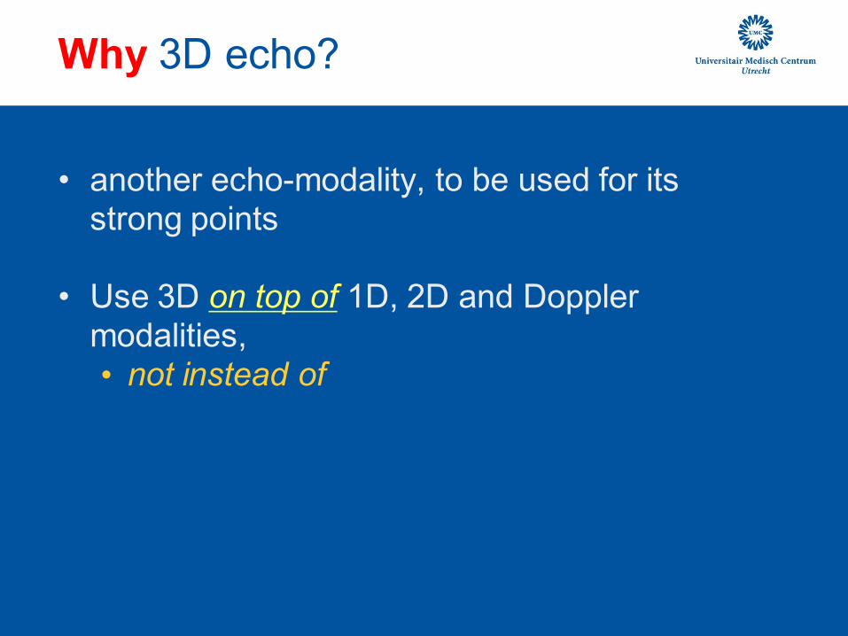
• another echo-modality, to be used for itsstrong points
• Use 3D on top of 1D, 2D and Dopplermodalities,• not instead of

3D echo in clinical practice
• Current applications • LV function• RV function• Morphology• 3D TEE
• Morphology – MV• Guiding interventions
• 3D & teaching

LV function

LV function
• Echo is the work horse of cardiacimaging
• In virtually all echo`s LV function is assessed
• M-mode: FS• 2D: EF - planimetry (Simpson)• 3D: EF - volumetrics

Reliability of M-mode and 2D bi-plane Simpson
• Good representation of LV function• Spherical shape of LV
• Normal septal motion
• In absence of regional wall motion abnormalities

Paradoxical septal movement
•FS 10% does not represent very poor LV function

Diastolic flattening of IVS = volume overload RV
•diastolic “D-sign”

Systolic D-sign = pressure overload RV

Reliability of M-mode and 2D planimetry
• Good representation of LV function• Spherical shape of LV• Normal septal motion• In absence of regional wall motion
abnormalities

Reliability of M-mode and 2D planimetry
• Good representation of LV function• Spherical shape of LV• Normal septal motion• In absence of regional wall motion
abnormalities
• Often not the case in patienst we are interested in• Regional wall motion abnormalities after MI• RV pathology in CHD patients

How good is echo in assessmentof LV function?

echo (M-Mode, 2D & 3D) – MRI -CT (volume in ml, EF in %)
•EDV •ESV •EF•Sugeng et al. Circulation, 2006. 114(7): p. 654-61.

Inter & intra-observer variability LVF
•Sugeng et al. Circulation, 2006. 114(7): p. 654-61.

RT 3D echo & LV function
• RT 3D echo • more reproducible (better correlation with
MRI)• more robust (less intra & inter-observer
variablity)than M-mode and 2D

RT 3D echo & LV function
• RT 3D echo • more reproducible (better correlation with
MRI)• more robust (less intra & inter-observer
variablity)than M-mode and 2D
• RT3D echo feasible in clinical practice?• How is it done in clinical practice?
• How much time does it take?

RT3D echo & Global LV function

RT3D echo & LV function
• Is it feasible in clinical practice?
• For which patients?

3D matrix transducer

RT 3-D data acquisition
Full matrix array transducer modules1. Real-time 3D, no `breath hold`. narrow
sector (60° x 30°)
2. `zoom mode` enlargement of subsegment of 3D data pyramid (30°x 30°sector)
3. Full volume = 90° x 90°(110° x 110°) pyramide volume

RT 3-D data acquisition
Full matrix array transducer modules1. Real-time 3D, no `breath hold`. narrow
sector (60° x 30°)
2. `zoom mode` enlargement of subsegment of 3D data pyramid (30°x 30°sector)
3. Full volume = 90° x 90°(110° x 110°) pyramide volume
Off-line analysis necessary

Full volume acquisition
• 4-7 subsequent subvolumes combined to a 90° x 90° pyramid volume
• ECG triggerd on R wave
• `breath-hold`
• Aquisition time: 5- 8 secs
• No actual real time

Full-volume 3D data set from the apex
•Limitations ultrasound 2D: also true for 3D

RT3D Analysis of LV volumes

RT3D Analysis of LV volumes

RT3D Analysis of LV volumes
•Adjustment of orthogonal axis

RT3D Analysis of LV volumes

RT3D Analysis of LV volumes

RT3D Analysis of LV volumes

RT3D Analysis of LV volumes

RT3D Analysis of LV volumes

• These anatomical landmarks are well defined• Straightforward• Easy to identify
• The endocardial border detection is automated• Based on algoritms
Little inter- and intra-observer variability

RT3D Analysis of LV volumes

RT3D Analysis of LV volumes

•< 5 min
RT3D Analysis of LV volumes

Conclusion: 3D echo & LVEF
• < 5 minutes acquisition & analysis:
• reproducible EF
• Better and faster than bi-plane Simpson
• More robust in your multi-echotec echo lab
Conclusion: 3D echo & LVEF
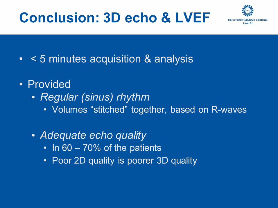
• < 5 minutes acquisition & analysis
• Provided• Regular (sinus) rhythm
• Volumes “stitched” together, based on R-waves
• Adequate echo quality• In 60 – 70% of the patients• Poor 2D quality is poorer 3D quality

Conclusion: 3D echo & LVEF
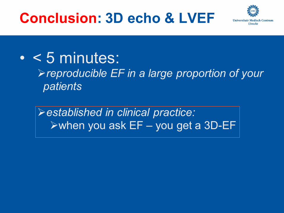
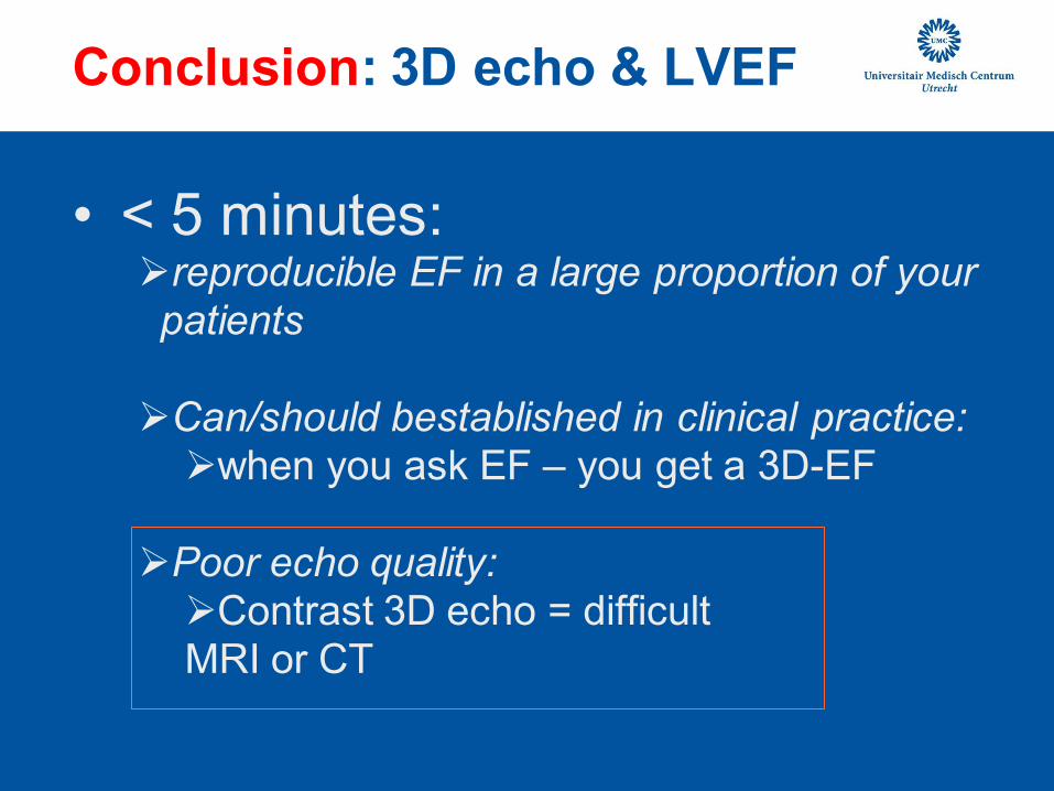
• < 5 minutes:reproducible EF in a large proportion of yourpatients
Conclusion: 3D echo & LVEF
• < 5 minutes:reproducible EF in a large proportion of yourpatients
established in clinical practice: when you ask EF – you get a 3D-EF
Conclusion: 3D echo & LVEF
• < 5 minutes:reproducible EF in a large proportion of yourpatients
Can/should bestablished in clinical practice: when you ask EF – you get a 3D-EF
Poor echo quality:Contrast 3D echo = difficultMRI or CT

Conclusion: 3D echo & LVEF
• < 5 minutes:reproducible EF in a large proportion of yourpatients
established in clinical practice: when you ask EF – you get a 3D-EF
Poor echo quality:Contrast 3D echo = difficultMRI or CT
If not feasible because of non-sr: ?

What is relatively new 3D & LV function?
• Regional LV volumes
• Parametric imaging
• Role regional volumes in CRT

Regional LV volumes

CRT; Regional volumes

•#17
•16 or 17 segments

•#17
•# Hundreds

“Parametric Imaging”imaging of a specific parameter in a 3D data set

“Parametric Imaging”imaging of a specific parameter in a 3D data set
Timing: CRT Excursion: ischaemia

Parametric Imagingparameter = timing

•Excursion PI = Representation of wall motion
Parametric Imagingparameter = excursion
(radial) Excursion PI
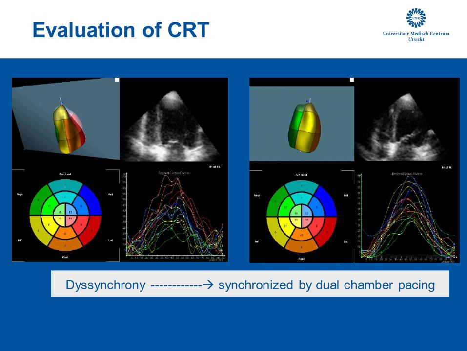
Evaluation of CRT
•LBBB •Biventricular pacingDyssynchrony ------------ synchronized by dual chamber pacing

3D echo & CRT
• Limited use so far
• Wall motion is assessed on basis of
endocardial border detetction
• No discrimination active – passive motion
• Not (necessarily) representative for
contraction/myocardial function

3D echo & CRT
• Limited use so far
• Wall motion is assessed on basis of
endocardial border detetction
• No discrimination active – passive motion
• Not (necessarily) representative for
contraction/myocardial function
Work in progress

What is new for RT3D echo & LV function?

What is new for RT3D echo & LV function?
• Philips has been market leader for manyyears
• Other vendors producing 3D paltforms• GE, Toshiba, Siemens/Acuson• Full-volume in a single heart beat• Actual real-time 3D for volumetrics
• 3D speckle tracking (Acuson)

Current method for full volume 3D
• Four or more R-R intervals required for full volume acquisition.
• This is not real time

Imaging
New Acuson (=Siemens) platform
Information Rate determines the image quality potential of an ultrasound system
Drives diagnostic confidence and speed of workflow
Novel Imaging Engine: 10 times the rate of other systems Up to 64 paralleled beams Up to 160 M voxels/sec
Enables: 90° x 90 ° volume at > 20 volumes/sec at depth of 16 cm 40° X 40° volume color flow at 20 volumes/sec at depth of 16cm

Full-volume acquisition in single heart beat
• Volume acquisition, 90x90 degree @ 20 vol/sec minimum
• Entire heart in one cardiac cycle rather than four cycles
• No stitching artifacts• Reduced impact of respiration • Reduced impact of
arrhythmia
• Real workflow improvements
• Broader clinical application
Imaging
* Not commercially available

Full-volume set from a single heart beat
• Acquisition is fast• Full-volume in on heart beat: currently trade-of with
• image resolution• sector width• Volume rate
• Solutions:• Flexi-volume (GE and Toshiba)• Much faster systems (Siemens)

Full-volume set from a single heart beat
• Sector width is important for encompassing the entire LV
• Resolution is not so important for volumetrics
• A step forward for volume measurements
• Also pts with a fib. or inabilty for breath-hold can have 3D LV-volumetrics

What else is new for the LV?

3D Speckle Tracking (3D ST)
• Cardiac motion is 3 dimensional, 2D Speckle Tracking(2DST) is limited because it cannot assess movement inthe 3rd dimension.
• The same technique used in 2DST can be applied to 3Ddata in time (4D) by tracking 3 dimensional cubictemplates through the cardiac cycle.

2D ST vs 3D ST:
In 3D ST: the speckle can be followed and analysed
In 2D ST: the speckle is moving out of the scanning plane: the scanning plane is analyzed

3D speckle:
• In 3DST, a speckle can actually be followed in 3D space• velocity, amplitude, direction: 3D vector• Unlimited number of vectors can be constructed
• The essence of myocardial function• Very complicated data• Difficult to visualize/analyse

3D speckle:
• In 3DST, a speckle can actually be followed in 3D space• velocity, amplitude, direction: 3D vector• Unlimited number of vectors can be constructed
• The essence of myocardial function• Very complicated data• Difficult to visualize/analyse
• The first “tries” in 3DST are sophisticated 2DST• Showing multiple 2D cross-section, derived from 3D
data set, throughout cardiac cycle

Insights in LV functionSystolic Radial strain: thickening
End systolic frame

Insights in LV functionSystolic Circumferential strain: shortening
End
systolic
frame

Insights in LV functionSystolic longitudinal strain: shortening
End
systolic
frame

•End systolic frame
Insights in LV functionRotation: (degrees°)

Insights in LV functionTwist (degrees °)
Twist = difference in rotation between segments

Insights in LV function torsion
Torsion (degrees °/cm)= Twist/Distanceof the C planes
•3DST provides relation between segments allowing calculation of the rotation value in 2 different SAX views in the same cardiac cycle and the distance between the
same two planes otherwise hard to do with 2DST.

RT3D echo & LV function: what is new?
• Increase in calculation power• Up to 10 x faster than existing systems
(containing 140 Pentium 4 processors stacked in the echo transducer)
• Full volume in a single heart beat• = actual real-time 3D
• 3-way trade-off between:volume, resolution and volume rate
• 3D speckle• Follow a speckle in space

What is new in 3D echo?
• RV volumes & EF

Assessment of RV function: 2D echo
•Courtesy dr Els
Pieper, Groningen

Assessment of RV function: 2D echo
•Jiang et al. Echocardiography 1997 Mar; 14(2): 189-206

Assessment of RV function: 2D echo
• Assessment of RV ejection fraction not possible with 2D echo MRI

Assessment of RV function: MRI
MRI is the current standard for assessment of RV volumes and function
Method of disk summation

Assessment of RV function: MRI

Assessment of RV function: MRI
End diastole End systole
•Limitations:
•- Availability
•- Costs
•- Time consuming acquisition and analysis
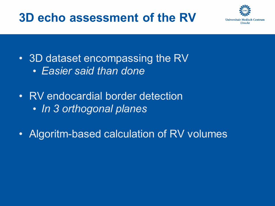
3D echo assessment of the RV
• 3D dataset encompassing the RV• Easier said than done
• RV endocardial border detection• In 3 orthogonal planes
• Algoritm-based calculation of RV volumes

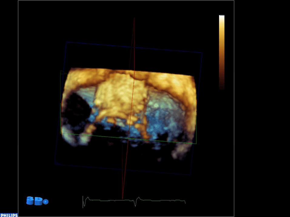
RT3DE: orthogonal views

RT3DE: acquisition dataset
• RV volumes and ejection fraction for the first time possible with echo!

3D & RV

Comparison between RT3DE and MRI
•Good correlations volumes & EF
•Heleen van der Zwaan et al,
JASE, in press

Results:
Reproducibility
Inter-
observer
Intra-
observer
RV EDV (ml) 5 ± 13 0 ± 10
RV ESV (ml) 10 ± 14 2 ± 8
RV EF (%) 9 ± 13 4 ± 9•* P < 0.001
0
5
10
15
20
25
Acquisition time Analysis time
RT3DE MRI
•* •*
•Heleen van der Zwaan et al,
JASE, in press

Results:
Reproducibility
Inter-
observer
Intra-
observer
RV EDV (ml) 5 ± 13 0 ± 10
RV ESV (ml) 10 ± 14 2 ± 8
RV EF (%) 9 ± 13 4 ± 9•* P < 0.001
0
5
10
15
20
25
Acquisition time Analysis time
RT3DE MRI
•* •*
•Heleen van der Zwaan et al,
JASE, in press

•Normal RV Tetralogy of Fallot

Conclusions
• 80% feasibility in patients with ConHD
• Good agreement between RT3DE and MRI; underestimation of volumes
• inter- en intra-observer values ± acceptable
• RV acquisition and analysis by RT3DE cost only few minutes

Conclusions
• 80% Feasibility in patients with ConHD
• Good agreement between RT3DE and MRI; underestimation of volumes
• inter- en intra-observer values ± acceptable
• RV acquisition and analysis by RT3DE cost only few minutes
Methodological differences RT3DE- MRI
Further research needed with the expectation that RT3DE will be clinically applicable

Analysis with same software:still differences
•Sugeng et al. JACC Cardiovasc imaging Jan 2010

RT3D echo: what is new?
• RT 3D TEE

Realtime TEE (X7-2t) matrix transducer:


•Mitral valve quatification-bookmark


3D TEE Aortic valve

3D TEE - pAVSD

3D TEE - pAVSD

RT 3D TEE: Conclusion
• Makes understanding of anatomy (MV) mucheasier
• Accessible for non-experts
• Unequivocal presentation of anatomy
• Contributes to the understanding of the MV, also for experts

3D echo & morphology
•Courtesy Heleen van der Zwaan

3D echo & morphology
• Current place of 3D echo for morpholgy:
• In selected patients
• For specific questions
• On top of 2D – not instead of
• Costs extra time
•Courtesy Heleen van der Zwaan

3D echo & morphology
• Current place of 3D echo for morpholgy:
• In selected patients
• For specific questions
• On top of 2D – not instead of
• Costs extra time
• Weigh – for every patient – whether the
extra time needed for RT3D echo will be
worth the effort. •Courtesy Heleen van der Zwaan

3D echo & morphology
• Current place of 3D echo for morpholgy:
• Not established yet
• No recommendations/guidelines
• 2 books will be out shortly:
• Buck& Monaghan
• Lang•Courtesy Heleen van der Zwaan

3D TTE: normal aortic valve

3D TTE: Bicuspid aortic valve

3D TTE – normal MV

3D TTE new generation

Full-volume set from a single heart beat
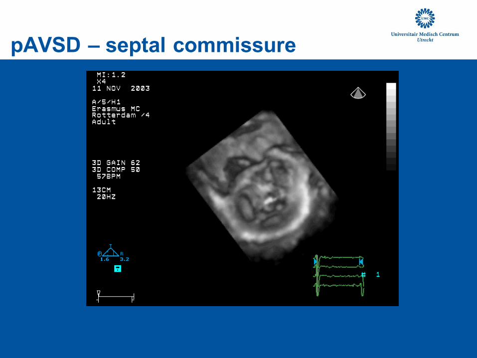
pAVSD – septal commissure

pAVSD – septal commissure

3D echo for morphology
• how to analyse the data?
• Dedicated software
• Every vendor its own system
• Do not communicate with each other
• Tomtec
• 3D data on a flat screen – missed opportunity

3D viewing of 3D data sets

I-space 3D viewing system

3D Personal Space System

3D viewing of 3D data sets
• Intuitive navigation through 3D data sets
• more accessible for non-(echo)experts
• Cardiac surgeons
• Interventional cardiologist
• Electro-physiologists


Creating surgical view of the heart
head toes

Mimicking atriotomy

Mimicking atrial septostomy

Conclusion 3D echo
• Ready for clinical practice for
• LV function – superior to 2D
• Intracardiac anatomy
• MV
• AoV
• TV
• Congenital HD
Additional information, on top of 2D echo, forselected patients

Conclusion 3D echo
• Work in progress
• RV function
• 3D full volume single heart beat
• 3D-speckle
• 3D display of 3D data

NHD, Willemstad, Curacao, 30 januari 2010
Thank you very much