Cultivation and Characterization of Cells from a...
17
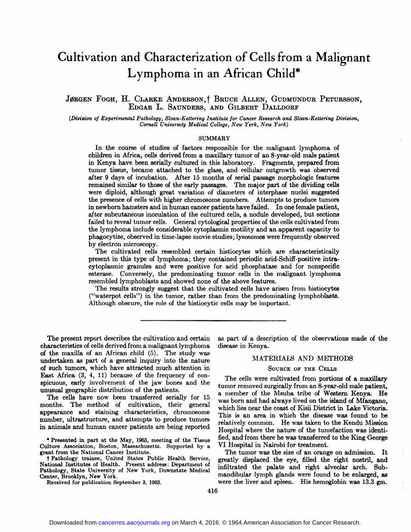
Cultivation and Characterization of Cells from a Malignant 0 Lymphoma in an African Child* J0RGEN FOGH, H. CLAEKE ANDERSON, f BRUCE ALLEN, GUDMUNDUR PETURSSON, EDGARL. SAUNDERS,AND GILBERT DALLDORF (Division of Experimental Pathology, Sloan-Kettering Institute for Cancer Research and Sloan-Kettering Division, Cornell University Medical College, New York, New York) SUMMARY In the course of studies of factors responsible for the malignant lymphoma of children in Africa, cells derived from a maxillary tumor of an 8-year-old male patient in Kenya have been serially cultured in this laboratory. Fragments, prepared from tumor tissue, became attached to the glass, and cellular outgrowth was observed after 9 days of incubation. After 15 months of serial passage morphologic features remained similar to those of the early passages. The major part of the dividing cells were diploid, although great variation of diameters of interphase nuclei suggested the presence of cells with higher chromosome numbers. Attempts to produce tumors in newborn hamsters and in human cancer patients have failed. In one female patient, after subcutaneous inoculation of the cultured cells, a nodule developed, but sections failed to reveal tumor cells. General cytological properties of the cells cultivated from the lymphoma include considerable cytoplasmic motility and an apparent capacity to phagocytize, observed in time-lapse movie studies; lysosomes were frequently observed by electron microscopy. The cultivated cells resembled certain histiocytes which are characteristically present in this type of lymphoma; they contained periodic acid-Schiff-positive intra- cytoplasmic granules and were positive for acid phosphatase and for nonspecific esterase. Conversely, the predominating tumor cells in the malignant lymphoma resembled lymphoblasts and showed none of the above features. The results strongly suggest that the cultivated cells have arisen from histiocytes ("waterpot cells") in the tumor, rather than from the predominating lymphoblasts. Although obscure, the role of the histiocytic cells may be important. The present report describes the cultivation and certain as part of a description of the observations made of the characteristics of cells derived from a malignant lymphoma disease in Kenya, of the maxilla of an African child (5). The study was undertaken as part of a general inquiry into the nature MATERIALS AND METHODS of such tumors, which have attracted much attention in SOURCEOF THE CELLS East Africa (3, 4, 11) because of the frequency of con- The ceUg ^ cultivated from rtions of a maxillary spicuous, early involvement of the jaw bones and the tumor removed ica% from an 8.year.old male patient, unusual geographic distribution of the patients & member rf th<j Mguba tribe rf Wegtem *; -^ The celte have now been transferred serially ior 15 wag bom and had &1 ]ived Qn the island rf Mf months. The method of cultivation, their general which lies near the coast of Kisii District in Lake Victoria, appearance and staining characteristics, chromosome Thig ig an are& ^ which ^ digease wag found to fce number, ultrastructure, and attempts to produce tumors re]ativel common, He was taken to the Kendu Mission m animals and human cancer patients are being reported Hospital where the nature rf ^ tumefaction was identi. * Presented in part at the May, 1963, meeting of the Tissue fied' and from there he was transferred to the King George Culture Association, Boston, Massachusetts. Supported by a VI Hospital in Nairobi for treatment, grant from the National Cancer Institute. The tumor was the size of an orange on admission. It t Pathology trainee, United States Public Health Service, greatly displaced the eye, filled the right nostril, and SÄÄ^^ infiit-tef r p,alaÃ-e rdrigf ar°lar ^ !ub- Center, Brooklyn, New York. mandibular lymph glands were iound to be enlarged, as Received for publication September 3, 1963. were the liver and spleen. His hemoglobin was 13.3 gm. 416 on March 4, 2016. © 1964 American Association for Cancer Research. cancerres.aacrjournals.org Downloaded from
Transcript of Cultivation and Characterization of Cells from a...
Cultivation and Characterization of Cells from a Malignant0Lymphoma in an African Child*
J0RGEN FOGH, H. CLAEKE ANDERSON, f BRUCE ALLEN, GUDMUNDUR PETURSSON,EDGARL. SAUNDERS,AND GILBERT DALLDORF
(Division of Experimental Pathology, Sloan-Kettering Institute for Cancer Research and Sloan-Kettering Division,Cornell University Medical College, New York, New York)
SUMMARY
In the course of studies of factors responsible for the malignant lymphoma ofchildren in Africa, cells derived from a maxillary tumor of an 8-year-old male patientin Kenya have been serially cultured in this laboratory. Fragments, prepared fromtumor tissue, became attached to the glass, and cellular outgrowth was observedafter 9 days of incubation. After 15 months of serial passage morphologic featuresremained similar to those of the early passages. The major part of the dividing cellswere diploid, although great variation of diameters of interphase nuclei suggestedthe presence of cells with higher chromosome numbers. Attempts to produce tumorsin newborn hamsters and in human cancer patients have failed. In one female patient,after subcutaneous inoculation of the cultured cells, a nodule developed, but sectionsfailed to reveal tumor cells. General cytological properties of the cells cultivated fromthe lymphoma include considerable cytoplasmic motility and an apparent capacity tophagocytize, observed in time-lapse movie studies; lysosomes were frequently observedby electron microscopy.
The cultivated cells resembled certain histiocytes which are characteristicallypresent in this type of lymphoma; they contained periodic acid-Schiff-positive intra-cytoplasmic granules and were positive for acid phosphatase and for nonspecificesterase. Conversely, the predominating tumor cells in the malignant lymphomaresembled lymphoblasts and showed none of the above features.
The results strongly suggest that the cultivated cells have arisen from histiocytes("waterpot cells") in the tumor, rather than from the predominating lymphoblasts.
Although obscure, the role of the histiocytic cells may be important.
The present report describes the cultivation and certain as part of a description of the observations made of thecharacteristics of cells derived from a malignant lymphoma disease in Kenya,of the maxilla of an African child (5). The study wasundertaken as part of a general inquiry into the nature MATERIALS AND METHODSof such tumors, which have attracted much attention in SOURCEOF THE CELLSEast Africa (3, 4, 11) because of the frequency of con- The ceUg ^ cultivated from rtions of a maxillaryspicuous, early involvement of the jaw bones and the tumor removed ica% from an 8.year.old male patient,unusual geographic distribution of the patients & member rf th<j Mguba tribe rf Wegtem *; -^
The celte have now been transferred serially ior 15 wag bom and had &1 ]ived Qn the island rf Mfmonths. The method of cultivation, their general which lies near the coast of Kisii District in Lake Victoria,appearance and staining characteristics, chromosome Thig ig an are& ^ which ^ digease wag found to fcenumber, ultrastructure, and attempts to produce tumors re]ativel common, He was taken to the Kendu Missionm animals and human cancer patients are being reported Hospital where the nature rf ^ tumefaction was identi.
* Presented in part at the May, 1963, meeting of the Tissue fied' and from there he was transferred to the King GeorgeCulture Association, Boston, Massachusetts. Supported by a VI Hospital in Nairobi for treatment,grant from the National Cancer Institute. The tumor was the size of an orange on admission. It
t Pathology trainee, United States Public Health Service, greatly displaced the eye, filled the right nostril, andSÄÄ^^ infiit-tefr p,alaÃerdrigfar°lar^ !ub-Center, Brooklyn, New York. mandibular lymph glands were iound to be enlarged, as
Received for publication September 3, 1963. were the liver and spleen. His hemoglobin was 13.3 gm.
416
on March 4, 2016. © 1964 American Association for Cancer Research. cancerres.aacrjournals.org Downloaded from

FoGH et al.—Cellsfrom a Malignant Lymphoma in African Child 417
per cent, white blood cells numbered 11,500, and platelets140,000 per cu. mm. His blood urea nitrogen was 32mg. per cent and alkaline phosphatase 8.8 Bodansky units.
The tumor was soft and friable and contained manyareas of necrosis. Histologie examination showed auniform structure of unorganized, large, immaturelymphoblasts with many scattered histiocytes.
CULTIVATIONOF CELL STRAIN
Tumor tissue obtained on March 1, 1962, in Nairobi,Kenya, was suspended as small pieces in LY medium (7)and carried to New York at close to body temperature.On March 3 it was prepared for cultivation as follows:the tumor tissue was minced into fragments less than 1cu. mm. with scalpels. The fragments were distributedinto a number of T-15 flasks containing different mediawith various concentrations of human and/or fetal bovineserum. In some of the culture flasks, attachment of theexpiant occurred after several days of incubation at37°C., but definite cellular growth was observed only after
9 days of incubation and only in one culture containingthe tumor cells in Puck's (14) medium N-16, 40 per cent;Evans' NCTC-109, 4 per cent; saline F, 41 per cent; fetal
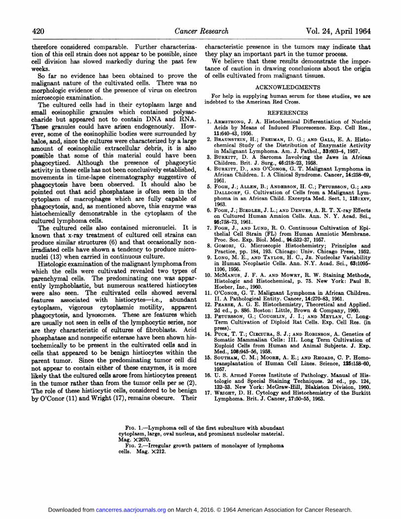
bovine serum, 15 per cent; glutamine, 8 mg. per cent;added penicillin and streptomycin. The outgrowth consisted of cells growing radially from the attached explant.Ten days later a nearly confluent sheet of cells had grownacross the culture flask, and at this stage cells were transferred, after treatment with 0.25 per cent trypsin, intoseveral other culture containers. The appearance of acell of the first subculture is shown in Figure 1, and thegrowth pattern of an established cell sheet, after severaltransfers of the cell strain, is seen in Figure 2.
Parallel cultures were inoculated into an x-radiatedsheet of primary human amnion cells (3000 r). Althougha temporary growth stimulation was observed, cultivationwith the feeder layer was not essential for long-termmaintenance.
Several weeks after initiation of the culture and afterfour passages the growth rate decreased, and the cellsappeared granular. At this time the medium was changedto consist of: basic medium, 60 per cent; fetal bovineserum, 20 per cent; and human serum, 20 per cent—whichenhanced the growth of the cultured lymphoma cells.Several weeks later the fetal bovine serum concentrationwas reduced to 15 per cent, and in this medium the cellshave continued to divide slowly. In the early stagesweekly transfers of one culture into at least two cultureswere possible; later the growth rate decreased, permittinga total of only twenty transfers during the first year ofcultivation.
METHODSOF CHAEACTERIZATIONOF THECULTUREDCELLS
Chromosome analysis.—To establish counts of thechromosomes cells grown on coverslips in Leighton tubeswere treated with 0.00025 per cent colchicine solution for1 hour and then with 0.9 per cent sodium citrate followedby air drying and fixation in 3:1 alcohol-acetic acidfixative. They were stained with 2 per cent acetic-orceine. After dehydration preparations were mounted
in Permount. Counts were made by means of a drawingapparatus.
Electron microscopy.—For micromorphologic studiesscraped cell sheets were fixed in 1 per cent phosphate-buffered osmium for 35 minutes, post-fixed in 10 per centneutral isotonic formalin, and dehydrated in a gradedseries of ethanols.
The material was embedded in butyl-methacrylate.Sections, 60-100 m^ in thickness, were cut with a Porter-Blum microtome, with a glass knife, and collected oncollodion-carbon-coated grids. Contrast was enhanced by"staining" with lead hydroxide.
The specimens were examined, and electron micrographs were taken at an instrument magnification of 7,200with an RCA-EMU-3FF electron microscope and the useof a 250-Mcondenser aperture and a 50-^ objective aperture.
Microcinematography.—For cine records, an Emdecotime-lapse unit combined with a Zeiss inverted microscopewith a 40 X phase-contrast objective was used to recordcells inoculated into a Sykes-Moore culture chamber.The speed was four frames per minute. Maintained at37°C., the culture fluid was changed every 24 hoursthroughout the 72-hour sequence.
Histochemical studies.—The materials studied includedcultivated cells grown as monolayers on coverslips and, forcomparison, stored and frozen (—20°C.) solid tissue
from the parent tumor which had given rise to the cultivated strain.
For routine histological examination the tissues werestained with hematoxylin and eosin after fixation in amixture of 10 per cent formalin, 80 per cent ethanol, and10 per cent distilled water. After similar fixation themethyl green-pyronin (9) stain was utilized for studyingthe distribution of DNA and RNA, the periodic acid-Schiff (PAS) (16) reaction for polysaccharides, and an oilred 0 (16) stain for lipide in the cultivated cells. Theacri dine orange stain (1) was also utilized to demonstrateDNA and RNA after fixation of the cultivated cells in amixture of 90 per cent ethanol and 10 per cent glacialacetic acid, and the Feulgen stain (10) was employed fordetection of DNA after fixation in a mixture of 1 partglacial acetic acid and 3 parts absolute ethanol. TheGomori cobalt sulfide method (8) was used for alkalinephosphatase, the Gomori lead sulfide method (8) for acidphosphatase, and the alpha-naphthyl acetate method (12)for non-specific esterase in cultivated cells which had beenfixed for 20 minutes in cold calcium-formal solution1 andin parent tumor tissue which had been fixed overnight incold calcium-formal and then sectioned with a freezingmicrotome.
RESULTSAppearance of cultivated cells.—From the earliest
passages, the cultivated cells exhibited a considerabledegree of morphological heterogeneity. Characteristically, the cells were large and bizarre and contained large,often irregularly shaped nuclei with prominent nucleoli.Many cells had multiple stellate processes; they tended to
110 per cent formalin with 0.4 gm. per cent calcium chlorideadded and brought to pH 7 by addition of 0.1 Msodium carbonate.
on March 4, 2016. © 1964 American Association for Cancer Research. cancerres.aacrjournals.org Downloaded from
418 Cancer Research Vol. 24, April 1964
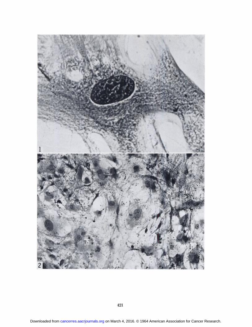
overlap and intertwine (Fig. 4), and their growth patterndid not simulate that of fibroblasta or epithelial cells. Astriking amount of extracellular eosinophilic debris wasoften seen scattered about in the vicinity of the cellaggregates. The nuclei varied in diameter from 12 to 80 M,and often presented an irregular lobulated outer margin(Fig. 5). This typical morphological appearance hasremained relatively unchanged during the entire period ofcultivation.
The appearance of the monolayer of cells dependedstrongly on the amounts and relative concentrations ofhuman and fetal bovine serum. In medium containing 20per cent human serum the growth was poor. An additionof 5-10 per cent fetal bovine serum improved the cellsheet only slightly. The most uniform-appearing cellsheets with well spread out cells of high density wereobserved in cultures containing 15 per cent or more fetalbovine serum, and an addition of human serum appearedadvantageous (Fig. 3). Results obtained after severaltransfers showed, as mentioned, that the optimal serumlevels for sustaining cell division were 15 per cent fetalbovine serum and 20 per cent human serum, althoughthe cells under these conditions formed a less uniformgrowth pattern.
A microcinematographic observation of the cells for aperiod of 72 hours after a transfer revealed an unusualdegree of cytoplasmic activity. Compared with otherhuman cell types studied in our laboratory by this technic(primary human amnion cells and FL cells), the culturedlymphoma cells exhibited very fast movements in alldirections in irregular patterns on the glass surface. Themost startling movements of some cells in the cultureincluded surface blebs which, due to constant protrusionand retraction in a rhythmic fashion, gave the impressionof a rotary movement, clockwise or counter-clockwise, ofthe peripheral parts of the cytoplasm. In other cells in thefield, however, the blebbing appeared in a nonrhythmicmanner, and such cells constantly changed shape withviolent motions. Some cell borders showed only undulating movements. The nuclear activity appeared to be lessthan that of the cytoplasm. Extracellular debris, oftenappearing as spherical cell fragments, seemed to beattracted to the cell surface, and occasionally such fragments appeared to be phagocytized.
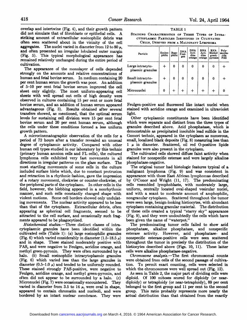
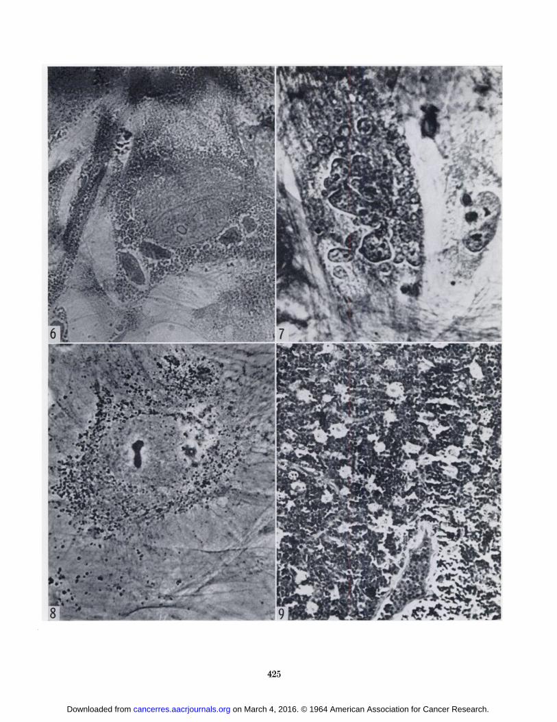
Histochemical studies.—Three distinct classes of intra-cytoplasmic granules have been identified within thecultivated cells (Table 1): (a) large eosinophilic granules(Fig. 6) which varied considerably in diameter (1.5-18.5 AI)and in shape. These stained moderately positive withPAS, and were negative to Feulgen, acridine orange, andmethyl green-pyronin. They were often surrounded by ahalo, (o) Small eosinophilic intracytoplasmic granules(Fig. 6) which varied less than the large granules indiameter (0.5-1.0 M)and tended to be uniformly rounded.These stained strongly PAS-positive, were negative toFeulgen, acridine orange, and methyl green-pyronin, andoften did not appear to be surrounded by a halo, (c)Micronuclei (Fig. 7) were occasionally encountered. Theyvaried in diameter from 3.5 to 14 p, were oval in shape,appeared to contain chromatin and nucleoli, and werebordered by an intact nuclear membrane. They were
TABLE 1STAININGCHARACTERISTICSOF THREE TYPES OP INTRA
CYTOPLASMICPARTICLESIDENTIFIEDIN CULTIVATEDCELLS,DERIVEDFROMA MALIGNANTLYMPHOMA
ParticleLarge
intracytoplasmicgranulesSmall
intracytoplasmicgranulesMicronucleiEosino-
philic++0Baso-philic00+DNAFeul-00+DNAAcridineorange00+RNA
Acridineorange00+RNAMethylgreen-pyronin00+Poly-saccha-ridePAS++0
Feulgen-positive and fluoresced like intact nuclei whenstained with acridine orange and examined in ultravioletlight.
Other cytoplasmic constituents have been identifiedwhich were separate and distinct from the three types ofgranules described above. Acid phosphatase, which isdemonstrable as precipitated insoluble lead sulfide in theGomori technic, appeared in the cytoplasm as numerous,small, localized black deposits (Fig. 8) measuring less than1 ti in diameter. Scattered, oil red O-positive lipidegranules were also present in the cytoplasm.
The cultivated cells showed diffuse faint activity whenstained for nonspecific esterase and were largely alkalinephosphatase-negative.
The original tumor had histologie features typical of amalignant lymphoma (Fig. 9) and was consistent inappearance with those East African lymphomas describedby O'Conor and Wright (11, 17). The predominating
cells resembled lymphoblasts, with moderately large,uniform, centrally located oval-shaped vesicular nuclei,and with a scant to moderate amount of amphophilic,nongranular cytoplasm. Scattered throughout the tumorwere seen large, benign-looking histiocytes, with abundantcytoplasm containing granular material. The distributionof these cells created a typical "starry sky" appearance
(Fig. 9), and they were undoubtedly the cells which havebeen given the name of "waterpot."
The predominating tumor cells failed to show acidphosphatase, alkaline phosphatase, and nonspecificesterase activity. However, acid phosphatase- andnonspecific esterase-positive cells were seen scatteredthroughout the tumor in precisely the distribution of thehistiocytes described above (Figs. 10, 11). These lattercells were alkaline phosphatase-negative.
Chromosome analysis.—The first chromosomal countswere obtained from cells of the second passage of cultivation. To permit exact counting, cells were selected inwhich the chromosomes were well spread out (Fig. 12).
As seen in Table 2, the major part of dividing cells werediploid. Of 100 mitoses scored for diploidy (or near-diploidy) or tetraploidy (or near-tetraploidy), 89 per centbelonged to the first group and 11 per cent to the secondgroup. This ratio probably represents more nearly theactual distribution than that obtained from the exactly
on March 4, 2016. © 1964 American Association for Cancer Research. cancerres.aacrjournals.org Downloaded from

FoGH et al.—Cellsfrom a Malignant Lymphoma in African Child 419
TABLE 2CHROMOSOMENUMBERSOF THE CULTUREDLYMPHOMACELLS
No.cells1227111No. chromosomes]294546474992
counted 33 cells. The quality of the preparations was notsufficient to determine whether the diploid cells weretruly euploid or whether abnormal chromosome rearrangements were present, not affecting the total chromosomenumber (quasi-diploidy).
A subsequent chromosome study was made on cells ofthe fifteenth-passage level. In these preparations mitosiswas rare, and reliable counts were obtained from only fourcells, of which three contained 46 chromosomes and onecontained 92 chromosomes. In each of these three diploidcells, five small acrocentrics could be distinguished, andthe cultured cells, therefore, could be recognized as beingof male origin. In the tetraploid cell a well illustratedendoreduplication with 46 pairs of chromosomes, or a totalof 92 chromosomes, was observed. The number of smallacrocentrics in that cell (10) was also consistent with themale origin of the cells.
The frequent observation of cells with lobated andmultiple nuclei and the great variation in nuclear sizeindicate a greater variation in chromosome number thanthat encountered from the counts. It is probable thatpolyploid or even heteroploid cells were present in agreater proportion than apparent from the data, sincesuch cells may divide much less frequently than diploidcells.
Attempts to produce tumors in heterologousand homologoushosts.—Tofurther test for possible malignant characteristics of the cultured cells, newborn Syrian hamsters andterminal human cancer patients were selected as therecipients most likely to develop tumors after inoculationsof these cells. The amounts of available cells limited theseinoculation experiments to one attempt with twelvehamsters, each of which received, subcutaneously, approximately IO6cells of the fourteenth passage; and three cancerpatients, each inoculated subcutaneously in the forearmwith 1 to 2 X 10' cells of passage 12, 16, and 18. Thehuman patients, selected by Dr. Chester Southam,volunteered to participate in this test which was carriedout by him and cooperators with the procedure they havepreviously adapted for such tests (15).
The hamsters, during the first 8 months of observation,have not developed tumors and have shown no signs ofdisease or growth retardation. One of the human recipients died from his own disease too early after inoculationof the cultured cells to permit an evaluation of growth atthe inoculation site. The second cancer patient developeda minor swelling during the first 10 days after inoculation,but compared with the control site where a large tumordeveloped from injection of H.Ep. #2 cells this minor
swelling was too small for biopsy, and the attempt withthis patient was therefore considered negative. The thirdtrial resulted in the development of a palpable nodule inone of two sites of inoculation of the cultured cells on theforearm of a 33-year-old female suffering from adeno-carcinoma of the cervix. The nodule was excised 15 daysafter inoculation, and microscopic sections of the biopsyfailed to reveal tumor cells; however, a number of histio-cytes were seen at the inoculation site. A simultaneousinjection into this patient of H.Ep. #2 cells resulted in alarger swelling.
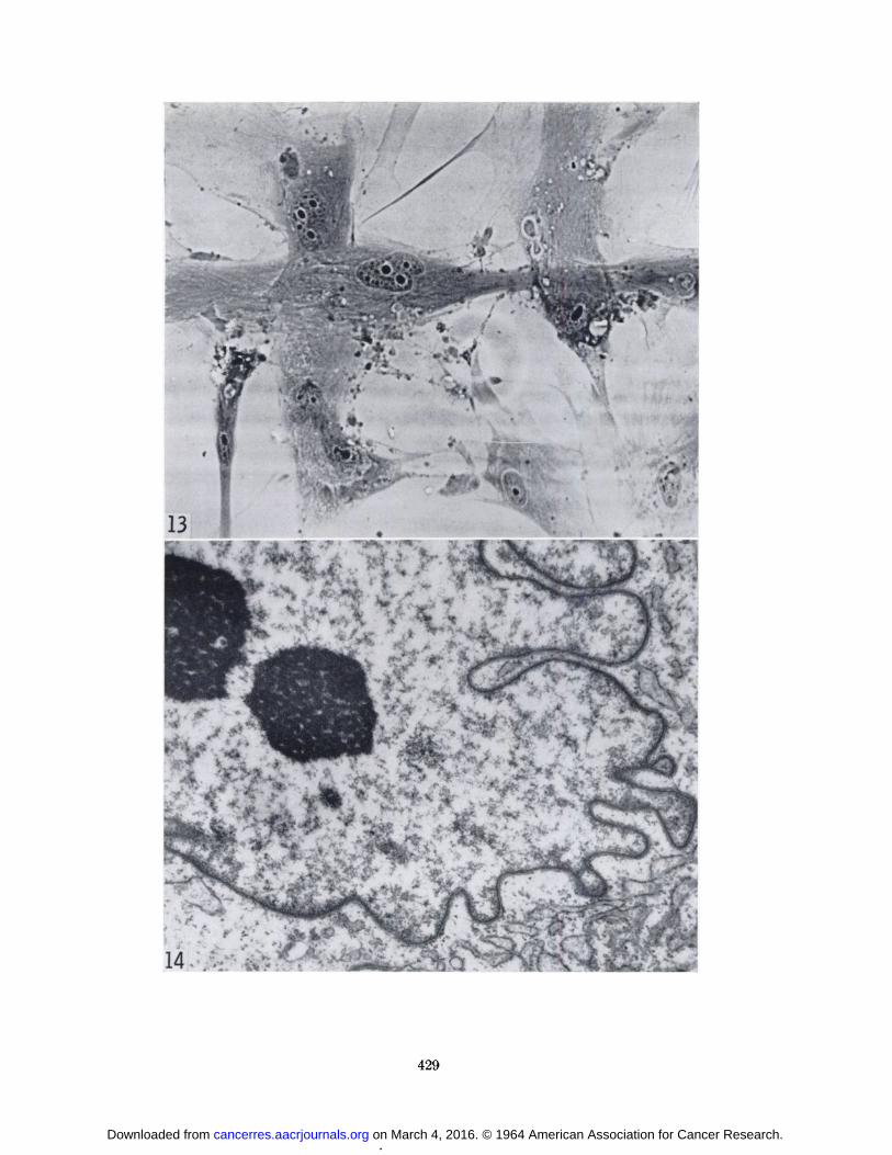
From the excised piece of tissue, cultures were established in our laboratory. Cells of these cultures showedsome resemblance to the originally cultured cells (Fig. 13),and serial transfers were possible. A chromosome analysisof the recultured cells showed them to be of diploidchromosome number, but of female sex. It may, therefore,be inferred that the latter cells originated from the recipient of the experimental inoculation, since this patientwas female, whereas the African patient was male.
Attempts to produce changes in various types of tissueculture (amnion cells, primary cultured or of the FL line,cells of Wistar diploid human embryonic lung strain WI26, the RECL-1 and RECL-6 strain of rat embryo cells[13], and primary mouse embryo cells) all gave negativeresults. No positive results were obtained by inoculatingnewborn hamsters or chicken chorioallantoic membrane,either directly with lymphoma culture supernatant or withsupernatants from "blind passages" in the other types of
cells.Electron microscopy.—Electron micrographs of the
cultured lymphoma cells (Figs. 14-16) showed irregularcell surfaces, rather large and irregular nuclei, and nucleoliappearing as irregular, coarse, granular, or filamentousmasses (Fig. 14). In the cytoplasm of many cells, theendoplasmic reticulum was prominent (Fig. 15); theassociated RNP granules appeared to be of normal sizeand density. Lysosomes and lipide bodies were frequentlyseen throughout the cytoplasm (Fig. 16).
Several different types of small vacuoles or granules wereobserved in the cytoplasm, but no structures of definiteviral morphology were found either in the cells or in theintercellular spaces.
DISCUSSION
The above represents the first reported cultivation ofcells from the malignant lymphoma of children in Africa.2The survival of the cells through multiple serial passageshas made possible their detailed characterization. Duringthe long period of cultivation no evidence of morphologicchange, preferential outgrowth of a specific cell type, orchromosome change has been observed. This appears toexclude in vitro transformation, and the different characterization studies, which, owing to the low rate ofdivision, had to be performed at many passage levels, are
1Fragments of two additional lymphomas were sent to TheSouth African Institute for Medical Research and were cultivated by J. H. S. Gear and J. M. Spence. One yielded cells which,in their opinion, resembled fibroblasta. One of us (G. D.) wasprivileged to examine these cells and found them more uniformand lacking certain features of our cells.
on March 4, 2016. © 1964 American Association for Cancer Research. cancerres.aacrjournals.org Downloaded from
420 Cancer Research Vol. 24, April 1964
therefore considered comparable. Further characterization of this cell strain does not appear to be possible, sincecell division has slowed markedly during the past fewweeks.
So far no evidence has been obtained to prove themalignant nature of the cultivated cells. There was nomorphologic evidence of the presence of virus on electronmicroscopic examination.
The cultured cells had in their cytoplasm large andsmall eosinophilic granules which contained polysac-charide but appeared not to contain DNA and RNA.These granules could have arisen endogenously. However, some of the eosinophilic bodies were surrounded byhalos, and, since the cultures were characterized by a largeamount of eosinophilic extracellular debris, it is alsopossible that some of this material could have beenphagocytized. Although the presence of phagocyticactivity in these cells has not been conclusively established,movements in time-lapse cinematography suggestive ofphagocytosis have been observed. It should also bepointed out that acid phosphatase is often seen in thecytoplasm of macrophages which are fully capable ofphagocytosis, and, as mentioned above, this enzyme washistochemically demonstrable in the cytoplasm of thecultured lymphoma cells.
The cultured cells also contained micronuclei. It isknown that x-ray treatment of cultured cell strains canproduce similar structures (6) and that occasionally non-irradiated cells have shown a tendency to produce micro-nuclei (13) when carried in continuous culture.
Histologie examination of the malignant lymphoma fromwhich the cells were cultivated revealed two types ofparenchymal cells. The predominating one was apparently lymphoblastic, but numerous scattered histiocyteswere also seen. The cultivated cells showed severalfeatures associated with histiocytes—i.e., abundantcytoplasm, vigorous cytoplasmic motility, apparentphagocytosis, and lysosomes. These are features whichare usually not seen in cells of the lymphocytic series, norare they characteristic of cultures of fibroblasts. Acidphosphatase and nonspecific esterase have been shown histochemically to be present in the cultivated cells and incells that appeared to be benign histiocytes within theparent tumor. Since the predominating tumor cell didnot appear to contain either of these enzymes, it is morelikely that the cultured cells arose from histiocytes presentin the tumor rather than from the tumor cells per se (2).The role of these histiocytic cells, considered to be benignby O'Conor (11) and Wright (17), remains obscure. Their
characteristic presence in the tumors may indicate thatthey play an important part in the tumor process.
We believe that these results demonstrate the importance of caution in drawing conclusions about the originof cells cultivated from malignant tissues.
ACKNOWLEDGMENTS
For help in supplying human serum for these studies, we areindebted to the American Red Cross.
REFERENCES
1. ARMSTRONG,J. A. Histochemical Differentiation of NucleicAcids by Means of Induced Fluorescence. Exp. Cell Res.,11:640-43, 1956.
2. BRAUNSTEIN, H.; FREIMAN, D. G.; AND GALL, E. A. Histochemical Study of the Distribution of Enzymatic Activityin Malignant Lymphoma. Am. J. Pathol., 33:603-4, 1957.
3. BURKITT, D. A Sarcoma Involving the Jaws in AfricanChildren. Brit. J. Surg., 46:218-23, 1958.
4. BURKITT, D., ANDO'CONOR, G. T. Malignant Lymphoma inAfrican Children. I. A Clinical Syndrome. Cancer, 14:258-69,1961.
5. FOGH, J.; ALLEN, B.; ANDERSON,H. C.; PETURSSON, G.; ANDDALLDORF, G. Cultivation of Cells from a Malignant Lymphoma in an African Child. Excerpta Med. Sect. 1, 118:xxv,1963.
6. FOGH, J.; BIEDLER, J. L.; ANDDENUES, A. R. T. X-ray Effectson Cultured Human Amnion Cells. Ann. N. Y. Acad. Sci.,96:758-73, 1961.
7. FOGH, J., AND LUND, R. O. Continuous Cultivation of Epithelial Cell Strain (FL) from Human Amniotic Membrane.Proc. Soc. Exp. Biol. Med., 94:532-37, 1957.
8. GOMORI, G. Microscopic Histochemistry; Principles andPractice, pp. 184, 193. Chicago: Univ. Chicago Press, 1952.
9. LONG, M. E., ANDTAYLOR, H. C., JR. Nucleolar Variabilityin Human Neoplastic Cells. Ann. N.Y. Acad. Sci., 63:1095-1106, 1956.
10. McMANUS, J. F. A. AND MOWRY, R. W. Staining Methods,Histologie and Histochemical, p. 75. New York: Paul B.Hoeber, Inc., 1960.
11. O'CoNOR, G. T. Malignant Lymphoma in African Children.II. A Pathological Entity. Cancer, 14:270-83, 1961.
12. PEARSE, A. G. E. Histochemistry, Theoretical and Applied.2d éd.,p. 886. Boston: Little, Brown & Company, 1960.
13. PETURSSON, G.; COUGHLIN, J. I.; AND MEYLAN, C. Long-Term Cultivation of Diploid Rat Cells. Exp. Cell Res. (inpress).
14. PUCK, T. T.; CIECIURA, S. J.; ANDROBINSON, A. Genetics ofSomatic Mammalian Cells: III. Long Term Cultivation ofEuploid Cells from Human and Animal Subjects. J. Exp.Med., 108:945-56, 1958.
15. SOUTHAM,C. M.; MOORE, A. E.; AND RHOADS, C. P. Homo-transplantation of Human Cell Lines. Science, 126:158-60,1957.
16. U. S. Armed Forces Institute of Pathology. Manual of Histologie and Special Staining Techniques. 2d éd.,pp. 124,132-33. New York: McGraw-Hill, Blakiston Division, 1960.
17. WRIGHT, D. H. Cytology and Histochemistry of the BurkittLymphoma. Brit. J. Cancer, 17:50-55, 1963.
FIG. 1.—Lymphoma cell of the first subculture with abundantcytoplasm, large, oval nucleus, and prominent nucleolar material.Mag. X2670.
FIG. 2.—Irregular growth pattern of monolayer of lymphomacells. Mag. X212.
on March 4, 2016. © 1964 American Association for Cancer Research. cancerres.aacrjournals.org Downloaded from
421
on March 4, 2016. © 1964 American Association for Cancer Research. cancerres.aacrjournals.org Downloaded from

FIG. 3.—Seriesof T-15 flasks demonstrating variation in appearance of the cell sheet with changes in serum content in t lieculture medium. Concentrations of fetal bovine and humanserum in per cent for the different cultures, from bottom to top,was as follows (fetal bovine/human): 25/10; 30/5; 35/0; 15/15;10/25; 15/0; 10/20; 5/30; 5/20; 0/20.
Flo. 4.—Hematoxylin and eosin stain of cultured lymphomacells growing on a coverslip. Note the irregular cell shapes andabundant cytoplasm which extends from the nucleus to formintertwining stellate processes. Also darkly staining granularextracellular debris is present. Mag. X340.
FIG. 5.—Feulgenstain of cultivated cells showing marked nuclear lobulation. Mag. X1085.
422
on March 4, 2016. © 1964 American Association for Cancer Research. cancerres.aacrjournals.org Downloaded from
423
on March 4, 2016. © 1964 American Association for Cancer Research. cancerres.aacrjournals.org Downloaded from
FIG. 6.—Periodicacid-Sehiff stain of cultivated lymphonia cellsshowing positively staining large intracytoplasmic granules,surrounded by halos, and also small intrucytoplasmic granules.Mag. X850.
FIG. 7.—Micronucleiare seen in the cultivated cell in the center, stained with heniatoxylin and eosin. Mag. X1350.
FIG. 8.—Gomori preparation for acid phosphatase demonstrating considerable enzyme activity, represented by blackintracytoplasmic granules of lead sulfide, in the cultivated lym-phoma cells. Mag. X850.
FIG. 9.—Low-power view of the lymphoma from which thecultivated cells originated shows predominating lymphoblasticcells with interspersed histiocytes creating a "starry sky" appearance (hematoxylin and eosin stain). Mag. X212.
424
on March 4, 2016. © 1964 American Association for Cancer Research. cancerres.aacrjournals.org Downloaded from
T^-"- *
**l*f*•*ïs>- «' éï ' ' ' "*•'*L~*\r5^/.:; I .,"'.!.>^^v'-vÃ
SÃŒa.:-ÃŒSP% ^?*4:
•,
Li»
0 ,'! •*O
425
on March 4, 2016. © 1964 American Association for Cancer Research. cancerres.aacrjournals.org Downloaded from
Fio. 10.—Acidphosphatase stain of the parent tumor showsenzyme activity restricted to the darkly staining histiocytes.Mag. X270.
FIG. 11.—Darkly staining, nonspecific esterase also appearsto be exclusively in the histiocytes. Mag. X'270.
FIG. 12.—Typicalmetaphase plate with 46 chromosomes fromcultured lympnoma cells. Mag. X2120.
426
on March 4, 2016. © 1964 American Association for Cancer Research. cancerres.aacrjournals.org Downloaded from

•*'• '"t A 2*
t * ' a1 : -4
"
12
427
on March 4, 2016. © 1964 American Association for Cancer Research. cancerres.aacrjournals.org Downloaded from

Fio. 13.—Cells cultured from biopsy of nodule resulting frominjection of the cultured lymphoma cells into a terminal humancancer patient. Mag. X340.
FIG. 14.—Electron micrographs of thin sections of cultured lymphoma cells. Mag. X 18,000. Large nucleus with lobes and incisions containing rather large, coarse, granulated nucleoli. Cluster of small vesicles at the cell border.
428
on March 4, 2016. © 1964 American Association for Cancer Research. cancerres.aacrjournals.org Downloaded from
r- i''•-•^^
14
429
on March 4, 2016. © 1964 American Association for Cancer Research. cancerres.aacrjournals.org Downloaded from
FIG. 15, 10.—Electron micrographs of thin sections of culturedlymphoma colls. Mag. X18,000.
FIG. 15.—Perinuclear concentric layers of rough endoplasmi?,reticulum tubules.
FIG. 1C.—Lysosomes (ly)), small vesicles, and unidentifiedmembrane-limited, fine, granular body in the perinuolear area(6).
4:;o
on March 4, 2016. © 1964 American Association for Cancer Research. cancerres.aacrjournals.org Downloaded from

- ' Xl'Vi
' Lv, .
'..- - - : V': ' A ^îir"
'- .•': .!f
,;-- v•.
f -, i---*«•:"*.. "
•Ã^
431
on March 4, 2016. © 1964 American Association for Cancer Research. cancerres.aacrjournals.org Downloaded from

1964;24:416-431. Cancer Res Jørgen Fogh, H. Clarke Anderson, Bruce Allen, et al. Lymphoma in an African ChildCultivation and Characterization of Cells from a Malignant
Updated version
http://cancerres.aacrjournals.org/content/24/3_Part_1/416
Access the most recent version of this article at:
E-mail alerts related to this article or journal.Sign up to receive free email-alerts
Subscriptions
Reprints and
To order reprints of this article or to subscribe to the journal, contact the AACR Publications
Permissions
To request permission to re-use all or part of this article, contact the AACR Publications
on March 4, 2016. © 1964 American Association for Cancer Research. cancerres.aacrjournals.org Downloaded from