
Crystal Deposition Diseases - pdfs.semanticscholar.org · No trauma or hiperuricemia. Dual Energy...
38
Crystal Deposition Diseases Dr. Marcelo Abreu HMD - PoA, Brazil
Transcript of Crystal Deposition Diseases - pdfs.semanticscholar.org · No trauma or hiperuricemia. Dual Energy...

Crystal Deposition Diseases
Dr. Marcelo Abreu
HMD - PoA, Brazil

Diagnosis: Pyrophosphate Arthropathy
Case 1. 58y, F, wrist pain for 2 weeks.
T2 fat saturation
• Inflammatory Arthritis
• CPPD crystals are pro-inflammatory particles
• Can cause synovial inflammation

Calcium Pyrophosphate Dihydrate (CPPD) Crystals
Normally Deposit in MSK System
CPPD sporadic deposit of CPPD is a common condition in the Elderly
• 8-10% of people aged 60 years. 20-40% at age 80y
Picture from UCSD Research Lab 2002
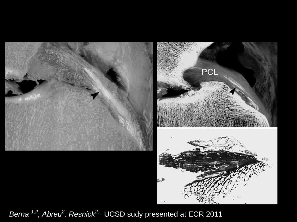
Berna 1,2, Abreu2, Resnick2, . UCSD sudy presented at ECR 2011
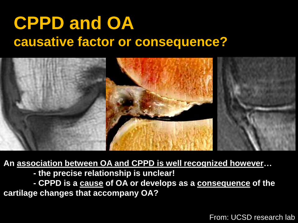
An association between OA and CPPD is well recognized however…
- the precise relationship is unclear!
- CPPD is a cause of OA or develops as a consequence of the
cartilage changes that accompany OA?
CPPD and OAcausative factor or consequence?
From: UCSD research lab

Neogi T, et al. Arthritis Rheum 2006
CPPD may be a marker of a reparative process by
metabolically active chondrocytes.
the suggestion that CPPD could be a marker of poor prognosis in knee OA was
not confirmed in several other longitudinal studies
100 patients who had undergone unilateral meniscectomy (20 year)
showed CPPD in 20% of operated knees compared with 4% of
contralateral unoperated knee
CPPD and OAcausative factor or consequence?
How accurate is MR imaging for CPPD deposits?

Faxitron radiograph of cadaver specimen Sagittal PD-weighted MR image of same spec
Abreu, Chung CB, Resnick D. CPPD crystalline deposits in the knee: anatomic, radiographic, MR imaging, and histologic study in cadavers. Skel Rad 2004
CHONDROCALCINOSIS
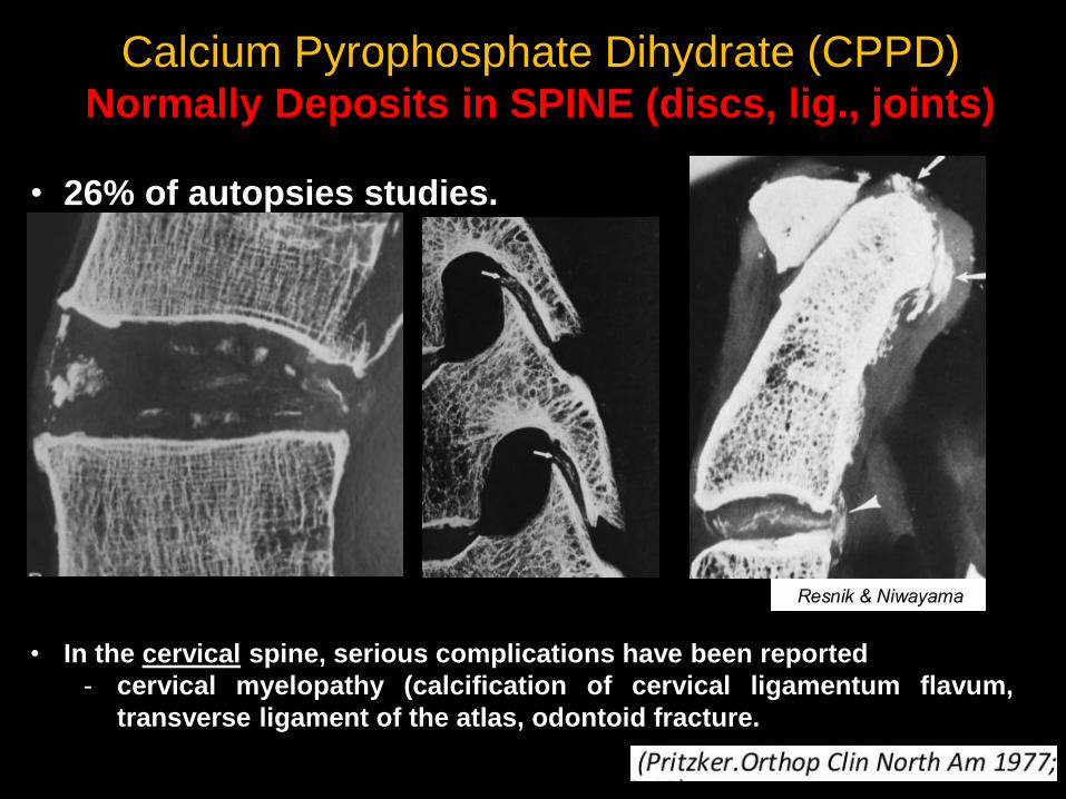
Calcium Pyrophosphate Dihydrate (CPPD) Normally Deposits in SPINE (discs, lig., joints)
• 26% of autopsies studies.
• In the cervical spine, serious complications have been reported
- cervical myelopathy (calcification of cervical ligamentum flavum,
transverse ligament of the atlas, odontoid fracture.
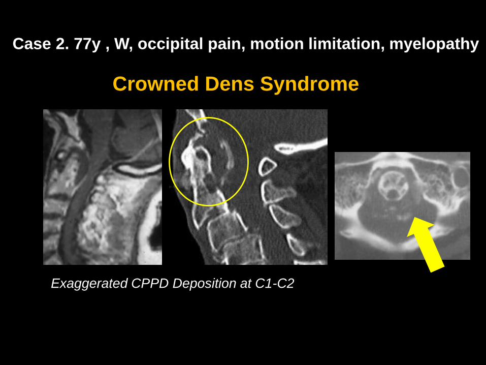
Exaggerated CPPD Deposition at C1-C2
Crowned Dens Syndrome
Case 2. 77y , W, occipital pain, motion limitation, myelopathy
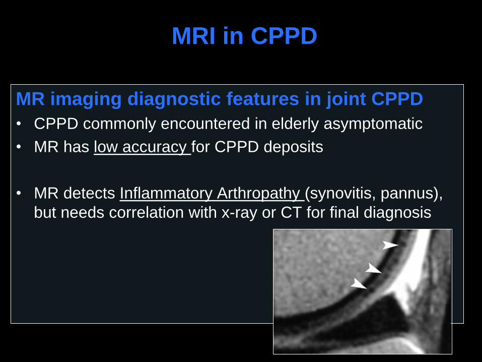
MRI in CPPD
MR imaging diagnostic features in joint CPPD
• CPPD commonly encountered in elderly asymptomatic
• MR has low accuracy for CPPD deposits
• MR detects Inflammatory Arthropathy (synovitis, pannus),
but needs correlation with x-ray or CT for final diagnosis

Case 3. 40y, M, Shoulder pain for 1 week.
Calcific Tendinitis (Hydroxyapatite)
T2 fat saturation
• Common in asymptomatic persons (when confined)
• Most commonly: Supraspinatus tendon insertion
• Less common: tendons of infraspinatus, subscapularis, deltoid,
wrist, elbow, gluteus maximus, knee, and neck.
• Inflammation and edema can occur

How accurate is MR imaging for Hydroxyapatite
deposits?
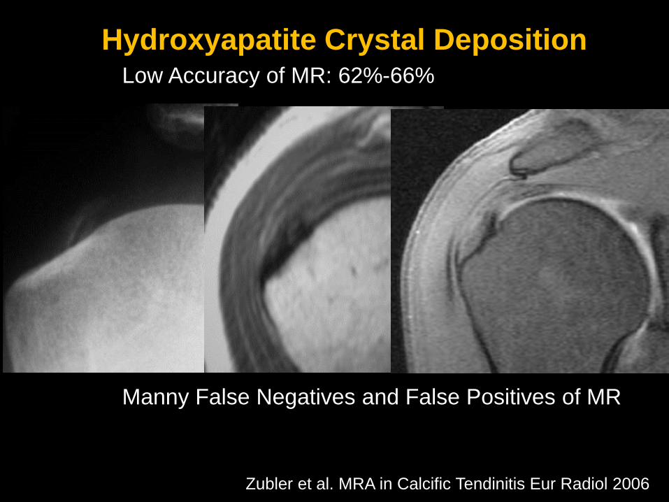
Low Accuracy of MR: 62%-66%
Manny False Negatives and False Positives of MR
Zubler et al. MRA in Calcific Tendinitis Eur Radiol 2006
Hydroxyapatite Crystal Deposition

Hydroxyapatite Crystal Deposition
Accuracy of MR increases with inflammation

tendon
BONE
Courtesy from D Resnick.

Case 4. 47y, F, Shoulder pain for 3 weeks.
T1 GRADIENT T2*
MR-Angio (GAD)
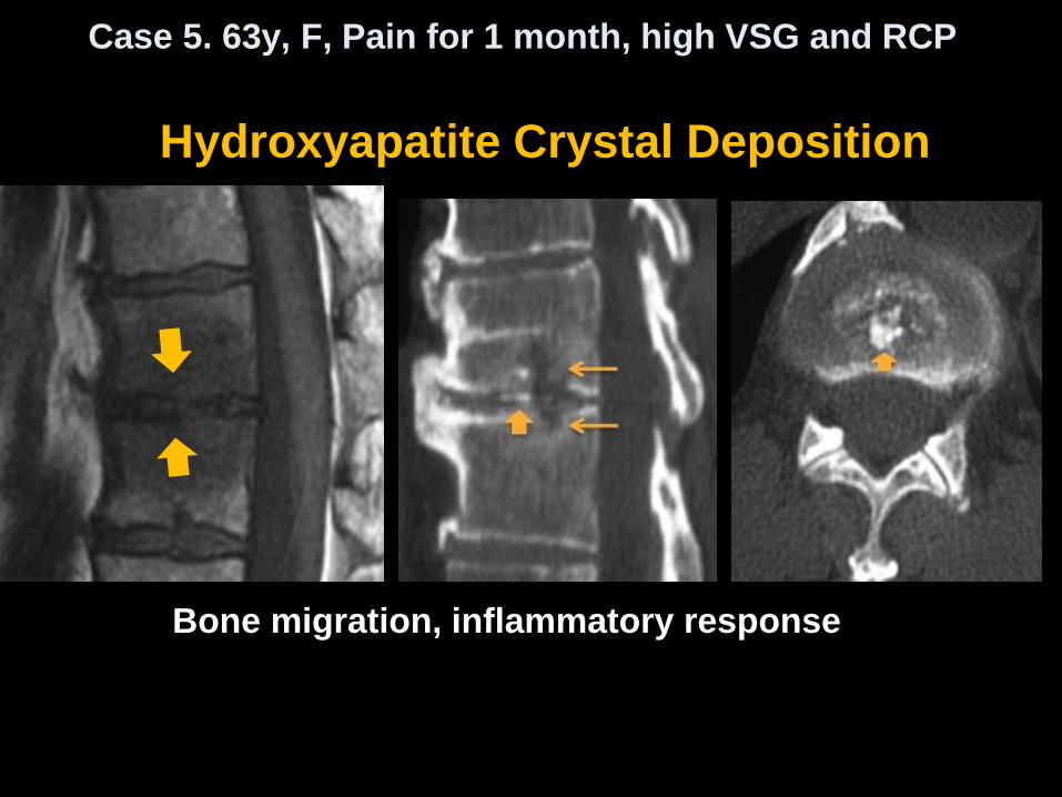
Case 5. 63y, F, Pain for 1 month, high VSG and RCP
Hydroxyapatite Crystal Deposition
Bone migration, inflammatory response

Can also be secondary to:
• Disc Steroid Injections
• Hemodialysis
• Ochronosis
Disc Hydroxyapatite Crystal Deposition• Intervertebral Disc Apatite
• Phosphocalcic Bruschite
• Apatite Rheumatism
CPPD


Round dense calcification
Cloud like appearance
Linear ``CROWNED DENS´´
Apatite x CPPDmorphology of calcification
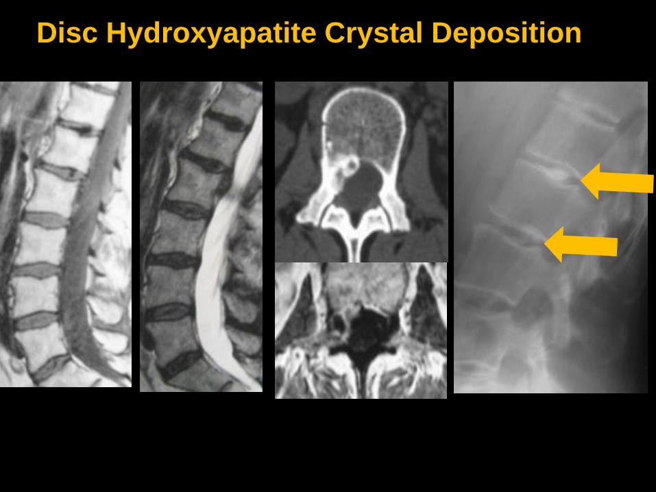
Disc Hydroxyapatite Crystal Deposition

Differentiate from:
Destructive Discovertebral Deg Disease (DDDD)
Charran, Puliccino V. Destructive discovertebral degenerative disease of the lumbar spine. Skel Rad 2012
Malalignment
Degenerative Disc Loss
End-plate failure
“bone sand” within the spinal canal.
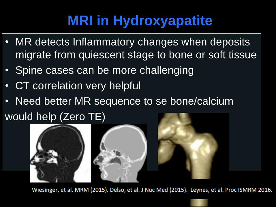
MRI in Hydroxyapatite
• MR detects Inflammatory changes when deposits
migrate from quiescent stage to bone or soft tissue
• Spine cases can be more challenging
• CT correlation very helpful
• Need better MR sequence to se bone/calcium
would help (Zero TE)
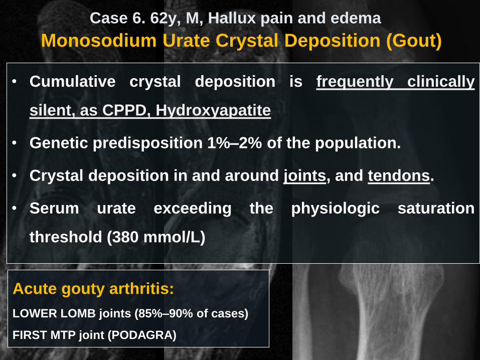
Case 6. 62y, M, Hallux pain and edema
Monosodium Urate Crystal Deposition (Gout)
• Cumulative crystal deposition is frequently clinically
silent, as CPPD, Hydroxyapatite
• Genetic predisposition 1%–2% of the population.
• Crystal deposition in and around joints, and tendons.
• Serum urate exceeding the physiologic saturation
threshold (380 mmol/L)
Acute gouty arthritis:
LOWER LOMB joints (85%–90% of cases)
FIRST MTP joint (PODAGRA)

• The disease has four phases:
1. Asymptomatic hyperuricemia
2. Acute
3. Intercritical
4. Chronic
Gout: Clinical
Advanced Stage of Disease

52y, M, Pain and edema lateral kneeMonosodium Urate Crystal Deposition (Gout)
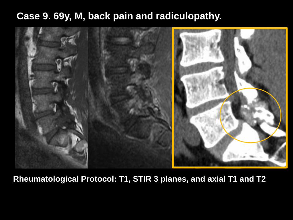
Case 9. 69y, M, back pain and radiculopathy.
Rheumatological Protocol: T1, STIR 3 planes, and axial T1 and T2

Case 10. 47y F, Cauda Equina Syndrome
• Hydroxyapatite Crystal Deposition
• Ossification of the Posterior Long Lig
• ``Bone Sand´´ in DDDD
• Gout
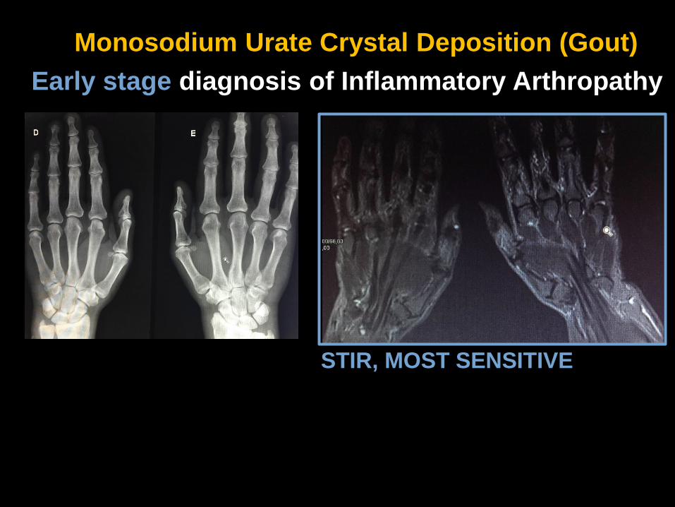
Monosodium Urate Crystal Deposition (Gout)
Early stage diagnosis of Inflammatory Arthropathy
STIR, MOST SENSITIVE

Monosodium Urate Crystal Deposition (Gout)
Early stage diagnosis of Inflammatory Arthropathy
STIR, MOST SENSITIVE MR-ANGIO, BETTER
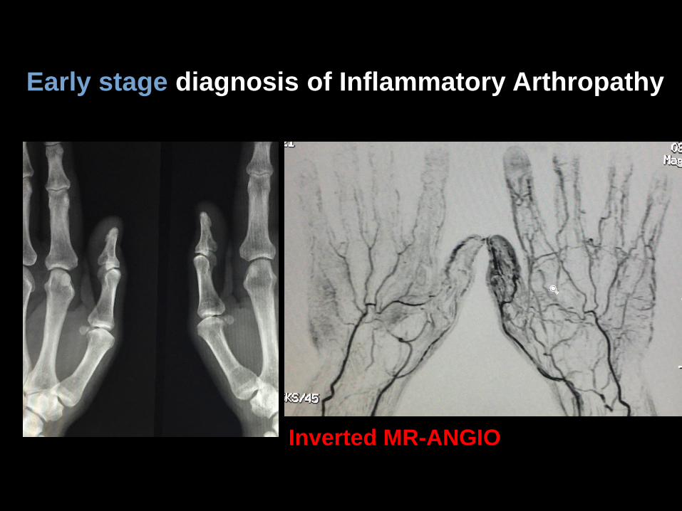
Inverted MR-ANGIO
Early stage diagnosis of Inflammatory Arthropathy

Case 7. 40, M, Local pain for 15 days. No trauma or hiperuricemia.
Dual Energy CT
Courtesy of Dr Skaf A
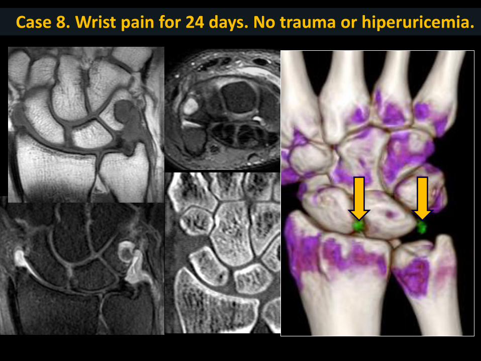
Case 8. Wrist pain for 24 days. No trauma or hiperuricemia.

COTICAL BONE
URATE
IODINE
Dual Energy CT in GOUT
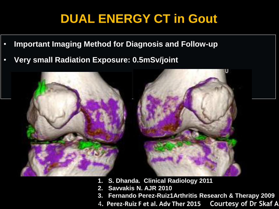
DUAL ENERGY CT in Gout
• Important Imaging Method for Diagnosis and Follow-up
• Very small Radiation Exposure: 0.5mSv/joint
1. S. Dhanda. Clinical Radiology 2011
2. Savvakis N. AJR 2010
3. Fernando Perez-Ruiz1Arthritis Research & Therapy 2009
4. Perez-Ruiz F et al. Adv Ther 2015 Courtesy of Dr Skaf A
Summary
Crystals Deposition Diseases
• CPPD, Hydroxyapatite and Urate: can be silent
• When activated, various clinical scenarios can be found:
– Acute/Chronic/Intercritical Inflammatory Arthritis
– Inflammatory Tendinopathy/Bursitis
– Back Pain, Compressive Myelopathy
MR imaging is a very useful imaging method for the
diagnosis of those diseases, most of the time together
with other methods like CT, DE-CT