Cryo-electron microscopy and tomography of biological ...Transmission electron microscopy (TEM) is...
15
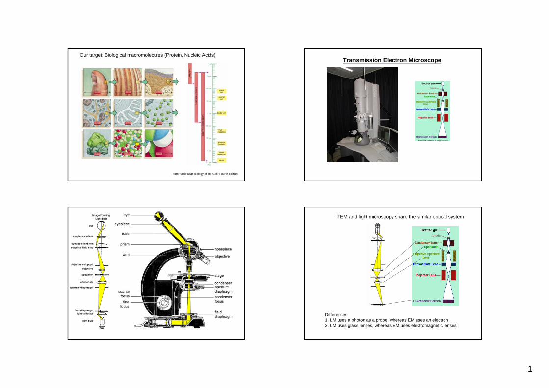
Lecture CIMST Winter School Cryo-electron microscopy and tomography of biological macromolecules 20.1.2011 9:00-9:45 in Y03G91 Dr. Takashi Ishikawa OFLB/005 Tel: 056 310 4217 e-mail: [email protected] Lab webpage: http://lbr.web.psi.ch/TI/TI_web.html 1. What can you see by TEM? Transmission electron microscopy (TEM) is an imaging technique. It can visualize biological, organic and inorganic materials as far as the specimen is thin enough for electrons to go through. Here in this lecture, we will focus on TEM technology to observe 3D structure of biological macromolecules. 2. Hardware Diagram of the transmission electron microscope is similar to that of a light microscope (LM). Both of them have a gun. Electrons or photons are emitted from the gun, penetrate the specimen, focused and enlarged by lenses (TEM uses electromagnetic lenses) and projected on a screen or a camera. Inside of the electron microscope is vacuum. 3. Image formation by TEM In principle, micrographs of TEM are 2D projections of the 3D objects. However, there are significant differences due to weak interaction between the electron beam and the specimen (i.e. TEM specimen is transparent against electrons). Therefore, instead of amplitude contrast LM utilizes, image formation of TEM depends on phase contrast which utilizes interference of electron waves. For phase contrast image must not be perfectly focused; there must be defocus. 4. Specimen preparation Since the contrast between biological materials (density ~1.4) and water (1.0) is poor, previously templates of heavy metal were made to enhance the contrast. With these methodologies (shadowing and negative stain) only the surface of the molecules is visualized. In 1980’s cryo-EM was developed. There ice-embedded biological macromolecules are observed directly at the liquid nitrogen temperature. 5. Resolution and radiation damage
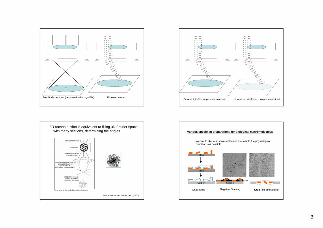
Transcript of Cryo-electron microscopy and tomography of biological ...Transmission electron microscopy (TEM) is...

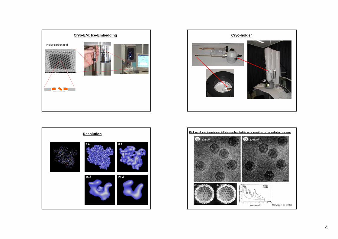
Lecture CIMST Winter School Cryo-electron microscopy and tomography of biological macromolecules 20.1.2011 9:00-9:45 in Y03G91 Dr. Takashi Ishikawa OFLB/005 Tel: 056 310 4217 e-mail: [email protected] Lab webpage: http://lbr.web.psi.ch/TI/TI_web.html 1. What can you see by TEM? Transmission electron microscopy (TEM) is an imaging technique. It can visualize biological, organic and inorganic materials as far as the specimen is thin enough for electrons to go through. Here in this lecture, we will focus on TEM technology to observe 3D structure of biological macromolecules. 2. Hardware Diagram of the transmission electron microscope is similar to that of a light microscope (LM). Both of them have a gun. Electrons or photons are emitted from the gun, penetrate the specimen, focused and enlarged by lenses (TEM uses electromagnetic lenses) and projected on a screen or a camera. Inside of the electron microscope is vacuum. 3. Image formation by TEM In principle, micrographs of TEM are 2D projections of the 3D objects. However, there are significant differences due to weak interaction between the electron beam and the specimen (i.e. TEM specimen is transparent against electrons). Therefore, instead of amplitude contrast LM utilizes, image formation of TEM depends on phase contrast which utilizes interference of electron waves. For phase contrast image must not be perfectly focused; there must be defocus. 4. Specimen preparation Since the contrast between biological materials (density ~1.4) and water (1.0) is poor, previously templates of heavy metal were made to enhance the contrast. With these methodologies (shadowing and negative stain) only the surface of the molecules is visualized. In 1980’s cryo-EM was developed. There ice-embedded biological macromolecules are observed directly at the liquid nitrogen temperature. 5. Resolution and radiation damage

Available discussion on the molecular structure depends on the level of resolution. Electron beams damage the specimen and limit the resolution. However, if the illumination is too dark, enough amount of contrast for image analysis cannot be obtained. The most essential and serious problem of 3D EM of biological molecules is this dilemma. 6. Various ways of three-dimensional reconstruction technique Here we will discuss four different approaches to reconstruct 3D structure of biological macromolecules from cryo-electron micrographs. 6-1. 2D crystal In the 2D crystal molecules array two-dimensionally. The Fourier transform of the image is a lattice. Thus, in the Fourier space, you can earn higher signal-to-noise ratio. Which enable the high quality data from lower illumination (i.e. lower radiation damage), resulting higher resolution (more than ten structures have been solved at the atomic resolution). However, this methodology demands, as X-ray crystallography, crystallization. Advantage, comparing to X-ray crystallography, is that we do not need heavy atoms, because direct images are recorded. For 3D information, tilted 2D crystals are inserted into the microscope with special holder. Since you cannot tilt 2D crystal at 90 degrees in the microscope, you will miss some information in the 3D Fourier space (missing cone). Example: bacteriorhodopsin, water channel. 6-2. Tubular crystal Molecules are arrayed as spirals. Since the signal will be spread along one dimensional lines (layer lines), the resolution is not so high as 2D crystals. All the necessary view angles for 3D reconstruction can be obtained from tubular crystal. Therefore, there is no missing information and tilt is not needed. Example: acetylcholine receptor, actin, microtubule 6-3. Single particle analysis This is a technique to calculate the 3D structure from many single particles (>5,000), determining their angles by computation. Since the molecules are dispersed in the solution, you can set solution condition freely, which allows you to see different stages of reaction, for example. However, the angle determination is complicated. Initially, you do not know either the final 3D structure or the view angles of individual particles. You must determine these two unknown factors at the same time. One approach is shown here: projection matching, which starts from a certain template and find the best-fitted view angle for each raw data particle. To generate initial templates, there are a few options. The resolution is evaluated using Fourier shell correlation. Example: Clp protease, ribosome 6-4. Electron tomography In single particle analysis, although you do not need crystals any more, you still average different particles, assuming they share the same structure. If not, the structural

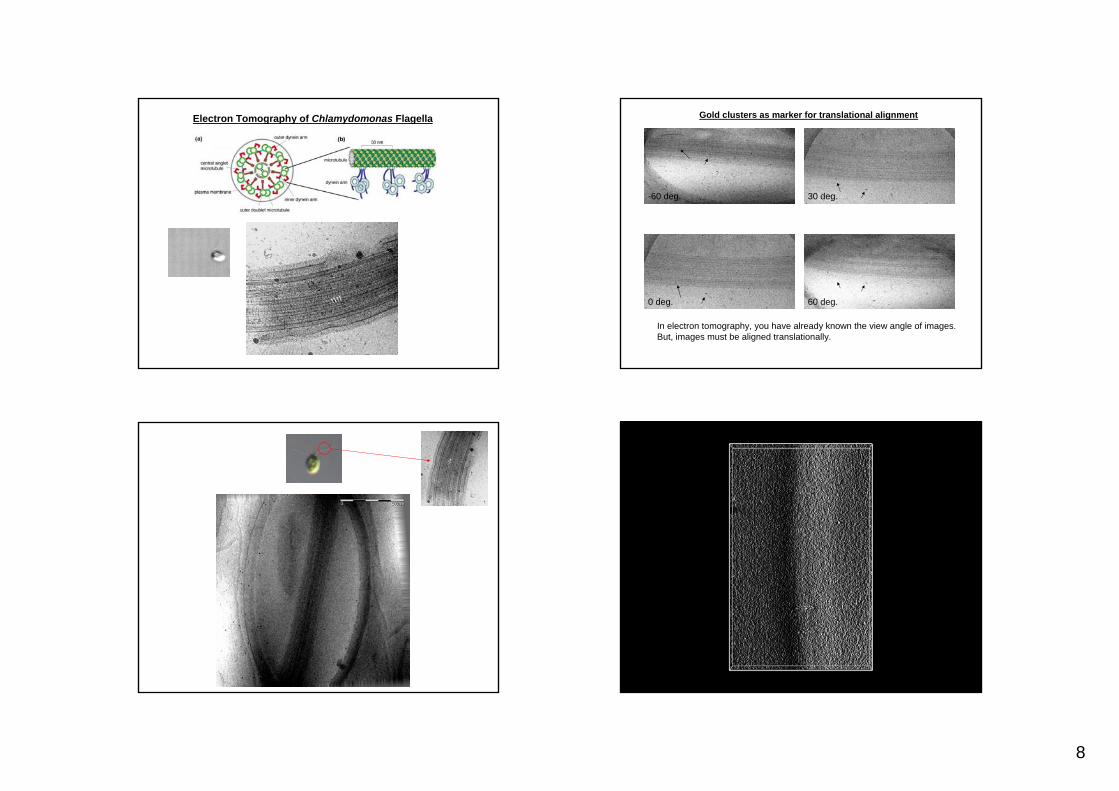
variability and flexibility are averaged out. Single particle analysis does not work for highly heterogeneous systems like whole cells or organelles, in which molecules are stacked vertically. Electron tomography is used for such specimens. You record the micrographs of the same specimen from various view angles by tilting the stage of the microscope. Since you control the tilt angle, you have no angle-determination problem, although you still have to align the origin of images. However, the resolution is limited because of multi radiation. Example: Eukaryotic flagella, whole cell tomography
References Many examples of structural biology by EM can be seen in Alberts et al. (2002) “Molecular biology of the cell, forth edition”, Garland Science. p. 560-570 (techniques), p. 949-969 (molecular motor research combining various methodologies including EM). There are some (but, not many) descriptions in Branden, C. and Tooze, J. (1999) “Introduction to structural biology, second edition” Garland. A good review on single particle analysis and electron tomography Baumeister, W. and Steven, A.C. (2000) “Macromolecular electron microscopy in the era of structural genomics” Trends in Biochemical Sciences 25, 624-631. Mathematical discussion on optics (including phase contrast, spherical aberration) Born, M. and Wolf, E. (1980) “Principles of optics” Pergamon Press Text books on single particle analysis Frank, J. (2006) “Three-dimensional electron microscopy of macromolecular assemblies” Oxford University Press.
1
Our target: Biological macromolecules (Protein, Nucleic Acids)
From “Molecular Biology of the Cell” Fourth Edition
Transmission Electron Microscope
From the material of Virginia Tech.
TEM and light microscopy share the similar optical system
Differences 1. LM uses a photon as a probe, whereas EM uses an electron2. LM uses glass lenses, whereas EM uses electromagnetic lenses

2
Inside of the electron microscope must be vacuum. The level of the vacuum varies, depending on the components (gun, column, camera etc.), and different pumps are used.
From the material of Virginia Tech.
Transmission Electron Microscopy (TEM)
Material Science
Biological Science1. Low Contrast2. High Sensitivity to the Electron Beam
You can see atoms directly
Why can’t we see the atom directly in biological TEM?
Wave–particle duality of electrons
L de Brogli proposed and G. P. Thomson proved that electron has wave-particle duality.
Screen
Interference of waves
3
Amplitude contrast (very weak with cryo-EM) Phase contrastDefocus: interference generates contrast In focus: no interference, no phase constrast
3D reconstruction is equivalent to filling 3D Fourier space with many sections, determining the angles
Baumeister, W. and Steven, A.C. (2000)
Various specimen preparations for biological macromolecules
Carbonstain
Mica
Negative Staining
Mica
Carbon
platinum
Shadowing Cryo (ice embedding)
We would like to observe molecules as close to the physiologicalconditions as possible.
4
Cryo-EM: Ice-Embedding
Holey carbon grid
Cryo-holder
Resolution
3 Å 8 Å
15 Å 20 Å
Biological specimen (especially ice-embedded) is very sensitive to the radiation damage
Conway et al. (1993)
6 e-/Å2 30 e-/Å2
6e-/Å2 30 e-/Å2

5
2D crystal
Stahlberg, H. et al. (2001)
3D view angles can be determined by diffraction pattern
Gyobu et al., (2006) J. Struct. Biol. 146, 325.
2D crystal: electron microscopy and electron diffraction
“Molecular Biology of the Cell”
Gonen et al. (2005) Nature 438, 633.
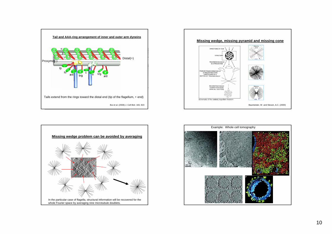
Missing wedge, missing pyramid and missing cone
Lucic et al. (2005) Annu. Rev. Biochem.

6
Tubular crystal
Kikkawa, T. et al. (1995)
Acetylcholine receptorUnwin (1993) J. Mol. Biol. 229, 1101
Diffraction of the tubular crystal is layer lines
Fourier
Miyazawa et al. (2003) Nature 423, 949.
Tubular crystal has no missing information.
Example of dynamic structural change: Actin / Myosin complex
Whittaker et al. (1995) Nature, 378, 748.

7
Clp enzyme w/o substrate
A AP
20 nm
Ishikawa et al. (2001)
Single particle analysis: 2D averagingAdvantage to reconstruct without crystallization: Free solution condition
ATP hydrolysis
with substrate (before reaction)
with substrate (after reaction)
Example of huge complexes: Ribosome
Spahn et al. (2001) 107, 373.
Single Particle Analysis: Projection Matching
Data
Classification based on Cross Correlation
Subaveragefor eachEuler angle
3D Reconstruction(New Reference)
Project
Initial Model
Final Reconstruction
Reproject
Iteration
5 nm
ClpA 3D ReconstructionSubstrates
ClpP
8
Electron Tomography of Chlamydomonas Flagella
-60 deg.
0 deg.
30 deg.
60 deg.
In electron tomography, you have already known the view angle of images.But, images must be aligned translationally.
Gold clusters as marker for translational alignment

9
3D alignment and averaging improve S/N3D Reconstruction of Chlamydomonas Flagella by electron cryo-tomography
3D Reconstruction of Chlamydomonas Flagella by electron cryo-tomographyOuter dynein arm(accelerator, force generator)
Averaging with 96nm periodicity reveals the IDA/ODA/RS/MT structure
96nm
Inner dynein arm(regulator)
Radial spokeMicrotubule
Bui et al. (2008) J. Cell Biol. 183, 923
10
Bui et al. (2008) J. Cell Biol. 183, 923
Tails extend from the rings toward the distal end (tip of the flagellum, + end)
Distal(+)Proxymal(-)
Tail and AAA-ring arrangement of inner and outer arm dyneinsMissing wedge, missing pyramid and missing cone
Baumeister, W. and Steven, A.C. (2000)
Missing wedge problem can be avoided by averaging
In the particular case of flagella, structural information will be recovered for the whole Fourier space by averaging nine microtubule doublets.
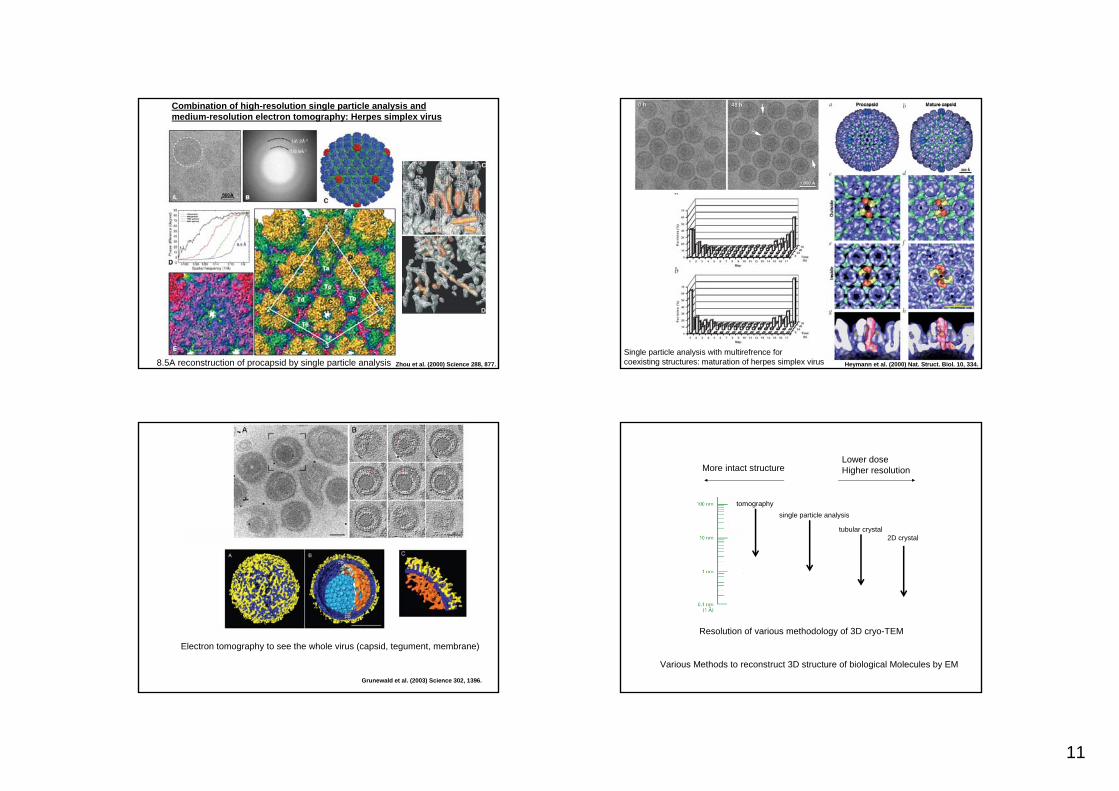
Example: Whole cell tomography
11
Combination of high-resolution single particle analysis and medium-resolution electron tomography: Herpes simplex virus
Zhou et al. (2000) Science 288, 877.8.5A reconstruction of procapsid by single particle analysis Heymann et al. (2000) Nat. Struct. Biol. 10, 334.
Single particle analysis with multirefrence for coexisting structures: maturation of herpes simplex virus
Grunewald et al. (2003) Science 302, 1396.
Electron tomography to see the whole virus (capsid, tegument, membrane)
Resolution of various methodology of 3D cryo-TEM
2D crystaltubular crystal
single particle analysistomography
Lower doseHigher resolutionMore intact structure
Various Methods to reconstruct 3D structure of biological Molecules by EM