Introduction to Biological Electron Microscopy · Introduction to Biological Electron Microscopy...
27
1 Introduction to Biological Electron Microscopy Andres Kaech Center for Microscopy and Image Analysis Scanning electron microscope (SEM) Transmission electron microscope (TEM) The types of electron microscopes Electron beam Specimen ~100 nm Electron beam Specimen Projection Surface Hela Cells
Transcript of Introduction to Biological Electron Microscopy · Introduction to Biological Electron Microscopy...

1
Introduction
to
Biological Electron Microscopy
Andres Kaech
Center for Microscopy and Image Analysis
Scanning electron microscope (SEM)Transmission electron microscope (TEM)
The types of electron microscopes
Electron beam
Specimen ~100 nm
Electron beam
Specimen
Projection Surface
Hela Cells
2
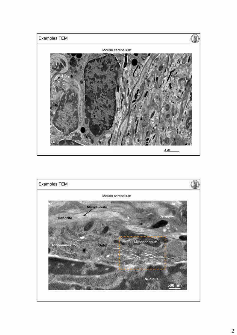
Examples TEM
Mouse cerebellum
Examples TEM
Mouse cerebellum
500 nm
Mitochondrium
Nucleus
GolgiRibosomes
SynapseDendrite
Microtubule
3
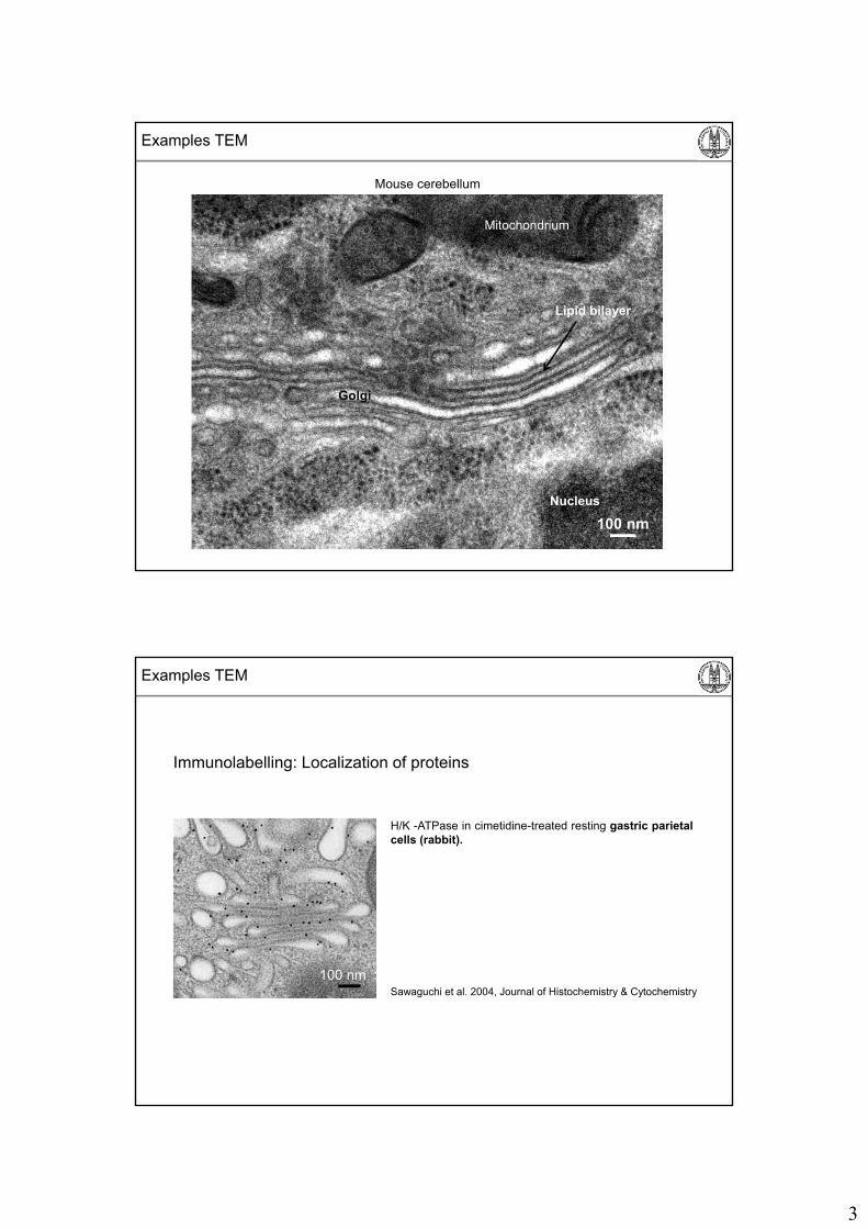
Examples TEM
Mouse cerebellum
100 nm
Mitochondrium
Nucleus
Golgi
Lipid bilayer
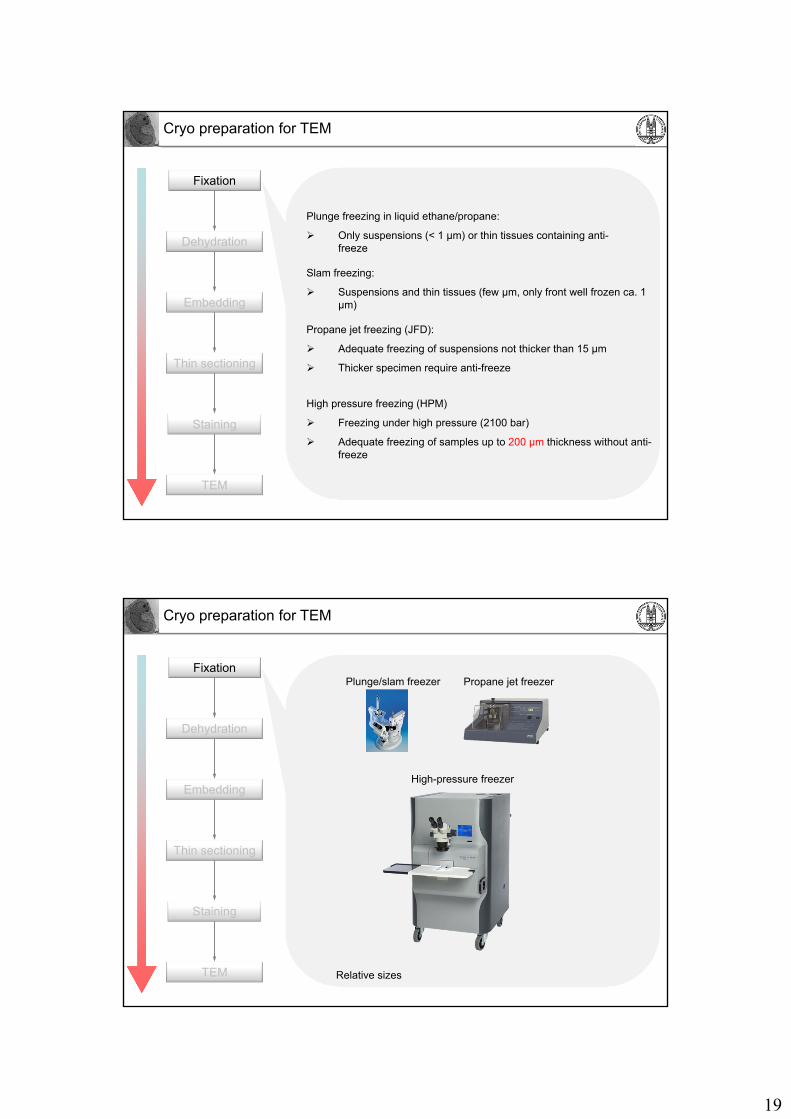
H/K -ATPase in cimetidine-treated resting gastric parietalcells (rabbit).
Immunolabelling: Localization of proteins
Sawaguchi et al. 2004, Journal of Histochemistry & Cytochemistry
100 nm
Examples TEM

4
Examples SEM
Mouse kidney
500 µm
Mouse kidney (glomerulus)
10 µm
Examples SEM

5
Pseudomonas aeruginosa
500 nm
Examples SEM
Wave-particle duality
Resolution depends on aperture and wavelength (Diffraction limited resolution)
Optical properties(Diffraction, chromatic abberation, spherical abberation, astigmatism etc.)
Abbe’s equation d = 0.61 λ/NA sin nNA
e-
Properties of electrons
Very similar to photons:

6
Resolution of biological objects limited by specimen preparation: Practical resolution: > 1 nm
TEM: 40 – 300 kV
Effective instrument resolution TEM: 0.5 nm (120 kV)
Effective instrument resolution SEM: 1 nm
Resolution of electron microscopes
The higher the energy of the electrons, the lower the wavelength, the higher the resolution
SEM: 0.5 – 30 kV
Widefield light microscopeTransmission electron microscope
Condenser lens
Objective lens
Projector lens
Specimen
Illumination
Final image
Transmission electron microscope vs. Widefield light microscope

7
Confocal laser scanning microscopeScanning electron microscope
Beam scanner
Detector
Lens system
Lens system
Specimen
Illumination
Scanning electron microscope vs. Confocal laser scanning microscope
Specimen holder
Specimen on a TEM grid
Specimen holders and stages - TEM
3 mm3 mm

8
Transmission electron microscope
Goniometer: x, y, z, r
Specimen size:
• 3 mm in diameter!
• Ca. 100 nm in thickness(electron transparent)
Specimen holders and stages - TEM
Specimen holders and stages - SEM
Scanning electron microscope
Specimen stage (x, y, z, r, tilt)
Objective lens
Stage
Specimen stub
Stub holder
Specimen size:
• 100 mm in diameter
• 2 cm in z-direction (not electron transparent)

9
Biology Electron microscope
Aqueous/hydrated
Soft
Light elements(C, O, H, N, S, P etc.)
“Large”
High vacuum
Electron beam
Sensitive to vibration/motion (High magnifications)
Not suitable for EM
Biological sample preparation for electron microscopy
Biology Electron microscope
High vacuum
Electron beam
Sensitive to vibration/motion (High magnifications)
Biological samples need to be transferred into a solid state...
...keeping the sample close to the native state
Not suitable for EM
Resistant to high vacuum
Resistant in electron beam
Thin – permeable for electrons(for TEM)
Contrast
Aqueous/hydrated
Soft
Light elements(C, O, H, N, S, P etc.)
“Large”
Biological sample preparation for electron microscopy

10
Biology Electron microscope
High vacuum
Electron beam
Sensitive to vibration/motion (High magnifications)
Any treatment changes the specimen!
Not suitable for EM
Resistant to high vacuum
Resistant in electron beam
Thin – permeable for electrons(for TEM)
Contrast
Aqueous/hydrated
Soft
Light elements(C, O, H, N, S, P etc.)
“Large”
Biological sample preparation for electron microscopy
2 cm
What is (was) this?
Biological sample preparation for electron microscopy

11
Dehydration
Critical Point Drying
Freeze-fractured/etched specimenFreeze-dried
specimen
Freeze-fracturing/Freeze-drying/Coating
RT-SEM
Low temperature processing
Low-temperature embeddingRT-embedding
RT-TEM, FIB-SEM
Cryo-Ultramicrotomy
Cryo-TEM
Cryo thin section
FROZEN SPECIMEN
Freeze-substitution
Cryo-SEM
RT specimen processing
Coating
RT-SEM
Ultramicrotomy
Staining
WARM SPECIMEN
High pressure freezing
Propane jet freezing
Plunge freezing
Embedding
Chemical fixation
thawing
Immunolabeling
Replica
RT-TEM
Preparation pathways overview
Bare grid technique
Sample preparation steps for TEM
Embedding
Fixation
Dehydration
Staining
Thin sectioning
TEM Requires thin specimen: 70 nm
Requires solid specimen (embedding in plastic)
Plastic only soluble in solvents (e.g. acetone)
Solvents dissolve biological matter

12
Embedding
Fixation
Dehydration
Staining
Room temperature processing for TEM
Thin sectioning
TEM
Stabilization of biological material
Chemical fixation (cross-linking) with Aldehydes, Ur2+…
Glutaraldehyde CH2 CH2 CH2 C
O
H
C
O
H
1 mm
1 mm
1 mm
1 mm3: penetration within 30-60 min at 20-37°C
Maximum size for good preservation
Formaldehyde
• Mainly used for electron microscopy
• Irreversible fixation and polymerisation
• Destroys antigens considerably:Not suitable for subsequent immunolabelling
• Needs to be prepared freshly (prevent polymerisation prior to fixation)
• Needs to be buffered (reaction causes drop of pH)
versusGlutaraldehyde
• Mainly used for LM
• Reversible fixation and polymerisation
• Destroys much less antigens than Glutaraldehyde
• Used for EM only in combination with Glutaraldehyde
• Needs to be prepared freshly (prevent polymerisation prior to fixation)
• Needs to be buffered (reaction causes drop of pH)
Room temperature processing for TEM

13
Room temperature processing for TEM
Osmiumtetroxide
• Cross linker of unsaturated lipids,proteins & phenolic compounds
• Provides contrast
• Can solubilise some proteins
Os
OO
O O
Embedding
Fixation
Dehydration
Staining
Thin sectioning
TEM
Post-fixation with OsO4
Embedding
Fixation
Dehydration
Staining
Room temperature processing for TEM
Thin sectioning
TEM
…often additional step after fixation:
Contrast enhancement – block staining withuranyl-acetate (Ur2+)

14
Embedding
Fixation
Dehydration
Staining
Thin sectioning
TEM
Room temperature processing for TEM
Substitution of water with solvent (ethanol, acetone)Usually performed with gradient of different concentrations.
Embedding
Fixation
Dehydration
Staining
Thin sectioning
TEM
Room temperature processing for TEM
Infusion with “plastic” formulation followed by polymerisation
Specimen embedded in Epon
• Plastic formulations consist of monomers, hardener, accelerator
• Polymerization by heat or UV light
• Epoxy resins, acrylic resins
• Note: Resins are toxic and allergenic

15
Embedding
Fixation
Dehydration
Staining
Thin sectioning
TEM
Room temperature processing for TEM
Cutting sections of ca. 70 nm -> electron transparent
Ultramicrotomy
Embedding
Fixation
Dehydration
Staining
Thin sectioning
TEM
Room temperature processing for TEM
Cutting sections of ca. 70 nm -> electron transparent

16
Room temperature processing for TEM
30 nm
70 nm
100 nm
150 nm
200 nm300 nm
Embedding
Fixation
Dehydration
Staining
Thin sectioning
TEM
Cutting sections of ca. 70 nm -> electron transparent
Embedding
Fixation
Dehydration
Staining
Thin sectioning
TEM
Contrast enhancement with heavy metals
Room temperature processing for TEM
UAc H2O Pb-citrate H2O5 min 30 sec 5 min 30 sec
Parafilm
Droplet with staining solution
Grid with sections facing down
• Uranium ions: phosphate groups of lipids (membrane contrast)
• Lead ions preferably bind to proteins

17
Embedding
Fixation
Dehydration
Staining
Thin sectioning
TEM
Interpretation/orientation
Room temperature processing for TEM
HEP2 cells infected with Chlamydia pneumoniae
Embedding
Fixation
Dehydration
Staining
Thin sectioning
TEM
Room temperature processing for TEM
Aldehydes: Slow (seconds to minutes), lots of artefacts like shrinkage… OsO4: Depolimerisation of proteins
ShrinkageConformational changes of proteinsLoss of lipids
Mechanical effectsLoss of LipidsShrinkage during polymerisation
Compression, knife marks
Staining artefacts (precipitation of heavy metals)
Interpretation mistakes

18
Embedding
Fixation
Dehydration
Staining
Thin sectioning
TEM
Cryo preparation for TEM
Cryo-ImmobilizationStabilization of biological material by freezing
Liquid water and vitrified water
Frozen water with ice crystals
Embedding
Fixation
Dehydration
Staining
Thin sectioning
TEM
Cryo preparation for TEM
Well frozen mouse cerebellum
Not well frozen mouse cerebellum
19
Embedding
Fixation
Dehydration
Staining
Thin sectioning
TEM
Cryo preparation for TEM
High pressure freezing (HPM)
Freezing under high pressure (2100 bar)
Adequate freezing of samples up to 200 µm thickness without anti-freeze
Plunge freezing in liquid ethane/propane:
Only suspensions (< 1 µm) or thin tissues containing anti-freeze
Propane jet freezing (JFD):
Adequate freezing of suspensions not thicker than 15 µm
Thicker specimen require anti-freeze
Slam freezing:
Suspensions and thin tissues (few µm, only front well frozen ca. 1 µm)
Embedding
Fixation
Dehydration
Staining
Thin sectioning
TEM
Cryo preparation for TEM
Relative sizes
Plunge/slam freezer Propane jet freezer
High-pressure freezer
20
Embedding
Fixation
Dehydration
Staining
Thin sectioning
TEM
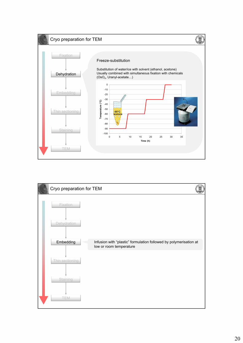
Freeze-substitution
Substitution of water/ice with solvent (ethanol, acetone)Usually combined with simultaneous fixation with chemicals(OsO4, Uranyl-acetate…)
Cryo preparation for TEM
-100
-90
-80
-70
-60
-50
-40
-30
-20
-10
0
0 5 10 15 20 25 30 35
Time (h)
Te
mp
era
ture
(°C
)
-90°Cacetone-90°Cacetone
Embedding
Fixation
Dehydration
Staining
Thin sectioning
TEM
Infusion with “plastic” formulation followed by polymerisation at low or room temperature
Cryo preparation for TEM

21
Embedding
Fixation
Dehydration
Staining
Thin sectioning
TEM
Same procedure as RT
Cryo preparation for TEM
Embedding
Fixation
Dehydration
Staining
Thin sectioning
TEM
Reduced extraction of cell constituentsReduced shrinkage
Mechanical effectsLoss of LipidsShrinkage during polymerisation
Compression, knife marks
Interaction of heavy metals with biology provides electron density
Interpretation/orientation
Cryo preparation for TEM
No RT fixation artefactsIce crystal damage possible

22
Specimen courtesy of Bettina Sobottka, Neurologische Klinik, University of Zurich
Room temperature vs. cryo preparation
500 nm
Conventionally fixed (glutaraldehyde) High pressure frozen
Thin sections of plastic embedded mouse cerebellum
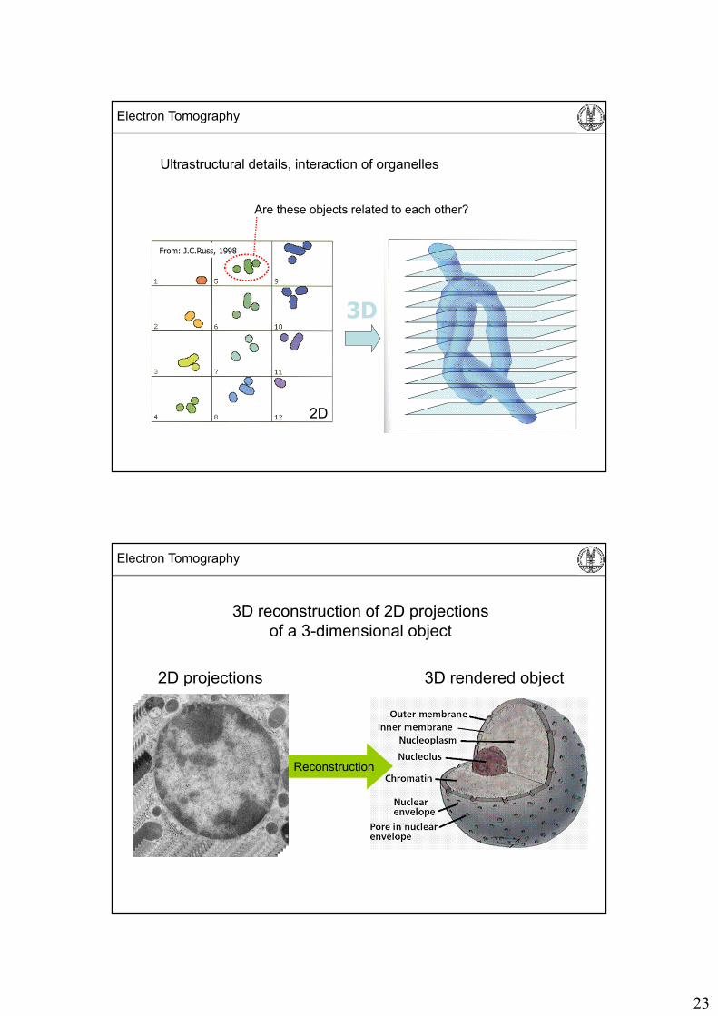
How do we get to 3D
23
3D
From: J.C.Russ, 1998
Ultrastructural details, interaction of organelles
Are these objects related to each other?
2D
Electron Tomography
3D reconstruction of 2D projectionsof a 3-dimensional object
2D projections 3D rendered object
Reconstruction
Electron Tomography

24
3D reconstruction based on:
I) Imaging of serial sections
Thickness of section for EM: 50 - 300 nm
Z resolution limited to section thickness
Imaging in TEM Imaging in SEM
Electron Tomography
Electron Tomography
3D reconstruction based on:
I) Imaging of serial sections
Focused ion beam SEMAblation of material with Ga-ions
Ultra-microtome in SEM

25
Electron Tomography
High-pressure frozen and embedded mouse brain (hippocampal slice culture)
II) Projection of a tilt series
Thickness of sections for tilt series: ~70 - 300 nm
Z resolution depending on number of different projections: 5 - 20 nm
NOTE: A combination of both methods is possible
3D high resolution of several successive sections
Imaging in TEM (RT or CRYO)
3D reconstruction based on:
Electron Tomography

26
Marsh 2005
Aligned projections of aPancreatic beta cell line: Double-tilt series, ±60°, 1.5° increment, section 400 nm thick
Electron Tomography
Calculation of tomograms based on projections – virtual sections
Marsh 2005
Electron Tomography

27
Rendering of segmented structures
Marsh 2005
Electron Tomography