Critical role of RAGE and HMGB1 in inflammatory heart … murine cardiac TnI induces severe...
10
Critical role of RAGE and HMGB1 in inflammatory heart disease Anna Bangert a,1 , Martin Andrassy a,1 , Anna-Maria Müller a , Mariella Bockstahler a , Andrea Fischer a , Christian H. Volz a , Christoph Leib a , Stefan Göser a , Sevil Korkmaz-Icöz b , Stefan Zittrich c , Andreas Jungmann a , Felix Lasitschka d , Gabriele Pfitzer c , Oliver J. Müller a,e , Hugo A. Katus a,e , and Ziya Kaya a,e,2 a Department of Internal Medicine III, University of Heidelberg, 69120 Heidelberg, Germany; b Department of Cardiac Surgery, University of Heidelberg, 69120 Heidelberg, Germany; c Institute of Vegetative Physiology, University of Cologne, 50931 Cologne, Germany; d Institute of Pathology, University of Heidelberg, 69120 Heidelberg, Germany; and e DZHK (German Centre for Cardiovascular Research), Partner Site Heidelberg/Mannheim, University of Heidelberg, 69120 Heidelberg, Germany Edited by Michael T. Lotze, University of Pittsburgh Cancer Institute, Pittsburgh, PA, and accepted by the Editorial Board November 30, 2015 (received for review November 12, 2015) Autoimmune response to cardiac troponin I (TnI) induces inflam- mation and fibrosis in the myocardium. High-mobility group box 1 (HMGB1) is a multifunctional protein that exerts proinflammatory activity by mainly binding to receptor for advanced glycation end products (RAGE). The involvement of the HMGB1–RAGE axis in the pathogenesis of inflammatory cardiomyopathy is yet not fully un- derstood. Using the well-established model of TnI-induced exper- imental autoimmune myocarditis (EAM), we demonstrated that both local and systemic HMGB1 protein expression was elevated in wild-type (wt) mice after TnI immunization. Additionally, phar- macological inhibition of HMGB1 using glycyrrhizin or anti-HMGB1 antibody reduced inflammation in hearts of TnI-immunized wt mice. Furthermore, RAGE knockout (RAGE-ko) mice immunized with TnI showed no structural or physiological signs of cardiac impairment. Moreover, cardiac overexpression of HMGB1 using adeno-associated virus (AAV) vectors induced inflammation in the hearts of both wt and RAGE-ko mice. Finally, patients with myocarditis displayed increased local and systemic HMGB1 and soluble RAGE (sRAGE) expression. Together, our study highlights that HMGB1 and its main receptor, RAGE, appear to be crucial factors in the pathogenesis of TnI-induced EAM, because inhibition of HMGB1 and ablation of RAGE suppressed inflammation in the heart. Moreover, the proinflammatory effect of HMGB1 is not nec- essarily dependent on RAGE only. Other receptors of HMGB1 such as Toll-like receptors (TLRs) may also be involved in disease pathogen- esis. These findings could be confirmed by the clinical relevance of HMGB1 and sRAGE. Therefore, blockage of one of these molecules might represent a novel therapeutic strategy in the treatment of au- toimmune myocarditis and inflammatory cardiomyopathy. myocarditis | cytokines | AAV I nflammatory cardiomyopathy is a relatively common cause of acute heart failure in the young, for which an efficient and specific therapy is lacking. Although most patients recover com- pletely, some present a deteriorating course. Recent work from our laboratory and others has focused on the dysregulation of the immune system as an essential modulator of disease induction and progression in heart failure (1, 2). In this context, release of cardiac troponin I (TnI) from damaged cardiomyocytes into the circulation is believed to trigger an autoimmune response to TnI (3, 4). Our group has established an animal model in which immunization with murine cardiac TnI induces severe myocardial inflammation and fibrosis, followed by severe heart failure (1). However, the ex- act pathomechanism and immune modulators involved in this inflammatory process are not yet fully elucidated. Extensive work has cast light on the role of high-mobility group box 1 (HMGB1) in the pathogenesis of infectious and noninfectious inflammatory diseases. HMGB1, first described as a DNA binding protein, has subsequently been associated with various pathological conditions such as cardiovascular disease (5, 6), cancer (7), and ischemia/reperfusion (I/R) injury (5). It is a key modulator of innate immune responses and regulates in part adaptive immunity (8). In response to cellular stress, HMGB1 acts as a damage-associated molecular pattern (DAMP) signal after passive release into the extracellular milieu during cell death or active secretion by mononuclear and other cell types (7). It binds to receptors such as receptor for advanced glycation end products (RAGE) and Toll-like receptors (TLRs) such as TLR-2 and -4, leading to the expression of inflammatory cytokines, chemokines, and corresponding receptors (9). Some studies describe differential effects of HMGB1- or RAGE-dependent signaling with regard to their concentration and release in a particular model or mode of application (10, 11). In a rodent model of myocardial infarction, exogenously ad- ministered HMGB1 had a beneficial effect on postinfarct myo- cardial remodeling (10). Kitahara and colleagues demonstrated reduced necrosis and smaller infarct size after myocardial in- farction in transgenic mice overexpressing HMGB1 (12). How- ever, in a murine model of I/R injury, our group recently showed that treatment of wild-type (wt) mice with recombinant HMGB1 increased the infarct size (6). In the same model of I/R injury, Significance Myocardial inflammation leads in many cases to cardiomyopathy and contributes to progressive heart failure. The exact patholog- ical mechanism of disease induction and progression in the setting of heart failure is unknown. High-mobility group box 1 (HMGB1), an evolutionarily abundant and highly conserved protein, pro- motes cardiac inflammation, and in turn immunity, as a damage- associated molecular pattern. HMGB1 stimulates immunity, at least in part, through interaction with its principal binding partner RAGE (receptor for advanced glycation end products). Here we show that HMGB1 and RAGE appear to be important components in cardiac troponin I-induced experimental autoimmune myocar- ditis as well as in patients with myocarditis. Both molecules rep- resent potential drug targets and show significant potential in heart failure treatment. Author contributions: A.B., M.A., C.H.V., O.J.M., and Z.K. designed research; A.B., A.-M.M., M.B., A.F., S.G., S.K.-I., A.J., F.L., G.P., and Z.K. performed research; S.K.-I., S.Z., A.J., F.L., G.P., and O.J.M. contributed new reagents/analytic tools; A.B., M.A., A.-M.M., M.B., A.F., C.H.V., C.L., S.G., S.Z., O.J.M., H.A.K., and Z.K. analyzed data; and A.B., M.A., A.-M.M., M.B., A.F., C.H.V., C.L., O.J.M., H.A.K., and Z.K. wrote the paper. The authors declare no conflict of interest. This article is a PNAS Direct Submission. M.T.L. is a guest editor invited by the Editorial Board. Freely available online through the PNAS open access option. 1 A.B. and M.A. contributed equally to this work. 2 To whom correspondence should be addressed. Email: [email protected]. This article contains supporting information online at www.pnas.org/lookup/suppl/doi:10. 1073/pnas.1522288113/-/DCSupplemental. www.pnas.org/cgi/doi/10.1073/pnas.1522288113 PNAS | Published online December 29, 2015 | E155–E164 IMMUNOLOGY AND INFLAMMATION PNAS PLUS
Transcript of Critical role of RAGE and HMGB1 in inflammatory heart … murine cardiac TnI induces severe...

Critical role of RAGE and HMGB1 in inflammatoryheart diseaseAnna Bangerta,1, Martin Andrassya,1, Anna-Maria Müllera, Mariella Bockstahlera, Andrea Fischera, Christian H. Volza,Christoph Leiba, Stefan Gösera, Sevil Korkmaz-Icözb, Stefan Zittrichc, Andreas Jungmanna, Felix Lasitschkad,Gabriele Pfitzerc, Oliver J. Müllera,e, Hugo A. Katusa,e, and Ziya Kayaa,e,2
aDepartment of Internal Medicine III, University of Heidelberg, 69120 Heidelberg, Germany; bDepartment of Cardiac Surgery, University of Heidelberg,69120 Heidelberg, Germany; cInstitute of Vegetative Physiology, University of Cologne, 50931 Cologne, Germany; dInstitute of Pathology, University ofHeidelberg, 69120 Heidelberg, Germany; and eDZHK (German Centre for Cardiovascular Research), Partner Site Heidelberg/Mannheim, University ofHeidelberg, 69120 Heidelberg, Germany
Edited by Michael T. Lotze, University of Pittsburgh Cancer Institute, Pittsburgh, PA, and accepted by the Editorial Board November 30, 2015 (received forreview November 12, 2015)
Autoimmune response to cardiac troponin I (TnI) induces inflam-mation and fibrosis in the myocardium. High-mobility group box 1(HMGB1) is a multifunctional protein that exerts proinflammatoryactivity by mainly binding to receptor for advanced glycation endproducts (RAGE). The involvement of the HMGB1–RAGE axis in thepathogenesis of inflammatory cardiomyopathy is yet not fully un-derstood. Using the well-established model of TnI-induced exper-imental autoimmune myocarditis (EAM), we demonstrated thatboth local and systemic HMGB1 protein expression was elevatedin wild-type (wt) mice after TnI immunization. Additionally, phar-macological inhibition of HMGB1 using glycyrrhizin or anti-HMGB1antibody reduced inflammation in hearts of TnI-immunized wtmice. Furthermore, RAGE knockout (RAGE-ko) mice immunizedwith TnI showed no structural or physiological signs of cardiacimpairment. Moreover, cardiac overexpression of HMGB1 usingadeno-associated virus (AAV) vectors induced inflammation inthe hearts of both wt and RAGE-ko mice. Finally, patients withmyocarditis displayed increased local and systemic HMGB1 andsoluble RAGE (sRAGE) expression. Together, our study highlightsthat HMGB1 and its main receptor, RAGE, appear to be crucialfactors in the pathogenesis of TnI-induced EAM, because inhibitionof HMGB1 and ablation of RAGE suppressed inflammation in theheart. Moreover, the proinflammatory effect of HMGB1 is not nec-essarily dependent on RAGE only. Other receptors of HMGB1 suchas Toll-like receptors (TLRs) may also be involved in disease pathogen-esis. These findings could be confirmed by the clinical relevance ofHMGB1 and sRAGE. Therefore, blockage of one of these moleculesmight represent a novel therapeutic strategy in the treatment of au-toimmune myocarditis and inflammatory cardiomyopathy.
myocarditis | cytokines | AAV
Inflammatory cardiomyopathy is a relatively common causeof acute heart failure in the young, for which an efficient and
specific therapy is lacking. Although most patients recover com-pletely, some present a deteriorating course. Recent work fromour laboratory and others has focused on the dysregulation ofthe immune system as an essential modulator of disease inductionand progression in heart failure (1, 2). In this context, release ofcardiac troponin I (TnI) from damaged cardiomyocytes into thecirculation is believed to trigger an autoimmune response toTnI (3, 4).Our group has established an animal model in which immunization
with murine cardiac TnI induces severe myocardial inflammation andfibrosis, followed by severe heart failure (1). However, the ex-act pathomechanism and immune modulators involved in thisinflammatory process are not yet fully elucidated.Extensive work has cast light on the role of high-mobility
group box 1 (HMGB1) in the pathogenesis of infectious andnoninfectious inflammatory diseases. HMGB1, first described asa DNA binding protein, has subsequently been associated withvarious pathological conditions such as cardiovascular disease (5,
6), cancer (7), and ischemia/reperfusion (I/R) injury (5). It is akey modulator of innate immune responses and regulates in partadaptive immunity (8). In response to cellular stress, HMGB1acts as a damage-associated molecular pattern (DAMP) signalafter passive release into the extracellular milieu during cell deathor active secretion by mononuclear and other cell types (7). It bindsto receptors such as receptor for advanced glycation end products(RAGE) and Toll-like receptors (TLRs) such as TLR-2 and -4,leading to the expression of inflammatory cytokines, chemokines,and corresponding receptors (9).Some studies describe differential effects of HMGB1- or
RAGE-dependent signaling with regard to their concentrationand release in a particular model or mode of application (10, 11).In a rodent model of myocardial infarction, exogenously ad-ministered HMGB1 had a beneficial effect on postinfarct myo-cardial remodeling (10). Kitahara and colleagues demonstratedreduced necrosis and smaller infarct size after myocardial in-farction in transgenic mice overexpressing HMGB1 (12). How-ever, in a murine model of I/R injury, our group recently showedthat treatment of wild-type (wt) mice with recombinant HMGB1increased the infarct size (6). In the same model of I/R injury,
Significance
Myocardial inflammation leads in many cases to cardiomyopathyand contributes to progressive heart failure. The exact patholog-ical mechanism of disease induction and progression in the settingof heart failure is unknown. High-mobility group box 1 (HMGB1),an evolutionarily abundant and highly conserved protein, pro-motes cardiac inflammation, and in turn immunity, as a damage-associated molecular pattern. HMGB1 stimulates immunity, atleast in part, through interaction with its principal binding partnerRAGE (receptor for advanced glycation end products). Here weshow that HMGB1 and RAGE appear to be important componentsin cardiac troponin I-induced experimental autoimmune myocar-ditis as well as in patients with myocarditis. Both molecules rep-resent potential drug targets and show significant potential inheart failure treatment.
Author contributions: A.B., M.A., C.H.V., O.J.M., and Z.K. designed research; A.B., A.-M.M.,M.B., A.F., S.G., S.K.-I., A.J., F.L., G.P., and Z.K. performed research; S.K.-I., S.Z., A.J., F.L.,G.P., and O.J.M. contributed new reagents/analytic tools; A.B., M.A., A.-M.M., M.B., A.F.,C.H.V., C.L., S.G., S.Z., O.J.M., H.A.K., and Z.K. analyzed data; and A.B., M.A., A.-M.M.,M.B., A.F., C.H.V., C.L., O.J.M., H.A.K., and Z.K. wrote the paper.
The authors declare no conflict of interest.
This article is a PNAS Direct Submission. M.T.L. is a guest editor invited by the EditorialBoard.
Freely available online through the PNAS open access option.1A.B. and M.A. contributed equally to this work.2To whom correspondence should be addressed. Email: [email protected].
This article contains supporting information online at www.pnas.org/lookup/suppl/doi:10.1073/pnas.1522288113/-/DCSupplemental.
www.pnas.org/cgi/doi/10.1073/pnas.1522288113 PNAS | Published online December 29, 2015 | E155–E164
IMMUNOLO
GYAND
INFLAMMATION
PNASPL
US

RAGE-deficient mice demonstrated significantly reduced myo-cardial damage compared with wt mice (6).The effect of HMGB1 and RAGE on the pathogenesis of cardiac
disorders is not only described in preclinical animal models but isalso investigated in human cardiac disorders. Different studies haverevealed an elevated HMGB1 level in patients with heart failurecorrelating with disease severity (13–17). In addition to this, somestudies have identified RAGE as a prognostic factor in human heartfailure (16, 18, 19).In the present study, we aimed to clarify the role of HMGB1 and
RAGE in an experimental model of murine autoimmune myocar-ditis. This experimental approach enables a reproducible sterilecardiac inflammation, as described previously (1, 20). Furthermore,the clinical relevance of both proteins should be investigated.Hence, we first studied the expression kinetics of HMGB1 in the
inflamed myocardium and serum of wt mice. Then, inhibition ofHMGB1 by glycyrrhizin (GL) was performed and heart tissue wasanalyzed in TnI-induced experimental autoimmune myocarditis(EAM) of wt mice. Additionally, RAGE knockout (RAGE-ko)mice were immunized with TnI to further study the role of RAGEsignaling in this EAM model. Finally, the role of the HMGB1–RAGE axis in our model was analyzed by an adeno-associatedvirus (AAV)9-mediated cardiac overexpression of HMGB1. In alast step, we studied the clinical relevance of HMGB1 and RAGEin patients with myocarditis.
ResultsTnI Immunization InducedMyocardial Inflammation and Fibrosis, IncreasedMyocardial HMGB1 Protein Expression, and Impaired Cardiac Function.First, an EAM was induced in mice (Fig. S1). TnI immunization
led to sustained myocardial inflammation and fibrosis, as evidencedby hematoxylin and eosin (HE) and Masson’s trichrome (MT)staining and evaluated as grade of inflammation/fibrosis, re-spectively [HE: TnI, day (d)21: 1.9 ± 0.35; d90: 2 ± 0.5; d270:2.3 ± 0.4; MT: TnI, d21: 1.2 ± 0.5; d90: 2.3 ± 0.5; d270: 2.3 ± 0.4].Control buffer-immunized group (CFA) showed neither inflamednor fibrotic tissue (Fig. 1 A and B).Furthermore, TnI immunization up-regulated myocardial HMGB1
protein expression on days 21, 90, and 270, as evaluated by quanti-tative analysis of immunohistochemical data. In heart tissues of CFA-immunized mice, no myocardial HMGB1 protein could be detected(Fig. 1 A and B).Western blot analysis showed a significantly increased myo-
cardial HMGB1 protein level in TnI-immunized mice comparedwith the CFA-treated group (Fig. 1C).The HMGB1 protein level in serum was also increased in TnI-
immunized mice compared with CFA groups over a period of270 d [TnI, d21: 36.4 ± 5.4 ng/mL; d90: 47.2 ± 11.3 ng/mL; d270:32.0 ± 5.6 ng/mL vs. CFA, d21: 19.3 ± 5.4 ng/mL, P < 0.05; d90:17.5 ± 1.7 ng/mL, P < 0.01; d270: 19.6 ± 9.9 ng/mL, not signif-icant (ns); Fig. 1D].
TnI Immunization Activated Specific Signaling Pathways. Signaltransducer and activator of transcription (STAT) 3 and c-JunN-terminal kinase (JNK) were analyzed by Western blot. Signifi-cantly increased levels of phosphorylated (p) compared with total(t) protein were detectable for STAT3 and JNK in the early phaseof inflammation (day 21) in the hearts of TnI-immunized mice. Atlater stages of disease (>day 90), p-STAT3 and p-JNK levels were
Fig. 1. TnI immunization increased HMGB1 protein level and activated specific signal transduction pathways in myocardium of wt mice. (A) Myocardialhistoscore of CFA- or TnI-immunized mice for inflammation (HE), fibrosis (MT), and HMGB1 expression on days 21, 90, and 270. (B) Corresponding macroscopicpictures (Left) and histopathological examinations (Right) of immunized mice. [Scale bars, 100 μm (columns 2–4) and 25 μm (column 5).] (C) Immunoblot andfold increase of HMGB1 compared with glyceraldehyde-3-phosphate dehydrogenase (GAPDH) in cardiac proteins of TnI-immunized mice on days 0, 21, 90,and 270. (D) Serum HMGB1 level in immunized mice. (E and F) Immunoblot and fold increase of p-STAT3 compared with t-STAT3 (E) and p-JNK compared witht-JNK (F) in cardiac proteins of TnI-immunized mice on days 0, 21, 90, and 270. Error bars indicate mean ± SEM. *P < 0.05, **P < 0.01, ***P < 0.005.
E156 | www.pnas.org/cgi/doi/10.1073/pnas.1522288113 Bangert et al.
decreased but still significantly increased compared with CFA-immunized mice (Fig. 1 E and F).
HMGB1 Inhibition Decreased TnI-Induced Myocardial Inflammation.To study the preventive and therapeutic effect of the HMGB1 in-hibition by glycyrrhizin, mice were treated with glycyrrhizin before(GL) and starting with day 14 (GL14) after TnI immunization.TnI-immunized mice treated with glycyrrhizin displayed im-
proved ejection fraction (EF) as well as significantly reducedhigh-sensitive troponin T (hs-TnT) levels and myocardial in-flammation compared with TnI-immunized and PBS-treatedanimals in both GL and GL14 groups (hs-TnT: PBS/TnI: 308.0 ±20.6 pg/mL vs. GL: 105.0 ± 12.6 pg/mL, P < 0.05, and vs. GL14:82.2 ± 22.5 pg/mL, P < 0.005; EF: PBS/TnI: 82.3 ± 1.7% vs. GL:93.5 ± 2.7%, P < 0.05, and vs. GL14: 83.2 ± 1.4%, ns; in-flammation score: PBS/TnI: 2.8 ± 0.2 vs. GL: 1.2 ± 0.5, P < 0.05,and vs. GL14: 0.8 ± 0.5, P < 0.05; Fig. 2 A–C).HMGB1 inhibition was also analyzed by the administration of
anti-HMGB1 antibody (Ab-HMGB1) as well as control antibody(Ab-Cont.) to TnI-immunized mice. Immunized mice treated withAb-HMGB1 showed a reduced hs-TnT level and inflammation aswell as an improved EF compared with Ab-Cont.–treated mice(hs-TnT: Ab-Cont.: 257.5 ± 91.5 pg/mL vs. Ab-HMGB1: 48.9 ±22.4 pg/mL, P < 0.1; EF: Ab-Cont.: 79.8 ± 2.9% vs. Ab-HMGB1:84.0 ± 1.0%, P < 0.1; inflammation score: Ab-Cont.: 1.6 ± 0.5 vs.Ab-HMGB1: 0.9 ± 0.3, P < 0.05; Fig. 2 D–F).
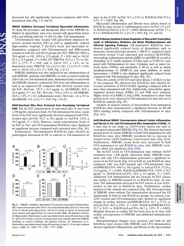
RAGE-Knockout Mice Were Protected from Developing TnI-InducedEAM. The hs-TnT concentration of untreated mice at day 0 was<100 pg/mL (detection limit). In TnI-immunized wt mice, serumlevels of hs-TnT were significantly elevated compared with CFA-treated mice (wt/TnI: 312.7 ± 56.1 pg/mL vs. wt/CFA: 124.0 ±10.2 pg/mL, P < 0.01). However, serum concentration levels ofhs-TnT in TnI- and CFA-immunized RAGE-ko mice displayedno significant increase compared with untreated mice (Fig. 3A).Furthermore, TnI-immunized RAGE-ko mice showed no
pathological alterations in EF in contrast to TnI-immunized wt
mice on day 21 (EF: wt/TnI: 74.1 ± 2.2% vs. RAGE-ko/TnI: 87.2 ±0.7%, P < 0.001; Fig. 3B).Myocardial inflammation and fibrosis were almost absent in
RAGE-ko mice on day 21 (inflammation score: wt/TnI: 1.9 ± 0.3vs. RAGE-ko/TnI: 0.2 ± 0.1, P < 0.005; fibrosis score: wt/TnI: 1.7 ±0.3 vs. RAGE-ko/TnI: 0.2 ± 0.1, P < 0.01; Fig. 3 C and D).
RAGE Deficiency Resulted in Down-Regulation of Myocardial TranscriptionLevels of Inflammatory Mediators and Matrix Metalloproteinases andAffected Signaling Pathways. TnI-immunized RAGE-ko miceshowed significantly reduced levels of chemokines such asmonocyte chemoattractant protein 1 (MCP-1), regulated uponactivation normal T-cell expressed and secreted (RANTES),macrophage inflammatory protein (MIP) 1α andMIP-1β, as well aschemokine (C-C motif) receptors (CCRs) such as CCR1/2/5 com-pared with TnI-immunized wt mice. Cytokines such as tumor ne-crosis factor (TNFα) and interleukin 6 (IL-6) as well as matrixmetalloproteinase 14 (MMP-14) and tissue inhibitor of metal-loproteinase 1 (TIMP-1) also displayed significantly reduced levelscompared with TnI-immunized wt mice (Fig. 3E).Next, the activity of STAT3 and Janus kinase 2 (JAK2) on day
21 was analyzed. We observed that RAGE-ko mice showed de-creased activation of STAT3/JAK2 signaling compared with wtmice when immunized with TnI. Additionally, extracellular signal-regulated protein kinase (ERK) 1/2 and JNK were analyzed.Higher levels of p-ERK1/2 and p-JNK, normalized to their totalprotein level, were observed in TnI-immunized wt mice but not inRAGE-ko animals (Fig. 3F).Analysis of nuclear extracts of myocardium from immunized
RAGE-ko mice demonstrated a significant decrease in NF-κBDNA binding activity compared with TnI-immunized wt mice(Fig. 3G).
AAV9-Mediated HMGB1 Overexpression Induced Cardiac Inflammationand Fibrosis in TnI- and CFA-Immunized Mice Independent of RAGE. Asa next step in our study, an AAV9-vector system was used tooverexpress myocardial HMGB1 (Fig. S1). We detected increasedprotein levels of cardiac HMGB1 in both CFA-immunized wt andRAGE-ko mice after HMGB1 treatment but no increase incontrol vector (AAV-luciferase; Luc)-treated mice (Fig. S2A).Furthermore, HMGB1 serum levels were increased in bothCFA-immunized wt and RAGE-ko mice after HMGB1 treat-ment, albeit not significant (Fig. S2B).The hs-TnT levels in CFA-immunized mice without AAV9
treatment were <100 pg/mL (detection limit). HMGB1 treat-ment and only CFA immunization generated a significant in-crease in hs-TnT levels (Fig. 4A) in both wt and RAGE-ko micecompared with Luc-AAV–treated and CFA-immunized mice(wt/HMGB1/CFA: 448.9 ± 50.5 pg/mL vs. wt/Luc/CFA: 112.0 ±12.0 pg/mL, P < 0.005; RAGE-ko/HMGB1/CFA: 510.0 ± 94.3pg/mL vs. RAGE-ko/Luc/CFA: 92.0 ± 4.9 pg/mL, P < 0.05).Additional TnI immunization did not increase hs-TnT releaseany further in HMGB1-treated wt or RAGE-ko mice. In con-trast, TnI immunization increased hs-TnT release in Luc-AAV–
treated wt but not in RAGE-ko mice. Furthermore, cardiacfunction of the animals was evaluated (Fig. 4B). Overexpressionof HMGB1 alone without TnI immunization decreased the EFsignificantly in both wt and RAGE-ko groups. In contrast, Luc-AAV–treated and CFA-immunized mice showed no significantchange in cardiac function (wt/HMGB1/CFA: 41.7 ± 9.7% vs.wt/Luc/CFA: 86.9 ± 0.9%, P < 0.01; RAGE-ko/HMGB1/CFA:53.7 ± 9.4% vs. RAGE-ko/Luc/CFA: 86.5 ± 1.0%, P < 0.005).Maximal decrease in cardiac EF has been seen in wt mice withcardiac overexpression of HMGB1 and additional immunizationwith TnI.Histopathological changes were assessed, and both wt and
RAGE-ko mice treated with HMGB1 and CFA immunizationshowed significant inflammation and fibrosis in the myocardium
Fig. 2. HMGB1 inhibition decreased TnI-induced myocardial inflammation.Mice were immunized with CFA or TnI and treated daily (GL21) or from day14 to 21 (GL14) with glycyrrhizin. (A) hs-TnT levels in serum of immunizedmice treated with glycyrrhizin or control buffer (PBS). (B) Ejection fraction.(C) Myocardial inflammation score was determined using HE staining on day21. (D) hs-TnT levels in serum of immunized mice treated with anti-HMGB1antibody or control antibody. (E) Ejection fraction. (F) Histoscore of in-flammation was determined using HE staining on day 21. Error bars indicatemean ± SEM. *P < 0.05, **P < 0.01, ***P < 0.005.
Bangert et al. PNAS | Published online December 29, 2015 | E157
IMMUNOLO
GYAND
INFLAMMATION
PNASPL
US

compared with Luc-AAV–treated mice (inflammation score:wt/HMGB1/CFA: 0.7 ± 0.2 vs. wt/Luc/CFA: 0.1 ± 0.1, P < 0.01;RAGE-ko/HMGB1/CFA: 1.5 ± 0.2 vs. RAGE-ko/Luc/CFA:0.3 ± 0.2, P < 0.01; fibrosis score: wt/HMGB1/CFA: 1.9 ± 0.2vs. wt/Luc/CFA: 0.4 ± 0.2, P < 0.005; RAGE-ko/HMGB1/CFA:2.8± 0.5 vs. RAGE-ko/Luc/CFA: 0.8± 0.1, P< 0.01; Fig. 4C andD).Hence, TnI-immunized RAGE-ko mice showed a significant
increase in myocardial inflammation and fibrosis when HMGB1was overexpressed. However, this effect was lower than in TnI-immunized and HMGB1-treated wt mice. Hearts treated withLuc-AAV and immunized with CFA were almost free from
proinflammatory and profibrotic structural changes. Representa-tive macroscopic and microscopic findings of AAV9 vectors(HMGB1 or Luc) and CFA- or TnI-immunized mice are shownin Fig. 4E.HMGB1 overexpression alone without TnI immunization led to
cardiac damage, inflammation, fibrosis, and deteriorated cardiacperformance. Therefore, all further animal studies were performedin CFA-immunized mice.
Myocardial AAV9-Mediated HMGB1 Overexpression Altered theTranscription of Diverse Immunomodulators, Activated IntracellularSignaling Pathways, and Enhanced NF-κB Binding Activity. HMGB1
Fig. 3. RAGE-ko mice were protected from developing TnI-induced EAM. Wt and RAGE-ko mice were immunized with CFA or TnI. Measurements were done onday 21. (A) hs-TnT levels in serum of immunized mice. (B) Ejection fraction. (C) Histoscore of inflammation and fibrosis in the hearts of immunized mice.(D) Representative macroscopic pictures (Left) and histopathological examinations (Right) of hearts stained with HE and MT. [Scale bars, 3 mm (column 2) and 100μm (columns 3 and 4).] (E) Myocardial mRNA levels of genes involved in cardiac inflammation. (F) Heart tissues were analyzed by Western blot, and fold increaseof phosphorylated compared with total STAT3, JAK2, ERK1/2, and JNK proteins was thereby detected. (G) Electrophoretic mobility shift assay was performed tomeasure the NF-κB binding activity in myocardium. Specificity of NF-κB binding activity was shown by including a 160-fold molar excess of unlabeled consensusNF-κB oligonucleotide (Cons.). Fold increase of NF-κB compared to Cons. was calculated. Error bars indicate mean ± SEM. *P < 0.05, **P < 0.01, ***P < 0.005.
E158 | www.pnas.org/cgi/doi/10.1073/pnas.1522288113 Bangert et al.

Fig. 4. HMGB1 treatment induced cardiac damage and affected activation of inflammation-related genes and signaling cascades in wt and RAGE-ko mice.Wt and RAGE-ko mice were treated with AAV9-HMGB1 or control vector (Luc) and were immunized with CFA or TnI. Measurements were done on day 21.(A) hs-TnT production in serum of immunized mice upon AAV9-vector treatment. (B) Ejection fraction. (C) Inflammation. (D) Fibrosis score. (E and F) Rep-resentative macroscopic pictures (Left) and histopathological examinations (Right) of hearts stained with HE and MT. [Scale bars, 3 mm (column 2) and 100 μm(columns 3 and 4).] (G) Myocardial mRNA levels were analyzed by qPCR. (H) Heart tissues were analyzed by Western blot, and fold increase of phosphorylatedcompared with total STAT3, JAK2, ERK1/2, and JNK proteins was thereby detected. (I) Electrophoretic mobility shift assay was performed to measure the NF-κBbinding activity in myocardium. Specificity of NF-κB binding activity was shown by including a 160-fold molar excess of unlabeled consensus NF-κB oligo-nucleotide (Cons.). Fold increase of NFkB compared to Cons. was calculated. Error bars indicate mean ± SEM. *P < 0.05, **P < 0.01, ***P < 0.005.
Bangert et al. PNAS | Published online December 29, 2015 | E159
IMMUNOLO
GYAND
INFLAMMATION
PNASPL
US
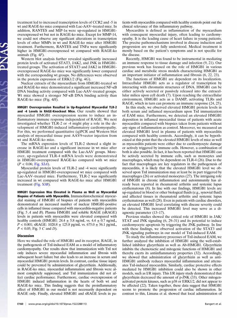
treatment led to increased transcription levels of CCR2 and -5 inwt and RAGE-ko mice compared with Luc-AAV–treated mice. Inaddition, RANTES and MIP-1α were up-regulated in HMGB1-overexpressed wt but not in RAGE-ko mice. Except for MMP-14,we could not observe any significant alterations in transcriptionlevels of other MMPs in wt and RAGE-ko mice after HMGB1treatment. Furthermore, RANTES and TNFα were significantlyhigher in HMGB1-overexpressed wt compared with RAGE-koanimals (Fig. 4F).Western blot analysis further revealed significantly increased
protein levels of activated STAT3, JAK2, and JNK in HMGB1-treated groups. The activation of STAT3 and JAK2 in HMGB1-overexpressed RAGE-ko mice was significantly lower comparedwith the corresponding wt groups. No differences were observedin the protein expression of ERK1/2 (Fig. 4G).Nuclear extracts of the myocardium from HMGB1-treated wt
and RAGE-ko mice demonstrated a significant increased NF-κBDNA binding activity compared with Luc-AAV–treated groups.Wt mice showed a stronger NF-κB DNA binding activity thanRAGE-ko mice (Fig. 4H).
HMGB1 Overexpression Resulted in Up-Regulated Myocardial TLR-2and -4 Levels in RAGE-Knockout Mice. Our results showed thatmyocardial HMGB1 overexpression seems to induce an in-flammatory immune response independent of RAGE. We nextinvestigated whether TLR-2 or -4 might play a role in the de-velopment of HMGB1-induced inflammatory cardiomyopathy.For this, we performed quantitative (q)PCR and Western blotanalysis of myocardial tissue post AAV9-vector injection fromwt and RAGE-ko mice.The mRNA expression levels of TLR-2 showed a slight in-
crease in RAGE-ko and a significant increase in wt mice afterHMGB1 treatment compared with the Luc-AAV groups. More-over, up-regulated TLR-4 mRNA levels were demonstratedin HMGB1-overexpressed RAGE-ko compared with wt mice(P < 0.06; Fig. S3A).Myocardial protein levels of TLR-2 and -4 were significantly
up-regulated in HMGB1-overexpressed wt mice compared withLuc-AAV–treated mice. Furthermore, TLR-2 was significantlyincreased in wt compared with RAGE-ko mice after HMGB1treatment (Fig. S3B).
HMGB1 Expression Was Elevated in Plasma as Well as MyocardialBiopsies of Patients with Myocarditis. Immunohistochemical myocar-dial staining of HMGB1 of biopsies of patients with myocarditisdemonstrated an increased number of nuclear HMGB1-positivecells in inflamed tissue compared with myocarditis-negative patients(Fig. 5 A and B). Plasma HMGB1 and soluble RAGE (sRAGE)levels in patients with myocarditis were elevated compared withhealthy controls (HMGB1: 24.4 ± 5.8 ng/mL vs. 4.4 ± 0.7 ng/mL,P < 0.001; sRAGE: 1020.8 ± 125.8 pg/mL vs. 675.0 ± 56.1 pg/mL,P < 0.05; Fig. 5 C and D).
DiscussionHere we studied the role of HMGB1 and its receptor, RAGE, inthe pathogenesis of TnI-induced EAM as a model of inflammatorycardiomyopathy. Our results show that immunization with TnI notonly induces severe myocardial inflammation and fibrosis withsubsequent heart failure but also leads to an increase in serum andmyocardial HMGB1 protein levels. In contrast, cardiac tissue injurycould be prevented by administration of glycyrrhizin. Additionally,in RAGE-ko mice, myocardial inflammation and fibrosis were al-most completely suppressed, and TnI immunization did not af-fect cardiac performance. Furthermore, cardiac overexpression ofHMGB1 induced inflammation in the hearts of both wt andRAGE-ko mice. This finding suggests that the proinflammatoryeffect of HMGB1 in our model is not necessarily dependent onRAGE only. Finally, elevated HMGB1 and sRAGE levels in pa-
tients with myocarditis compared with healthy controls point out theclinical relevance of this inflammatory pathway.Myocarditis is defined as inflammation of the myocardium
with consequent myocardial injury, often leading to cardiomy-opathy. It is the leading cause of heart failure in young patients.The precise pathomechanisms involved in disease induction andprogression are not yet fully understood. Medical treatment ismostly based on the patient’s symptoms and is not specific forthe disease.Recently, HMGB1 was found to be instrumental in mediating
an immune response to tissue damage and infection (9, 21). Ourprevious work has focused on the role of HMGB1 in variouscardiac and metabolic stress models characterizing HMGB1 asan important initiator of inflammation and fibrosis (6, 22, 23).The functions of HMGB1 are dependent on its localization.
Intracellular HMGB1 acts as a regulator of transcription byinteracting with chromatin structures of DNA. HMGB1 can beeither actively secreted or passively released into the extracel-lular milieu upon cell death (7). Upon reaching the extracellularenvironment, HMGB1 acts as a DAMP, activating TLRs andRAGE, which in turn can promote an immune response (24, 25).In this study, we observed elevated HMGB1 protein levels in
both serum and inflamed myocardium upon TnI immunizationof EAM mice. Furthermore, we detected an elevated HMGB1deposition in inflamed myocardial tissue of patients with acutemyocarditis compared with biopsies taken from patients withoutany signs of inflammation as control. Moreover, we measured anelevated HMGB1 level in plasma of patients with myocarditiscompared with healthy controls. Accordingly, it can be hypoth-esized at this point that the elevated HMGB1 levels of mice as wellas myocarditis patients were either due to cardiomyocyte damageor actively triggered by immune cells. However, a combination ofboth is also possible. In fact, it has been reported that HMGB1 canbe actively secreted by immune cells, such as monocytes andmacrophages, which may be dependent on TLR-4 (26). Due to thefact that macrophages are key regulators in the pathogenesis ofmyocarditis, it is likely that the elevated HMGB1 level we ob-served upon TnI immunization may at least be in part triggered bymacrophages (26) or activated monocytes (27). The intriguing roleof HMGB1 in chronic inflammation and autoimmunity has al-ready been reported in rheumatoid arthritis and systemic lupuserythematosus (8). In line with our findings, HMGB1 levels areup-regulated in blood or other biological fluids (e.g., synovial fluid)and affected tissues in rheumatoid arthritis and systemic lupuserythematosus as well (28). Even in patients with cardiac disorders,an elevated HMGB1 level correlating with disease severity couldbe detected. This increased HMGB1 level may serve as a di-agnostic parameter (13–17).Previous studies showed the critical role of HMGB1 in JAK/
STAT and JNK signaling (6, 29–31) and its potential to inducecardiomyocyte apoptosis by triggering the JNK pathway. In linewith these findings, we observed activation of the STAT3 andJNK signaling pathways in our model of TnI-induced EAM.To study the inflammatory processes of TnI-induced EAM, we
further analyzed the inhibition of HMGB1 using the well-estab-lished inhibitor glycyrrhizin as well as Ab-HMGB1. Glycyrrhizininhibits the chemotactic and mitogenic functions of HMGB1 andthereby exerts its antiinflammatory properties (32). Accordingly,we showed that administration of glycyrrhizin as well as anti-HMGB1 antibody reduces myocardial inflammation and attenu-ates TnI-induced myocarditis. Similarly, cardiac protective effectsmediated by HMGB1 inhibition could also be shown in othermodels, such as I/R injury. This I/R injury study demonstrated thatglycyrrhizin decreased the amount of p-JNK (33). Other mitogen-activated protein kinases, such as p38 or ERK1/2, did not appear tobe affected (22). Taken together, these data suggest that HMGB1seems to promote the progression of cardiac inflammation. Incontrast to this, Limana et al. showed that local administration of
E160 | www.pnas.org/cgi/doi/10.1073/pnas.1522288113 Bangert et al.

low doses of HMGB1 induced an improvement in cardiac functionas well as myocardial regeneration after myocardial infarction (10).These controversial data may be ascribed to different experimentalsetups in the two studies, which is already discussed by Ramasamyet al. (29). Moreover, the type of cell death seems to influence theHMGB1 protein level, which is important for the induction of ei-ther cardiac repair or remodeling (30).In our study, we further investigated the role of RAGE in TnI-
induced autoimmune myocarditis. Several studies point out theactivation of RAGE by HMGB1 (7, 24). Altered protein levels ofRAGE isoforms in patients suffering from heart failure alsorefer to the clinical relevance of this pathway (16, 18, 19).Therefore, based on the EAM model, our results show for thefirst time, to our knowledge, that RAGE-ko mice are almostcompletely protected from developing TnI-induced autoimmunemyocarditis. Ablation of RAGE resulted in significantly de-creased levels of several inflammatory mediators such as
chemokines, chemokine receptors, MMPs, and cytokines, whichare important for the pathogenesis of autoimmune myocarditis(20). Additionally, NF-κB binding activity was reduced in im-munized RAGE-ko mice.RAGE-dependent sustained NF-κB activation has been im-
plicated in various chronic inflammatory diseases (34, 35). Thedownstream effects on regulating transcriptional levels ofproinflammatory mediators upon RAGE ligation have beenreviewed recently, and our results strongly suggest that the pro-tective effect observed in RAGE-ko mice was due to suppressionof RAGE downstream signaling (36). Sirois and colleagues showedthat RAGE promotes DNA uptake into endosomes and lowers theimmune recognition threshold for the activation of TLR-9, theprincipal DNA-recognizing transmembrane signaling receptor(37). This potentially can lead to undesirable autoimmunedisorders such as systemic lupus erythematosus (38).
Fig. 5. Myocarditis patients displayed increased myocardial expression of HMGB1 and elevated levels of HMGB1 and sRAGE in plasma. (A) The numberof nuclear HMGB1-positive cells was increased in the myocardium of patients with myocarditis (n = 13) compared with those of the control group (n = 11).(B) Representative histopathological stainings (HMGB1 and HE) of endomyocardial biopsies (three biopsies for each patient shown). [Scale bars, 500 μm(columns 1–3) and 100 μm (column 4).] (C and D) Patients with myocarditis (n = 10) displayed significantly elevated levels of HMGB1 (C) and sRAGE (D) in theirplasma compared with healthy controls (n = 11). Error bars indicate mean ± SEM. *P < 0.05, ***P < 0.005.
Bangert et al. PNAS | Published online December 29, 2015 | E161
IMMUNOLO
GYAND
INFLAMMATION
PNASPL
US
Data on the role of the HMGB1–RAGE axis in lymphoidcells, especially in an autoimmune setting, remain scarce. Initialfindings were published by Tian et al., who demonstrated acritical role for HMGB1 in RAGE-dependent activation ofplasmacytoid dendritic cells and B cells in concert with TLR-9driving autoimmune processes (39). It has also previously beenshown that RAGE deficiency affected maturation and migra-tion of dendritic cells and their cross-talk with natural killer(NK) cells, which is pivotal for sustaining innate immunity andinitiating a subsequent adaptive immune response (40, 41). Inaddition, RAGE deficiency has been linked to drastically re-duced T-cell activation and differentiation, leading to sup-pressed B-cell response (40, 42).This led to the hypothesis that the blunted immune response
in RAGE-ko mice could be caused by inhibited activation ofB-cells or reduced inflammatory cell recruitment and chemotaxis.More research needs to be done to identify the exact signalingmechanisms and cells involved in this RAGE-mediated process.To further analyze the role of HMGB1 signaling in TnI-induced
EAM, we overexpressed HMGB1 specifically in the heart using anAAV9-vector system. In AAV9 vectors, the HMGB1 expressioncassette is under the control of a cytomegalovirus (CMV)-enhanced 260-bp myosin light chain (MLC)-2v promoter, whichensures cardiomyocyte-specific expression (43, 44). Here weobserved myocardial inflammation and fibrosis independent ofTnI immunization in both HMGB1-overexpressing wt and inRAGE-ko mice, suggesting that HMGB1 may induce cardiacinflammation by mechanisms independent of RAGE. Therewere no signs of inflammation in other organs. Our resultsstrongly suggest that the inflammatory response we observed inHMGB1-overexpressing RAGE-ko animals seems to be at leastin part triggered by the activation of STAT3, JNK, and JAK2,because we observed elevated levels of those signaling moleculesin HMGB1-overexpressing, CFA-immunized RAGE-ko mice.Because the observed proinflammatory effect of HMGB1 wasnot dependent on RAGE, we assessed whether TLRs might beinvolved. TLRs as well as RAGE belong to the family of pattern-recognition receptors (PRRs) and are components of innateimmunity (7, 24, 25). TLRs are known to be involved in sensingdifferent exogenous foreign molecular products, known asPAMPs (pathogen-associated molecular patterns), whereasRAGE is predominantly involved in the recognition of endoge-nous molecules (7). Indeed, we observed an up-regulation ofTLR-2 and -4 when HMGB1 was overexpressed. In this context,Paulus et al. showed recently that polyethylene particles, whichact as a PAMP, induce TLR-2 up-regulation in the synovial layerof mice (45). Furthermore, HMGB1–nucleosome complexes areknown to activate the immune response through TLR-2 signaling(46).Because HMGB1 is known to act as a DAMP when passively
released to the extracellular milieu upon tissue injury/destruction, wehypothesize that in our model it up-regulates TLR-2 and -4 eitherdirectly by intercalating TLR transcription via its DNA binding ca-pacity or indirectly by affecting cytokine expression levels.Both HMGB1-treated and CFA-immunized wt and RAGE-ko
groups displayed increased mRNA levels of nearly all measured cy-tokines/chemokines and their receptors. We could not show anysignificant difference between these groups, and hence TLRs mightbe responsible for this release. In fact, several studies showed thatmainly TLR-4 is required for HMGB1-mediated cytokine production(47). We also detected significantly enhanced RANTES,MCP-1, andMIP-1α mRNA levels in HMGB1-treated wt in comparison withRAGE-ko mice. This may indicate an additional RAGE-, TLR-2–,or other receptor-dependent cytokine activating pathway. In thiscontext, Bianchi et al. showed a RAGE-dependent up-regulation ofchemokines, including RANTES and MIP-1α, in microglia (48).Another study showed that an HMGB1–nucleosome complex me-diated inflammatory cytokine activation through TLR-2 (46).
Because HMGB1 can also bind to other cytokines, such as IL-1β,the inflammatory response can also be mediated through thereceptors of partner molecules (7). We further observed a sig-nificant difference between wt and RAGE-ko HMGB1-treatedmice in TLR-2 protein levels. These effects may indicate an in-teraction between RAGE and TLR-2. This could be possible dueto an interaction between RAGE and a typical TLR adaptorprotein, MyD88 (31, 49). Furthermore, this could also indicatethe presence of increased HMGB1 immune complexes causedduring necrosis, such as HMGB1–DNA (39) mediated by RAGEand HMGB1–nucleosome, which mainly signals through TLR-2(46).Moreover, increased TLR-4 protein levels were observed in
both HMGB1-treated wt and RAGE-ko mice. These might in-dicate that the HMGB1 in our study is in a reduced form, whichmight stimulate TLR-4 and finally results in enhanced cytokinerelease (24, 47). Additional studies are strongly needed to clearlyunravel the role of HMGB1–receptor activity in the pathogenesisof inflammatory cardiomyopathy.In summary, we showed that there is a significant expression of
HMGB1 in the myocardium of TnI-immunized wt mice. This wasassociated with increased HMGB1 levels in the serum of thesemice. Inhibition of HMGB1 resulted in protection of tissuedamage in TnI-induced EAM. Furthermore, ablation of RAGEnearly abolished TnI-induced myocardial inflammation/fibrosisand consecutive heart failure. This protective effect might beassociated with impaired activation of NF-κB and decreasedmRNA expression levels of inflammatory cytokines, chemokines,and chemokine receptors. In addition, our study demonstratesthat myocardial overexpression of HMGB1 alone seems to beable to induce myocardial inflammation independent of RAGE.HMGB1 and sRAGE seem to play an important role also inpatients with acute myocarditis, because we demonstrated ele-vated HMGB1 and sRAGE levels in plasma and increased ex-pression of HMGB1 in myocardial biopsies of patients sufferingfrom acute myocarditis.Additional studies are necessary to further investigate the exact
pathomechanism of these findings. Our study provides valuableinformation on the role of HMGB1 and RAGE in the pathogenesisof myocardial inflammation. Targeting the HMGB1–RAGE axismight be a promising novel tool for the treatment of inflammatorycardiomyopathy.
Materials and MethodsMice. Female A/J wild-type mice were obtained from Envigo. RAGE-ko micewere backcrossed to the A/J background for at least six generations (50).All mice were maintained in the animal facility unit of the University ofHeidelberg. In all experiments, 5- to 6-wk-old female mice were used. TheAnimal Care and Use Committee of the University of Heidelberg approvedall procedures involving the use and care of animals (German Animal Pro-tection Code G-142/09).
Preparation of Recombinant Murine Cardiac Troponin I. The expression ofmurine cardiac troponin subunit I was performed via Escherichia coli and pu-rified as described previously (51). In addition to purification via ion-exchangechromatography, TnI was applied to a cardiac troponin C affinity column asa second purification step (52). TnI-containing fractions were dialyzed against1 mmol/L HCl, and then lyophilized and stored at −80 °C.
Immunization with TnI. All mice were treated with an s.c. injection of 100 μLemulsion, which contained either 150 μg murine cardiac TnI in supple-mented complete Freund’s adjuvant with 5 mg/mL Mycobacterium tuber-culosis H37Ra (Sigma) or 1× PBS control buffer in adjuvant alone. We namedthese control buffer-immunized groups “CFA.”
Glycyrrhizin Treatment. Glycyrrhizin (10 mg/kg; Sigma) was administered dailyby i.p. injection, from day 0 to 21 or from day 14 to 21, in addition to TnI orCFA immunization (days 0 and 7). Mice were killed on day 21. Buffer solution(1× PBS) was used as control. The dose of glycyrrhizin was chosen on thebasis of our pilot studies.
E162 | www.pnas.org/cgi/doi/10.1073/pnas.1522288113 Bangert et al.

Anti-HMGB1 Antibody Treatment. Ab-HMGB1 (anti-HMGB1 antibody 2G7) waskindly provided by Kevin Tracey, The Feinstein Institute for Medical Research,Manhasset, NY. As a control antibody, mouse IgG2b-κ (Sigma) was used. Thewt mice were treated i.p. with 50 μg antibody every second day for 3 wk inaddition to the TnI immunization (days 0 and 7) and were killed on day 21.
Plasmid Construction and AAV9 Vector. Gene synthesis of a codon-optimizedmurine HMGB1 sequence (National Center for Biotechnology Informationreference no. AAH64790.1) was performed by GeneArt. At the 5′ end a re-striction site for BamHI and a Kozak sequence were added, whereas at the 3′end a restriction site for BsrGI was introduced. The cDNA of Renilla luciferasewas amplified from the plasmid pGL4 from Promega with primers carrying aBamHI site at the 5′ end and a BsrGI site at the 3′ end. Both cDNAs wereinserted using BamH1/BsrGI sites into the vector genome plasmid pdsCMV-MLC260-EGFP (44), resulting in pdsCMV-MLC260-HMGB1 (AAV9-HMGB1;HMGB1) and pdsCMV-MLC260-luciferase (AAV9-luciferase; Luc). Self-com-plementary AAV9 vectors were generated by cotransfection of the re-spective genome plasmids with pDP9rs, a derivate from pDP2rs (53) with theAAV9 cap gene from p5E18-VD2-9. Vectors were purified as described be-fore (43, 44).
Experimental Setup. The experimental setup for all groups is summarized inFig. S1 and Table S1.
i) Wt mice were immunized with either TnI or CFA on days 0, 7, 60, and 245and killed on day 21, 90, or 270, as described previously (1).
ii) HMGB1 inhibition was analyzed in immunized wt mice by using glycyr-rhizin over a period of 21 d (TnI/GL) and for a therapeutic approach fromday 14 to 21 (TnI/GL14). Immunization with TnI or CFA was done on days0 and 7 and mice were killed on day 21.
Furthermore, HMGB1 inhibition was analyzed in immunized wt micetreated with Ab-HMGB1 or Ab-Cont. Immunization with TnI was performedon days 0 and 7 and mice were killed on day 21.
iii) Wt or RAGE-ko mice were immunized with either TnI or CFA on days0 and 7 and killed on day 21.
iv) Wt or RAGE-ko mice received a systemic single dose of 0.5–1.0 × 10¹²
AAV9-HMGB1 vector or AAV9-luciferase vector by i.v. injection via thetail vein 2 wk before (day −14) the initial TnI immunization on day 0. Asecond immunization was performed on day 7. Thirty-five days postAAV9-vector transfer, all mice were killed for further analysis.
Determination of hs-TnT, HMGB1, and sRAGE Serum/Plasma Protein Levels. hs-TnT assay was used as an indicator for myocardial damage in serum of mice21 d after treatment with either TnI or CFA. Collected serum samples werediluted (1:20) with cold 0.9% NaCl solution. The hs-TnT was measured by theelectrochemiluminescence method (ECLIA; Elecsys 2010 analyzer). Details ofthe test principle have been described before (54).
For a quantitative determination of HMGB1protein in serum/plasma, a high-sensitivity sandwich ELISA (Shino-Test) was performed according to the man-ufacturer’s instructions (55). ELISA for detection of sRAGE levels (BioVendor) inplasma was performed according to the manufacturer’s instructions.
Histopathology and Immunohistochemistry. Mice were killed by cervical dis-location and all hearts were removed, fixed in formalin [10% (vol/vol)], andsubsequently embedded in paraffin. Sections of 3- to 5-μm thickness werecut and stained with hematoxylin and eosin to determine the level of in-flammation. Additionally, Masson’s trichrome staining was performed todetect collagen deposition to assess the grade of fibrosis. Five sections ofeach heart were inspected in a double-blinded manner by two independentinvestigators by light microscopy to evaluate inflammation and fibrosis.
Immunohistochemical HMGB1 staining of murine and human heart sec-tions was performed with rabbit polyclonal anti-HMGB1 antibody as theprimary reagent (1:250; Abcam) and biotinylated donkey anti-rabbit anti-body (1:200; Dianova) as the secondary reagent. An avidin-biotin-alkalinephosphatase system (ABC-AP; Dako) was used according to the manufac-turer’s instructions. Parallel incubations with nonimmune IgGs of the rele-vant species served as negative control.
HE-, MT-, and HMGB1-stained sections were analyzed as described in TableS2. To quantify the HMGB1 expression in human heart sections, TMARKERsoftware was used as previously described (56).
Quantitative PCR. An RNeasy Mini Kit (Qiagen) was used according to themanufacturer’s instructions to isolate total RNA from hearts. One microgram
RNA was used to synthesize cDNA (iScript cDNA Synthesis Kit; Bio-Rad).Quantitative PCR was then performed using 25 ng cDNA in a 20-μL reactionvolume. A denaturation step at 95 °C for 5 min was carried out, followed by40 cycles of denaturation at 95 °C for 10 s and annealing at 60 °C for 30 s.The primer sequences are listed in Table S3. Quantification was done byusing a QuantiFast SYBR Green PCR Kit (Qiagen) according to the manu-facturer’s instructions and using an iCycler iQ2 Detection System (Bio-Rad).The measured gene expressions were normalized to the expression of thereference gene L32.
Echocardiography. Echocardiographic measurements of the first experimentalapproach (i) were performed with an ATL HDI 9000 (Philips) ultrasonographwith a 10-MHz annular array transducer as previously described (1).
In experimental approaches (ii), (iii), and (iv), echocardiography wasperformed using a VisualSonics Vevo 2100 System, 30-MHz linear MicroScantransducer (MSH400), especially optimized for cardiovascular experiments inmice. Parasternal long-axis projection cine loops were acquired at the levelof clear visible walls of the aortic annulus. Furthermore, parasternal short‐axis cine loops were performed at the level of the papillary muscles. Storeddata of the B-mode and M-mode images were executed and analyzed by asingle operator using a proprietary software package provided with theVevo 2100 platform. The presented percentage ejection fraction was mea-sured in the long axis and calculated using formulas of the Cardiac Mea-surement Package (Vevo 2100 1.5.0). Echocardiography was conducted ina blinded manner.
Western Blotting. For Western blot analysis, flash-frozen heart tissues wereground in RIPA buffer (0.1% SDS, 1.0% Triton X-100, 0.1% sodium deoxy-cholate, 140 mM NaCl) supplemented with Halt Protease and PhosphataseInhibitor Cocktail (Life Technologies). The tissue suspension was passed threetimes through a 25G needle to lyse the membrane. The tubes were centri-fuged at 18,000 × g for 15 min. The supernatant was used for the de-termination of protein concentration using Protein Assay BradfordReagent (Bio-Rad).
Twenty micrograms of total myocardial protein extract per lane was sep-arated on NuPAGE Bis-Tris gels (Invitrogen) and transferred to a polyvinylidenefluoride (PVDF) transfer membrane (Immobilon-P; Millipore). The membranewas first blocked [5% (wt/vol) bovine serum albumin/Tris-buffered saline] for1 h at room temperature (RT) and then incubated with a primary antibody for1 h at RT (Table S4). After repeated washing steps, the blots were incubatedwith secondary antibody (horseradish peroxidase-coupled anti-rabbit IgG,1:3,000) for 1 h at RT. Protein bands were detected using a chemiluminescenceagent (GE Healthcare). Quantitative analysis was performed with ImageJsoftware (NIH). GAPDH served as a loading control.
Electrophoretic Mobility Shift Assay. The binding activity of p65-NF-κB innuclear protein extracts (70 μg) of heart tissue was assayed with 32P radio-actively labeled oligonucleotide sequences (5′-AGT TGA GGG GAC TTT CCCAGG C-3′) at 50,000 cpm by autoradiography. The experiment is based onelectrophoretic mobility of a protein–nucleic acid complex, which migratesmore slowly than the corresponding free nucleic acid, and was performed aspreviously described (57).
Human Samples. Endomyocardial biopsies from patients with myocarditisaccording to the Dallas criteria were used for staining (HMGB1 and HE). Inaddition, blood samples were taken for HMGB1 and sRAGE detection. Bi-opsies taken from patients with no signs of inflammation and blood samplesfrom healthy donors served as controls.
The study was conducted in accordance with the Declaration of Helsinkiand was approved by the local ethics committee of the University of Hei-delberg. All subjects included in the study providedwritten informed consent.
Statistical Analysis. Results are expressed as mean ± SEM. Data were analyzedusing the Mann–Whitney U and two-way ANOVA tests. Values of P <0.05were considered statistically significant and are marked as *P < 0.05,**P < 0.01, ***P < 0.005.
ACKNOWLEDGMENTS. The authors acknowledge Dr. Angelika Bierhaus andher collaborators for sharing the RAGE-ko mice, Dr. Kevin Tracey (TheFeinstein Institute for Medical Research) for kindly providing the anti-HMGB1 antibody, and Dr. Tanja Weis as well as Dr. Alamara Karimi forproviding the human plasma samples. We thank Kirsten Keilbach, RenateÖttl, Vesna Vukovic, Karl Varadi, Patrick Heger, and Annette Buttler fortechnical assistance, and David Stanmore for critically reading the manu-script. This work was supported by the Deutsche Forschungsgemeinschaft
Bangert et al. PNAS | Published online December 29, 2015 | E163
IMMUNOLO
GYAND
INFLAMMATION
PNASPL
US
Research Grants AN 403/2-1 (to Z.K. and M.A.), in part KA 1797/7-1 (to Z.K.)and the Sonderforschungsbereich 612 (to G.P.). Furthermore, the DZHK
(German Centre for Cardiovascular Research) and the BMBF (German Minis-try of Education and Research) (Z.K. and O.M.) partly supported this work.
1. Göser S, et al. (2006) Cardiac troponin I but not cardiac troponin T induces severeautoimmune inflammation in the myocardium. Circulation 114(16):1693–1702.
2. Kaya Z, et al. (2001) Contribution of the innate immune system to autoimmunemyocarditis: A role for complement. Nat Immunol 2(8):739–745.
3. Eriksson S, Halenius H, Pulkki K, Hellman J, Pettersson K (2005) Negative interferencein cardiac troponin I immunoassays by circulating troponin autoantibodies. Clin Chem51(5):839–847.
4. Leuschner F, et al. (2008) Absence of auto-antibodies against cardiac troponin I pre-dicts improvement of left ventricular function after acute myocardial infarction. EurHeart J 29(16):1949–1955.
5. Oozawa S, et al. (2008) Effects of HMGB1 on ischemia-reperfusion injury in the ratheart. Circ J 72(7):1178–1184.
6. Andrassy M, et al. (2008) High-mobility group box-1 in ischemia-reperfusion injury ofthe heart. Circulation 117(25):3216–3226.
7. Sims GP, Rowe DC, Rietdijk ST, Herbst R, Coyle AJ (2010) HMGB1 and RAGE in in-flammation and cancer. Annu Rev Immunol 28:367–388.
8. Andersson U, Tracey KJ (2011) HMGB1 is a therapeutic target for sterile inflammationand infection. Annu Rev Immunol 29:139–162.
9. Lotze MT, Tracey KJ (2005) High-mobility group box 1 protein (HMGB1): Nuclearweapon in the immune arsenal. Nat Rev Immunol 5(4):331–342.
10. Limana F, et al. (2005) Exogenous high-mobility group box 1 protein induces myo-cardial regeneration after infarction via enhanced cardiac C-kit+ cell proliferationand differentiation. Circ Res 97(8):e73–e83.
11. Rossini A, et al. (2008) HMGB1-stimulated human primary cardiac fibroblasts exert aparacrine action on human and murine cardiac stem cells. J Mol Cell Cardiol 44(4):683–693.
12. Kitahara T, et al. (2008) High-mobility group box 1 restores cardiac function aftermyocardial infarction in transgenic mice. Cardiovasc Res 80(1):40–46.
13. Kohno T, et al. (2009) Role of high-mobility group box 1 protein in post-infarctionhealing process and left ventricular remodelling. Cardiovasc Res 81(3):565–573.
14. Liu T, et al. (2015) Increased serum HMGB1 level may predict the fatal outcomes inpatients with chronic heart failure. Int J Cardiol 184:318–320.
15. Volz HC, et al. (2012) HMGB1 is an independent predictor of death and hearttransplantation in heart failure. Clin Res Cardiol 101(6):427–435.
16. Wang LJ, et al. (2011) Increased serum high-mobility group box-1 and cleaved re-ceptor for advanced glycation endproducts levels and decreased endogenous secre-tory receptor for advanced glycation endproducts levels in diabetic and non-diabeticpatients with heart failure. Eur J Heart Fail 13(4):440–449.
17. Yan XX, et al. (2009) Increased serum HMGB1 level is associated with coronary arterydisease in nondiabetic and type 2 diabetic patients. Atherosclerosis 205(2):544–548.
18. Lu L, et al. (2009) Increased glycated albumin and decreased esRAGE levels are relatedto angiographic severity and extent of coronary artery disease in patients with type 2diabetes. Atherosclerosis 206(2):540–545.
19. Koyama Y, et al. (2008) Soluble receptor for advanced glycation end products (RAGE)is a prognostic factor for heart failure. J Card Fail 14(2):133–139.
20. Kaya Z, et al. (2008) Identification of cardiac troponin I sequence motifs leading toheart failure by induction of myocardial inflammation and fibrosis. Circulation118(20):2063–2072.
21. Bianchi ME (2007) DAMPs, PAMPs and alarmins: All we need to know about danger.J Leukoc Biol 81(1):1–5.
22. Volz HC, Kaya Z, Katus HA, Andrassy M (2010) The role of HMGB1/RAGE in in-flammatory cardiomyopathy. Semin Thromb Hemost 36(2):185–194.
23. Andrassy M, et al. (2011) HMGB1 as a predictor of infarct transmurality and functionalrecovery in patients with myocardial infarction. J Intern Med 270(3):245–253.
24. Harris HE, Andersson U, Pisetsky DS (2012) HMGB1: A multifunctional alarmin drivingautoimmune and inflammatory disease. Nat Rev Rheumatol 8(4):195–202.
25. Yang H, Tracey KJ (2010) Targeting HMGB1 in inflammation. Biochim Biophys Acta1799(1-2):149–156.
26. Wang F, et al. (2010) Fas (CD95) induces rapid, TLR4/IRAK4-dependent release of pro-inflammatory HMGB1 from macrophages. J Inflamm (Lond) 7:30.
27. Gardella S, et al. (2002) The nuclear protein HMGB1 is secreted by monocytes via anon-classical, vesicle-mediated secretory pathway. EMBO Rep 3(10):995–1001.
28. Magna M, Pisetsky DS (2014) The role of HMGB1 in the pathogenesis of inflammatoryand autoimmune diseases. Mol Med 20:138–146.
29. Ramasamy R, Yan SF, Schmidt AM (2008) Stopping the primal RAGE reaction inmyocardial infarction: Capturing adaptive responses to heal the heart? Circulation117(25):3165–3167.
30. Vogel S, et al. (2015) Necrotic cell-derived high mobility group box 1 attracts antigen-presenting cells but inhibits hepatocyte growth factor-mediated tropism of mesen-chymal stem cells for apoptotic cell death. Cell Death Differ 22(7):1219–1230.
31. Rojas A, et al. (2013) The receptor for advanced glycation end-products: A complexsignaling scenario for a promiscuous receptor. Cell Signal 25(3):609–614.
32. Mollica L, et al. (2007) Glycyrrhizin binds to high-mobility group box 1 protein andinhibits its cytokine activities. Chem Biol 14(4):431–441.
33. Zhai CL, et al. (2012) Glycyrrhizin protects rat heart against ischemia-reperfusion in-jury through blockade of HMGB1-dependent phospho-JNK/Bax pathway. ActaPharmacol Sin 33(12):1477–1487.
34. Bierhaus A, et al. (2004) Loss of pain perception in diabetes is dependent on a re-ceptor of the immunoglobulin superfamily. J Clin Invest 114(12):1741–1751.
35. Schmidt AM, Yan SD, Yan SF, Stern DM (2001) The multiligand receptor RAGE as aprogression factor amplifying immune and inflammatory responses. J Clin Invest108(7):949–955.
36. van Zoelen MA, Achouiti A, van der Poll T (2011) RAGE during infectious diseases.Front Biosci (Schol Ed) 3:1119–1132.
37. Belinsky GS, et al. (2014) Patch-clamp recordings and calcium imaging followed bysingle-cell PCR reveal the developmental profile of 13 genes in iPSC-derived humanneurons. Stem Cell Res (Amst) 12(1):101–118.
38. Horton CG, Pan ZJ, Farris AD (2010) Targeting Toll-like receptors for treatment of SLE.Mediators Inflamm 2010:498980.
39. Tian J, et al. (2007) Toll-like receptor 9-dependent activation by DNA-containingimmune complexes is mediated by HMGB1 and RAGE. Nat Immunol 8(5):487–496.
40. Dumitriu IE, et al. (2005) Release of high mobility group box 1 by dendritic cellscontrols T cell activation via the receptor for advanced glycation end products.J Immunol 174(12):7506–7515.
41. Semino C, Angelini G, Poggi A, Rubartelli A (2005) NK/iDC interaction results in IL-18secretion by DCs at the synaptic cleft followed by NK cell activation and release of theDC maturation factor HMGB1. Blood 106(2):609–616.
42. Chen Y, et al. (2008) RAGE ligation affects T cell activation and controls T cell dif-ferentiation. J Immunol 181(6):4272–4278.
43. Müller OJ, et al. (2006) Improved cardiac gene transfer by transcriptional and trans-ductional targeting of adeno-associated viral vectors. Cardiovasc Res 70(1):70–78.
44. Müller OJ, Schinkel S, Kleinschmidt JA, Katus HA, Bekeredjian R (2008) Augmentationof AAV-mediated cardiac gene transfer after systemic administration in adult rats.Gene Ther 15(23):1558–1565.
45. Paulus AC, et al. (2014) Polyethylene wear particles induce TLR 2 upregulation in thesynovial layer of mice. J Mater Sci Mater Med 25(2):507–513.
46. Urbonaviciute V, et al. (2008) Induction of inflammatory and immune responses byHMGB1-nucleosome complexes: Implications for the pathogenesis of SLE. J Exp Med205(13):3007–3018.
47. Yang H, et al. (2010) A critical cysteine is required for HMGB1 binding to Toll-likereceptor 4 and activation of macrophage cytokine release. Proc Natl Acad Sci USA107(26):11942–11947.
48. Bianchi R, Kastrisianaki E, Giambanco I, Donato R (2011) S100B protein stimulatesmicroglia migration via RAGE-dependent up-regulation of chemokine expression andrelease. J Biol Chem 286(9):7214–7226.
49. Sakaguchi M, et al. (2011) TIRAP, an adaptor protein for TLR2/4, transduces a signalfrom RAGE phosphorylated upon ligand binding. PLoS One 6(8):e23132.
50. Bierhaus A, et al. (2001) Diabetes-associated sustained activation of the transcriptionfactor nuclear factor-kappaB. Diabetes 50(12):2792–2808.
51. Krüger M, Pfitzer G, Stehle R (2003) Expression and purification of human cardiactroponin subunits and their functional incorporation into isolated cardiac mousemyofibrils. J Chromatogr B Analyt Technol Biomed Life Sci 786(1-2):287–296.
52. al-Hillawi E, Minchin SD, Trayer IP (1994) Overexpression of human cardiac troponin-Iand troponin-C in Escherichia coli and their purification and characterisation. Twopoint mutations allow high-level expression of troponin-I. Eur J Biochem 225(3):1195–1201.
53. Grimm D, Kay MA, Kleinschmidt JA (2003) Helper virus-free, optically controllable,and two-plasmid-based production of adeno-associated virus vectors of serotypes 1 to6. Mol Ther 7(6):839–850.
54. Andrassy M, et al. (2012) HMGB1 is associated with atherosclerotic plaque composi-tion and burden in patients with stable coronary artery disease. PLoS One 7(12):e52081.
55. Yamada S, Yakabe K, Ishii J, Imaizumi H, Maruyama I (2006) New high mobility groupbox 1 assay system. Clin Chim Acta 372(1-2):173–178.
56. Schüffler PJ, et al. (2013) TMARKER: A free software toolkit for histopathological cellcounting and staining estimation. J Pathol Inform 4(Suppl):S2.
57. Andrassy M, et al. (2006) Posttranslationally modified proteins as mediators of sus-tained intestinal inflammation. Am J Pathol 169(4):1223–1237.
E164 | www.pnas.org/cgi/doi/10.1073/pnas.1522288113 Bangert et al.


![Severe valvular and congenital heart diseases in adults€¦ · term condition] scheme, severe valvular heart disease. Valvular heart diseases are very diverse and require different](https://static.fdocuments.us/doc/165x107/600678272dffc94bfc1e40e5/severe-valvular-and-congenital-heart-diseases-in-term-condition-scheme-severe.jpg)