Investigation into the role of HMGb1 in relation to ...
234
Investigation into the role of HMGb1 in relation to myofibroblasts and cancer cells exposed to various conditions Sikander Sharma A thesis submitted in partial fulfilment of the requirements of Liverpool John Moores University for the degree of Doctor of Philosophy July, 2015
Transcript of Investigation into the role of HMGb1 in relation to ...
Investigation into the role of HMGb1 in relation to myofibroblasts and cancer cells exposed to
various conditions
Sikander Sharma
A thesis submitted in partial fulfilment of the requirements of Liverpool John Moores University for the degree of Doctor of
Philosophy
July, 2015

Table of Contents
Acknowledgement.......................................................................................................... i
Abstract .......................................................................................................................... ii
List of Figures .............................................................................................................. iii
List of Abbreviations .................................................................................................... vi
Chapter 1: Cancer
1.1. Carcinogenesis ............................................................................................................. 1
1.1.1. Metastasis in cancer ............................................................................................ 3
1.1.2. Colon adenocarcinoma ....................................................................................... 5
1.1.3. Hypoxia ............................................................................................................... 7
1.2. The tumour microenvironment ..................................................................................... 11
1.2.1. Extracellular matrix (ECM) remodelling ............................................................. 14
1.2.2. Cellular cross talk in the tumour microenvironment ........................................... 18
1.2.3. The interstitial fluid pressure in tumours ............................................................ 19
1.2.4. Hypoxia and glucose deprivation in solid tumours ............................................. 20
1.3. High mobility group box 1 (HMGb1) protein ................................................................. 22
1.3.1. Role of HMGb1 in disease ................................................................................ 25
1.3.2. The role of HMGb1 in cancer ............................................................................ 29
1.3.3. HMGb1 and its receptors .................................................................................. 32
1.3.4. Receptor for advanced glycation end products (RAGE) .................................... 33
1.3.5. RAGE and Tumour Microenvironment .............................................................. 36

1.3.6. RAGE in cancer ................................................................................................ 37
1.3.7. RAGE and HMGb1 ........................................................................................... 40
1.3.8. Toll like Receptors ............................................................................................ 42
1.3.9. TLRs and HMGb1 ............................................................................................. 43
1.4. Myofibroblasts ............................................................................................................. 45
1.4.1. Myofibroblasts and cancer ................................................................................ 50
1.4.2. Role of Myofibroblasts in metastasis ................................................................. 53
1.4.3. Interplay between myofibroblasts, the ECM and growth factors ........................ 55
1.4.4. Myofibroblasts differentiation in the tumour microenvironment .......................... 57
1.5. Aims and objectives .................................................................................................... 59
Chapter 2: General Materials and Methods
2.1. Recombinant proteins, antibodies, antibiotics and cells ............................................... 61
2.2. Cell culture .................................................................................................................. 61
2.2.1. Cryopreservation ............................................................................................. 62
2.2.2. Treatment of CCD18 cells with recombinant HMGb1 ...................................... 63
2.2.3. Treatment of CCD18 cells with recombinant HMGb1 and PI3K or
MEK1/2 inhibitors (LY294002 or U0126) ......................................................... 64
2.2.4. Assessment of proliferation using the neutral red uptake (NRU) assay ........... 65
2.2.5. Assessment of toxicity and proliferation using the
MTT (3-(4, 5-dimethylthiazol-2-yl)-2, 5-diphenyltetrazolium bromide) assay ... 66
2.3. Protein Assay .............................................................................................................. 67
2.3.1. Sample preparation from CCD18, HT29, MCF-7, EJ138 and A549 cells ...... 67
2.3.2. The extraction of proteins from the cells ...................................................... 68
2.3.3. Determination of the sample protein concentration ....................................... 69
2.3.4. Sodium dodecyl sulphate polyacrylamide gel electrophoresis (SDS–PAGE). 70

2.3.5. Western blotting ............................................................................................ 71
2.3.6. Blot development .......................................................................................... 73
2.3.7. Dot Blotting ................................................................................................... 74
2.3.8. Quantitative Analysis of Western Blots ......................................................... 74
2.4. Migration assay ........................................................................................................... 75
2.5. Invasion assay ............................................................................................................ 77
2.6. Statistics ...................................................................................................................... 77
2.7. Table of inhibitors and antibodies used in Migration and Invasion assays ................... 78
Chapter 3: Investigation into the role of recombinant protein HMGb1 in CCD18 myofibroblasts proliferation
3.1. Introduction ................................................................................................................. 79
3.2. Aims and Objectives .................................................................................................... 82
3.3. Materials and Methods ................................................................................................ 82
3.3.1. CCD18 myofibroblasts cell culture ................................................................ 82
3.3.2. Treatment of CCD18 cells with recombinant HMGb1 and PI3K and
ERK1/2 inhibitors U0126 and LY294002) ................................................... 83
3.3.3. Assessment of proliferation using the neutral red uptake (NRU) assay ......... 84
3.3.4. Assessment of toxicity and proliferation using the MTT (3-(4,5-
dimethylthiazol-2-yl)-2,5-diphenyltetrazolium bromide) assay ....................... 84
3.4. Results ........................................................................................................................ 85
3.4.1. HMGb1 triggers proliferation in CCD18 myofibroblasts ................................. 85
3.4.2. HMGb1 triggers proliferation in CCD18 myofibroblasts via MEK1/2
pathway ........................................................................................................ 91
3.4.3. HMGb1 triggers proliferation in CCD18 myofibroblasts via the PI3K
pathway ........................................................................................................ 94

3.5. Discussion ................................................................................................................... 97
Chapter 4: Investigation into the release of HMGb1 from cancer cells exposed to various micro environmental stress conditions 4.1. Introduction ............................................................................................................... 102
4.2. Aims .......................................................................................................................... 104
4.3. Material and methods ................................................................................................ 105
4.3.1. Cell culture.................................................................................................. 105
4.3.2. Sample preparation .................................................................................... 105
4.3.3. The extraction of cellular proteins ............................................................... 106
4.3.4. Determination of cellular protein concentration ........................................... 106
4.3.5. Dot Blot analysis for the detection of HMGb1, RAGE and TLR-4 ................ 106
4.3.6. SDS–PAGE and western blot analysis for the detection of
HMGb1, RAGE and TLR-4 ........................................................................ 107
4.4. Results ...................................................................................................................... 107
4.4.1. The release of HMGb1 is triggered by glucose deprivation in HT29 colon
adenocarcinoma cells ................................................................................. 107
4.4.2. The release of HMGb1 in response to lack of glucose is a phenomenon in
common with other cancer cell types .......................................................... 110
4.4.3. Glutamine deprivation also stimulated the release of HMGb1 ..................... 112
4.4.4. The CC18 myofibroblast cells express advanced glycation end product
(RAGE) ...................................................................................................... 114
4.4.5. The CCD18 myofibroblast cells express toll like receptor-4 (TLR-4) ........... 116
4.5. Discussion ................................................................................................................. 117
Chapter 5: Role of HMGb1 in myofibroblasts migration and invasion
5.1. Introduction ............................................................................................................... 123
5.2. Aims and objective .................................................................................................... 127
5.3. Material and Methods ................................................................................................ 128
5.3.1. Cell culture ................................................................................................. 128
5.3.2. Migration assay.......................................................................................... 128
5.3.3. Invasion assay ........................................................................................... 129
5.4. Results (Migration Assays) ....................................................................................... 130
5.4.1. Culture medium from HT29 cancer cells starved of glucose triggers
Migration in CCD18 myofibroblast cells ...................................................... 130
5.4.2. HMGb1 released from HT29 colon adenocarcinoma cells triggers migration
in CCD18 myofibroblasts ............................................................................ 134
5.4.3. HMGb1 triggers migration in myofibroblasts cells via RAGE signalling ....... 136
5.4.4. HMGb1 present in the conditioned medium triggers migration of
myofibroblasts via TLR-4 signalling ............................................................ 140
5.4.5. HMGb1 triggers migration in CCD18 myofibroblasts via MEK1/2 and
PI3K pathways ........................................................................................... 143
5.4.6. CCD18 myofibroblast cells invade through the matrigel matrix in
response to conditioned medium ................................................................ 146
5.4.7. HMGb1 in conditioned medium triggers invasion in CCD18 myofibroblast
cells ............................................................................................................ 149
5.4.8. RAGE and TLR-4 facilitate CCD18 myofibroblast cells to invade through
matrigel matrix ........................................................................................... 152
5.4.9. The invasion in CCD18 myofibroblast cells take place via activation of the
MEK1/2 and PI3K signalling pathways ........................................................ 156

5.4.10. MMP-2 but not MMP-9 is produced and secreted by CCD18 Myofibroblast
cells ............................................................................................................ 159
5.5. Discussion ................................................................................................................. 162
Chapter 6: Conclusions and future work
6.1 Major Findings ............................................................................................................ 170
6.2 Conceptual Advances ................................................................................................. 176
References: ............................................................................................................... 188

i
Acknowledgment
I would like to express my special appreciation and thanks to my advisor Dr. Andrew Evans,
you have been an incredible mentor for me. I would like to thank you for encouraging my
research and for allowing me to grow as a scientist. Your advice on both research as well as on
my career have been priceless. I have learnt a lot from you and without your help I would not
have finished my thesis successfully. I would also like to thank my second supervisor Dr Elaine
Hemers. Elaine, I will always be grateful to you for your all help in troubleshooting problems I
encountered throughout my research especially when no one was around and I was about to
give up. I am particularly indebted to my both mentors Andrew and Elaine for their constant faith
in my lab work. I also like to thank my third supervisor Dr Glynn Hobbs for serving as my mentor
whenever I need him. A special thanks to Dr Vicky Anderson for her kind support during my
research at LJMU. It is not sufficient to express my gratitude with only a few words.
Words cannot express how grateful I am to my mother, father, brother, sister-in-law, father-in-
law and my mother-in-law for all of the sacrifices that you’ve made on my behalf. Your prayer
for me was what sustained me thus far. I would also like to thank all of my friends who
supported me in writing, and incented me to strive towards my goal. At the end I would like to
express appreciation to my beloved wife Urvashi. Her love and support without any complaint
and regret has enabled me to complete this PhD project. She took the responsibility to bear our
all living expenses and worked day and night. Even when she was ill, she would not tell me so
as to enable me to concentrate on my research. Urvashi, I owe my every achievement to you.

ii
Abstract
A dynamic stroma that changes with alterations occurring in the epithelium is fundamental for
the maintenance of epithelial tissue. Glucose starvation, anoxia and acidosis are characteristic
features of the central core of most solid tumours. Myofibroblasts are stromal cells present in
many such solid tumours including those of the colon, and they are known to be involved in all
stages of tumour progression. HMGb1 is a nuclear protein that plays an important role in
nucleosome stabilisation and gene transcription. Whilst HMGb1 is a nuclear protein, it has been
reported to takes part in the immune system when passively released by necrotic cells or
actively secreted by inflammatory cells such as dendritic cells in the extracellular milieu. The
data presented in this thesis suggests that the microenvironmental condition of glucose
starvation is responsible for the active release of HMGb1 from different types of cancer cell
lines (HT29, MCF-7 and A549) under normoxic conditions. Recombinant HMGb1 (10ng/ml) was
shown to trigger proliferation in myofibroblasts cells via activation of PI3K and MEK1/2.
Conditioned medium collected from glucose deprived HT29 cells was shown to stimulate
migration and invasion of colonic myofibroblasts, and these processes were significantly
inhibited by immunoneutralising antibodies to HMGb1, RAGE and TLR-4, along with specific
inhibitors of PI3K and MEK1/2. In addition, MMP-2 and MMP-9 two major degraders of
basement membrane were investigated for their involvement in CCD18 myofibroblasts
migration and invasion in matrigel membrane invasion assay setup. The data suggested that
these proteases are not upregulated in this set up and thought not to play any major role in the
migration and invasion of myofibroblast cells. There remains a possibility for other proteases
being released from myofibroblasts and subsequent digestion of matrigel matrix in this set up.
Together these data suggest that HMGb1 released from the cancer cells under glucose
starvation is involved in stimulating the CCD18 myofibroblasts migration and invasion and that
this was through activation of RAGE and TLR-4, resulting in activation of the MAPK and PI3K
signalling pathways. Thus, this study suggests that HMGb1 may be released by cancer cells in
areas of low glucose in solid tumours with the resulting activation of myofibroblasts. Therefore,
HMGb1 may be considered as potential therapeutic target to inhibit solid tumour growth.

iii
List of Figures
Figure 1.1: ................................................................................................................................................. 9
Schematic representation of a typical solid tumour.
Figure 1.2: ............................................................................................................................................... 13
Schematic representation of tumour stroma in microenvironment showing tumour cells
(transparent), fibroblasts (in yellow), myofibroblasts (green) and endothelial cells (in grey).
Figure 1.3: ............................................................................................................................................... 17
Schematic representation of tumour stroma in microenvironment showing ECM degradation followed by escaping of cancer and stromal cells.
Figure 1.4. ............................................................................................................................................... 24
Structure of HMGb1 protein.
Figure 1.5: ............................................................................................................................................... 27
Release of HMGb1 as a danger signal during inflammation.
Figure 1.6: ............................................................................................................................................... 34
Structure of RAGE (full length):
Figure 1.7: ............................................................................................................................................... 48
TGF-β1 mediated transdifferentiation of myofibroblasts from fibroblasts.
Figure 2.1 ................................................................................................................................................ 72
Preparation of a gel/nitrocellulose transfer sandwich
Figure 3.1: ............................................................................................................................................... 86
CCD18 myofibroblasts growth curve showing growth over 7 days period.
Figure 3.2: ............................................................................................................................................... 87
HMGb1 induced proliferation in CCD18 myofibroblasts (NRU assay- HMGb1 using low concentration).
Figure 3.3: ............................................................................................................................................... 89
HMGb1 induced proliferation in CCD18 myofibroblasts (NRU assay- HMGb1 using high concentration).

iv
Figure 3.4: ............................................................................................................................................... 90
HMGb1 induced proliferation in CCD18 myofibroblasts (MTT assay- HMGb1 single dose) at 48h.
Figure 3.5: ............................................................................................................................................... 92
Analysis of the toxicity caused by U0126 (MEK1/2 inhibitor) to CCD18 myofibroblast cells using MTT assay at 48h.
Figure 3.6: ............................................................................................................................................... 93
The inhibitory effect of U0126 when combined with HMGb1 on CCD18 cells.
Figure 3.7: ............................................................................................................................................... 95
Analysis of the toxicity caused by LY294002 (PI3K inhibitor) to CCD18 myofibroblast cells using MTT assay at 48h.
Figure 3.8: ............................................................................................................................................... 96
Statistical analysis of inhibitory effect of LY294002 when combined with HMGb1 on CCD18 cells.
Figure 4.1: ............................................................................................................................................. 109
Western blot showing release of HMGb1 in the culture medium of HT29 cells.
Figure 4.2: ............................................................................................................................................. 111
Western blot showing the release of HMGb1 from three different cancer cell lines in normoxia.
Figure 4.3: ............................................................................................................................................. 113
Western blot showing the release of HMGb1 in the culture medium from HT29 cells under glucose and glutamine deprived and normoxic conditions for 48h.
Figure 4.4: ............................................................................................................................................. 115
Western blot confirming the presence of RAGE in the CCD18 cell lysates.
Figure 4.5: ............................................................................................................................................. 116
Western blot confirming the presence of TLR-4 in CCD18 cell lysates.
Figure 5.1: ............................................................................................................................................. 133
CCD18 myofibroblast cells migration assay in response to HT29 conditioned medium.
Figure 5.2: ............................................................................................................................................. 136
CCD18 myofibroblasts migration assay in response to HMGb1 present in the glucose free conditioned medium of HT29 cells.
v
Figure 5.3: ............................................................................................................................................. 138
Effect of inhibiting RAGE and HMGb1 by neutralising antibodies on CCD18 myofibroblast cells migration.
Figure 5.4: ............................................................................................................................................. 142
Comparative analysis of CCD18 myofibroblast cells migration assay in response to HT29 conditioned medium and blocking TLR-4 or HMGb1/TLR-4 complex using immunoneutralising anti-HMGb1 or a combination of anti-HMGb1 and anti-TLR-4 antibodies.
Figure 5.5: ............................................................................................................................................. 145
Comparative analysis of CCD18 migration assay in response to the treatment with HT29 conditioned medium and MEK1/2 and PI3K inhibitors in the medium
Figure 5.6: ............................................................................................................................................. 148
CCD18 myofibroblasts invasion assay in response to the treatment with HT29 conditioned medium with and without glucose and fresh medium without glucose.
Figure 5.7: ............................................................................................................................................. 151
CCD18 myofibroblasts invasion assay in response to HT29 conditioned medium and anti-HMGb1 antibodies in the medium.
Figure 5.8: ............................................................................................................................................. 154
CCD18 myofibroblasts invasion assay in response to the treatment with HT29 conditioned medium and anti-RAGE and anti-TLR-4 antibodies in the medium.
Figure 5.9: ............................................................................................................................................. 158
Effect of MEK1/2 and PI3K inhibitors on CCD18 myofibroblasts invasion in response to HT29 conditioned medium as chemoattractant.
Figure 5.10: ........................................................................................................................................... 160
Western blot showing the release of MMP-2 and MMP-9 from CCD18 cells.
Figure 6.1: ............................................................................................................................................. 185
Proposed mechanism of HMGb1 induced proliferation, migration and invasion in myofibroblasts
Figure 6.2: ............................................................................................................................................. 187
Proposed activation of the pathways involved in proliferation, migration and invasion of myofibroblasts cells.
vi
List of Abbreviations
(α-SMA) α-smooth muscle actin
(A549) Human Lung cancer cells
(AP1) Activator protein-1 transcription factor
(APS) Ammonium persulphate
(ATP) Adenosine triphosphate
(β-FGF) Fibroblast growth factor beta
(CAFs) Cancer associated fibroblasts
(CCD18) Human colonic myofibroblasts
(CDC-42) Cell division cycle-42
(CDK) Cyclin dependent kinase
(CLE) Cutaneous lupus erythematosus
(CTAD) C-terminal transactivation domain
(CXCR4) Chemokine CXC receptor 4
(DC) Dendritic cells
(ECM) Extracellular matrix
(ED-A) Extra domain A
(EGF) Epidermal growth factor
(EGF) Epidermal growth factor
(EJ138) Human bladder cancer cells
(EMMPRIN) Extracellular matrix metalloproteinase inducer
(ERK1/2) Extracellular- signal related kinases
(esRAGE) Expressed secretory RAGE
(FAK) Focal adhesion kinases

vii
(FCS) Foetal calf serum
(FRAP) FKBP-rapamycin associated protein
(HER2) Human epidermal growth factor receptor 2
(HGF) Hepatocyte growth factor
(HIF) Hypoxia-inducible factor
(HMG) High mobility group proteins
(HSCs) Hepatic stellate cells
(HT29) Human colon adenocarcinoma cells
(ICAM-1) Intracellular adhesion molecule 1
(IFN-γ) Interferon gamma
(IGF) Insulin like growth factors
(IGF2) Insulin like growth factor 2
(IL) Interleukin
(IRAK1) Interleukin-1 receptor-associated kinase 1
(IRAK2) Interleukin-1 receptor-associated kinase 2
(IRAK4) Interleukin-1 receptor-associated kinase 4
(LPS) Lipopolysaccharides
(MAPK) Mitogen activated protein kinase
(MCF-7) Human breast cancer cells
(Mdm2) Mouse double minute 2
(MEK1/2) Mitogen-activated protein kinase kinase 1/2
(MMP) Matrix metalloproteinase
(MSCs) Mesenchymal cells
(MTT) (3-(4, 5- dimethylthiazol-2-yl)-2, 5-diphenyltetrazolium bromide
(NCAM) Neural cell adhesion molecule
viii
(NF-ҡB) Nuclear factor kappa B
(NRU) Neutral red uptake
(NSCLC) Non-small cell lung cancer
(NtRAGE) N-truncated RAGE
(PBS) Phosphate buffer saline
(pDCs) Plasmacytocoid dendritic cells
(PDGF) Platelet derived growth factor
(PHDs) Prolyl-4 hydroxylases
(PI3K-AKT) Phosphatidylinositide 3-kinases-protein kinase B
(PTEN) Phosphatase and tensin homolog
(pVHL) von Hippel-Lindau protein
(RAG1/2) Recombination activating proteins 1/2
(RAGE) Receptor for advanced glycation end products
(RIPA) Radioimmunoprecipitation assay buffer
(ROS) Reactive oxygen species
(SDF) Stromal cell-derived factor
(SDS-PAGE) Sodium dodecyl sulphate polyacrylamide gel electrophoresis
(SEM) Standard error of the mean
(SFU) S-fluorouracil
(TBS) Tris-Buffered Saline
(TEMED) Tetramethylethylenediamine
(TGF-β) Transforming growth factor-beta
(TGFBR1) Type I transforming growth factor beta receptor
(TGFBR2) Type II transforming growth factor beta receptor
(TIMPs) Tissue inhibitors of metalloproteinases

ix
(TIRAP) Toll-interleukin 1 receptor domain containing adaptor protein
(TLR) Toll like receptor
(TNF-α) Tumour necrosis factor-α
(TRAF-6) TNF receptor associated factor-6
(VCAM-1) Vascular cell adhesion molecule 1
(VDA) Vascular disrupting agents
(VEGF) Vascular endothelial growth factor
1
Chapter 1
Introduction
1. Cancer
Cancer is a disease associated with morbidity and mortality and is characterised by
uncontrolled proliferation of cells within the body. There were 14.1 million new cases and 8.2
million deaths recorded in 2012 worldwide. Out of 8.2 million, lung cancer, liver cancer and
colorectal cancer contributed to 1.6 million, 745000 and 723000 deaths respectively worldwide
in 2012 (Ferlay et al. 2015). In spite of many advances in cancer research, the mortality
associated with cancer is still a major concern. In the last couple of decades, the understanding
of cancer at the molecular level has lead to the identification of a number of new drug targets for
the development of novel drug therapies. Some of these drugs such as cyclin dependent kinase
(CDK) inhibitors and brentuximab vedotin (antibody drug conjugate) targeted at CD30 antigen
have already entered for use in clinics (de Claro et al. 2012; Sánchez-Martínez et al. 2015).
Unfortunately, most cancers are still associated with mortality in human population around the
world (Hanahan and Weinberg 2000).
1.1 Carcinogenesis
Carcinogenesis in humans can be induced by any one or combination of certain chemical,
biological or physical damage that causes genetic changes to occur in the cells. The process of
carcinogenesis can be divided into three different stages; initiation, promotion, and progression.
2
The first stage of carcinogenesis is initiation, which can be a result of irreversible genetic
alteration (Balmain and Brown 1988; Sugimura and Ushijima 2000; Califano et al. 2015). The
stages of initiation have been studied in a number of experimental models in vivo. The genetic
changes that occur during the process of initiation could arise from one or more simple
mutations such as transitions, transversions, or small deletions (Goodman et al. 1991). Thus,
the first stage in the development of cancer (initiation) is a common phenomenon that can occur
spontaneously in humans. However, based on the studies carried out on rat and mouse
systems, it appears that most initiated cells usually do not go on to develop into cancer, but may
remain quiescent in the organism for a lifetime. It is believed that adults carry many initiated
cells that do not develop into cancer (Pitot and Dragan 1991; Bajaj et al. 2015).
The second stage of carcinogenesis is ‘promotion’ which does not involve changes in the
structure of DNA, however it can change the expression of the genome. During the ‘promotion’
stage, a promoting agent (ligand) binds to a specific receptor which results in an altered
expression of genes (Dragan and Pitot 1992). This change in expression of genes is regulated
by the availability of the receptor and other promoting molecules in the cells. Therefore, it is
likely that specific promoting agents would promote specific subsets of initiated cells (Pardal et
al. 2003). Interestingly, it has been shown in many experimental models in vivo that the process
of carcinogenesis does not always involve the stage of promotion. For example, if the dose of a
promoting agent is substantially high, the stages of initiation and even promotion can be
bypassed. However, in humans the stage of promotion during carcinogenesis is normally easily
identified (Pitot et al. 1989).
The final stage of carcinogenesis is progression. This is characterised by multiple molecular
changes within the genome and karyotypic instability. The normal cells undergoing cell division

3
regulate the structure of their genome and karyotype but cancer cells are unable to do so
(Fearon and Vogelstein, 1990). The malignant cells have capability of repeatedly altering the
structure of their genome. This genomic shuffling becomes the basis for increased growth and
metastatic potential, the ability to escape immune surveillance and acquired drug resistance
(Stark 1986). A number of molecular targets for the different stages of carcinogenesis have
been studied. These include proto-oncogenes, cellular oncogenes and tumour suppressor
genes. However, alterations in both alleles of the tumour suppressor genes are found only in
the progression stage of carcinogenesis (Vanden et al. 1990).
1.1.1 Metastasis in cancer
In spite of many advances in the cancer chemotherapy and better clinical outcomes, metastatic
spread remains a big hurdle in treatment of this disease (Hanahan and Weinberg 2000).
Metastasis is the process of movement of tumour cells from one organ to another distant organ
within the body. Metastatic spread is considered as a major cause of treatment failure in most
cancers. For example, approximately 80% of patients who died of prostate cancer had clinical
evidence of bone metastasis (Coleman 2006). Cancer metastasis involves complex interactions
between tumour cells and other cells with the tumour microenvironment followed by movement
of the tumour cells via the blood or lymphatic system, eventually seeding in distant organs
(Fokas et al. 2007). The migration of tumour cells might be effected by a combination the
mobility of the cells together with the attractions induced by cytokines or other molecules and/or
availability of right nutrients at the migratory site (seed and soil theory). Early in vivo work by
Hart and Fidler (1980) demonstrated the seed and soil effect where melanoma cells were
injected into the circulation of mice and tumour growth appeared in the lungs. Surprisingly,
metastatic lesions did not develop at the site of where cells were injected. This suggested that

4
sites of metastatic spread are characterised by the microenvironment of the specific host tissue
such as lungs where cancer cells are provided with all the nutrients necessary for their growth
(Hart and Fidler 1980; Fokas, Engenhart-Cabillic et al. 2007).
The steps of pathogenesis of metastatic spread include invasion in the local host tissue,
lymphatic penetration by malignant cells and finally detachment. It has been established that
once malignant cells enter into the lymphatic system, they can also find their way to blood
vessels (Steeg 2006). Therefore, metastatic spread can be classified into an orderly sequence;
invasion, intravasation, circulation, extravasation and colonization. The lymphatic channels are
thin walled; thus, provide a negligible resistance to the penetration by the tumour cells. This is
one of the reasons why lymphatic system has been considered as common pathway for the
tumour cells to enter into the circulation (Chambers et al. 2002). Once the tumour cells have
made their way into the lymphatic system, they can detach themselves and be carried away or
remain proliferative at the site of invasion (Fidler 2003). Some tumour cells are aggregated by
cell interactions and form large cluster of cells. Such types cells have increased potential to
form tumours after their arrest into the circulation (Tanaka et al. 1977). In addition, during the
circulation phase, tumour cells can interact with other tumour cells, platelets, lymphocytes and
other host cells. The metastatic tumours have always been a cause of concern for successful
cancer chemotherapy because of high intolerance of the drugs administered targeting more
than one tumour type or acquired drug resistance (Carmeliet and Jain 2000; Khozin et al.
2015).

5
1.1.2 Colon adenocarcinoma
Colon adenocarcinoma is listed amongst the top ten malignancies in many countries. This colon
cancer accounts for approximately 55,000 deaths every year in USA (Sanson‐Fisher et al.
2000). Half of the patients who undergo surgical removal of the cancer encounter complications
linked with metastatic spread and are consequently not expected to survive (Kamangar et al.
2006). The management of this disease include adjuvant therapy with the synthetic drug, S-
fluorouracil (SFU). However, a number of side effects have been seen in patients taking SFU
(Shepherd 2003).
Colon cancer is a result of several genetic and epigenetic alterations, which drive the
transformation of normal colonic epithelial cells to colon adenocarcinoma cells. This process is
called colon carcinogenesis. During this process, the genetic and epigenetic changes have
direct impact on molecular signature of cancer cells in which they occur. The understanding
about the molecular genetics of colon cancer has revealed that colon carcinogenesis is
multistep process characterized by genomic (Fearon and Vogelstein 1990). The genetic or
epigenetic alterations in colon cancers activate oncogenes (genes that encodes for proteins like
growth factors, growth factor receptors, transducers of growth factor responses and
transcription factors that induce growth factors induced gene transcription) or suppress tumour
suppressor genes (genes that regulate DNA damage repair, cell cycle arrest, mitogenic
signalling, cell differentiation, migration and programmed cell death) in various signalling
pathways such as mitogen activated protein kinase cascade (RAF-RAS-MAPK), transforming
growth factor-beta (TGF-β) and Phosphatidylinositide 3-kinases-protein kinase B (PI3K-AKT)
pathways. There are three forms of genomic instability been reported in colon cancers: 1)

6
microsatellite instability 2) chromosomal instability (gain and losses of chromosomal regions)
and 3) chromosomal translocations (Shih et al. 2001; Vurusaner et al. 2012; Lovén et al. 2013).
Previously, it was believed that adenomatous polyps were transformed into malignant tumours.
However, now it has been suggested that hyperplastic polyps may transform into malignant
tumours via adenoma to adenocarcinoma progression route (Jass 2004). It has been suggested
that colorectal cancer evolves multiple molecular pathway with different morphological and
clinical characteristics and two most common pathways are chromosomal instability and
microsatellite instability. It has been shown that molecular and morphologically heterogenic
hyperplastic polyps do exist and one with extensive DNA methylation are likely to have
significant malignant potential (Jass 2007).
Reportedly, about 75% of colon cancers are resistant to the growth inhibitory effect of
transforming growth factor (TGF-β) (Elliott and Blobe 2005). TGF-β is a tumour suppressor that
mediates its effects via a heteromeric receptor complex. This heteromeric receptor complex
consists of type I transforming growth factor beta receptor (TGFBR1) and type II transforming
growth factor beta receptor (TGFBR2). Upon activation, these receptors phosphorylate down-
stream signalling proteins such as Smad2, Smad3, PI3K and p38-MAPK (Markowitz and
Roberts 1996). The downstream transcriptional targets of TGF-β are genes involved in
proliferation, extracellular matrix (ECM) production and apoptosis. Therefore, considering the
central role of TGF-β in colon cancer, it may be considered as a logical target for deregulation
of colon cancer (Fynan and Reiss 1993).

7
1.1.3 Hypoxia
Hypoxia (low oxygen levels) plays an important role in tumour progression and has been
considered as an established prognostic factor in solid tumours (Dhani et al. 2015). Most solid
tumours are characterised by disorganised vasculature that is needed for oxygen and nutrient
supply (Chung et al. 2010). Typically, hypoxia in tumours occurs at a distance of 100-200µM
from the nearest vasculature (Figure 1.1) (Pugh and Ratcliffe 2003). Those cells that do not get
enough oxygen and nutrients can eventually become necrotic or undergo apoptosis. However,
this is not always the case in hypoxic tumours. The response to meet the stress of low oxygen
tension in cells is facilitated by a transcription factor known as hypoxia-inducible factor (HIF).
The transcription factor HIF-1 is composed of HIF-1α and HIF-1β subunits. Although, HIF-1β is
constitutively expressed however the expression of HIF-1α is induced by hypoxic cells where
oxygen concentration is less than 6% in their microenvironment (Semenza 2003). Under
hypoxic conditions, HIF-1 gets activated after the stabilization of HIF-1α subunit, however under
normoxic conditions, HIF-1α is rapidly degraded via ubiquitinated proteasomal degradation and
is virtually undetectable (Pugh and Ratcliffe 2003).
A number of HIF-1 regulated genes have been shown to play critical roles in cellular response
to hypoxia, glycolysis, erythropoiesis, angiogenesis and vascular remodelling. In tumour cells
loss of p53 activity has been shown to increase HIF-1α expression and transcription of
downstream target genes including vascular endothelial growth factor (VEGF). In addition,
activation of certain signalling pathways such as PI3K and the serine/theronine kinases protein
B (AKT) and FKBP-rapamycin associated protein (FRAP) has been shown to induce the
expression of HIF-1α and VEGF mRNA under normoxic conditions (Zhong et al. 2000; Li et al.

8
2015). Phosphatase and tensin homolog (PTEN) protein is encoded by PTEN gene is a tumour
suppressor. Loss of PTEN activity also leads to increased expression of HIF-1α (Courtnay et al.
2015). Although, molecular mechanism by which cells senses hypoxia followed by HIF-1α
stabilisation is unclear but there are experimental evidences suggesting the requirement of
superoxide generated in mitochondrion followed by hydrogen peroxide for the induction of HIF-1
activity (Michiels et al. 2002).
The regulatory mechanism behind HIF-1 stability, expression and associated pathophysiological
consequences remains an active area of research. In normoxia, oxygen dependent prolyl-4
hydroxylases (PHDs) hydroxylate oxygen dependant degradation domain (ODD) of HIF-1α.
This hydroxylated HIF-1α bonds with von Hippel-Lindau protein (pVHL) followed by 26S-
proteasomal degradation (Ivan et al. 2001). The von Hippel-Lindau (VHL) protein has been
shown to play critical role in the ubiquitination of HIF-1α. The protein binds to the stabilization
domain of HIF-1α. It has been suggested that metal chelators such as cobalt chloride or
desferrioxamine (iron chelator) facilitate HIF-1α stabilization by dissociated VHL from HIF-1α.
Interestingly, hypoxia does not cause dissociation of VHL from HIF-1α, rather in hypoxia, HIF-
1α gets stabilised because its iron containing PHDs require oxygen as co-factor. Whilst oxygen
is the limiting substrate for hydroxylation, under many pathophysiological conditions, PHD
activity is also modulated by limiting iron and ascorbate availability (Brahimi-Horn and
Pouysségur 2007). Another regulatory mechanism involves transactivation domain that is
present on the C-terminus of HIF-1α. This domain is known as C-terminal transactivation
domain (CTAD). The hydroxylation of CTAD renders the HIF-1α to the p300 co-activator which
prevents transactivation of HIF-1α. This results in stabilisation of HIF-1α (Hirsilä et al. 2003).

9
Figure 1.1: Schematic representation of a typical solid tumour. There are well vascularised regions on and inside the periphery of tumour and seminecrotic areas towards the centre of the tumour. There is anoxic, acidic and glucose starved necrotic core in the centre of tumour (Koh and Powis 2012).
Hypoxia can affect tumour growth in positive way by making tumour cells adapt to survive the
local oxygen and nutrient deprived conditions. For example, hypoxia drives increased anaerobic
glycolysis facilitated by increased levels of glycolyic enzymes, glucose transporters and neo-
vascularisation (Pouysségur et al. 2006). Although, a clear explanation of pro-apoptotic and
anti-apoptotic hypoxia is still lacking, however the interplay between p53 (which appears to be
hypoxia inducible) and HIF-1 is can not be neglected (Carmeliet et al. 1998).
Low glucose and
low oxygen
Cancer cells
Semi-necrotic
/hypoxic region
Necrotic and
acidic core
Vascularised regions
10
The tumour suppressor p53 gene undergoes mutational inactivation in many cases of solid
tumour. Under hypoxic conditions, p53 undergoes post-translational modifications and gets
stabilised. Upon stabilisation, p53 become active and promote cell cycle regulation and
apoptosis (Oren et al. 2002). Under severe hypoxic or anoxic conditions, p53 interacts with HIF-
1α directly or via mouse double minute 2 (Mdm2) -E3 ubiquitin-protein ligase pathway. In
addition, in severe hypoxia or anoxia induced accumulation of p53 has been shown to inhibit
HIF-1 transcriptional activity via Mdm2 targeted proteasomal degradation (Honda and Yasuda
1999). In principle, once p53 is activated, it either suppresses or destroys HIF-1α. Therefore,
increased expression of p53 and thus destruction of HIF-1α might induce apoptosis (An et al.
1998).
It has been observed that cells surviving oxygen deficiency also usually survive apoptosis
induced by chemotherapy (Schmaltz et al. 1998). This idea has been supported by the finding
where vascular endothelial growth factor (VEGF) neutralizing antibodies blocked the anti-
apoptotic effect of hypoxia on HepG2 cells (Baek et al. 2000). It has been shown that cancer
cells secrete some factors that inhibit endothelial cell apoptosis by activating the extracellular-
signal related kinases ERK1/2 pathway (Reinmuth et al. 2001). In addition, a number of growth
factors such as VEGF, insulin like growth factor 2 (IGF2) and TGF-β can activate signal
transmission that can lead to HIF-1α expression and cell survival (Tabatabai et al. 2006). This
also includes hypoxia-induced platelet derived growth factor (PDGF) signalling and activation of
the PI3K/Akt pathway. Therefore, HIF-1 can also stimulate autocrine signalling pathways crucial
for cell survival under hypoxia (Zhang et al. 2003). It has been suggested that hypoxic tumours
are more likely to acquire resistance against radiation and chemotherapy. In addition, these
hypoxic tumour cells are more aggressive and have increased potential for invasion and
metastasis than the normal tumour cells (Otrock et al. 2009).
11
1.2 The tumour microenvironment
The tumour microenvironment has been reported to play important roles in the progression of
cancer (McAllister and Weinberg 2014). The tumour microenvironment is a complex system and
consists of many cell types such as endothelial cells, pericytes, smooth-muscle cells,
fibroblasts, myofibroblasts, neutrophils and other granulocytes, mast cells, macrophages and
dendritic cells (Brennecke et al. 2015). These cells in addition to cancer cells might contribute to
the acquisition of hallmark traits by creating ‘tumour microenvironment’ described by Hanahan
and Weinberg previously. In addition, lymphatic vascular system plays a crucial supporting role
in the microenvironment of metastatic tumours (Farber and Rubin 1991; Lee et al. 2015).
Whilst tumour associated fibroblasts and myofibroblasts are normal cells, they also support
cancer in a positive way by releasing growth factors that are necessary for tumour growth (De
Wever et al. 2008; Nagasaki et al. 2014). In addition, these cell types also constitute a
substantial part of tumour stroma, which has an important role in the maintenance of tissue
homeostasis (Figure 1.2) (Alcaraz and Roca-Cusachs 2015). The tumour stroma in the
microenvironment provides structural support and facilitates the cross talk between the cells
(Ohtani 1998).
Another key constituent of tumour microenvironment is the extracellular matrix (ECM), which is
an important regulator of normal tissue behaviour. The ECM is typically composed of collagen,
laminin, fibronectin and proteoglycans (Bosman and Stamenkovic 2003). The ECM separates
the endothelium and underlies endothelial cells, pericytes, fibroblasts, myofibroblasts and other
cell types (Figure 2). In normal tissue, ECM’s role is to maintain homeostasis, which helps to

12
prevent the formation of a neoplasm (Kalluri 2003). However, in the tumour stroma, this is not
achieved because of remodelling of the ECM. The ECM remodeling is mediated by a number of
matrix degrading enzymes such as serine, cysteine, matrix metallproteases (MMPs) and
endoglyosidases such as heparanase (Vlodavsky et al. 2002).
13
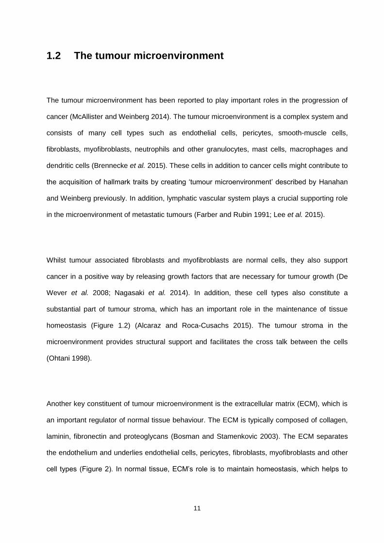
Figure 1.2: The tumour stroma in microenvironment showing tumour cells (transparent), fibroblasts (in yellow), myofibroblasts (green) and endothelial cells (in grey). The necrotic areas are displayed in blue colour. Hypoxia, acidic core and lack of glucose are three hallmarks of tumour microenvironment displayed from centre to middle regions. The MMPs released from stromal cells such as fibroblasts and myofibroblasts degrade ECM (shown in purple and blue colours). Hypoxia induced stabilization of HIF-1 (red areas) contribute to promote tumour cells growth and angiogenesis by facilitating the release of growth factors such as VEGF.
HIF-1
MMPs and other
growth factors
Endothelial cells
Hypoxic areas
Necrotic core
Low glucose and
acidic core
Fibroblasts
Myofibroblasts
Vascularised region
Extracellular matrix

14
1.2.1 Extracellular matrix remodelling
The ECM is an important element of tumour microenvironment and an important regulator of
normal tissue behaviour. One of the common aspects of many solid tumours is desmoplasia.
This is caused by morphologically and functionally altered tumour stroma in the tumour
microenvironment (Whatcott et al. 2015). This desmoplastic response is mediated by a number
of growth factors and cytokines such as TGF-β and PDGF. It has been shown that both are
responsible for the induction of signalling cascade that modulates the tumour stroma (Micke
2004). The altered expressions of α-smooth muscle actin (α-SMA), vimentin and desmin in
fibroblasts are typical biomarkers for desmoplastic reaction in tumour microenvironment
(Elenbaas and Weinberg 2001).
In the tumour microenvironment, the ECM is remodelled to support the neoplastic cells to
proliferate and to allow them to enter into the circulation. The remodelling of ECM characterised
by structural disruption or modification of ECM components which modulates the cell’s ability to
survive, proliferate and migrate (Figure 3). The ECM remodelling is mediated by cysteine
proteases (cathepsin B) (Yan et al. 1998), aspartic proteases (cathepsin D), serine proteases
(elastase and uPA) (Van den Steen et al. 2001) and MMPs (Matzner et al. 1985; Stamenkovic
2003).
It has been shown that most solid tumours exhibit increased expression of proteases that are
correlated with tumour progression. However, during ECM remodelling, most of these proteases
are produced by stromal cells of the host tissue. For example, in human breast cancer, stromal

15
fibroblasts have been reported to produce MMP-11 and urokinase plasminogen activator (uPA)
(Pupa et al. 2002). In addition, MMP-1, 2 and 3 have been reported to be localised on the
fibroblasts located in close proximity of invading cancer cells (Gomez et al. 1997). For example,
extracellular matrix metalloproteinase inducer (EMMPRIN), a membrane bound glycoprotein is
produced by cancer cells. The production of EMMPRIN stimulates stromal cells to synthesise
MMPs in the microenvironment (Tang, et al. 2004). In addition, aspartic protease cathepsin B is
found on plasma membrane of tumour cells and is correlated with invasion (Gangoda et al.
2015). A study has revealed that EMMPRIN stimulates MMP-1 production by fibroblasts
followed by binding of MMP-1 to EMMPRIN on tumour cells. This process facilitates
degradation of ECM by tumour cells (Biswas et al. 1995).
In tumour microenvironment, cancer cells stimulate stromal cells (fibroblast, myofibroblasts,
endothelial cells, pericytes etc) to produce proteases which cleave matrix components (Figure
1.3). This cleavage or proteolysis of ECM modifies focal adhesion and cytoskeleton of ECM and
triggers the release of focal adhesion kinases (FAK) (Fashena and Thomas 2000). In addition,
the proteolysis of ECM components also exposes the binding sites such as integrin binding
sites available on ECM. Furthermore, the laminin receptor (67LR), which is overexpressed by
various tumour cells, interacts with integrins and modulates the interaction between tumour
cells and laminin. This process has been shown to facilitate the metastasis of tumour cells
(Martignone et al. 1993).
The ECM remodelling has a significant effect on tumour cell behaviour by stimulating various
signalling pathways and molecules. A number of epidermal growth factor (EGF) family
cytokines such as TGF-β, PDGF, fibroblast growth factor beta (b-FGF) and interferon gamma
16
(IFN-ᵞ) bind to various components of ECM and remain inactive until they get signals from
matrix proteases (Stamenkovic 2003; Park et al. 1993). Therefore, an increase in the protease
activity on the ECM can lead to the activation and release of various growth factors that can
stimulate tumour cells in the tumour microenvironment (Streuli 1999). The remodelling of the
ECM also has a crucial role in angiogenesis. Once released from endothelial cells, the
angiogenic factors like VEGF can be stored in the ECM. All these processes are initially guided
by tumour cells but later on involve the microenvironment and the host cells to participate, thus
disrupting intracellular signalling between tumour cells and the ECM within the tumour
microenvironment (Park et al. 1993). The tumour cells remodel the matrix to override and
modulate the homeostatic arrangement within the microenvironment. Moreover, the
composition of tumour stroma is equally important for therapeutic response as the tumour
phenotype is (Pupa et al. 2002).
17
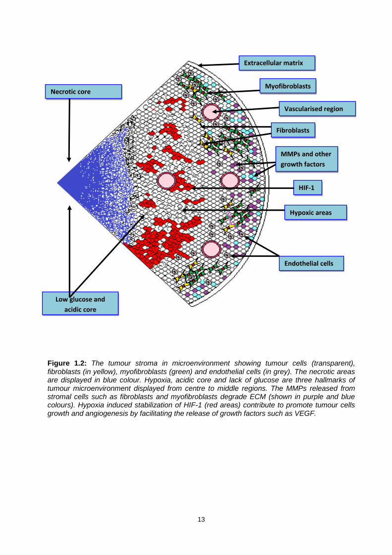
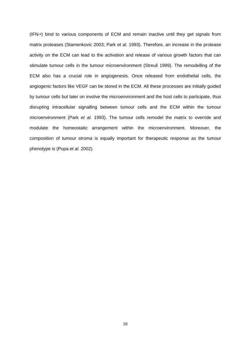
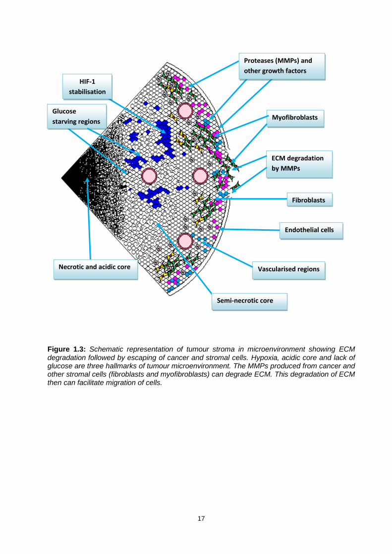
Figure 1.3: Schematic representation of tumour stroma in microenvironment showing ECM degradation followed by escaping of cancer and stromal cells. Hypoxia, acidic core and lack of glucose are three hallmarks of tumour microenvironment. The MMPs produced from cancer and other stromal cells (fibroblasts and myofibroblasts) can degrade ECM. This degradation of ECM then can facilitate migration of cells.
Glucose
starving regions Myofibroblasts
ECM degradation
by MMPs
HIF-1
stabilisation
Necrotic and acidic core Vascularised regions
Semi-necrotic core
Proteases (MMPs) and
other growth factors
Endothelial cells
Fibroblasts

18
1.2.2 Cellular cross talk in the tumour microenvironment
The cross talk between the cells in tumour microenvironment is linked with the progression of
the disease. For example, hepatic sinusoidal endothelial cells enhance the interaction between
hepatocytes and kupffer cells to exchange nutrients that is needed for neo-vascularisation
within tumours (Bhatia et al. 1999). It has been established that tumour cells promote VEGF
expression in hypoxic conditions (Levy et al. 1996). Therefore, hypoxia induced VEGF
expression and subsequent formation of neo-vascularisation promotes cell survival under harsh
conditions within the microenvironment.
The expression of some angiogenic factors depends on organ specific cell-cell cross talk within
the microenvironment. For example, interleukin-8 (IL-8) has been shown to induce proliferation
of endothelial cells (Li et al. 2003). In contrast, the expression of IL-8 was inhibited in melanoma
and hepatocyte co-cultures. Therefore, the expression of angiogenic factors in tumour cells is
directly linked with individual organ specific microenvironments (Desbaillets et al. 1997).
The epithelial and mesenchymal cells are the drivers of differentiation and development.
Furthermore, it has been shown that changes in the stroma can promote epithelial
transformation (Park et al. 2000). For example, mesenchymal and epithelial cells regulate the
ovarian cycle before and during the reproductive period. During the menopause, any loss in the
cross talk between mesenchymal and epithelial cells is correlated with the promotion of ovarian
cancer. Therefore, any interference in the cell-cell interaction during embryogenesis might be
responsible for the onset of cancer (Aboseif et al. 1999).
19
The cellular cross talk is directly linked with motility, survival and invasion. For example,
integrins present between the cells and the ECM are responsible for the adhesion of cells to the
ECM. A disruption in the integrin mediated adhesion facilitates cellular translocation and
apoptosis (Frisch et al. 1996). Similarly, the pro-survival and pro-invasive signals are needed for
survival and invasion. For example, integrins activate a number of downstream molecules such
as FAK and MAPK/ERK pathways. The phosphorylated FAK is necessary for the survival of
cells combating anoikis (programmed cell death due to lack of cell-cell or cell-matrix interaction)
(Frisch et al. 1996).
A number of other pathways such as PI3K, Ras, Rac, Rho and cell division cycle-42 (CDC-42)
are involved in the regulation of cell motility within the tumour microenvironment. However, most
of these pathways overlap in facilitating the process of motility, invasion and survival in cancer
and stromal cells (Xue et al. 2000).
1.2.3 The interstitial fluid pressure in tumours
The central core of solid tumours is often necrotic due to a lack of nutrients and anoxia resulting
from a poor blood supply. However, there is a viable rim on the periphery of this necrotic zone,
below basement membrane of most solid tumours. Many chemotherapeutic drugs have been
developed to target these cells and also the tumour vasculature (Shaked et al. 2006).
In spite of many advances in the target-oriented chemotherapy, most therapeutic agents are
unable to kill the cells of the viable rim of tumours. The viable rim encapsulates the hypoxic and

20
necrotic core of solid tumour and thought to be one of the reasons why most drugs cannot
penetrate and reach to the central core of tumours (Jain 1987; Minchinton and Tannock 2006).
There could be a couple of explanations for the viable rim escaping effective treatment. One
could be that the microenvironment within the tumour is dynamic and keep changing. This might
result in an increase in the interstitial fluid pressure and cells from central core coming out and
contribute in the formation of rim. Another explanation could be linked with the work done by
Yuval Shaked et al. (2006) which suggested that vascular disrupting agents (VDA) may induce
acute mobilisation of circulating endothelial cells in tumours. These cells may home to the
viable rim in the tumours and may remain there after the therapy (Shaked et al. 2006).
1.2.4 Hypoxia and glucose deprivation in solid tumours
The multistep development of most solid tumours involves six hallmarks of cancer reported by
Hanahan and Weinberg previously. Recent conceptual advances have added two emerging
hallmarks of cancer. These are reprogramming of energy metabolism and evading immune
destruction. Unlike apoptosis or autophagy, necrosis in tumours might have direct impact on
tumour growth and metastasis because necrotic cells might explode and release growth factors
and other nutrients into the host tumour microenvironment. (Hanahan and Weinberg 2011). It is
established that hypoxia is a common phenomenon that occurs in most solid tumours.
Apparently, most hypoxic cells are resistant to radiotherapy and to a number of
chemotherapeutic agents. It has been shown that there is a correlation between the oxygen
levels and the response of tumours to the radiation therapy. In addition, the oxygen deprivation
is also correlated to the metastatic potential of the tumour cells. Hypoxia has been considered

21
as one of three hallmarks of tumour microenvironment. The other two are glucose deprivation
and acidosis (Tannock 1972; Schlappack et al. 1991; Hanahan and Weinberg 2011).
The microenvironment of solid tumour is not only characterised by hypoxia but also the
formation of acidic environment within solid tumours (Raghunand et al. 2014). In solid tumours,
lactic acid formation and hydrolysis of adenosine triphosphate (ATP) have been found to be
acidic and a wider range of pH (5.85-7.68) has been observed in different regions of the same
malignant tissue with median pH of 7.0. This pH is generally lower than the surrounding normal
tissue where median pH is 7.5. The lower pH or acidosis is correlated with the decreased
radiation sensitivity of solid tumours. In addition, it has been shown that the cytotoxic effect of
doxorubicin (a DNA intercalating agent) and mitoxantrone (type II topoisomerase inhibitor) was
reduced at low extracellular pH (Wike-Hooley et al. 1984; Mahoney et al. 2003).
It is clear that neoplastic tissues utilise a large quantity of glucose. For example, Walker
carcinomas in rats were found to contain 0-5mg/ml of glucose in tumour interstitial fluid when
compared to the normal subcutaneous interstitial fluid of rats with 90-100mg/ml of glucose
(Woodward and Hudson 1954; Gullino et al. 1964). This might suggest that glucose passing
through the tumour vasculature is rapidly utilised by the neoplastic cells (Wike-Hooley et al.
1984; Graff 2014). The effect of glucose deprivation and acidosis has been shown to increase
metastasis in murine tumour cell lines in vivo. However, the underlying mechanism of glucose
deprivation induced metastatic spread remains unknown (Schlappack, et al. 1991). Glucose
deprivation and hypoxia have been shown to induce the accumulation of proteasomes in the
nucleus of cancer cells (HT29 colon cancer cells). Proteasome is a major site for protein
degradation, which plays a key role in the proteolysis to maintain homeostasis. It has been

22
shown that glucose deprivation and hypoxia can modulate the intracellular location of
proteasomes. Inhibiting protein degradation by selective inhibitor of proteasome has been
shown to restore the sensitivity to topoisomerase-IIα targeted drugs in vitro and in vivo.
Therefore, an increased proteolysis in the nucleus of cells could be the survival strategy of
cancer cells to survive under hypoxia and glucose deprivation (Ogiso et al. 1999).
1.3 High mobility group box 1 (HMGb1) protein
The high mobility group (HMG) proteins are chromosomal proteins, named according to their
ability to move electrophoretically through polyacrylamide gels. There are three main known
superfamilies; HMGa, HMGb and HMGn. The HMGb superfamily has a functional sequence
motif, which is called DNA binding box (Bustin 2001). HMGb1 is a non-histone protein involved
in stabilization of nucleosomes and the bending of DNA, which facilitates gene transcription
(Lange et al. 2008). In addition, HMGb1 is responsible for modulating the activity of steroid
hormone receptors by participating in the maintenance of nucleosome structure. As supporting
evidence, HMGb1-deficient mice died shortly after birth, possibly because of inactivation of
glucocorticoid receptor responsive genes (Wei et al. 2003). The HMGb1 is expressed in all cells
of vertebrates and in yeast, plants and bacteria (Bustin et al. 1990). The cellular localisation of
HMGb1 could be tissue specific with notably high levels found in lymphoid tissues and testis. In
addition, an increased level of HMGb1 has been seen in cytoplasm of the cells of the liver and
the brain. However, HMGb1 concentration is usually more in the nuclei of the cells than the
cytoplasm (Mosevitsky et al. 1989).

23
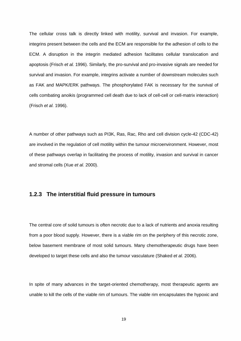
Structurally, HMGb1 is composed of three main domains; the A box domain and B box domain
are homologous DNA binding pockets and a negatively charged chain of 30 amino acids
(Figure 1.4) (Chen et al. 2004; Ellerman et al. 2007). The A box domain plays an important role
for its antagonistic and anti-inflammatory effect whereas the B box domain has a role similar to
that of proinflammatory cytokines (Andersson, Erlandsson-Harris et al. 2002) (Figure 1.4). The
amino acids 150-183 on the acidic tail are responsible for binding to the receptor for advanced
glycation end products (RAGE) (Figure 1.4) (Ellerman et al. 2007). The HMGb1 binds to the
minor grove of DNA without sequence specificity, and induces bends in the helical structure of
the DNA. The formation of this complex facilitates interaction between DNA and other factors
such as p53, nuclear factor kappa β (NF-kβ), recombination activating proteins 1/2 (RAG1/2)
and some steroid hormone receptors (Bianchi 2004). The HMGb1 also interacts with other
molecules including RNA, Lipopolysaccharides (LPS) or endotoxins and IL-1β. Thus HMGb1
may play a key role in facilitating the arrangement of many nucleoprotein complexes (Bianchi
and Manfredi 2014; Keyel 2014).
24
Figure 1.4. Structure of HMGb1 protein. HMGb1 is a 215–amino acid (AA) protein of ∼30 kDa. HMGb1 is composed of three domains: two positively charged domains (A box and B box) and a negatively char Acidic tail. A and B boxes are DNA-binding domains and B box is also responsible for cytokine activity by inducing macrophage secretion of proinflammatory cytokines. This cytokine activity is antagonized by recombinant A box. The protein structure involved in the binding of HMGb1 with RAGE is located between amino acid residues 150 and 183 (Ellerman et al. 2007).

25
1.3.1 Role of HMGb1 in disease
HMGb1 does not possess signal sequence and therefore, does not transverse the endoplasmic
reticulum. However, it is released actively by various cells such as macrophages, pituicyte and
erythroleukemia cells (Tang et al. 2007). In addition, HMGb1 is passively released from necrotic
or damaged cells but not from cells undergoing apoptosis, these apoptotic cells retain HMGb1
within their nuclei (Zong et al. 2004). Therefore, apoptotic cells do not trigger inflammation even
after the loss of the membrane (Bonaldi et al. 2003). Hence, HMGb1 can be considered as a
critical stimulus of inflammation during cell death (Scaffidi et al. 2002). The HMGb1 has two
lysine-rich nuclear localization sequences which direct its movement towards the nucleus of the
cell (Bonaldi et al. 2003). Whilst it is a nuclear protein, HMGb1 is released from the cells under
certain conditions to take part in the inflammatory process (Frank et al. 2015; Zhu et al. 2015).
During inflammation, extracellular HMGb1 activates infiltrating macrophages via the RAGE
receptor. In addition, activated macrophages/monocytes are also responsible for the release of
HMGb1 in the extracellular milieu (Figure 1.5) (Andersson et al. 2002; Huttunen and Rauvala
2004). There are three main steps have been suggested for HMGb1 secretion; 1) exit from the
nucleus to the cytoplasm, 2) translocation from the cytoplasm into cytoplasmic organelles, and
3) exocytosis. Macrophages/monocytes upon activation by proinflammatory cytokines acetylate
HMGb1 at their lysine-rich nuclear localization sequences. This leads to the translocation of
HMGb1 into the cytoplasmic vesicles followed by extracellular release (Gardella et al. 2002),
(Figure 1.5).

26
It has also been suggested that HMGb1 is involved in various diseases including autoimmune
disorders, sepsis, chronic inflammatory disease and cancer (Wang et al. 2004; Sims et al.
2009). An increase in the concentration of HMGb1 has been observed in the plasma and
epithelial lining fluids in patients with acute lung injury (Ueno et al. 2004). In addition, HMGb1,
TNF-α and IL-1 β have been suggested to be involved in the pathogenesis of cutaneous lupus
erythematosus (CLE). In CLE, HMGb1 forms a pro-inflammatory loop between tumour necrosis
factor-α (TNF-α) and IL-1β to sustain prolonged inflammation, suggesting an important role in
this inflammatory autoimmune disorder (Barkauskaite et al. 2007). A study revealed that anti-
HMGb1 antibody inhibited synovial inflammation by blocking HMGb1 in an experimental model
of arthritis. However, this inhibition was independent of TNF-α pathway suggesting that TNF-α
is not the only pathway necessary for extracellular release of HMGb1 (Pullerits et al. 2008).

27
Figure 1.5: Release of HMGb1 as a danger signal during inflammation. The figure shows a diagrammatic illustration of potential pathways for HMGb1 release leading to inflammatory responses. HMGb1 can be released extracellularly by passive secretion from any necrotic cell or by active secretion from activated macrophages/monocytes (Andersson et al. 2002).
HMGb1 has been reported to be responsible for the impairment of intestinal barrier function in
mice (Sappington et al. 2002). In addition, HMGb1 play an important role in increasing ileal
mucosa permeability and bacterial translocation to lymph nodes (Sappington et al. 2002).
Furthermore, elevated levels of HMGb1 have been observed in synovial fluids of experimental
animal models of arthritis (Andersson and Erlandsson‐Harris 2004). In addition, a clinical study
has revealed that synovial fluid of rheumatoid arthritis patients had more elevated levels of
HMGb1 than that of osteoarthritis patients (Taniguchi et al. 2003).

28
The hemorrhagic shock is characterised by activation of inflammatory cytokines. A clinical
report revealed elevated levels of HMGb1 in the blood circulation of hemorrhagic shock
patients. However, the elevated levels of HMGb1 went to normal as clinical conditions improved
(Fan et al. 2007). In addition, the serum HMGb1 level goes significantly higher in sepsis
patients compared to normal healthy volunteers. This observation was constant when
compared to patients who died of sepsis versus the patients who had survived after recovering
from sepsis (Yang et al. 2004).
Recently, the effect of HMGb1 on fibroblasts and keratinocytes has been explored. It has been
shown that cytokine activity of HMGb1 stimulates keratinocyte scratch wound healing in vitro
(Ranzato et al. 2009). In addition, HMGb1 was shown to induce proliferation and migration of
keratinocytes via ERK1/2 pathway (Ranzato et al. 2010). To further support these findings, anti-
RAGE antibody and selective MEK1/2 inhibitor (PD98059) were used to inhibit the HMGb1-
RAGE-ERK1/2 pathway. This resulted in the inhibition of HMGb1 induced wound healing.
Therefore, HMGb1 can be considered as a potential therapeutic target for the development of
drugs for chronic inflammatory disease, chronic inflammatory autoimmune disorders and severe
wounds (Ranzato et al. 2010). It has also been shown that HMGb1 stimulates vascularisation in
chicken embryo chorioallantoic membrane in vivo, which suggests that HMGb1 plays an
important role in angiogenesis (Mitola et al. 2006). In addition, HMGb1 triggers proliferation and
migration of glioblastoma cells via ERK1/2 activation (Bassi et al. 2008). However, the
proliferation of cells can not be considered as marker for the expression of HMGb1 in vivo (Ller
et al., 2004).

29
1.3.2 The role of HMGb1 in cancer
Hanahan and Weinberg in 2000 proposed a model that defined six hallmarks of cancer. These
are 1) unlimited replicative potential, 2) ability to develop new blood vessels, 3) escape from
surveillance, 4) insensitivity to inhibitors of growth, 5) self-sufficiency in growth signals and 6)
invasion and metastasis (Hanahan and Weinberg 2000). Recently, seventh hallmark has been
proposed which is inflammation. In addition, dysregulated cellular energies within tumour
microenvironment and evading immune destruction are two additional emerging hallmarks that
most or may be all tumours exhibit (Hanahan and Weinberg 2011). All these hallmarks of
cancer are linked with the levels, localisation and alterations in HMGb1 (Mantovani 2008;
Mantovani et al. 2008). Therefore, HMGb1 is now important to understand the molecular
biology of cancer. Several solid tumours including melanoma, prostate cancer, breast cancer,
pancreatic cancer and colon cancer exhibit markedly elevated levels of HMGb1 (Völp et al.
2006). These elevated levels of HMGb1 are associated with tumour formation, proliferation and
metastasis and chemotherapeutic response. The presence of HMGb1 in the extracellular
medium of cells is indicative of stress conditions (Lotze and Tracey 2005).
The HMGb1 has dual role in cancer. The first role is correlated with neovascularisation in solid
tumours (Campana et al. 2008). A rapidly growing tumour causes reduction in the microvessel
density followed by formation of necrotic areas within the tumour. The necrotic areas within
tumour not only produce angiogenic factor such as VEGF but also attract macrophages. The
macrophages have been reported to release HMGb1 in stress conditions such as necrosis. In
addition, HMGb1 binds to its receptor RAGE and activates NF-ƘB. Upon activation, NF-ƘB
upregulates the production of certain cytokines and angiogenic factors in endothelial cells (van
30
Beijnum et al. 2008). The direct inhibition of HMGb1 with immunoneutralising antibodies has
been reported to inhibit angiogenesis in vivo and in vitro (van Beijnum et al. 2006). It has been
suggested that HMGb1 might have a direct impact on migration of cells because of its ability to
modulate the adhesive properties of the cells and ECM components (Ellerman et al. 2007). In
addition, HMGb1 can enhance invasive and metastatic potential of tumour cells via the NF-κB
pathway (Sasahira et al. 2008).
The other role of HMGb1 in cancer is related to the immune response against tumours. There is
evidence that HMGb1 is released in response to specific chemotherapy or radiation therapy
induced conditions, and this is thought to promote immune response against tumours
(Campana et al. 2008). Extracellular HMGb1 has been shown to activate dendritic cells in
murine models. Upon activation, these dendritic cells facilitate immune response against
immunogenic apoptotic lymphoma cells (Ronchetti et al. 1999). In addition, HMGb1 has also
been shown to trigger anti-neoplastic response from T cells (Campana et al. 2008). HMGb1 has
been shown to induce antigrowth signals within tumours. These antigrowth signals usually act in
two ways; a) cells may escape from the proliferation cycle and enter into the stage of
quiescence (G0 phase) and b) permanent loss of proliferative potential by some alternations in
their environment (Hanahan and Weinberg 2011). Contrary to this, it has been shown that MCF-
7 cells overexpressing HMGb1 progress through to S2 phase when compared with HMGb1
knockdown MCF-7 cells (Yoon et al. 2004). However, other work has shown that HMGb1
overexpression has been shown to suppress the growth of MCF-7 xenografts in nude mice
(Jiao 2007).
31
Recent work has suggested an important role of HMGb1 in the survival of myeloid derived
suppressor cells followed by suppressing the immune response against tumours. These
myeloid derived suppressor cells are found in abundance in most cancers and are
representative of suppressed immune system (Parker et al. 2015). The extracellular HMGb1
due to its cytokine, chemokine and growth factor activity acts as protumour protein however, it
does possess an ability to sustain genome stability during tumorigenesis. Thus, whilst HMGb1
may promote tumour cell survival during the early stages of chemotherapy however on the
contrary intracellular HMGb1 led inhibition of autophagy might increase the effectiveness of
anticancer treatment (Kang et al. 2013).
HMGb1 activates various signalling pathways such as MAPKs, protein kinase B (AKT) and
PI3K. These pathways play important roles in proliferation and migration of normal cells and
cancer cells. Supporting evidence includes activation of the PI3K pathway by HMGb1 in
neutrophils and colon cancer cells (Kuniyasu et al. 2003). Furthermore, HMGb1 has been
shown to induce toll like receptor-4 (TLR-4) mediated activation of MyD88-IRAK4-p38 and
Myd88-IRAK4-AKT pathways. The activation of HMGb1-RAGE complex has been shown to
activate NF-ƘB, JNK kinases and ERK1/2 pathways (Degryse, et al. 2001). In addition to RAGE
receptor, HMGb1 binding with TLR-2 and TLR-4 receptors also has been shown to activate the
NF-КB pathway. This suggests that NF-КB is an important signalling pathway that requires
activation by HMGb1 (van Beijnum et al. 2008).

32
1.3.3 HMGb1 and its receptors
The HMGb1 has been shown to modulate its cytokine like activity by interacting with multiple
receptors including receptor for advanced glycation end products (RAGE) (Kokkola et al. 2005),
toll like receptor 2 and 4 (TLR-2 and TLR-4) (Curtin et al. 2009; Kim et al. 2013), chemokine
CXC receptor 4 (Schiraldi et al. 2012), and T cell immunoglobulin mucin 3 (TIM-3) (Baghdadi et
al. 2013). However, RAGE is the most commonly researched receptor for HMGb1. This
receptor belongs to immunoglobulin superfamily and is expressed in many cells including
monocytes, macrophages, smooth muscle cells, dendritic cells (DC) and endothelial cells. The
receptor for advanced glycation products is a receptor for multiple ligands that can therefore be
activated by different ligands including HMGb1. However, the effects of this activation are
dependent of the type of the cell this receptor is expressed upon. Activation of RAGE in
monocytes/macrophages has been reported to trigger inflammatory response, neoplastic
transformation and metastasis in neuroepithelial tumour cells (Taguchi et al. 2000). The
fibroblasts have been shown to express RAGE and the activation of RAGE on fibroblasts was
correlated with proliferation and migration of fibroblasts in tumour microenvironment (Liu et al.
2010; Rojas et al. 2010). It has been shown that RAGE overexpression is associated with
chronic degenerative disease and cancer (Tanaka et al. 2000). In addition, activation of RAGE
by HMGb1 is associated with increased expression of the anti-apoptotic B-cell lymphoma-2
(Bcl-2) gene, which then inhibits apoptosis (Bierhaus et al. 2005). The protein HMGb1 has been
shown to interact with TLR-2 and TLR-4 and the activation of TLR-4 by HMGb1 was shown to
inhibit migration of enterocytes and endothelial cells (Dai et al. 2010; Bauer et al. 2013).

33
1.3.4 Receptor for advanced glycation end products (RAGE)
The receptor RAGE known to play important roles in the many diseases such as diabetes,
arthritis, Alzheimer’s disease and cancer is localized on chromosome 6 in humans and mice
(Wang et al. 2012).. The advanced glycation end products (AGEs) are common ligands for
RAGE and involved in various diseases such as hyperglycaemia, renal failure and other
inflammatory diseases. The NƐ-carboxymethylysin (CML) is one of the most common AGE
ligands that can bind to RAGE (Wang et al. 2012). Apart from the AGEs, RAGE interacts with
other ligands such as the S100 family of molecules (Schmidt et al. 2000). The S100 family of
molecules have been reported to play important roles in inflammatory diseases. In addition,
amyloid-β-peptide and β fibrils are other ligands of RAGE involved in the development of
Alzheimer’s disease. Interestingly, RAGE itself might not be overexpressed in most cancers but
its ligands such as S100 family and HMGb1 are generally overexpressed in most type of
cancers. However, amyloid-β-peptide and β fibrils have not been reported in cancers but they
are overexpressed in neurological disorders (Hofmann et al. 1999).

34
Figure 1.6: Structure of RAGE: A) The structure of RAGE consists of one V and 2 C (C1 and C2) domains. There is also a transmembrane domain which joins cytoplasmic tail below the surface membrane of cells. B) Three dimensional structure showing V, C1 and C2 domains of RAGE (Lin et al. 2009; G Fritz 2011)
The structure of RAGE consists of one V-type and two C-type domains, a short transmembrane
domain and a cytoplasmic tail (Figure 1.6) (Bierhaus et al. 2005). There are three major RAGE
isoforms have been reported; full-length RAGE, expressed secretory RAGE (esRAGE) and N-
truncated RAGE (NtRAGE). However, a few other splice variants of RAGE have also been seen
in pathological conditions. The mRNA of these splice variants lacks N terminal and C terminal
(Yonekura et al. 2003). These splice variants have been seen on various cells including

35
endothelial cells and pericytes. However, the function of these isoforms is not fully understood
(Schmidt et al. 1994).
The esRAGE has similar immunoglobulin domains as that of a full length RAGE but lacks exons
10 and 11 that encode the transmembrane domain of full length RAGE. This is because
esRAGE does express intron 9, which has a stop codon within the sequence. Therefore this
stop codon does not allow expression of exons 10 and 11. The esRAGE has been reported to
be secreted by cultured cells into the extracellular environment (Cheng et al. 2005). In the
extracellular environment, esRAGE acts as decoy for many ligands which also bind to full length
RAGE (Mahajan and Dhawan 2013). Before the discovery of esRAGE, a synthetic version of
esRAGE was produced in baculovirus system, which was used as decoy for RAGE ligands.
This system was used to inhibit the activation of RAGE. This synthetic version of esRAGE
molecule has also been termed soluble RAGE (sRAGE) (Hofmann et al. 1999).
The NtRAGE has a stop codon which is located on intron 1 causing the loss of exons 1 and 2.
Therefore, NtRAGE lacks the v-type immunoglobulin domain. However, it is expressed on
plasma membrane. Han et al. detected a 42kDa protein which was similar to the full length
RAGE on the plasma membrane (Han et al. 2011). However, considering the fact that NtRAGE
lacks a signal peptide, the mechanism behind its expression on plasma membrane is unclear.
In addition, its expression on plasma membrane does not interfere with AGE stimulated effects.
However, its overexpression does correlate with inhibition in migration of endothelial cells.
Therefore, it appears that NtRAGE might interact with other ligands and can interfere with
various signalling pathways involving RAGE (Yonekura et al. 2003).

36
1.3.5 RAGE and Tumour Microenvironment
Many important cells of the microenvironment such as endothelial cells, pericytes,
macrophages, fibroblasts and myofibroblasts are known to express RAGE (Logsdon, Fuentes
et al. 2007). Therefore, RAGE ligands generated by cancer cells are likely to influence tumour
microenvironment. Similarly, these cells in the tumour microenvironment can also produce
RAGE ligands and these ligands can interact with RAGE on cancer cells (Rouhiainen et al.
2013). The most important role of RAGE that influences the tumour microenvironment is its
ability to influence angiogenesis. It is established that angiogenesis play important roles in
proliferation and migration of cancer cells, endothelial cells and pericytes (Papetti and Herman
2002). A number of RAGE ligands including HMGb1 have been shown to influence endothelial
cells (Gupta and Massagué 2006). In addition, HMGb1 overexpression was correlated with
angiogenesis in colon cancer and inhibiting HMGb1 abrogated the formation of new vessels
(Schlueter et al. 2005). Furthermore, expression of RAGE was correlated with VEGF (an
angiogenic stimulator) expression on endothelial cells. Also, a number of RAGE ligands have
reported to induce other angiogenic factors such as IL-8. Therefore, it appears that RAGE and
RAGE ligands are key players that participate in the tumour microenvironment (Folkman 1990).
Fibroblasts have been shown to express RAGE in tumour microenvironment. It is clear that the
number of fibroblasts present is correlated with increased cancer progression (Rojas et al.
2010). In addition, they produce growth factors and ECM molecules which facilitate
angiogenesis within the tumour microenvironment. RAGE has also been reported to influence
fibroblasts by upregulating fibroblasts growth factor (FGF). In addition, activation of RAGE on
37
fibroblasts was correlated with proliferation and chemotaxis of fibroblasts (Liu et al. 2010).
However, the correlation of RAGE with cancer-associated myofibroblasts is unknown.
The macrophages are another major cell type stimulated by the activation of RAGE within the
microenvironment. The macrophages exert their immunomodulatory effect by presenting
tumour-associated antigens to T-cells and also by expressing immuno-stimulatory cytokines.
The RAGE activity has been reported to stimulate macrophage function which was correlated
with inflammation (Hofmann et al. 1999; Hasegawa et al. 2003).
1.3.6 RAGE in cancer
RAGE is expressed in many solid tumours including ovarian, lung, prostate, colonic, brain and
melanomas. In addition, elevated levels of RAGE have been reported in many pathological
conditions such as diabetes, arthritis and Alzheimer’s disease (Hofmann et al. 2002; Fukami et
al. 2015; Liu et al. 2015). These elevated levels are directly linked with the activation of NF-Кβ
which has been shown to play central role in activating downstream signalling in many tumours
(Pikarsky et al. 2004). Therefore, this could be one of the reasons why most tumours have
elevated levels of RAGE. However, there is conflicting evidence regarding the overexpression
of RAGE in cancers. Some studies support the overexpression of RAGE in many cancers
including colon, prostate and gastric (Sasahira et al. 2005; Sparvero et al. 2009). Contrary to
this, no increase in the levels of RAGE in colon and pancreatic cancers has been reported by
other researchers (Bartling et al. 2005). In addition, significantly low levels of RAGE have been
observed in lung cancer. In particular, the levels of full length RAGE and esRAGE were

38
significantly low in non-small cell lung cancer (NSCLC). However, RAGE ligands have been
reported to be increased in most tumours (Bartling et al. 2005).
There is conflicting evidence about the involvement of RAGE in lung cancer as reduced levels
of RAGE were reported in NSCLC. In addition, esRAGE, which is an antagonist for the full
length RAGE is downregulated in NSCLC. Furthermore, the overexpression of full-length RAGE
was correlated with reduction in tumour size (NCI-H358 lung cancer cells) in vivo (Logsdon et
al. 2007). However, several RAGE ligands such as HMGb1, S100A12 and S100P are
overexpressed in the lung cancer (Diederichs et al. 2004). Furthermore, S100A4
overexpression was correlated with poor prognosis resulted from metastatic spread of
pulmonary adenocarcinoma (Zou et al. 2004).
The role of RAGE in breast cancer is not fully understood. However, RAGE ligands appeared to
play important roles in progression of the disease. For example, S100A4 has been shown to
play crucial role in breast cancer growth. In addition, S100A4 negative patients’ survival rate
was much higher than those of S100A4 positive patients (Rudland et al. 2000). Also, elevated
levels of HMGb1 were observed in human primary breast carcinoma. Therefore, there is much
evidence that supports the involvement of RAGE ligands in breast cancer. However, it is
nuclear that if these ligands mediate their effect via RAGE in breast cancer (Lum and Lee
2001).
The receptor for advanced glycation end products along with its ligands has been reported to be
elevated in many stages of prostate cancer. For example, S100A8 and S100A9 have been
reported to be overexpressed in human prostate cancer. The receptor (RAGE) has been shown

39
to be expressed by prostate cancer cells and its ligands S100A8 and S100A9 are apparently
also secreted by prostate cancer cells (Gebhardt et al. 2006). In addition, S100s appeared to
play key role in migration of prostate cancer cells by activating NF-Кβ and MAPK pathway in
vitro. Also, elevated levels of HMGb1 and RAGE have been reported in prostate cancer tissue
when compared with normal prostate tissue (Ishiguro et al. 2005). Furthermore, HMGb1 and
RAGE overexpression was observed in the PC3 prostate cancer cell line, which was related to
androgen deprivation induced cancer cell invasion (Kuniyasu et al. 2003). Moreover, RAGE
expression has been shown to be increased at various stages of colon cancer progression
(Sasahira et al. 2005).
This receptor (RAGE) has been reported to play important role in a neoplastic model of IL-10
null mouse. The administration of sRAGE (decoy for RAGE ligands) significantly reduced the
neoplasia related inflammation in IL-10 null mouse model (Berg et al. 1996). In addition, S100P
and RAGE complex triggered proliferation and migration of SW480 colon cancer cells and
inhibiting S100P/RAGE abrogated proliferation and migration in the SW480 cells. Furthermore,
the expression of S100A4 was correlated with the invasive potential of the colon cancer cells as
S100A4 is specifically overexpressed in invasive carcinoma. All these findings suggest that
RAGE might play an important role in the interface between the inflammation and
carcinogenesis (Yammani et al. 2006).

40
1.3.7 RAGE and HMGb1
RAGE is a multi-ligand receptor that binds to various molecules including HMGb1 and the S100
family of proteins. Structurally, RAGE has two N-glycosylation sites on its V domain. These N-
glycans undergo carboxylation which enhances the ability of the receptor to bind to HMGb1 with
subsequent signal transduction. The carbohydrate groups are then removed by PNGase F and
therefore do not alter the conformation of protein complex. Upon confirmation, the protein
complex (receptor-ligand complex) can initiate signal transduction necessary for cell growth
(Wilton et al. 2006).
There is evidence that supports the involvement of RAGE and HMGb1 complex in the cancer.
For example, blockade of HMGb1 and RAGE by immunoneutralising antibodies suppressed
tumour growth in murine model of lung cancer (Schlueter et al. 2005). In addition, HMGb1 has
been shown to induce proliferation and neovascularization in endothelial cells (Chavakis et al.
2007). Other research has shown that HMGb1 plays an important role in the activation of DCs.
This activation is mediated via TLR-4 (another receptor for HMGb1). However, HMGb1 induced
maturation and migration of DCs was shown to involve activation of RAGE via HMGb1 binding
(Dumitriu et al. 2005).
Various cell surface receptors such as RAGE and TLRs play important roles in signal
transduction. It was observed that nucleosomes containing HMGb1, derived from secondary
necrotic cells, play an important role in inflammation by activating macrophages and DCs which
also express RAGE and TLRs. The mechanism of action of this signal transduction was

41
explored using macrophages with defective RAGE and TLRs. Interestingly, the deletion of TLR-
2 abrogated the activation of macrophages and DCs. This suggests when HMGb1 is associated
nucleosome, the signal transduction will be strictly through TLR-2 (Urbonaviciute et al. 2008).
Increased concentration of DNA containing complexes have been observed in autoimmune
disorders such as Lupus. The HMGb1 interaction with DNA containing immune complexes has
been extensively studied where these complexes bind to HMGb1 enter into the endosomal
pathway of plasmacytocoid dendritic cells (pDCs) via CD32 mediated uptake where it
encounters with TLR-9. Since, HMGb1 can directly bind to DNA, it is possible that when
HMGb1 is bound to DNA it would signal through RAGE and the activation of TLR-9-MyD88
pathway (Tian et al. 2007).
The ligand binding of RAGE involves the activation of two major pathways which are
CDC42/Rac and MAPK. It is evident that the activation of the MAPK pathway leads to the
activation of NF-kβ and neurite outgrowth. The CDC42 pathway has been implicated in HMGb1
induced migration in cancer cells. The HMGb1 induced activation of RAGE lead to the
activation of CDC42 and Rac1 and facilitates changes in the cytoskeleton. To support this, NF-
kβ gene expression assay was carried out. It was found that expression of dominant negative
RAGE eliminated the NF-kβ activation induced by HMGb1. Therefore, HMGb1 induced
activation of RAGE was responsible for NF-kβ activation followed by neurite outgrowth
(Huttunen et al. 1999). In addition, HMGb1 mediated activation of RAGE may amplify the
expression of RAGE with subsequent activation of NF-kβ. The NF-kβ responsive genes play
key role in inflammatory process (such as intracellular adhesion molecule 1 (ICAM-1), vascular
cell adhesion molecule 1 (VCAM-1) and few other cytokines e.g., TNFα, IL-1β and IL-8).
Therefore, HMGb1 induced activation of RAGE can induce and sustain a pro-inflammatory
phenotype (Park et al. 2003; Fiuza, Bustin et al. 2003). Apparently, RAGE was the first
42
receptor identified for HMGb1 binding but blocking RAGE did not abrogate the effect of HMGb1
completely in many experimental models. This suggested that an additional receptor for HMGb1
must exist. Subsequently, Toll like receptors were identified as HMGb1 binding receptors (Park
et al. 2004).
1.3.8 Toll Like Receptors
The tolls like receptors (TLRs) are conserved proteins that help cells of the innate immune
system to respond to endogenous danger molecules by activating intracellular signalling
pathways. TLR signalling might require the receptors to dimerize or oligomerize for signal
transduction to take place. Endothelial cells have been shown to express different TLRs on their
membranes such as TLR-4, TLR-2 and TLR-9. These are multi-ligand receptors and when
associated with HMGb1 and play important roles in activating immune response against
inflammation via MyD88 pathway (Nogueira-Machado et al. 2011). However, these receptors
can bind to endotoxin or LPS and gram positive and gram negative bacteria. For example, TLR-
4 is the main receptor for endotoxin and also for gram-negative bacteria whereas TLR-2 is a
receptor for gram-positive bacteria and also for fungi. Both play important roles in host response
to fungal or bacterial infections (Yu et al. 2006).

43
1.3.9 TLRs and HMGb1
Various blood borne pathogens and cytokines usually target endothelial cells. Subsequently,
endothelium expresses TLRs and gets activated. In particular, the activation of endothelium in
response to microbial stimulation is mediated by TLR-2 (Iwasaki and Medzhitov 2004). In
addition, it has also been shown that the activation of TLR-2 may lead to the activation and
translocation of NF-kβ (Smith et al. 2003). The involvement of Rac1 and PI3K has been
reported to activate TLR-2 in fimbrillin stimulated monocytes. The Rac1 and PI3K were
recruited to the cytoplasmic domain of TLR followed by subsequent signal transduction
(Harokopakis et al. 2006). The recruitment of Rac1 and PI3K resulted in CD18-dependent
adhesion to ICAM-1. In addition, the dominant negative variant of PI3K and Rac1 inhibited the
expression of NF-kβ reporter gene. This suggested an important role of PI3K and Rac1 in TLR-
2 signalling (Arbibe et al. 2000).
Toll like receptor 2 has been reported as main receptor for HMGb1 in RAW264.7 macrophages.
This was detected by using fluorescence resonance energy transfer (FRET) and
immunoprecipitation assays. In addition, it was shown that the stimulation of HMGb1 increased
TLR-2 mediated activation of NF-kβ. Furthermore, various dominant negative downstream
regulators of TLR-2 such as MyD88, toll-interleukin 1 receptor domain containing adaptor
protein (TIRAP), Interleukin-1 receptor-associated kinase 1 (IRAK1), Interleukin-1 receptor-
associated kinase 2 (IRAK2), Interleukin-1 receptor-associated kinase 4 (IRAK4), TNF receptor
associated factor-6 (TRAF-6) and p38 inhibited HMGb1 induced activation of NF-kβ (Park et al.
2006). The interaction of HMGb1 was also substantiated by the finding where HMGb1 induced
TNFα release was compromised in MyD88-/- and TLR-2-/- mice in comparison to wild type

44
mice. This suggests that HMGb1 interaction with TLR-2 can induce other pathways that may
cross talk at several levels. However, this ultimately results in the activation of NF-kβ (Yu et al.
2006).
The ligand HMGb1 can signal through either TLR-2 or TLR-4. The signalling through TLR-4 has
been shown to play important role for MyD88 and IRAK. To investigate the contribution of
downstream proteins, RAW 264.7 macrophages were transduced with NF-kβ dependent
luciferase reporter and dominant negative forms of MyD88, TIRAP, IRAK1, IRAK2 and IRAK4. It
was found that dominant negative MyD88, TIRAP, IRAK1, IRAK2 and IRAK4 inhibited HMGb1
induced NF-kβ dependent luciferase reporter gene activity. In addition, it has been shown that
PI3K can directly interact with TLR-4. Therefore, these data suggested key roles of these
proteins in TLR-4 signalling cascade (van Beijnum et al. 2008; Park et al. 2006).
The signalling cascades that occur in TLR-2 and TLR-4 activation overlap at many points.
However, the differentiation between two pathways might be visible at the level of proteins
bound to the TIR domain of TLR. In addition, it has been shown that TLR-4 signalling could be
MyD88 dependent or independent. However, in both cases, NF-kβ dependent gene expression
will be seen followed by TLR activation. NF-kβ is a transcription factor belongs to five-member
family of hetro or homodimers. These dimers remain in the cytoplasm in an inactive form by the
inhibitor of kappa β kinase (Ikβ). The phosphorylation of Ikβ is required for activation of NF-kβ
(Park et al. 2004).
The HMGb1 binding to TLR-4 is important in many pathological conditions including
inflammation. This interaction triggers the activation of proinflammatory cascade. For example,

45
HMGb1 produced in tissues and serum during the inflammation triggers the release of TLR-4
dependent TNF-α (Tsung et al. 2005). In addition, HMGb1 has been shown to induce migration
in vascular smooth muscle cells via TLR-4 dependent PI3K/Akt pathway (Inoue et al. 2007).
The molecular understanding of HMGb1-TLR-4 interaction in inflammation may have significant
implications for designing targeted therapeutics to suppress HMGb1 mediated tissue injury.
Anti-HMGb1 antibody is one such example that has been highly effective in tissue injuries to
animals with sepsis, ischemia and collagen induced arthritis (Yang et al. 2010). Though, TLR-4
has been implicated in migration and proliferation of neutrophil and vascular smooth muscle
cells (Reaves et al. 2005; Rönnefarth et al. 2006; Pi et al. 2013). However, its role in cancer-
associated myofibroblasts is not clear.
1.4 Myofibroblasts
Myofibroblasts are one type of mesenchymal cell that constitute a major part of stroma.
Myofibroblasts are spindle shaped cells transiently found in early to mid-phase wound tissue
and predominantly derived from mesenchymal stem cells (Quante et al. 2011). By secreting
cytokines, growth factors and extracellular matrix proteins and proteases, they play important
role in inflammation, repair and fibrosis (Marangoni et al. 2015). In addition, myofibroblasts play
important roles in connective tissue remodelling by synthesising ECM components and
providing cytoskeletal support from smooth muscle cells. It is well established that
myofibroblasts contribute toward connective tissue remodelling by exerting contractile force,
synthesise ECM component and undergo apoptosis in wound healing. However, in
desmoplastic situations, myofibroblasts may persist and cause organ failure (Hinz et al. 2007).

46
The tumour associated myofibroblasts provide that mechanical environment (contractile force
and ECM remodelling) which promotes tumour progression (Mareel et al. 2009).
The contractile force is necessary for generating tissue contraction. The myofibroblasts
generate this contractile force by expressing alpha smooth muscle actin (α-SMA) encoded by
the ACTA gene. This force is stronger than other forces generated by other isoforms of actin in
myofibroblasts cells. The contractile force exerted by myofibroblasts followed by ECM synthesis
and connective tissue remodelling is irreversible and thus can produce prolonged contractures
(Follonier et al. 2010). It has been shown that myofibroblasts use a lockstep mechanism of
contractile events that results in strong contractions mediated by RhoA/Rho-associated kinase
in vitro. This study supported strong isomeric contraction generated in myofibroblast stressed
collagen (Castella et al. 2010).
The origin of myofibroblasts remains controversial as it has been suggested that myofibroblasts
originate from progenitor stem cells whereas some other studies suggest that they
transdifferentiate from tissue fibroblasts (Gabbiani 1996; Powell et al. 1999), (Figure 1.7).
Furthermore, pericytes and vascular smooth muscles share a close anatomical relationship,
suggesting more than one route for transdifferentiation of myofibroblasts (Powell 2000).
The transdifferentiation of fibroblasts to myofibroblasts involves proto-myofibroblasts that
express extra domain A (ED-A) variant of fibronectin at the cell surface (Figure 1.7). The proto-
myofibroblasts are an intermediate between fibroblasts and myofibroblasts and have been
shown to form actin containing stress fibres (Tomasek et al. 2002). These cells are capable of
generating contractile forces necessary to close the wound during the healing process.
47
Transforming growth factor β1 (TGF-β1) is a multifunctional cytokine that plays a central role in
natural wound healing process. It has been shown that TGF-β1 increases the expression of
ED-A fibronectin and modulates the differentiation of proto-myofibroblasts into myofibroblasts
with de-novo expression of α smooth muscle actin (αSMA) (Figure 1.7) (Tomasek et al. 2002).
Proto-myofibroblasts are different to myofibroblasts as myofibroblasts exhibit a higher
organisation of extracellular fibronectin in fibrils, hence can generate greater contractile force
than proto-myofibroblasts in vivo (Tomasek et al. 2002). Their positivity for α-SMA has been
considered as a most widely used biomarker. However, it is a misconception that
myofibroblasts must express α-SMA to be ‘myofibroblasts’. The most important defining nature
of myofibroblasts is the de novo development of stress fibre and contractile force (Follonier et
al. 2010).
Certain other factors such as interleukin-1 (IL-1), tumour necrosis factor α (TNFα), PDGF and
TGF-β play an important role at various stages of transdifferentiation of myofibroblasts (Kovacs
and DiPietro 1994). TGF-β has been shown to be the most important cytokine for the
transdifferentiation of myofibroblasts from fibroblasts. Myofibroblasts have been shown to have
high levels of αSMA (Vaughan et al. 2000) (Figure 1.7). Although myofibroblasts share many of
the features of fibroblasts and smooth muscle cells, smoothelin and caldesmon (components of
smooth muscles) are absent in myofibroblasts (Van der Loop et al. 1996). In addition, several
other modulators of the myofibroblast phenotype have been suggested such as endosialin in
tumour associated fibroblasts, osteopontin in dermal fibroblasts and periostin (Gullberg and
Reed 2011; (Lenga et al. 2008). Expression of these biomarkers has been reported as being
unique to the myofibroblast phenotypes (Vi et al. 2009).

48
Figure 1.7: TGF-β1 mediated transdifferentiation of myofibroblasts from fibroblasts. Proto-myofibroblasts share similar features of myofibroblasts but are weaker in generating the contractile force necessary for wound healing process. Myofibroblasts exhibit de novo expression of α SMA (Tomasek et al. 2002).
Myofibroblasts play a significant role during the inflammatory response as they can produce
both chemokines and cytokines (Hogaboam et al. 1998). The myofibroblasts induced secretion
of cytokines and chemokines can augment or downregulate the inflammatory response
(Tetsuka et al. 1994). In addition, myofibroblasts express key adhesion molecules such as
vascular cell adhesion molecule (VCAM), intracellular adhesion molecule (ICAM) and neural

49
cell adhesion molecule (NCAM). This suggests that the cells like lymphocytes, mast cells and
neutrophils may get attached onto myofibroblasts to take part in the inflammatory response
(Schroeder and Page 1972).
Myofibroblasts have been shown to play major roles in the pathogenesis of renal interstitial
fibrosis (Yang and Liu 2001; Liu 2006). Although it is clear that myofibroblasts play important
role in the pathogenesis linked to renal failure, the origin of renal myofibroblasts is poorly
understood. There are several progenitor cells that have been suggested such as local
fibroblasts, circulating fibrocytes and epithelial cells residing via epithelial to mesenchymal
transition (EMT) (Zeisberg and Kalluri 2004). However, the presence of epithelial progenitor
cells via EMT route remains controversial (Inoue et al. 2009).
The role of myofibroblasts in lung fibrosis has been established. The lung fibrosis may involve
chronic pulmonary obstructive disease, pleural fibrosis and pulmonary hypertension. Indeed,
many studies have shown that lung myofibroblasts are derived from bone marrow progenitor
cells and lung epithelium cells. However, some studies contradicted the involvement of lung
myofibroblasts in lung fibrosis. For example, there was no contribution from myofibroblasts in
acute lung injury involving intra-tracheal instillation of bleomycin in vivo (Rock et al. 2011). The
involvement of myofibroblasts in lungs may be linked with chronicity and irreversibility of the
fibrotic process. The fate of myofibroblasts of any origin in lungs may determine the onset of
normal healing process or end-stage fibrosis (Rock et al. 2011).
Myofibroblasts have been shown to escape immune surveillance and become apoptosis-
resistant in many pathological conditions including liver fibrosis and cancer. The mechanism for
50
establishment of the apoptosis-resistant myofibroblast phenotype is not clear (Thannickal et al.
2004). However, various mechanisms have been suggested to play important roles in
prolonged survival of myofibroblasts including their escape from immune surveillance. For
example, CD34+ stellate precursor cells induce differentiation to myofibroblasts from
lymphocytes and plasmocytes. The CD34+ stellate cells induce apoptosis in Fas-ligand
positive lymphocytes. Therefore, apoptosis inducing Fas-ligand dependent signals would not be
available to myofibroblasts. This escape from surveillance would consolidate myofibroblasts
recruitment and enhance fibrosis. In addition, NF-Кβ has also been shown to induce
antiapoptotic effects in myofibroblasts in liver fibrosis (Wallach-Dayan et al. 2007).
It has also been shown that factors responsible for myofibroblast differentiation can also
promote myofibroblast survival. For example, NADPH oxidase enzyme has been reported to
promote TGF-β induced myofibroblast differentiation and the production of ECM components
(Amara et al. 2010).
1.4.1 Myofibroblasts and cancer
The stroma is fundamental for the maintenance of epithelial tissue. The stroma also changes
with alterations that occur in the epithelium. In cancer, these changes maintain an environment
that supports proliferation and metastasis. It is established that desmoplasia, inflammation, and
angiogenesis make a favourable environment for invasion and metastasis to occur (Mareel and
Leroy 2003). In normal conditions, fibroblasts exhibit little ECM or cell matrix contacts. However,
in the event of tissue injury or inflammation, fibroblasts transdifferentiate into myofibroblasts and
migrate to damaged tissue and help in the formation of the ECM by producing collagen (Hinz et
51
al. 2007). As previously mentioned, myofibroblasts also secrete MMPs, major degraders of
basement membrane, which also play an important role in the formation of new blood vessels
(angiogenesis). Angiogenesis requires degradation of the basement membrane and the
remodelling of the ECM (Rundhaug 2005).
It is evident that myofibroblasts are present in colon cancer, breast cancer, lung cancer,
prostate cancer and pancreatic cancers (De Wever and Mareel 2003; Cho et al. 2012).
However, their origin is controversial. Indeed, a number of origins of cancer-associated
myofibroblasts have been suggested such as resident fibroblasts and bone marrow derived
mesenchymal stem cells. In addition, endothelial cells, epithelial cancer cells, smooth muscle
cells, adipocyte and stellate cells have also been suggested. This diverse possible origin of
myofibroblasts may be related to the heterogeneous myofibroblast population that is observed
within tumours (De Wever et al. 2008).
Myofibroblasts have been reported to refine the information about the response to cancer
chemotherapy and prognostic outcome in patients with stromal tumours. For example,
increased levels of α-SMA by myofibroblasts predict reoccurrence of disease in colorectal
cancer (De Wever et al. 2008). The oncogenes derived paracrine signals trigger tumour growth
and invasion. These signals may lead to myofibroblasts attraction, proliferation and invasion
however identifying particular signals that influence myofibroblasts to infiltrate and proliferate on
tumour site should be investigated (De Wever et al. 2004). The gene expression profile of
tumour associated host cells including cancer associated fibroblasts and myofibroblasts
resulted in prognostic signatures which were associated with poor survival in primary breast
cancers. In the same study, the wound response signature obtained from 50 fibroblasts cultures
52
contained genes associated with tissue remodelling, migration and angiogenesis. In addition,
breast cancer patients whose tumours expressed the wound response signature had a reduced
survival rate than those patients whose tumours showed no expression of wound response
signature. The protein types presented in wound response signature were expressed by
myofibroblasts. This suggested an important role of myofibroblasts at prognostic front (Bissell
and Hines 2011). In addition, myofibroblasts have shown to produce low density lipoprotein
receptor (LOXL2) and α-SMA. The expression of PDGF-β, LOXL2, and CD10 in cancer
associated with fibroblasts has been correlated with poor prognosis in breast, colon and
pancreatic cancers (Paulsson, et al. 2009).
The interaction between two key cytokines (TGF-β and PDGF) and myofibroblasts appear to
play an important role in cancer progression. It has been shown that PDGF is crucial for the
development of proto-myofibroblasts at various stages of inflammation and during the wound
healing process. However, PDGF does not induce the expression of αSMA in myofibroblasts
(Tomasek et al. 2002).
Cancer cell derived TGF-β, an indirect pro-invasive factor has been shown to modulate
myofibroblasts differentiation in stromal tumours (Mareel and Leroy 2003). In addition, TGF-β
has been considered as tumour suppressor for its growth and cell cycle progression inhibitory
properties in early stage tumours. However, late stage tumour cells not only become resistant
to growth inhibition phenomenon induced by TGF-β but also start actively secreting TGF-β
(Derynck et al. 2001). It has been observed that TGF-β has strong chemoattractant properties
for fibroblasts. As a result, fibroblasts migrate towards TGF-β secreting cancer cells. This leads
to transdifferentiation from fibroblasts into cancer associated myofibroblasts (Postlethwaite et al.
53
1987). The α-SMA negative fibroblasts do not stimulate invasion however after the TGF-β
induced conversion into α-SMA positive myofibroblasts, they do (Watson et al. 2014). In
addition, reactive oxygen species (ROS) induced oxidative stress has been shown to trigger
myofibroblasts differentiation and secretion of pro-inflammatory cytokines such as IL-6 and IL-8
in primary tumours (Lane et al. 2011).
1.4.2 Role of myofibroblasts in metastasis
The tumour microenvironment plays an important role in the final outcome of cancer
chemotherapy. The tumour stromal cells such as cancer associated fibroblasts (CAFs) have
been shown to promote tumour proliferation and angiogenesis. Indeed, the inflammatory
immune cells such dendritic cells and macrophages are highly recruited in carcinomas and play
important roles in tumour growth and metastasis (Allinen et al. 2004) (Coussens and Werb
2002). Moreover, the likely role of myofibroblasts in tumour invasion front has been supported
by a study which revealed that the myofibroblasts population gradually increases with the
invasive stages of many cancers (Nakayama et al. 1998). In support of previous findings, it has
been shown that CAFs play a significant role in the enhancement of metastatic spread by
modulating α-SMA expression. For example, increased expression of α-SMA was seen in
myofibroblasts present in human epidermal growth factor receptor 2 (HER2) positive breast
cancers (Toullec et al. 2010). In addition, inactivation of the tumour suppressor, phosphatase
and tensin homolog (Pten), resulted in increased tumour growth followed by ECM remodelling
in stromal fibroblasts of tumours in mouse mammary glands (Trimboli et al. 2009).

54
There is evidence that support the involvement of stromal myofibroblasts in tumour
development. For example, stromal myofibroblasts have been shown to stimulate growth in
breast cancer cells in vivo. In addition, they have been shown to promote invasion of breast,
pancreatic and squamous carcinoma cells in vitro (Casey et al. 2008; Hwang et al. 2008). The
mesenchymal cells and their derivatives including myofibroblasts have been shown to promote
breast cancer metastasis (Hu et al. 2008). The stromal myofibroblasts have been considered as
predictors of human disease outcome and their abundance is correlated with poor survival in
colorectal cancer (Tsujino et al. 2007).
The myofibroblasts population was significantly higher in breast adenocarcinomas and lung
cancers that had metastasised to lymph nodes. For example, HER2 type invasive
adenocarcinoma metastasising to lymph nodes have the highest proportion of myofibroblasts
compared with basal or luminal (BLC or Lum-A) type cancers. This suggested that
myofibroblasts population is correlated with tumour growth and metastasis (Toullec et al. 2010).
Myofibroblasts express genes that are responsible for the release of certain invasion associated
factors, ECM proteins and protease. For example, the CAFs have been reported to express
collagens, MMPs, cell adhesion molecules and cytoskeleton components. In addition,
myofibroblasts have been shown to trigger deposition and proteolysis of ECM molecules which
may facilitate migration of cancer cells (Gaggioli et al. 2007). Interestingly, the increased density
of collagen present in myofibroblasts is directly correlated with tumour formation and this
increased density of collagen is correlated with the invasive potential of lung cancers
(Provenzano et al. 2008). These data further suggest an important of role of stromal
myofibroblasts in cancer metastasis.
55
1.4.3 Interplay between myofibroblasts, the ECM and growth factors
Unlike normal stroma, tumour stroma exhibits an altered ECM and increased numbers of
fibroblasts and myofibroblasts. In addition, the composition and amount of ECM generated may
vary amongst tumours (Hinz and Gabbiani 2010). Myofibroblasts are known to play important
roles in the formation and repair of the ECM. Recently, it has been accepted that the
mechanical force generated by myofibroblasts is dependent upon the expression of αSMA. This
mechanical force plays an important role in tissue remodelling, cytokine synthesis and ECM
component production (Hinz and Gabbiani 2010). The ECM is made up of a mixture of matrix
proteins including collagen and other glycoproteins and proteoglycans. Most of these proteins
are produced by myofibroblasts and play important roles in parenchymal or epithelial cell
differentiation (Fullár et al. 2015). This occurs by persistent accumulation of contractile cells
(fibroblasts and myofibroblasts) and the formation of collagen in the ECM. This provides a
scaffold for tissue formation and growth. This phenomenon is called fibrosis and usually
resistant to treatments however, most of the time in cancer the ECM is remodelled (Hinz and
Gabbiani 2010).
The mechanical resistance or contractile force generated during the formation of ECM with the
action of TGF-β has been considered as primary stimulus for myofibroblasts differentiation. It
has been shown that highly contractile cytoskeleton of myofibroblasts is stiffer than normal ECM
stiffness threshold. It is known that various fibrotic organs and cancerous tissues exceed this
threshold support the presence of myofibroblasts in pathological conditions (Hotary et al. 2000).
The tissue stiffness has been reported as a consequence of myofibroblasts induced ECM
remodelling. In addition, it has been shown that an increased liver stiffness lead to the activation

56
of fibroblastic cells such myofibroblasts and the accumulation of collagen in vivo. This
suggested that early mechanical changes are sufficient to induce contraction cascade (Georges
et al. 2007). In addition, myofibroblasts have been shown to activate TGF-β from stiffened ECM
by integrin mediated contraction. Therefore, there could be a direct link between mechanical
and chemical factors regulating myofibroblasts differentiation which can cause fibrosis (Wipff et
al. 2007).
The degradation of the ECM is a physiologically regulated phenomenon and is important for
tissue repair and remodelling. However, a dysregulated degradation of ECM can become a
cause of many diseases including cancer (Nagase et al. 2006). The matrix metalloproteinase
(MMP 1-3), major degraders of basement membranes and the matrix, are also secreted by
myofibroblasts. However, these MMPs are inhibited by endogenous tissue inhibitors of
metalloproteinases (TIMPs) (Leivonen et al. 2013). Thus, a balance between MMPs and TIMPs
is important for ECM remodelling in the tissue. These MMPs are classified on the basis of
substrate they degrade; for example, MMP-1 digests collagen type I, II, and III, MMP-2 digests
collagen type I, III and IV and MMP-3 degrades laminin, fibronectin and casein. However, many
growth factors may bind to proteoglycans or collagens and thereby can neutralise the effect of
MMPs temporarily (Benyon et al. 1996).
The myofibroblasts also secrete growth factors, which play important roles in initiating cell
mobility and proliferation through paracrine and autocrine mitogenic effect and also in the
induction of cell differentiation and sometimes apoptosis. It has been shown that certain
inflammatory cytokines, eicosanoids and growth factors released from damaged tissue may act
more proximally on myofibroblast and influence myofibroblasts to secrete additional growth
57
factors in vivo (Powell et al. 1999). For example; hepatocyte growth factor (HGF) and
keratinocyte growth factor (KGF) are secreted by myofibroblasts in response to IL-1 (Chedid et
al. 1994). It has also been observed that growth factors such as epidermal growth factor (EGF)
and TGF-α, insulin like growth factors (IGF-I and IGF-II), fibrosis growth factors (FGF-1, FGF-2,
KGF), IL-6 and IL-11 are secreted by myofibroblasts and act as major paracrine growth factors
for epithelial and parenchymal cells (Bamba et al. 2003) . In addition, CXC family of cytokines
such as IL-6 and FGF family (FGF-1 and FGF-2) secreted by myofibroblasts are particularly
important as they possess angiogenic properties necessary for wound healing (Powell et al.
1999).
1.4.4 Myofibroblasts differentiation in the tumour microenvironment
In the tumour microenvironment, cancer cells interact with local and distant host cells. This
interaction may alter the whole system and trigger tumour initiation, invasion and metastasis.
This is evident by desmoplastic reactions, response to inflammation by macrophages,
lymphocytes and dendritic cells and neovascularisation in primary tumours (Mareel and Leroy
2003). In the tumour microenvironment, myofibroblasts induced secretion of pro-inflammatory
cytokines may have an impact on the recruitment of inflammatory cells. These inflammatory
cells may influence the invasive potential of tumour cells. For example, cancer associated
fibroblasts isolated from multistep skin tumourigenesis expressed activated levels of IL-6 (Erez
et al. 2010).

58
The tumour microenvironment undergoes continuous remodelling in primary tumours that
initiates a metastatic niche and the recruitment of host cells. This suggests an important role for
circulating secretory products in the microenvironment. The primary tumour signals to the bone
marrow and triggers the release of mesenchymal cells (MSCs). The MSCs then enter into the
circulation and are recruited to the tumour site by stromal cell-derived factor-1α (SDF-1α). At
the tumour site, TGF-β triggers the MSCs to be converted into myofibroblasts (Quante et al.
2011).
Hypoxia, one of the hallmarks of the tumour microenvironment has also been shown to induce
the transdifferentiation of fibroblasts to myofibroblasts. Hypoxia induced stabilisation of HIF-1α
has been shown to play important role in this process. The inhibition of HIF-1α in junD deficient
fibroblasts resulted in reduced expression of αSMA containing stress fibre. This suggested that
HIF-1α is crucial for regulating contractile features of fibroblasts which would then
transdifferentiate into myofibroblasts (Toullec et al. 2010).
JunD, a functional component of the activator protein-1 transcription factor (AP1), has key roles
in controlling angiogenesis and damage from oxidative stress. The inactivation of JunD results
in the accumulation of ROS in cells and tissues. In addition, junD inactivation facilitates tumour
growth followed by extensive modification in the tumour stroma including differentiation of
myofibroblasts from fibroblasts. In the tumour stroma, junD deletion caused fibroblasts to adopt
myofibroblastic features such expression of α-SMA with high levels of adherens and focal
junctions. This also included the enhanced expression of vinculin, tensin and focal adhesion
kinase (FAK) contents (Gerald et al. 2004).

59
A large population of granulin-secretory cells are also found in tumours. The granulin
accumulation in tumours converts resident fibroblasts to myofibroblasts and promotes tumour
growth (Elkabets et al. 2011). The secretory cells in the tumour microenvironment recruit
hematopoietic stem cells from the bone marrow, which can promote myofibroblast
differentiation at distant sites. For example, TGF-β present in exosomes can convert fibroblasts
into myofibroblasts in distant organs. The exosomes are secreted by most cell types including
cancer cells (Pitt et al. 2014). Thus TGF-β is always in abundance in most cancers and can
influence the stromal cells within the microenvironment. It has also been suggested that TGF-β
might facilitate metastasis by producing fibronectin followed by trans-differentiation from proto-
myofibroblasts to myofibroblasts (De Wever et al. 2004). During the process of ECM
remodelling, cancer associated fibroblasts or myofibroblasts secrete various MMPs which are
associated with ECM degradation. Some myofibroblasts cells can move along with metastatic
cancer cells (Figure 1.3) to the distant sites including lungs. These migratory myofibroblasts
have been shown to produce pro-survival signals in lung site (Duda et al. 2010). Therefore,
transformed fibroblasts or cancer-associated myofibroblast may influence the tumour
microenvironment and contribute towards malignant transformation in solid tumours. Although
myofibroblastic contribution has been established in malignancies, the role of HMGb1 in
myofibroblasts and the subsequent consequences have not been reported thus far. Therefore, it
is logical to investigate the role of HMGb1 in myofibroblasts as this may be linked with invasion
and metastasis of tumour cells.
1.5 Aims and objectives
This research is based on the hypothesis that HMGb1, a novel non-histone chromatin protein
plays important roles in the stimulation of tumour associated myofibroblasts. Upon stimulation,

60
these myofibroblasts may proliferate in situ followed by their invasion and migration to distant
sites. This HMGb1 led migration and invasion of tumour associated myofibroblasts might
facilitate the migration of other neighbouring cells including cancer cells. Therefore, it is also
likely that the levels of HMGb1 in tumour microenvironment are directly related to metastatic
potential of those tumours.
The role of HMGb1 in the stimulation of tumour associated myofibroblasts has not been
reported yet. Thus, the initial aims of this study will be to investigate if the recombinant HMGb1
can stimulate CCD18 myofibroblast cells to proliferate in vitro. In addition, the pathways
involved in the proliferation will be investigated. It is known that HMGb1 is released from
necrotic cancer cells however, if there is any connection of other microenvironmental stresses
such as glucose starvation/hypoxia/lack of other nutrients such as glutamine with the release of
HMGb1 is not clear. These stress conditions represent typical intra-tumoral stresses and very
common in tumour microenvironment. Therefore, an investigation will be carried out to check if
HMGb1 is released extracellularly from a panel of cell lines when exposed to different
microenvironmental stress conditions. An investigation into the role of extracellular HMGb1 will
be carried out to check if this extracellular HMGb1 released from different cancer cells under
stress conditions can stimulate CCD18 myofibroblast cells to migrate and invade. In addition,
the involvement of HMGb1 receptors such as RAGE and TLRs along with downstream
signalling pathways such as PI3K and ERK1/2 will also be investigated. The involvement of
receptors and/or downstream pathways will be validated by the inclusion of selective
antagonists in each study to provide ‘proof of concept’ based outcomes. Based on the findings,
a possible mechanism for tumour spread involving proliferation, migration and invasion of
myofibroblast cells along with possible activation of some downstream pathways can be
proposed.

61
Chapter 2
2. General Materials and Methods
2.1 Recombinant proteins, antibodies, antibiotics and cells
Human recombinant HMGb1, anti-RAGE, anti-TLR4, and anti-MMP-2 and anti-MMP-9 primary
antibodies was obtained from R&D Systems. Anti-mouse, anti-goat and anti-rabbit secondary
antibodies were also obtained from R&D Systems. The MEK1/2 inhibitor (U0126) and PI3K
inhibitor (LY294002) were obtained from Cell Signaling Technology®. HT29 colon
adenocarcinoma cells, EJ138 bladder cancer cells, A549 lung cancer cells, MCF-7 breast
cancer cells and CCD18 myofibroblast cells were purchased from American Type Culture
Collection (ATCC).
2.2 Cell culture
Myofibroblasts (CCD18) cells were cultured in T75 cm2 flasks in MEM Eagle complete medium
(Minimum Essential Medium Eagle) (Sigma Aldrich) supplemented with 10% foetal calf serum
(FCS) (BioSera), 1% glutamine (GlutaMax-100x) (Gibco Ltd) and 1% antibiotic/antimycotic
solution (100x) (Gibco Ltd) at 37°C, 5% CO2 and 95% humidity. The cells were passaged when
they had reached about 85-90% confluence. To passage the cells, the complete medium was
aspirated off and cells were washed twice with phosphate buffered saline (PBS). The PBS was
then aspirated off and cells were treated with 1ml of Trypsin-EDTA solution (0.025%(w/v))
62
(Invitrogen). The complete medium contains many substances including Mg2+ and Ca2+, which
are trypsin inhibitors. Hence, washing cells with PBS prior to the treatment with trypsin is
important. After the treatment with trypsin, cells were incubated at 37°C until the cells had
detached from the flask surface. The trypsin-EDTA solution was then neutralized by the addition
of 5ml of complete medium and this cell suspension was used to seed more flasks.
Other cancer cell lines that were used such as HT29 colon adenocarcinoma cells, A549 lung
cancer cells, MCF-7 breast cancer cells and EJ138 bladder cancer cells were maintained using
RPMI1640 complete medium consisting of RPMI1640 (Sigma Aldrich) supplemented with 10%
foetal calf serum (FCS) (BioSera), 1 % glutamine (GlutaMax-100x) (Gibco Ltd) and 1%
antibiotic/antimycotic solution (100x) (Gibco Ltd) at 37°C, 5% CO2 and 95% humidity.
2.2.1 Cryopreservation
Cells were routinely stored in liquid nitrogen for later use. After removing the medium from a
T75 cm2 flask (85-90% confluent), the cells were washed with PBS twice to remove the traces
of complete medium before treating with 1ml of Trypsin-EDTA. The flask was then allowed to
incubate at 37°C, 5% CO2 and 95% humidity for a few minutes until the cells had detached from
the surface of the flask. The trypsin was then neutralized by the addition of 5ml of complete
medium. The cell suspension was then transferred to a universal tube and centrifuged at
1000rpm for 5 minutes at 4°C. The supernatant was then carefully discarded without disturbing
the cell pellet. The cell pellet was then resuspended in 1ml of freeze mix (70% RPMI medium,
20% FCS and 10% dimethyl sulphoxide (DMSO). DMSO is a cryoprotectant and protects the

63
cells from rupture caused by the formation of ice crystals. The cryovial was then transferred to
isopropyl alcohol containing ‘Mister Frosty’ which was slowly cooled to -80°C in a freezer before
being transferred to liquid nitrogen for long term storage.
2.2.2 Treatment of CCD18 cells with recombinant HMGb1
To determine the effect of HMGb1 on proliferation of CCD18 myofibroblasts, cell suspensions of
CCD18 cells (6x10⁴ cells/ml) were used to seed 24-well plates (1ml/well). The plates were
incubated at 37°C and 5% CO2 for 24 hours to allow the cells to adhere and begin proliferating.
After this period, the complete medium was aspirated from each well and the cells were then
washed twice with PBS. Serum free MEM Eagle medium was added to each well (1ml/well) and
plates were allowed to incubate for a further 24 hours at 37°C, 5% CO2 and 95% humidity. The
next day, cells were treated (1ml/well) for 96 hours with a range of concentrations of
recombinant HMGb1 (R&D systems) (0.01, 0.05, 0.1, 0.5, 1, 5, 10, 50 and 100ng/ml) made up
in serum free MEM Eagle (four wells per treatment per 24-well plate). In addition, four wells per
plate were treated with serum free MEM Eagle medium as controls (serum free medium without
HMGb1).

64
2.2.3 Treatment of CCD18 cells with recombinant HMGb1 and PI3K or
MEK1/2 inhibitors (LY294002 or U0126)
In order to investigate the likely pathway/s that HMGb1 might utilise to signal proliferation,
experiments were carried out using HMGb1 alone or in the presence of Phosphatidylinositide 3-
kinases (PI3K) or mitogen-activated protein kinase kinase (MEK1/2) inhibitors. To achieve this,
CCD18 myofibroblasts were seeded (6x10⁴ cells/ml) into 24-well plates (1ml/well). The plates
were incubated at 37°C and 5% CO2 for 24 hours to allow the cells to adhere and begin
proliferating. After this period, the complete medium was aspirated from each well and the cells
were then washed twice with PBS. MEM Eagle (serum free) was added to each well (1ml/well)
and plates were allowed to incubate for a further 24 hours at 37°C, 5% CO2 and 95% humidity.
The next day, the medium was removed and cells were treated (1ml/well) for 48 hours with
HMGb1 (10ng/ml) alone, HMGb1 (10ng/ml) with LY294002 (5µM) and HMGb1 (10ng) with
U0126 (50µM) (R&D systems) made up in serum free MEM Eagle medium (8 wells per
treatment per 24-well plate with either U0126 or LY294002). In addition, eight wells were
treated with serum free MEM Eagle only as controls (serum free medium without HMGb1,
LY294002 or U0126). The 24-well plate treatment scheme was planned using 8 wells for
controls (serum free medium), 8 wells for HMGb1 (10ng/ml in serum free medium) and 8 wells
for combination treatment with HMGb1 (10ng/ml) and U0126 (50µM/ml) in serum free medium).
Similar treatment scheme was implemented for HMGb1 and LY2904002 treatment which
included 8 wells for controls (serum free medium), 8 wells for HMGb1 (10ng/ml in serum free
medium) alone and 8 wells for combination treatment with HMGb1 (10ng/ml and LY2904002
(5µM/ml) in serum free medium).
65
2.2.4 Assessment of proliferation using the neutral red uptake (NRU)
assay
The neutral red uptake assay has traditionally been viewed as an assay of toxicity however, it
can also be used as an assay of proliferation. Neutral red (NR) is a weakly cationic dye which
enters into the living cells by non-ionic diffusion through the cell membrane. Once internalised,
the dye then accumulates in the lysosomes of living cells. The NR can then be extracted using
1% acetic acid and 50% ethanol in dH2O. The absorption spectrum of the supernatant
containing NR can be measured at 540nm (Borenfreund and Puerner 1985; Borenfreund and
Puerner 1986).
A neutral red medium mix was made immediately prior to use by the addition of 380ul of neutral
red solution (Sigma Aldrich, N2889) to 25ml of pre-warmed (37°C) MEM Eagle serum free
medium to achieve a working concentration of 50ug/ml (w/v). The HMGb1 treatment medium
was aspirated off from the cells. The neutral red medium mix was added (1ml/well) and the 24-
well plates were returned to the incubator for 2 hours for the dye to be taken up by the cells.
After 2 hours, the cells were washed with PBS (1ml/well) once and neutral red de-stain (50%
ethanol, 49% dH2O and 1% glacial acetic acid) was added (350µl/well) to lyse the cells and
release the dye. Complete cell lysis and a homogenous colour was achieved by gentle shaking
on a plate shaker. Aliquots of 100µl (x 3 per well) of resulting coloured solution were transferred
to wells of 96-well plates. The absorbance of the accumulated neutral red was measured at
540nm using the Titertek Multiscan plate reader. The readings from treated cells were
compared to those of non-treated control cells and these results expressed as percentages
taking the control mean value as 100%.

66
2.2.5 Assessment of toxicity and proliferation using the MTT (3-(4, 5-
dimethylthiazol-2-yl)-2, 5-diphenyltetrazolium bromide) assay
The MTT assay developed by Mossman (1983) can also be used to determine cytotoxicity or
proliferation of cells in response to treatment with substances. In the ideal world, colorimetric
assay for living cells should utilize a colourless substrate which can be converted into a
coloured product by any living cell. The tetrazolium salt measures the activity of various
dehydrogenase enzymes and it is cleaved in active mitochondria. MTT is cleaved by all living
and metabolically active cells however it is not the case with dead cells. Therefore, the reaction
will only occur in the living cells. The amount of formazan produced by active cells is more than
the cells underwent senescence. Therefore, it can measure the activation even if the cells are
not proliferating (Mosmann 1983).
The MTT solution was made prior to use by the addition of 2.5ml of MTT solution (5mg/ml)
(Sigma Aldrich, M5655) to 25ml of pre-warmed (37°C) minimum essential medium eagle serum
free medium (working concentration 0.5mg/ml (w/v)). The HMGb1, U0126 and LY294002
treatment medium was aspirated off from the cells. The MTT mix was added (1ml/well) and the
24-well plates were returned to the incubator for 2 hours for the dye to be taken up by the cells.
After 2 hours, 2-propanol was added (350µl/well) to lyse the cells and release the dye.
Complete cell lysis and a homogenous colour were achieved with help by gentle shaking on a
plate shaker. Aliquots of 100µl (x 3 per well) of resulting coloured solution were transferred to
the wells of 96-well plates. The absorbance of the accumulated MTT solution was measured at
540nm using the Titertek Multiscan plate reader. The readings from treated cells were

67
compared to those of non-treated control cells and these results expressed as percentages
taking the control mean value as 100%.
2.3 Protein Assay
2.3.1 Sample preparation from CCD18, HT29, MCF-7, EJ138 and A549
cells
CCD18 myofibroblast cells, HT29 colon adenocarcinoma cells, A549 lung cancer cells, EJ138
bladder cancer cells and MCF-7 breast cancer cells were seeded into 17 x T25 cm2 flasks at a
density of 4.0x10⁵ per flask and allowed to grow overnight at 37°C, 5% CO2 and 95% humidity.
The following day, medium was removed and replaced with serum free medium (5ml/flask) and
incubated for 24h. After 24h, the medium was aspirated off and replaced with fresh medium
(RPMI 1640) with glucose (1.8 g/L) (9 flasks) and without glucose (8 flasks). The glucose free
flasks were either incubated in normoxic or anoxic conditions (4 flasks for each condition). The
flasks with glucose were also treated to the same conditions (4 flasks for each condition). The
remaining flask was used as day 0 control (serum free medium with glucose). The medium was
decanted into a labelled universal tube. This medium was immediately frozen at -80°C. For
each of the following 4 days, 1 flask of cells from each condition (anoxia or normoxia with or
without glucose) was removed and the medium again stored at -80°C. Between day 2 and 4, in
some of the conditions, some cells appeared to be floating in the medium, these were
presumed to be dead cells. These cells were removed from the medium by centrifugation
(1,000rpm for 5 min) before the medium was frozen at -80°C. The medium was collected for
investigation at 3 different occasions for each set of flasks.

68
In addition, HT29, A549, MCF-7 and EJ138 cells were treated with and without glutamine to
assess the release of HMGb1 in low nutrient conditions. Two flasks of each cell type was
treated with either glutamine (200mM/L) in serum free medium (RPMI 1640) or glutamine and
serum free medium and incubated in normoxic conditions for 48h. The protocol for the collection
of medium remained same as described above.
2.3.2 The extraction of proteins from the cells
The extraction of proteins from the flasks of cells (HT29 and CCD18) was carried out. To
achieve this, a lysis buffer was prepared by adding phosphatase inhibitor cocktail set-II (10µl)
and protease cocktail set-II (10µl) in RIPA buffer (980 µl) (Calbiochem reagents). To each flask,
100µl of lysis buffer was added and the cells were scraped using a cell scraper and transferred
into a labelled eppendorf tube. The contents of the eppendorf tube were then homogenised for
30 seconds using a pellet pestle motor (Kontas). The eppendorf was then left on ice for 30min
to achieve complete lysis. After 30 minutes, the tube was centrifuged for 3 minutes
@10,000rpm at 4°C. The supernatant was carefully collected without disturbing the cell pallet in
a labelled eppendorf tube before determining the protein concentration. This process was
repeated for the cells from all flasks.

69
2.3.3 Determination of the sample protein concentration
To determine the protein concentration of the cell lysates and conditioned medium from each
fl;ask, the BioRad DC protein assay was used. The BioRad protein assay is a colorimetric
assay, which is modified from protein measurement technique using Folin phenol reagent
described more than 60 years ago (Lowry, Rosebrough et al. 1951). This assay uses three
different reagents namely reagent A (alkaline copper tartrate solution), reagent B (diluted Folin
reagent) and reagent S (10% sodium dodecyl sulfate surfactant solution). This protein assay is
based on the blue colour which is generated between the reaction of proteins with copper and
the subsequent reduction of Folin by copper were amino acids, tyrosine and tryptophan are the
reduced products. These reduced products upon loss of oxygen generate blue colour which is
measured at 650-750nm. Bovine serum albumin (BSA) was prepared as a stock solution
(1mg/ml) in dH2O. This was then used to make up protein standards (0, 0.05, 0.1, 0.2, 0.3, 0.5,
0.75 and 1mg/ml).
To determine the protein concentration in the cell lysates, samples of each cell lysate (5µl) and
each of the BSA standards (5μl) were added to wells of a 96-well plate (in triplicate). BioRad
reagent ‘AS’ was prepared by adding reagent S (120µl) to reagent A (6ml). Reagent AS (25µl)
was then added to each well of the 96-well plate. Then BioRad reagent B (200µl) was added to
each well and the reagents were mixed well on orbital shaker for 10 minutes for the colour to
develop. The absorbance was read on 690nm on a plate reader (Titertek multiscan) and
background reading was measured for blanks using dH2O. A BSA standard curve was prepared
and the protein concentration of each sample was determined from this. The protein assay was
carried out to ensure that all samples used for western blot were of approximately equal total
70
protein content. The same process was repeated to determine protein concentration in
conditioned medium obtained from flasks of HT29 cells.
2.3.4 Sodium dodecyl sulphate polyacrylamide gel electrophoresis
(SDS–PAGE)
Sodium dodecyl sulphate polyacrylamide gel electrophoresis (SDS–PAGE) was carried out
using the BioRad mini protean II electrophoresis system to separate the protein samples.
Before pouring the gel, the gap (1.5mm) between the glass plates was filled with dH2O to check
for leakage. The dH2O was removed and resolving gel was made up (Protogel (30%), 6ml; 4x
resolving buffer, 5ml; dH2O, 8.78ml; 10% (w/v) Ammonium persulphate (APS), 200µl;
Tetramethylethylenediamine (TEMED), 20µl) and rapidly mixed without incorporating air
bubbles quickly poured between the plates. The resolving gel buffer was then overlaid with 1ml
of water saturated isobutanol and allowed to set. Isobutanol was used to prevent oxygen from
getting into the gel which could oxidise it and inhibit polymerization. After the gel was set, this
was removed and the top of the gel was washed with dH2O and this was removed and the
surface of the gel was carefully dried using filter paper. The stacking gel was again rapidly
mixed without incorporating air (Protogel (30%), 1.3ml; 4 x stacking buffer, 2.5ml; bromophenol
blue (0.05 mg/ml), 100µl; dH2O, 6ml; 10% (w/v) APS, 50µl; TEMED, 10µl) was then poured on
the top of the gel and a comb (10 well space) was inserted in the gel to make spaces for the
protein sample wells. The gel was left for 30 minutes to set.
The samples were then diluted appropriately with Radioimmunoprecipitation assay (RIPA)
buffer to make equal concentrations of total protein content (equalising to lowest protein

71
concentration of all the samples used on that occasion). The samples were prepared by mixing
50µl of sample with 10µl of 6x Laemmli loading buffer-reducing (1.5% SDS, 10% glycerol,
62.5Mm Tris-HCL (pH6.8), 0.0025% bromophenol blue, 2% β-mercaptoethanol). The samples
were then denatured at 100˚C for 4 minutes. The gel was submerged in 1 x running buffer
(25mM Tris, 192mM Glycine, and 0.1% (w/v) SDS, at pH8.3), and 45µl of each sample was
loaded in each well in the stacking gel. The gel was run for 30 minutes at 26mA, or until the dye
front had reached the bottom of the gel. Whilst the gel was running, 1 litre of transfer buffer was
prepared by combining 100ml of 10x transfer buffer (Tris/Glycine Buffer, BioRad), 200ml
methanol, and 700ml dH2O. Once the gel had run, the gel was removed from the
electrophoresis apparatus and soaked for about 15 minutes in transfer buffer.
2.3.5 Western blotting
A BioRad transfer tank was used to transfer the proteins from the gel to a nitrocellulose
membrane (Amersham Hybond-ECL). Following electrophoresis, gels were allowed to soak in
transfer buffer for 30 min to remove an excess salt. The nitrocellulose membranes and filter
papers were cut in a size to cover the entire surface of the gel. The nitrocellulose membranes
and filter papers were pre-soaked in transfer buffer for 20 min and a gel sandwich was prepared
as illustrated in figure 2.1.

72
(+ Positive charge) (Clear plastic)
Foam pad
Filter paper
Nitrocellulose membrane
Gel
Filter paper
Foam pad
(- Negative charge) (Black plastic)
Figure 2.1 Preparation of a gel/nitrocellulose transfer sandwich
Note: The arrows denote direction of protein migration with charge
First a sponge was laid on black part of the gel holder, followed by filter paper, gel,
nitrocellulose membrane, filter paper and finally another sponge, flooding with transfer buffer
with each layer. A pasteur pipette was used as a rolling pin to remove any air bubbles from
between the layers. A cooling block and the gel sandwich were fitted into the transfer tank and
the tank filled with transfer buffer. The transfer of proteins was achieved running the gel at a
constant 350 mA for 70 minutes.
One litre of Tris-Buffered Saline (TBS) (20mM Tris, 500mM sodium chloride, at pH 7.5) was
made up. Nitrocellulose blots were washed (3 x 10 minutes) with TBS with 0.1% (v/v) Tween 20
(T) added (TBS-T). All non-specific protein binding sites were then blocked by incubating with
73
5% (w/v) non-fat milk (Marvel) in TBS-T for 1 hour at room temperature. Blocking buffer was
poured off and the blot incubated with 10ng/ml mouse anti-human HMGb1 primary anybody
(R&D Systems) or goat anti-human RAGE (R&D Systems) or TLR-4 antibody (R&D Systems)
in 5% (w/v) Marvel (non-fat dry milk) in TBS-T at 4oC overnight. The following day, the blot was
washed with TBS-T (3 x 10 minutes). A secondary antibody (anti-mouse/goat/rabbit) conjugated
to horseradish peroxidise (R&D systems) was diluted in 5% (w/v) Marvel in TBS-T (1 in 1000)
and the blot was incubated in this for 1 hour at room temperature. The blot was then washed in
TBS-T (3 x 10 minutes).
2.3.6 Blot development
Equal volumes each of ECL reagents (SuperSignal West Pico, Thermo Scientific) were mixed
and added to the blot for 5 minutes on a shaking platform. Filter paper was used to remove
excess reagent and the blot was placed on glass plate and wrapped in Clingfilm. The glass
plate was placed into a cassette (Kodak) and taken to a dark room. Hyperfilm (Kodak) was
loaded under the glass plate and the orientation of the blot was recorded. The cassette was
then closed and the Hyperfilm exposed for the appropriate length of time. After this, the
Hyperfilm was removed and developed and fixed (Kodak) according to the manufacturer’s
protocol.
74
2.3.7 Dot Blotting
To determine the working concentration of anti HMGb1, anti-RAGE and Anti-TLR-4 antibodies,
a dot blot analysis was carried out prior to the western blot for each protein type. The
nitrocellulose membrane was cut to an appropriate size enough for 8-10 sample dots. The
samples were then spotted (2µl) onto the membrane. The nitrocellulose membrane was then
allowed to air-dry for 2 hours. From this point on, nitrocellulose blots were treated and
developed in the same way as western blots (see section 2.3.5).
2.3.8 Quantitative Analysis of Western Blots
The intensity of individual bands of western blots was determined by analysing scanned images
of membranes using ImageJ software. The intensity is expressed as a percentage in terms of
“normalised integrated density” of the control band for each blot. The “integrated density” is
defined as the sum of the pixels in the image. The control was allocated an arbitrary value of
100%.
75
2.4 Migration assay
It is hypothesised that HMGb1 is released from cancer cells into the culture medium, particularly
in conditions of glucose starvation. It is also possible that this release of HMGb1 might trigger
migration of myofibroblasts. Therefore, a chemotaxis assay (migration assay) was set up. HT29
colon adenocarcinoma cell lines were seeded in 2 xT75cm2 (2x10⁶ cells per ml). Both flasks
were incubated over night at 37°C, 95% humidity and 5% CO2 to allow the cells to adhere. The
following day, the complete medium was aspirated off and cells were washed twice with PBS.
The medium on the cells was replaced with serum free medium with glucose in one flask and
without glucose in the other flask. After 48 h, both lots of ‘conditioned medium’ (glucose
containing and glucose free used medium) were collected into labelled universals and to be
used to test their chemoattractant properties using the migration assay.
The CCD18 cell suspension was made in serum free medium (4.0x10⁴ per ml). In a 24-well
plate, glucose free and glucose containing conditioned medium were added (2 wells of each,
0.75ml per well). The CCD18 myofibroblast cells were then seeded (0.5ml per insert) in 4
chamber inserts (Biocoat®, 8µm pores) and 4 inserts were put into the 4 wells in the 24 well
plate and ensured that insert bottom is touching the conditioned medium in the wells. The plate
was incubated for 20h in the incubator at 37°C, 95% humidity and 5% CO2.
Note: Versene was used to detach myofibroblasts cells undergoing migration and invasion
assays

76
After the 20h incubation period, the 24-well plates were removed and cells which had migrated
were stained for counting. Fixation and staining steps were carried out using ReaStain Quick-
Diff kit. Each chamber insert was dried with cotton bud from inside and dipped in fixative
solution for 2 minutes followed by red stain (Reastain Quick-Diff Red) and blue stain (Reastain
Quick-Diff Blue) for 2 minutes each. Each chamber insert was then rinsed thoroughly and left
upside down position on a filter paper to dry for 30 minutes. Once the chamber inserts are
dried, membranes were cut out (using a scalpel and tweezers), and mounted on a microscope
glass slide using a small drop of ‘Microll’ immersion oil followed a cover slip. The membranes
were viewed using inverted fluorescence and phase contrast microscope (Olympus IMT2) with
camera (QImaging-MP3.3-RTV-CLR-10) and images of random fields of vision were captured
from each membrane (x20 magnification) using Image Pro Plus software. The cells that had
migrated through the membrane were counted using ImageJ cell counter software and the
average of the 5 fields of vision for each membrane calculated.
In an effort to determine which cytokines or pathways are involved in the migratory response,
the assay was also carried out using known inhibitors of specific cytokines or pathways. These
were added to the myofibroblast cell suspension used in the inserts or conditioned medium
used in the wells. The ERK1/2 inhibitor U0126 and PI3K inhibitor LY294002 used to block the
respective pathways (see table 2.7). These inhibitors have been shown to selectively inhibit
these pathways (Satoh, Nakatsuka et al. 2000; Gottschalk, Doan et al. 2005).

77
2.5 Invasion assay
CCD18 myofibroblasts invasion assays were performed using 8µm pore matrigel matrix
Biocoat® inserts according to the manufacturer’s instruction (Becton Dickinson). The CCD18
cell suspension was made in serum free medium (6.0x10⁴ per ml). The myofibroblasts cells
were plated onto (0.5ml per insert) inserts in a 24-well plate supplied by the manufacturer. The
inserts were put into the wells of 24-well plate. HT29 conditioned medium was used as
chemoattractant and plated onto the lower chamber (0.5ml) and ensured that inserts were
touching the conditioned medium (see section 2.4). The plate was incubated for 22h in the
incubator at 37°C, 95% humidity and 5% CO2. The myofibroblasts invasion, in response to the
conditioned medium with and without various inhibitors and antibodies were determined by their
inclusion in the lower chamber of the 24-well plate. Cells invaded through the matrigel matrix
membrane were detected on the lower surface by using ReaStain Quick-Diff staining method
and visualised using at x20 magnification under a microscope using the protocol described for
migration assay above (same as section 2.4). In an effort to determine which cytokines or
pathways are involved in myofibroblast invasion, the assay was also carried out using known
inhibitors of specific cytokines or pathways added to the myofibroblast cell suspension used in
the inserts or conditioned medium used in the wells. (see table 2.7)
2.6 Statistics
Results are presented as mean and +SEM. The results were compared using t-test where
appropriate and were considered significant at p<0.05.

78
2.7 Table of inhibitors and antibodies used in migration and
invasion assays
Inhibitor Name Manufacturer Working
Concentration
Assay Compartment Used
Anti-HMGb1 antibody R&D Systems 5µg/ml Inserts and wells
Anti-RAGE antibody R&D Systems 8µg/ml Inserts
Anti-TLR-4 antibody R&D Systems 2µg/ml Inserts
U0126
(MEK1/2 inhibitor)
Cell Signalling 50µM/L Inserts and wells
LY294002
(PI3K inhibitor)
Cell Signalling 5µM/L Inserts and wells

79
Chapter 3
3. Investigating the role of HMGb1 in stimulating
CCD18 myofibroblasts proliferation
3.1 Introduction
The high mobility group box 1 protein is a member of the HMGb superfamily. It is expressed in
all vertebrate and non-vertebrate cells, yeast, plants and bacteria (Bustin et al. 1990). HMGb1 is
a non-histone protein involved in stabilization of nucleosomes and the bending of DNA, which
facilitates gene transcription (Lange et al. 2008). Whilst HMGb1 is a nuclear protein, it is
released from the cells under certain conditions to take part in the inflammatory process. During
inflammation, extracellular HMGb1 activates infiltrating macrophages, via the RAGE receptor. In
addition, activated macrophages/monocytes are also responsible for the release of HMGb1 in
the extracellular milieu (Andersson et al. 2002; Huttunen and Rauvala 2004).
It has also been suggested that HMGb1 is involved in various diseases including autoimmune
disorders, sepsis, chronic inflammatory disease and cancer (Wang et al. 2004; Sims et al.
2009). An investigation into the influence of HMGb1 on fibroblasts and keratinocytes has been
explored recently, where HMGb1 was shown to act as a cytokine and stimulate keratinocyte
scratch wound healing in vitro (Clericuzio et al. 2013). The HMGb1 activates various signalling
pathways such as MAPKs, protein kinase B (AKT) and PI3K. These pathways play important
roles in proliferation and migration of cells involved in wound healing such as fibroblasts and
keratinocytes (Taboubi et al. 2007; Song et al. 2012). For example, HMGb1 was shown to

80
induce proliferation and migration of keratinocytes via ERK1/2 pathway (Ranzato et al. 2009).
To further support these findings, Ranzto et al. (2009) used anti-RAGE antibodies and selective
MEK1/2 inhibitor (PD98059) to inhibit HMGb1-RAGE-ERK1/2 pathway. The inhibition of MEK1/2
by PD98059 abrogated HMGb1 induced wound healing. Both PI3K and MEK1/2 pathways have
also been suggested to play important roles in proliferation of colonic myofibroblasts cells
(Hemers et al. 2005). In addition, the pathways have also been implicated in proliferation and
migration of cancer cells. Bassi et al. (2008) investigated the role of HMGb1 in glioblastoma
cells in vitro. The results suggested that HMGb1 triggers proliferation and migration of
glioblastoma cells via ERK1/2 activation.
Ranzato et al. (2010) suggested that HMGb1 could be considered as a potential therapeutic
target for the development of drugs for chronic inflammatory disease, chronic inflammatory
autoimmune disorders and severe wounds. Although HMGb1 has been shown to induce
proliferation and migration in various cancer cells including glioblastoma and colon cancer cells,
its role and involvement with myofibroblasts has not been explored thus far.
The myofibroblasts have been shown to play important roles in wound healing and inflammation
(Lin et al. 2015). They share similar characteristic features as those of fibroblasts however get
differentiated by de novo expression of α-SMA and stress fibres during the transition from
quiescent fibroblasts to myofibroblasts. Other mechanisms may include epithelial to
mesenchymal or endothelial to mesenchymal transition (Zeisberg et al. 2007). It has been
established that myofibroblasts play important roles in the formation and repair of the ECM. For

81
example, myofibroblasts have been shown to be involved in connective tissue remodelling by
synthesising ECM components and providing cytoskeletal support from smooth muscle cells
(Klingberg et al. 2013). Unlike normal stroma, tumour stroma exhibits an altered extracellular
matrix (ECM) and an increased expression of myofibroblasts. However, the composition and
amount of ECM generated may vary amongst tumours (Mehner and Radisky 2013). Although
myofibroblasts are important cells in the tumour ECM, their role in tumour proliferation remains
unclear. In addition, little is known about how they interact with tumour cells and why they are
found generally found on the tumour periphery.
Myofibroblasts play a significant role during inflammatory response as they can produce both
chemokines and cytokines. This production of cytokines and chemokines by myofibroblasts can
augment or down regulate the inflammatory response (Tetsuka et al. 1994). Many pathological
conditions such as liver fibrosis and cancer are characterised by elevated levels of
myofibroblasts which are able to escape immune surveillance and become apoptosis-resistant
(Barcellos-de-Souza et al. 2013; Mallat and Lotersztajn 2013). Therefore, it is possible that
CAFs may receive continuous proliferative signals within the tumour microenvironment and
subsequently may contribute to tumour progression.
We hypothesise that HMGb1, which is a nuclear protein is released from cancer cells in
extracellular milieu within microenvironment and trigger proliferation in myofibroblasts.

82
3.2 Aims and objectives
The aims of this study were to:
a) Determine if HMGb1 can induce proliferation in CCD18 myofibroblast cells.
b) Determine if the effect seen was time and/or dose dependent.
c) Investigate the intracellular signalling pathways involved in HMGb1 induced proliferation
of myofibroblasts.
3.3 Materials and methods
3.3.1 CCD18 myofibroblasts cell culture
The myofibroblasts (CCD18) cells were cultured in T75 cm2 flasks in complete medium
(minimum essential medium eagle) (Sigma Aldrich, Dorset UK) supplemented with 10% foetal
calf serum (FCS) (BioSera, East Sussex UK), 1% glutamine (GlutaMax-100x) (Gibco Ltd,
Warrington UK) and 1% antibiotic/antimycotic solution (100x) (Gibco Ltd) at 37°C, 5% CO2 and
95% humidity. The cells were passaged when they had reached 85-90% confluence. While
growing CCD18 myofibroblast cells, it was observed that the growth slowed down after passage
12. Therefore, all assays were carried out between passage number 2 and 10.

83
3.3.2 Treatment of CCD18 cells with HMGb1 and PI3K and ERK1/2
inhibitors (U0126 and LY294002)
Cell suspensions of CCD18 myofibroblasts (5x10⁴cells/ml) were used to seed into 24-well plates
(1ml/well). The plates were incubated at 37°C and 5% CO2, 95% humidity for 24 hours to allow
the cells to adhere and begin proliferating. After this period, the complete medium was aspirated
from each well and the cells were then washed twice with phosphate buffer saline (PBS).
Minimum essential medium (MEM) eagle (serum free with no other supplement) was added to
each well and plates were allowed to incubate for a further 24 hours at 37°C, 5% CO2 and 95%
humidity. The next day, medium was aspirated off and cells were treated (1ml/well) for 96 hours
with a range of concentrations of recombinant HMGb1 (R&D systems) (0.01, 0.05, 0.1, 0.5, 1, 5,
10, 50 and 100ng/ml) made up in serum free minimum essential medium eagle (four wells per
treatment). In addition, four wells were treated with serum free-minimum essential medium eagle
only as controls (serum free medium without any supplement and HMGb1). For proof of
concept inhibitor assay, cells were treated (1ml/well) for 48 hours with HMGb1 (10ng) alone and
HMGb1 (10ng) with LY294002 (5µM) and HMGb1 (10ng) with U0126 (50µM) (R&D systems)
made up in serum free - minimum essential medium eagle (8 wells per treatment). In addition,
eight wells were treated with serum free-minimum essential medium eagle only as controls
(serum free medium without HMGb1).

84
3.3.3 Assessment of proliferation using the NRU assay
The CCD18 myofibroblast cells have population doubling time of 4-5 days (Figure 3). Therefore,
CCD18 cells were treated for 96h with HMGb1 to give sufficient time for the proliferation. The
controls were washed with PBS three times and treated with serum free medium 24h prior to the
treatment and the medium was changed to fresh serum free medium on the day of treatment to
ensure unavailability of other nutrients to the cells. A neutral red medium mix was made
immediately prior to use by the addition of 380ul of neutral red solution (Sigma Aldrich, N2889)
to 25ml of pre-warmed (37°C) minimum essential medium eagle serum free medium (a working
neutral red concentration of 50ug/ml (w/v)). The HMGb1 treatment medium was aspirated off
from the cells. The neutral red medium mix was added (1ml/well) and the 24-well plates were
returned to the incubator for 2 hours for the dye to be taken up by the cells. After 2 hours, the
cells were washed with PBS (1ml/well) once and neutral red de-stain (50% ethanol, 49% dH2O
and 1% glacial acetic acid) was added (350µl/well) to lyse the cells and release the dye.
Complete cell lysis and a homogenous colour were achieved with help by gentle shaking on a
plate shaker. Aliquots of 100µl (x 3 per well) of resulting coloured solution were transferred to
wells of 96-well plates. The absorbance of the accumulated neutral red was measured at 540nm
using a Titertek Multiscan plate reader.
3.3.4 Assessment of toxicity and proliferation using the MTT assay
The MTT solution was made prior to use by the addition of 2.5ml of MTT solution (5mg/ml)
(Sigma Aldrich, M5655) to 25ml of pre-warmed (37°C) minimum essential medium eagle serum

85
free medium (a working MTT concentration of 0.5mg/ml (w/v)). The HMGb1, U0126 and
LY294002 treatment medium was aspirated off from the cells. The MTT mix was added
(1ml/well) and the 24-well plates were returned to the incubator for 2 hours for the dye to be
taken up by the cells. After 2 hours, 2-propanol was added (350µl/well) to lyse the cells and
release the dye. Complete cell lysis and a homogenous colour were achieved with help by
gentle shaking on a plate shaker. Aliquots of 100µl (x 3 per well) of resulting coloured solution
were transferred to wells of 96-well plates. The absorbance of the accumulated MTT solution
was measured at 540nm using a Titertek Multiscan plate reader.
3.4 Results
3.4.1 HMGb1 triggers proliferation in CCD18 myofibroblasts
The HMGb1 has been shown to play critical roles as cytokines and danger signalling molecules.
HMGb1 has been shown to be released from necrotic cells but not apoptotic cells and play
multiple roles in extracellular milieu. In addition, HMGb1 has been shown to induce proliferation
certain other cancer cells such as glioblastoma and normal cells such as endothelial cells.
However, the role of HMGb1 in myofibroblasts has not been explored. Thus, determining
whether HMGb1 can trigger proliferation in CCD18 myofibroblasts would be of interest.
Therefore, the proliferation assays (NRU and MTT) were carried out to test the proliferative
effect of recombinant HMGb1 on CCD18 myofibroblast cells.

86
Figure 3.1: CCD18 myofibroblasts growth curve showing growth over 8 days period. The cells were grown in MEM complete medium in 8x T25 cell culture flasks. The cells were trypsinised and manually counted on haemocytometer. The assay was carried out three times at three different occasions using different passage number of the cells each time.
The log phase of the growth of CCD18 myofibroblast cells was determined. The CCD18
myofibroblast cells were seeded at a density of 3x10⁴ cells/ml in eight T25cm2 flasks. Each flask
was sacrificed every 24h from day 0-8, where day 0 being the day when cells were seeded. The
cells were trypsinised and counted on a haemocytometer. The population doubling time of
CCD18 myofibroblasts was around 3-5 days where log phase started when cells reached a
density of approximately 4x10⁴ cells/ml (Figure 3.1).
0
10000
20000
30000
40000
50000
60000
70000
80000
90000
0 1 2 3 4 5 6 7 8
Cell n
um
ber
Day
***
***
***
** *
*** *** **
P<0.001=*** P<0.05=* P<0.01=**

87
Figure 3.2: HMGb1 induced proliferation in CCD18 myofibroblasts (NRU assay- HMGb1 using low concentration). Recombinant HMGb1 was used in a dose spectrum of 0ng/ml - 0.5ng/ml, where 0ng/ml served as control (serum free medium without HMGb1). The absorbance of accumulated NRU dye was measured at 540nm using the plate reader. HMGb1 appeared to trigger some proliferation at 0.01 and 0.05ng/ml but this was not statistically significant (P>0.05). The standard error of the mean (SEM) displayed as error bars. The student t-test was used to determine P values. n=12 from 3 different occasions.
To determine the proliferative effect of HMGb1 on myofibroblasts cells, CCD18 myofibroblast
cells were treated with HMGb1 with different concentrations for 96h before assessment using
the NRU assay. It was observed that HMGb1 triggers proliferation in myofibroblasts in a dose-
range of 0.1 to 50ng/ml.
0
20
40
60
80
100
120
140
160
0 0.01 0.05 0.1 0.5
% M
yofi
bro
bla
sts
pro
lifer
atio
n (
10
0%
= c
on
tro
l)
HMGb1 (ng/ml)
* *
P<0.05=*

88
The proliferation assays were carried out in two parts comprising of lower (0.01-0.5ng/ml) or
higher doses (1-100ng/ml) of recombinant HMGb1. During the testing of lower doses, HMGb1
appeared to stimulate myofibroblasts cells in a dose ranging from 0.1 to 0.5ng/ml (Figure 3.2).
This proliferation was statistically significant (P<0.05). Although, 0.05ng/ml of HMGb1also
triggered proliferation in myofibroblasts cells however, this proliferation was lower than that of
0.1ng/ml HMGb1 and was not statistically significant either. In addition, no difference was
observed between the proliferation induced by the treatment with 0.1ng/ml and 0.5ng/ml of
HMGb1 (Figure 3.2). Thus, higher dose of HMGb1 were tested for further investigation.

89
Figure 3.3: HMGb1 induced proliferation in CCD18 myofibroblasts (NRU assay- HMGb1 using high concentration). Recombinant HMGb1 was used in dose spectrum of 0, 1, 5, 10, 50 and 100ng/ml where 0ng/ml served as control (serum free medium without HMGb1). The absorbance of accumulated NRU solution was measured at 540nm using the plate reader. The SEM displayed as error bars. The student t-test was used to determine P values. n=29 from 8 occasions.
0
20
40
60
80
100
120
140
160
0 1 5 10 50 100
% M
yofi
bro
bla
sts
pro
lifer
atio
n (
10
0%
=co
ntr
ol)
HMGb1 (ng/ml)
***
*** *** ***
P<0.001=***

90
Figure 3.4: HMGb1 induced proliferation in CCD18 myofibroblasts (MTT assay- HMGb1 single dose) at 48h. Recombinant HMGb1 was used at 10ng/ml and 0ng/ml served as control (serum free medium without HMGb1). The absorbance of accumulated MTT solution was measured at 540nm using the plate reader. The SEM displayed as error bars. The student t-test was used to determine P values. n=30 from 8 occasions.
In the higher dose segment, the cells were treated with 1, 5, 10, 50 and 100ng/ml of HMGb1
(Figure 3.3). All four (1ng, 5ng, 10ng, 50ng) out of five doses significantly triggered the
proliferation in CCD18 myofibroblast cells. Although, the results from all four (1ng, 5ng, 10ng
0
20
40
60
80
100
120
140
Control HMGb1
%P
rolif
erat
ion
(1
00
%=c
on
tro
l)
HMGb1 (10ng/ml)
P<0.001=***
***

91
and 50ng/ml) doses of HMGb1 were statistically significant however 10ng/ml appeared to be the
most effective dose. The reason for choosing 10ng/ml as most effective dose was the highest
statistical significance (P<0.001) and lowest SEM value amongst all other doses which triggered
proliferation. Approximately 30% increase in the proliferation of the myofibroblasts was
observed when compared to the controls (serum free media) at 96h.
The 10ng/ml treatment with HMGb1 was also carried out on these cells using less exposure
time (48h) and proliferation was assessed this time using the MTT assay (Figure 3.4). The
reason for using MTT assay this time was to confirm that proliferative effect observed was not
an anomaly found with the NRU assay. In addition, MTT being taken up by mitochondrion of
living cells could measure different endpoints of proliferation. However, this resulted in a very
similar proliferative response on the cells as seen after the 96h treatment (Figure 3.3).
3.4.2 HMGb1 triggers proliferation in CCD18 myofibroblasts via
MEK1/2 pathway
These results show that recombinant HMGb1 can trigger proliferation in CCD18 myofibroblasts,
but the mechanisms or pathways involved are not known. Therefore, an investigation into the
involvement of key pathways was carried out. A number of pathways involving MEK1/2 have
been implicated in cell proliferation. The MEK1/2 is a type of MAPK/ERK kinase and they have
been implicated in a variety of proliferation pathways in cancer and normal cells (Liang et al.
2011; Li et al. 2015; Liu et al. 2015). Therefore, to determine if MEK1/2 is activated during
HMGb1 induced proliferation, a selective inhibitor of MEK1/2 (U0126) was used. This was
carried out using the MTT toxicity and proliferation assay.

92
Figure 3.5: Analysis of the toxicity caused by U0126 (MEK1/2 inhibitor) to CCD18 myofibroblast cells using MTT assay at 48h. The SEM displayed as error bars. The student t-test was used to determine P values. n=12 from 3 occasions.
0
20
40
60
80
100
120
control U0126@50µM
% S
urv
ival
(1
00
%=c
on
tro
l)
Treatment with U0126@50µM/L for 48h

93
Figure 3.6: The inhibitory effect of U0126 when combined with HMGb1 on CCD18 cells. The proliferation triggered by HMGb1 was 1/3 fold compared to the controls (serum free medium without HMGb1) in CCD18 myofibroblast cells. U0126 (50µM/L) completely abrogated the proliferative effect of HMGb1 in CCD18 cells at 48h when combined with HMGb1 (10ng/ml). The SEM displayed as error bars. The student t-test was used to determine P values. n=12 from 3 occasions.
0
20
40
60
80
100
120
140
control HMGb1 HMGb1+U0126
% P
rolif
erat
ion
(1
00
%=c
on
tro
l)
Treatment with HMGb1@10ng/ml and U0126@50µM/L for 48h
***
P<0.05=* P<0.001=***
*
94
To determine a non-toxic dose of U0126, various concentration of U0126 were used to treat
CCD18 myofibroblasts for 48h and toxicity was assessed using the MTT assay. The results
suggested that U0126 (50µM/L) is not toxic to CCD18 myofibroblasts at 48h (Figure 3.5). This
non-toxic dose (50µM/L) was used to inhibit the MEK1/2 pathway when combined a proliferative
dose of HMGb1 (10ng/ml) in serum free medium. The CCD18 myofibroblast cells were treated
with HMGb1 (10ng/ml) alone (as positive control), in combination with U0126 (50µM/L) or with
serum free medium only (as negative control) for 48h before assessment using the MTT assay
(Figure 3.6).
The proliferation triggered by HMGb1 (positive control) was approximately 30% compared to the
negative control (serum free medium without HMGb1) in CCD18 myofibroblast cells at 48h.
However, U0126 (50µM/L) completely abrogated the proliferative effect of HMGb1 in CCD18
cells at 48h when combined with HMGb1@10ng/ml. Therefore, results obtained from MTT
assays suggested that HMGb1 induced proliferation in CCD18 myofibroblasts involves the
MAPK/ERK pathway.
3.4.3 HMGb1 triggers proliferation in CCD18 myofibroblasts via the
PI3K pathway
In addition to MEK1/2, role of intracellular signal transducer enzymes (PI3Ks) was explored
using MTT toxicity and proliferation assays on CCD18 myofibroblasts. PI3Ks are a family
of enzymes involved in cellular functions such as cell growth, proliferation, differentiation,

95
motility, survival and intracellular trafficking, which in turn are involved in cancer (Foukas et al.
2010). They have been implicated in variety of proliferation pathways in cancer and normal cells
(Ohashi et al. 2015; Song et al. 2015).
Figure 3.7: Statistical analysis of the toxicity caused by LY294002 (PI3K inhibitor) to CCD18 myofibroblast cells using MTT assay at 48h. The SEM displayed as error bars. The treatment with LY294002 was slightly toxic at 5µM/Lat 48h. The student t-test was used to determine P values. n=12 from 3 occasions.
0
20
40
60
80
100
120
control LY294002@5µM
% S
urv
ival
(1
00
%=c
on
tro
l)
LY294002@5µM/L treatment for 48h
*
P<0.05=*

96
Figure 3.8: Statistical analysis of inhibitory effect of LY294002 when combined with HMGb1 on CCD18 cells. The proliferation triggered by HMGb1 was 1/3 fold compared to the controls (serum free medium without HMGb1) in CCD18 myofibroblast cells. The student t-test was used to determine P values. This proliferation was statistically significant (P<0.001). The SEM displayed as error bars.
Therefore, to investigate if PI3Ks are activated during HMGb1 induced proliferation, a selective
inhibitor of PI3K (LY294002) was used. In addition, toxicity of LY294002 was determined using
MTT toxicity assay on CCD18 cells. The results suggested that LY294002 (5µM/L) does not
cause cell death in myofibroblasts at 48h (Figure 3.7). This non-toxic dose (5µM/L) was used to
inhibit the PI3K pathway and combined with HMGb1 proliferative dose (10ng/ml) in serum free
medium. The myofibroblasts cells were treated with HMGb1 (10ng/ml) alone and in combination
0
20
40
60
80
100
120
140
control HMGb1 HMGb1 + LY294002
% P
rolif
erat
ion
(1
00
%=c
on
tro
l)
Treatment with HMGb1@10ng/ml, LY294002@5µM/L for 48h
***
P<0.001=***

97
with LY294002 (5µM/L) for 48h and absorbance of accumulated MTT solution was measured at
540nm (Figure 3.8).
The results from MTT assays suggested that HMGb1 induced proliferation in CCD18
myofibroblasts involves PI3K pathway. The proliferation triggered by HMGb1 was 1/3 fold
compared to the controls (serum free medium without HMGb1) in CCD18 myofibroblast cells.
However, when combined with HMGb1@10ng/ml, LY294002 (5µM/L) completely abrogated the
proliferative effect of HMGb1 in CCD18 cells at 48h (Figure 3.8).
3.5 Discussion
Myofibroblasts are present in colon cancer, breast cancer, lung cancer, prostate cancer and
pancreatic cancer (Martin et al. 1996; Tuxhorn et al. 2002; Allinen et al. 2004; Bailey et al. 2008)
and they are also useful in predicting the prognostic outcome in response to cancer
chemotherapy (De Wever and Mareel 2003). However, the origin of cancer associated
myofibroblasts is still controversial. Indeed, multiple origins of cancer associated myofibroblasts
have been suggested such as transition from resident fibroblasts within tumour or from bone
marrow derived mesenchymal stem cells. It has been proposed that the multiple origin of
myofibroblasts may be related to the heterogeneous myofibroblasts population observed within
tumours (De Wever et al. 2008).
Myofibroblasts have been shown to stimulate growth in breast cancer cells in vivo. In addition,
they have also appeared to promote invasion of breast, pancreas and squamous carcinoma
cells in vitro (Casey et al. 2008; Hwang et al. 2008). It has been shown that myofibroblasts

98
promote invasive breast carcinoma in situ (Hu et al. 2008). Furthermore cancer epithelial cells
and their derivatives, including myofibroblasts have shown to promote breast cancer metastasis
(Muehlberg et al. 2009). In addition, stromal myofibroblasts have been considered as predictors
of human disease outcome and their abundance is correlated with poor survival in colorectal
cancer (Tsujino et al. 2007).
Several solid tumours including melanoma, prostate cancer, breast cancer, pancreatic cancer
and colon cancer exhibit markedly elevated levels of HMGb1 (Völp et al. 2006). These elevated
levels of HMGb1 are associated with tumour formation, proliferation and metastasis and
chemotherapeutic response (Sims et al. 2009). In addition, HMGb1 is involved in various
diseases including autoimmune disorders, sepsis and chronic inflammatory disease (Wang et al.
2004; Sims et al. 2009). HMGb1 has been shown to trigger proliferation in many cells such as
periodontal ligament fibroblasts, endothelial cells, keratinocytes and glioblastoma cells (Bassi et
al. 2008; Ranzato et al. 2009; Chitanuwat et al. 2013; Hayakawa et al. 2015). Furthermore,
HMGb1 has been shown to induce migration in endothelial cells and glioblastoma cells. HMGb1
activates various signalling pathways such as MAPK-AKT and PI3K (He et al. 2012; Kim et al.
2012). These pathways play an important role in the proliferation and migration of tumour cells.
It is evident that HMGb1 activates the PI3K pathway in neutrophils and colon cancer cells
(Kuniyasu et al. 2003).
The role of HMGb1 in myofibroblasts has not been explored thus far. Considering the
proliferative effect of HMGb1 on other cell types such as dendritic cells, endothelial cells and
glioblastoma cells, it was hypothesised that HMGb1 may trigger proliferation in myofibroblasts.
Therefore, NRU proliferation assay was carried out using recombinant HMGb1 in a range of

99
doses (0-100ng/ml) for 96h. This dose and time range of HMGb1 has been shown to trigger
proliferation in fibroblasts recently (Chitanuwat et al. 2013). The NRU assay revealed that
HMGb1 treatment for 96h was able to trigger approximately a 30% increase in proliferation in
CCD18 myofibroblasts over a range of doses over 96h (0.1ng/ml to up to a concentration of
50ng/ml) (Figures 3.2 and 3.3). However, 10ng/ml of HMGb1 appeared to trigger proliferation
with the minimum variation in data (SEM). With 10ng/ml HMGb1, in keeping with the other
doses within the effective range, an increase of approximately 30% in the cell number was seen
when compared to the controls (Figures 3.2 and 3.3).
The NRU assay measures viability/proliferation of cells by the ability of the cells to take up the
neutral red dye into the cellular lysosomes within the cells (Repetto et al. 2008). However, it is
prudent to carry out a 2nd viability assay that measures a different endpoint to ensure confidence
in findings. Unlike NRU assay, MTT assay measure viability/proliferation of cells by the ability of
the cells to enzymatically reduce MTT solution to a blue crystalline formazan product. The
previous NRU proliferation assays showed that HMGb1 could trigger proliferation in CCD18
myofibroblasts over 96h. However, this time the MTT assay was used to assess proliferation at
48h (HMGb1 10ng/ml). The similar dose (10ng/ml) and exposure time (48h) of HMGb1 has been
shown to trigger proliferation in smooth muscle cells of atherosclerotic plaques previously (Porto
et al. 2006).
The results from MTT assay show that HMGb1 (10ng/ml) triggers proliferation in CCD18
myofibroblast cells at 48h. This proliferation was observed as similar to that observed at 96h
(approximately a 30% increase in proliferation) (Figure 3.4). In addition, U0126 (MEK1/2
inhibitor) and LY294002 (PI3K inhibitor) have been shown to inhibit their respective pathways in

100
24-48 hour in various cell lines including fibroblasts (DeSilva et al. 1998; Wu et al. 2006). Thus,
proliferation/inhibition assays were carried out using MTT at 48h to ensure sufficient time for
measurable proliferation and inhibition to occur. The inhibitory effect on proliferation of cells
including keratinocytes and lymphocytes using U0126 and LY294002 has previously been
described at 48h using 10, 25 and 50µM/L of U0126 (Dolci et al. 2001; Yano et al. 2003) or 1, 5
and 10µM/L of LY294002 (Dolci et al. 2001; Du et al. 2001; Squires et al. 2003). Using this
information, MTT assays were carried to find a non-toxic dose in CCD18 myofibroblasts of each
of the inhibitors at 48h that might be an effective inhibitor. MTT assay revealed that U0126 at
50µM/L was non-toxic to CCD18 myofibroblasts (Figure 3.5). Similarly, a 5µM/L dose of
LY294002 appeared to be non-toxic to CCD18 myofibroblasts (Figure 3.7). These non-toxic
doses were selected to study whether MAPK/ERK (also known as MEK1/2) and PI3K pathways
are involved in HMGb1 triggered proliferation in CCD18 myofibroblasts.
The CCD18 myofibroblast cells were treated with HMGb1 alone or in combination with U0126 or
LY294002. The results obtained from MTT assays revealed that U0126 (50µM/L) completely
abrogated the proliferative effect of HMGb1 on myofibroblasts cells (Figure 3.6). However, a
slightly toxic effect was also seen using the combination of HMGb1 with U0126 (a reduction of
about 10% in cell viability compared to control (P<0.05) however, this was not seen when
CCD18 cells were treated with U0126 (50µM/L) alone. This suggested that U0126 (50µM/L) was
not toxic to myofibroblasts cells. U0126 is described as a highly selective inhibitor of MEK1/2
however, its selectivity has been questioned recently by some research groups (Bain et al.
2007; Qin et al. 2010). Thus, the marginal toxic effect of combination treatment of HMGb1 and
U0126 might be linked to the blockade of additional proliferative pathways involved in HMGb1
induced proliferation in myofibroblasts cells.

101
The proliferation triggered by HMGb1 in CCD18 myofibroblasts was also abrogated by
LY294002, the selective inhibitor of PI3K (Figure 3.8). LY294002 was non-toxic to CCD18
myofibroblast cells when used alone at a concentration of 5µM/L (Figure 3.7). This dose of
LY294002 successfully blocked the proliferative effect of HMGb1 at 10ng/ml on myofibroblast
cells. The results obtained from MTT assay suggest that MEK1/2 or MAPK/ERK and PI3K
pathways are involved in HMGb1 induced proliferation in CCD18 myofibroblast cells. These
results are in line with previous observations establishing these pathways as most common
signalling pathways involved in the proliferation and migration of various cells including colon
cancer cells (Greenhough et al. 2007; Lai et al. 2010; Lee et al. 2010). Considering the
contribution of myofibroblasts in the proliferation of tumours, these results provide a new insight
of myofibroblasts biology in relation to the tumour microenvironment. The HMGb1 and its role in
cancer has been explored to some extent, but it would be interesting to see how HMGb1
influenced normal cells such, as how myofibroblasts can contribute to the tumour spread after
getting proliferative signals from HMGb1. In this study, an investigation into the role of HMGb1
in proliferation of myofibroblast cells was carried out and based on the results it is now clear that
HMGb1 triggers proliferation in myofibroblast cells. In addition, this HMGb1 induced proliferation
involves the activation of MEK1/2 and PI3K pathways.

102
Chapter 4
4. Investigation into the release of HMGb1 from cancer cells exposed to various microenvironmental stress conditions
4.1 Introduction
The HMGb1 is a non-histone protein involved in stabilization of nucleosomes and the bending
of DNA and hence facilitating gene transcription (Lange et al. 2008). HMGb1 binds to the minor
grove of DNA without sequence specificity, and thereby induces bends in the helical structure of
the DNA. The formation of HMGb1-DNA complex facilitates interaction between DNA and other
factors such as p53, NF-kβ, recombination activating proteins 1/2 (RAG1/2) and some hormone
receptors (Bianchi 2004). Whilst HMGb1 is a nuclear protein, it is released from the cells under
certain conditions to take part in the inflammatory process. During inflammation, extracellular
HMGb1 activates infiltrating macrophages via the RAGE receptor. In addition, activated
macrophages/monocytes are also responsible for the release of HMGb1 in the extracellular
milieu (Andersson et al. 2002). Furthermore, HMGb1 is passively released by necrotic cells
(Huttunen and Rauvala 2004; Andersson et al. 2002). The release of HMGb1 involves following
steps; 1) exit from the nucleus to the cytoplasm, 2) translocation from the cytosol in to
cytoplasmic organelles, and 3) exocytosis. Macrophages/monocytes upon activation by
proinflammatory cytokines acetylate HMGb1 at lysine-rich nuclear localization sequences. This
leads to the translocation of HMGb1 into the cytoplasmic vesicles followed by extracellular
release (Gardella et al. 2002)

103
Several solid tumours including melanoma, prostate cancer, breast cancer, pancreatic cancer
and colon cancer exhibit noticeably elevated levels of HMGb1 (Völp et al. 2006; Gnanasekar et
al. 2009; Kostova et al. 2010). These elevated levels of HMGb1 are associated with tumour
formation, proliferation and metastasis and chemotherapeutic response (Lotze and Tracey
2005). It has been suggested that HMGb1 might have a direct impact on migration of cells
because of its ability to modulate the adhesive properties of the cells and ECM components
(Ellerman et al. 2007).
Receptor for advanced glycation end products (RAGE) is the most commonly known receptor
for HMGb1. The receptor belongs to immunoglobulin superfamily and is expressed in many
cells including monocytes, macrophages, smooth muscle cells, dendritic cells and endothelial
cells. RAGE is multi-ligand receptor that can be activated by several ligands including HMGb1.
However, the effects of this activation are dependent of the type of the cell it is expressed upon.
Activation of RAGE in monocytes/macrophages has been reported to trigger the inflammatory
response and to trigger neoplastic transformation and metastasis in neuroepithelial tumour cells
(Taguchi et al. 2000). It has been shown that RAGE overexpression is associated with chronic
degenerative disease and cancer (Tanaka et al. 2000; Onyeagucha et al. 2013). The HMGb1-
RAGE complex has recently been shown to enhance ATP production in tumours which then
facilitates tumour proliferation and migration (Kang et al. 2013). RAGE is not the only receptor
for HMGb1 and others include the toll-like receptors 2 and 4 (TLR-2 and TLR-4). Park et al.
(2004) demonstrated that blockage or knockdown of these receptors resulted in decreased
HMGb1 activation in vitro and in vivo (Park et al. 2004). In addition, HMGb1 has been shown to
activate TLR-4 and p38MAPK pathways which play an important role in mediating acute lung
injury (Yang et al. 2013). The effect of extracellular HMGb1 followed by its binding to RAGE and
TLR-4 which also involves activation of NF-κB has been implicated in tumour cell growth and
migration (Palumbo et al. 2007). However, the release of HMGb1 in the extracellular
104
environment and subsequent effects on myofibroblast cells in the tumour stroma remain
unclear. In addition, the release of HMGb1 in response to low glucose conditions has not been
explored.
4.2 Aims
The aims of this study were to:
a) Investigate whether HMGb1 is released from HT29 colon carcinoma cells in response to
different stress conditions such as anoxia and/or the absence of nutrients such as
glucose or glutamine.
b) Investigate if the release of HMGb1 is cell line specific or it is a common phenomenon
that occurs in most of other cancer cell lines.
c) Investigate if CCD18 myofibroblast cells express RAGE and TLR-4 (receptors for
HMGb1) and therefore might interact with HMGb1 released from tumour cells.
105
4.3 Material and methods
4.3.1 Cell culture
Myofibroblasts (CCD18) cells, HT29 colon adenocarcinoma cells, A549 lung cancer cells,
EJ138 bladder cancer cells and MCF-7 breast cancer cells were cultured in T75 cm2 flasks in
complete medium (minimum essential medium eagle and RPMI1640) (Sigma Aldrich)
supplemented with 10% foetal calf serum (FCS) (BioSera) and 1% antibiotic/antimycotic
solution (100x) (Gibco Ltd) at 37°C, 5% CO2 and 95% humidity as discussed in section 2.2.
4.3.2 Sample preparation
HT29 colon adenocarcinoma cells were seeded into 17 x T25 cm2 flasks at a density of 4.0x10⁵
per flask and allowed to grow overnight at 37°C, 5% CO2 and 95% humidity. The following day,
medium was removed and replaced with serum free medium (5ml/flask) and incubated for 24h.
After 24h, the medium was aspirated off and replaced with fresh medium (RPMI 1640) with
glucose (9 flasks) and without glucose (8 flasks). The glucose free flasks were either incubated
in normoxic or anoxic conditions (4 flasks for each condition). The flasks with glucose were also
treated to the same conditions (4 flasks for each condition). The remaining flask was used as
day 0 control (serum free medium with glucose). The medium from this flask was collected into
a labelled universal tube and stored at -80oC for later analysis. In addition, two flasks of each
cell types (A549 lung cancer cells, MCF-7 breast cancer cells and EJ138 bladder cancer cells)
were treated with serum free medium with and without glucose in normoxic conditions for 48h
and medium were collected in labelled universal tubes and stored at -80oC. In addition, HT29

106
cells were treated with and without glutamine incubated in normoxic conditions for 48h to
assess the release of HMGb1 in low nutrient conditions. The protocol for the collection of
medium remained same as discussed in section 2.3.1. The myofibroblast cells (CCD18) were
treated with serum free medium for one day before incubating in normoxia with and without
glucose for 24 and 48h. The medium from CCD18 cells were discarded and proteins were
extracted from the cells to investigate if RAGE and TLR-4 are present on the myofibroblasts.
This investigation was carried out using western blot analysis.
4.3.3 The extraction of cellular proteins
The extraction of proteins from the flasks of cells (CCD18) was carried out as discussed in
2.3.2.
4.3.4 Determination of cellular protein concentration
To determine the protein concentration of the cell lysates and conditioned medium, a BioRad
DC protein assay was used (see section 2.3.3).
4.3.5 Dot Blot analysis for the detection of HMGb1, RAGE and TLR-4
A dot blot analysis was carried out to optimise the concentration of primary antibodies against
HMGb1, RAGE and TLR-4 (See section 2.3.7).

107
4.3.6 SDS–PAGE and western blot analysis for the detection of
HMGb1, RAGE and TLR-4
The protein extracts from CCD18 myofibroblasts and the culture medium from HT29, MCF-7,
EJ138 and A549 were prepared as described in section 2.3.2 and subjected to western blot
analysis (section 2.3.4). The samples were incubated with appropriate antibodies listed in
section 2.7.
4.4 Results
4.4.1 The release of HMGb1 is triggered by glucose deprivation in
HT29 colon adenocarcinoma cells
The extracellular HMGb1 has been shown to play important role in many diseases including
cancer. A number of cells including inflammatory immune cells actively secrete HMGb1 as an
immune response (Lu et al. 2014). However, necrotic cells have been reported to release
HMGb1 passively in the extracellular environment (Scaffidi et al. 2002). In addition, HMGb1 has
previously been shown to be released from cancer cells in hypoxic conditions (Kang et al.
2013). However, the release of HMGb1 under low glucose conditions has never been reported.
It has been established that most solid tumours are acidic and hypoxic at placed within their
tumour mass (Pellegrini et al. 2014). In addition, low glucose areas are characteristic feature of
an aggressive tumour mass (Laderoute et al. 2006). Most solid tumours are composed of
number cells with major part consisting of stromal cells such as myofibroblasts (Micke 2004).
Thus, it was hypothesised that certain cancer cells may release HMGb1 into their
108
microenvironment and this HMGb1 might stimulate tumour grow by stimulating resident
myofibroblasts. To this notion, a western blot analysis was developed to investigate if HT29
colon adenocarcinoma cells release HMGb1 into their culture medium under different stress
conditions commonly observed in tumour microenvironment.
Our western blot analysis showed that HT29 cell release HMGb1 into their culture medium. This
release of HMGb1 is triggered by glucose deprivation under normoxic conditions. This study
compared the release of HMGb1 from HT29 cells under glucose deprivation or normal levels of
glucose in anoxic and normoxic conditions. Surprisingly, the amount of HMGb1 released from
the colon adenocarcinoma cell line under normoxic conditions without glucose was greater than
the anoxic conditions with or without glucose. This release was triggered between 24 to 48h.
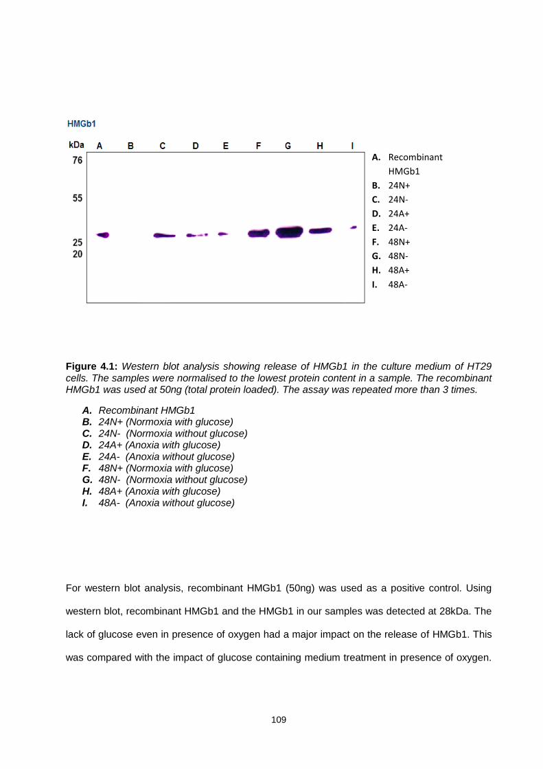
109
Figure 4.1: Western blot analysis showing release of HMGb1 in the culture medium of HT29 cells. The samples were normalised to the lowest protein content in a sample. The recombinant HMGb1 was used at 50ng (total protein loaded). The assay was repeated more than 3 times.
A. Recombinant HMGb1 B. 24N+ (Normoxia with glucose) C. 24N- (Normoxia without glucose) D. 24A+ (Anoxia with glucose) E. 24A- (Anoxia without glucose) F. 48N+ (Normoxia with glucose) G. 48N- (Normoxia without glucose) H. 48A+ (Anoxia with glucose) I. 48A- (Anoxia without glucose)
For western blot analysis, recombinant HMGb1 (50ng) was used as a positive control. Using
western blot, recombinant HMGb1 and the HMGb1 in our samples was detected at 28kDa. The
lack of glucose even in presence of oxygen had a major impact on the release of HMGb1. This
was compared with the impact of glucose containing medium treatment in presence of oxygen.
A. Recombinant
HMGb1
B. 24N+
C. 24N-
D. 24A+
E. 24A-
F. 48N+
G. 48N-
H. 48A+
I. 48A-

110
There appeared to be a major quantitative difference amongst these two conditions with more
HMGb1 being released when cells were treated with no glucose conditions (Figure 4.1). In
addition, the presence of glucose appeared to trigger the release of HMGb1 but only after 24h
(Figure 4.1). Interestingly, figure 4.1 also shows that harsher condition such as lack of oxygen
(anoxia) did not show any significant impact on the release of HMGb1 when compared to
oxygen rich conditions.
4.4.2 The release of HMGb1 in response to lack of glucose is a
phenomenon in common with other cancer cell types
Western blot analysis from this study showed that glucose deprivation triggers the release of
HMGb1 in HT29 cells (Figure 4.1). This release was greater than the release of HMGb1 in
response to anoxia, a condition which is a known cause of HMGb1 release. To investigate
whether this HMGb1 release in response to low glucose levels was specific to the colon
adenocarcinoma cell line or whether it is a common phenomenon, other cancer cells were
analysed. MCF-7 (breast adenocarcinoma), EJ138 (bladder carcinoma) and A549 (lung
carcinoma) cells were treated with similar conditions and their culture medium at 48h were
collected. A western blot was carried out to determine whether these cell lines followed the
same pattern of HMGb1 release in response to glucose deprivation in HT29 cells.
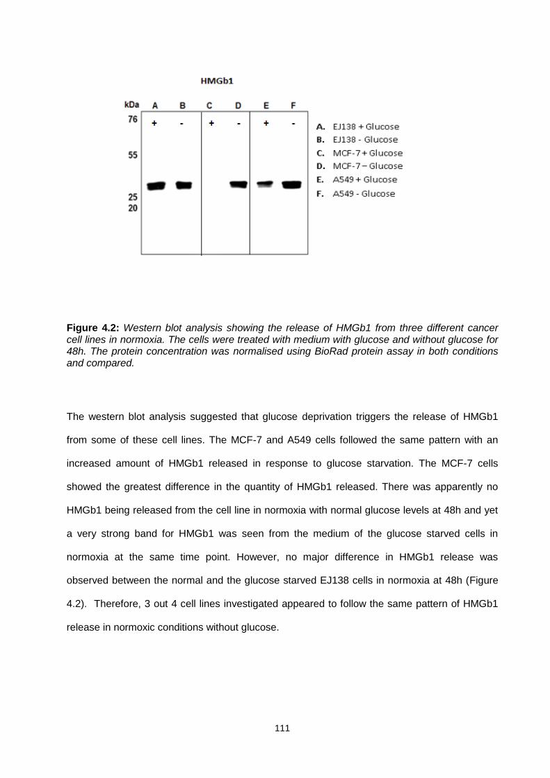
111
Figure 4.2: Western blot analysis showing the release of HMGb1 from three different cancer cell lines in normoxia. The cells were treated with medium with glucose and without glucose for 48h. The protein concentration was normalised using BioRad protein assay in both conditions and compared.
The western blot analysis suggested that glucose deprivation triggers the release of HMGb1
from some of these cell lines. The MCF-7 and A549 cells followed the same pattern with an
increased amount of HMGb1 released in response to glucose starvation. The MCF-7 cells
showed the greatest difference in the quantity of HMGb1 released. There was apparently no
HMGb1 being released from the cell line in normoxia with normal glucose levels at 48h and yet
a very strong band for HMGb1 was seen from the medium of the glucose starved cells in
normoxia at the same time point. However, no major difference in HMGb1 release was
observed between the normal and the glucose starved EJ138 cells in normoxia at 48h (Figure
4.2). Therefore, 3 out 4 cell lines investigated appeared to follow the same pattern of HMGb1
release in normoxic conditions without glucose.

112
4.4.3 Glutamine deprivation also stimulated the release of HMGb1
The results from western blot analysis suggested that HMGb1 was released from a selection of
cancer cells including HT29, MCF-7, and A549 in glucose deprivation conditions under
normoxia. However, whether HMGb1 also released in response to other nutrient deprived
conditions is unclear. Therefore, the release of HMGb1 from HT29 cells was compared
between normal and glutamine deprived conditions at 48h.
The western blot was set up in two sets; one was to compare the release of HMGb1 in
response to glutamine deprived versus normal medium. The other one was to compare HMGb1
release in medium deprived of glutamine versus glucose. The results from the first set of
western blot suggest that HMGb1 is released in glutamine deprived culture medium of HT29
cells (Figure 4.3 A).This release of HMGb1 was compared to glutamine deprived and glutamine
containing medium. It was found that there was no major difference in the release of HMGb1 in
glutamine deprived conditions compared to normal glutamine conditions. However, the second
set of western blot comparing the release of HMGb1 between from HT29 cells in glucose and
glutamine deprived medium revealed that only glucose deprivation triggers the release of
HMGb1 (Figure 4.3 B).
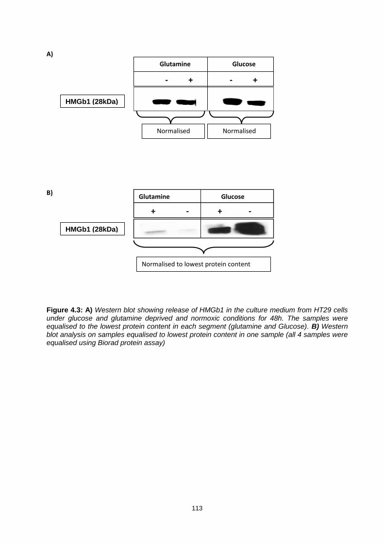
113
A)
B)
Figure 4.3: A) Western blot showing release of HMGb1 in the culture medium from HT29 cells under glucose and glutamine deprived and normoxic conditions for 48h. The samples were equalised to the lowest protein content in each segment (glutamine and Glucose). B) Western blot analysis on samples equalised to lowest protein content in one sample (all 4 samples were equalised using Biorad protein assay)
HMGb1 (28kDa)
- + - +
Glutamine Glucose
+ - + -
Glutamine Glucose
HMGb1 (28kDa)
Normalised Normalised
Normalised to lowest protein content

114
4.4.4 The CC18 myofibroblast cells express advanced glycation end
product (RAGE)
Receptor for advanced glycation end products, a multiligand receptor and is also a receptor for
HMGb1. The HMGb1 and RAGE complex has been implicated in many diseases including
cancer (Nienhuis et al. 2009; Sims et al. 2009; Dzaman et al. 2015). The results from western
blot analysis confirmed the presence of HMGb1 in the conditioned medium of cancer cells
including HT29 cells. This study has shown that recombinant HMGb1 triggers proliferation in
myofibroblasts cells. Therefore, there is a possibility that HMGb1 induced proliferation of
myofibroblasts cells involves activation of RAGE which might be present on the membrane of
CCD18 myofibroblast cells.
The CCD18 myofibroblast cells were treated with glucose free and normal glucose conditions in
normoxia and anoxia for 24h and 48h and the cell lysates were collected for protein analysis.
The protein concentration was equalised so that all samples contained the same concentration
of total cellular protein before western blot analysis on all samples. Detection of GAPDH was
used as loading control for the samples. The results from western blot analysis suggested that
RAGE is present on myofibroblasts cells. The samples included with and without glucose in
normoxic and with anoxic conditions. All conditions including 48h normoxia without glucose
showed some levels of RAGE. There was no significant difference observed between any
specific treatment but relatively higher levels of RAGE appeared to be correlated with high
glucose levels (Figure 4.4). The ligand HMGb1 controlled upregulation or downregulation of
RAGE would be difficult to investigate in this case because RAGE is a multiligand receptor and
might be interacting with other ligands as well as HMGb1. Similarly, HMGb1 might be
interacting with other receptors which may be present on myofibroblasts. Therefore, HMGb1

115
signalling in response to glucose deprivation might involve one or more other receptors which
are present on myofibroblasts. Although, this study was focused on investigating similar
conditions which triggered the release of HMGb1 from HT29 cells but there was always a
possibility for HMGb1 binding to certain receptors which may not be upregulated in similar
conditions as those with HT29 cells. Those receptors may include RAGE and TLR-4 amongst
others. However, this experiment confirmed that CCD18 myofibroblast cells express RAGE in
all treatment conditions (Figure 4.4).
Figure 4.4: Western blot confirming the presence of RAGE in the CCD18 cell lysates. The cells were treated with different conditions for 24 and 48h. The protein concentration was normalised using BioRad assay and GAPDH was used as loading control.
A. 24N+ (Normoxia with glucose) B. 24N- (Normoxia without glucose) C. 24A+ (Anoxia with glucose) D. 24A- (Anoxia without glucose) E. 48N+ (Normoxia with glucose) F. 48N- (Normoxia without glucose) G. 48A+ (Anoxia with glucose)
A B C D E F G
RAGE
GAPDH
H
42KDa
40KDa
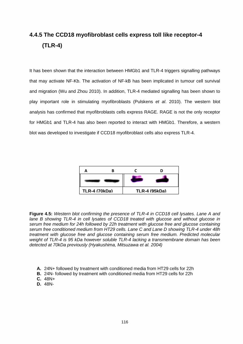
116
4.4.5 The CCD18 myofibroblast cells express toll like receptor-4
(TLR-4)
It has been shown that the interaction between HMGb1 and TLR-4 triggers signalling pathways
that may activate NF-Kb. The activation of NF-kB has been implicated in tumour cell survival
and migration (Wu and Zhou 2010). In addition, TLR-4 mediated signalling has been shown to
play important role in stimulating myofibroblasts (Pulskens et al. 2010). The western blot
analysis has confirmed that myofibroblasts cells express RAGE. RAGE is not the only receptor
for HMGb1 and TLR-4 has also been reported to interact with HMGb1. Therefore, a western
blot was developed to investigate if CCD18 myofibroblast cells also express TLR-4.
Figure 4.5: Western blot confirming the presence of TLR-4 in CCD18 cell lysates. Lane A and lane B showing TLR-4 in cell lysates of CCD18 treated with glucose and without glucose in serum free medium for 24h followed by 22h treatment with glucose free and glucose containing serum free conditioned medium from HT29 cells. Lane C and Lane D showing TLR-4 under 48h treatment with glucose free and glucose containing serum free medium. Predicted molecular weight of TLR-4 is 95 kDa however soluble TLR-4 lacking a transmembrane domain has been detected at 70kDa previously (Hyakushima, Mitsuzawa et al. 2004)
A. 24N+ followed by treatment with conditioned media from HT29 cells for 22h B. 24N- followed by treatment with conditioned media from HT29 cells for 22h C. 48N+ D. 48N-
TLR-4 (70kDa) TLR-4 (95kDa)
A B C D
117
The results from western blot confirmed that myofibroblasts cells express TLR-4. The cells were
treated with glucose and without glucose for 24h in the absence of serum. The expression of
TLR-4 was observed in both conditions (Figure 4.5).The cells were also treated with serum free
medium with glucose for 24h followed by 22h treatment with medium taken from HT29 grown
for 48h in the absence of glucose. As western blot analysis showed that TLR-4 is present in
stress conditions such as lack of glucose and then it is possible that it might interact with
HMGb1 to initiate signalling cascade in microenvironment. The TLR-4 was observed in at two
different places at 95kDa with 48h treatment with glucose free and glucose rich conditions and
70kDa with fresh medium and HT29 conditioned medium treatment (24h+22h) (Figure 4.5). The
predicted molecular weight of TLR-4 is 95kDa and therefore the 70kDa could be proteolytic
degraded product that lacks transmembrane domain. The soluble TLR-4 which lacks
transmembrane domain has been identified at 70kDa previously (Hyakushima et al. 2004).
4.5 Discussion
Tumour hypoxia facilitates the release of intracellular moieties including HMGb1 from tumours.
It has also been shown that hypoxic regions of tumours attract macrophages that have strong
pro-angiogenic properties which then facilitate the release of HMGb1. This facilitates more
effective proliferation and metastatic spread in tumours (Ellerman et al. 2007). There is an
increasing body of evidence that supports the hypothesis that neoplastic tissues utilise a large
quantity of glucose. The effect of glucose deprivation and acidosis has been shown to increase
metastasis in murine tumour cell lines in vivo. However, the underlying mechanism of glucose
deprivation induced metastatic spread remains unknown (Schlappack et al. 1991; Xie and
Huang 2003). Glioblastoma cells do not release HMGb1 in their culture medium. However,

118
necrotic glioma cells can release HMGb1 after its translocation from nucleus to cytosol (Bassi et
al. 2008). The release of HMGb1 from immune cells as danger signal is well documented
(Manfredi et al. 2009; Pisetsky 2011; Ganz et al. 2015). However, the release of HMGb1 from
cancer cells under glucose starving conditions has, so far, not been reported.
A western blotting method was developed to detect the release of HMGb1 from HT29 cells.
The western blot results showed that HMGb1 is released from HT29 cells when exposed to
different stress conditions such as anoxia and/or the absence of glucose. Interestingly, absence
of glucose appeared to stimulate the release of HMGb1 even in presence of oxygen at early
time points (24 to 48h) in our system (Figure 4.1). This release of HGMb1 was compared to the
release of HMGb1 in absence of oxygen. It was found that glucose deprivation is the main
stimulus which facilitates the release of HMGb1 from HT29 cells.
The release of HGMb1 in anoxic conditions though appeared to be time dependent but it was
slower than the normoxic conditions with glucose starvation. In addition, in an attempt to
investigate the release of HMGb1 at higher time points (72 and 96h) it was found that number of
cells were floating in the flasks. These cells were presumed to be dead cells. In that case, even
if HMGb1 was released, being a nuclear protein, it was apparent that HMGb1 would have
passively been released or spilled out from the dead cells into the culture medium. Therefore,
carrying out an investigation using western blot would not represent the true data as actively
and passively released HMGb1 can not be differentiated in terms of structure and molecular
weight. Therefore, a western blot was not carried out on those samples.
119
The other aim was to investigate whether this release of HMGb1 is cell line specific or it’s a
common phenomenon that occurs in other cell lines derived from other types of tumour. The
hypoxia induced HMGb1 release has been seen in a number of cells such as RAW264.7
macrophages, WM9 melanoma cells and hepatocytes (Wang et al. 2004; Tsung et al. 2007; Ito
et al. 2007). The results from western blot analysis showed that the release of HMGb1 under
glucose free normoxic conditions is not cell line specific and other cells such as MCF-7 and
A549 followed the same pattern. However, EJ138 did not appear to release more HMGb1 in
glucose free conditions. The levels of HMGb1 observed in the culture medium taken from
EJ138 were not different in both glucose containing and glucose free conditions.
It has been documented that hypoxic or semi hypoxic regions inside the tumours are glucose
deprived. However, glutamine is also an important nutrient for the cells to compete their cycle.
The nutrient deprived conditions such as lack of glutamine in solid tumours have been shown to
induce autophagy in the cells which in turn helps tumour cells to proliferate in the
microenvironment (Ye et al. 2010). It has been shown that the glioblastoma cells that are
destined to die (autophagy) release HMGb1 in their culture medium without the lysis of
membrane or necrosis (Thorburn et al. 2008). Therefore, there is a possibility that HT29 cells
were undergoing autophagy but it can not be confirmed because of poor understanding of the
mechanism of autophagic cell death. However, the drivers of autophagic cell death such as lack
of nutrients (glutamine) and oxygen were tested. Glutamine has been considered as major
regulator of autophagy and lack of glutamine may promote cell death via apoptosis (Sakiyama
et al. 2009). Therefore, it was also investigated whether this release of HMGb1 was triggered by
absence of the nutrient glutamine or whether release was specific to the absence of the nutrient
glucose. The results from our western blot showed that HMGb1 is released from both glutamine
deprived and glutamine containing conditions and there was no major difference in release

120
observed between the two conditions. However, when glutamine deprived and glutamine
containing conditions were compared with glucose deprived and glucose containing conditions,
it was found that glucose deprived conditions serve as major stimulus which facilitates the
release of HMGb1. These findings are novel and have not been published thus far.
Receptor for advanced glycation end products (RAGE) is the most commonly known receptor
for HMGb1. RAGE is multi-ligand receptor, expressed in many cells including monocytes,
macrophages and smooth muscle cells can be activated by several ligands including HMGb1.
The activation of RAGE in monocytes/macrophages has been reported to trigger the
inflammatory response and to trigger neoplastic transformation and metastasis in
neuroepithelial tumour cells (Taguchi et al. 2000). A recent study has shown that HMGb1 binds
to RAGE and not to the TLR-4 to promote resistance to Melphan by inducing beclin-1
dependent autophagy (Tang et al. 2010). The data from our proliferation assays demonstrated
that HMGb1 triggers proliferation in myofibroblasts cells where blocking both MEK1/2 and PI3K
pathways with selective inhibitors abrogated the proliferative effect of HMGb1 in CCD18
myofibroblast cells (Figure 3.6 and 3.8). Recently, it has been shown that HMGb1-RAGE
complex increases the production of ATP, which is required by tumour cells to grow and migrate
to the distant sites. This production of ATP was blocked by either inhibiting RAGE or HMGb1 by
immunoneutralising antibodies which resulted in diminished proliferation and migration of
tumour cells (Kang et al. 2013). The data from our western blot suggested that RAGE is
expressed on myofibroblasts cells.
Although, the expression of RAGE was not correlated with the release of HMGb1 in different
stress conditions from cancer cells but it was detected in all stress conditions such as glucose

121
deprivation and/or anoxia. Thus, there is a possibility for the activation of HMGb1-RAGE and
subsequent activation of downstream signalling which facilitates proliferation in myofibroblasts
cells such as activation of MAPK/ERK pathway in the HMGb1 induced proliferation in
myofibroblasts shown in this study. Recently, it has been shown that RAGE expression is
upregulated at various stages of transdifferentiation from hepatic stellate cells (HSCs) to
myofibroblasts (Wang et al. 2013). However, here it is not confirmed yet whether HMGb1
induced proliferation of myofibroblasts involves activation of HMGb1-RAGE complex. The
HMGb1-RAGE complex has been successfully detected by co-immunoprecipitation technique
recently (Lai et al. 2013; Kang et al. 2014). Therefore, to further validate the interaction of
RAGE and HMGb1 to trigger proliferation in myofibroblasts, co-immunoprecipitation should be
carried out myofibroblasts cell lysates followed by affinity chromatography to elute RAGE or
HMGb1 or both. The co-immunoprecipitation is a technique which detects the interaction of a
protein with other proteins in a sample (Kaboord and Perr 2008).
RAGE is not the only receptor for HMGb1 and others include the toll-like receptors 2 and 4
(TLR-2 and TLR-4). Park et al. (2004) demonstrated that blockage or knockdown of these
receptors resulted in decreased HMGb1 activation in vitro and in vivo (Park et al. 2004). The
activation of HMGb1-TLR-4 complex has been shown to trigger migration in HSC cells via the
activation of PI3K-AKT pathway (Wang et al. 2013). The results from our western blot
suggested that TLR-4 is present on myofibroblasts cells treated with both conditions i,e. glucose
containing and glucose deprived conditions for 48h. In addition, we also investigated whether
TLR-4 is expressed on the CCD18 cells treated with serum fee medium for 24 hr then treated
for 22h with used serum free medium taken from HT29 after 48h. These conditions mimic the
migration assay conditions where inhibition TLR-4 significantly affected the migration response
in myofibroblasts cells (explained in chapter 5).

122
The results from our western blot suggested that TLR-4 is present on the myofibroblasts cells
that underwent treatment mimicking (treatment with HT29 culture medium) the migration assay
treatment. However, the protein bands for TLR-4 were also observed at 70kDa which is
presumably a proteolytic degraded product of TLR-4. This 70kDa band corresponding TLR-4
has previously been observed by others (Hyakushima et al. 2004). The role of HMGb1-TLR-4
complex has been implicated in predicting the outcome of cancer chemotherapy. The activation
of this complex is correlated with relapse after the anthracycline based chemotherapy in breast
cancer patients (Apetoh et al. 2007). The activation of HMGb-TLR-4 complex on dendritic cells
was detected by co-immunoprecipitation technique (Apetoh et al. 2007). Therefore, it would be
logical approach to apply co-immunoprecipitation to selectively detect the HMGb-TLR-4
complex in myofibroblast cell lysates. This would further confirm whether HMGb1 induced
proliferation and migration of myofibroblasts cells involved the activation of HMGb1-TLR-4
complex.
123
Chapter 5
5. Role of HMGb1 in myofibroblasts migration and invasion
5.1 Introduction
Presence of stromal cells such as fibroblasts and myofibroblast in solid tumours plays an
important role in predicting the metastatic behaviour of solid tumours. It has been established
that stromal cells play important roles in carcinogenesis. The stromal myofibroblasts have been
considered as predictors of human disease outcome and their abundance is correlated with
poor survival in colorectal cancer (Tsujino et al. 2007). The likely role of myofibroblasts in
tumour invasion has been explored in the past and it was found that the myofibroblasts
population gradually increases with the invasive stages of many cancers (Nakayama et al.
1998). The expression of α-SMA, which is a biomarker for myofibroblasts, has been correlated
with enhanced metastatic spread of tumours whose characterisation involves CAFs. For
example, an increased expression of α-SMA has been seen in fibroblasts present in HER2
breast cancers (Toullec et al. 2010).
There is evidence that support the involvement of stromal myofibroblasts in tumour
development. For example, myofibroblasts have been shown to stimulate proliferation in breast
cancer cells in vivo (Khanna et al. 2015). In addition, they have also been shown to promote
invasion of breast, pancreas and squamous carcinoma cells in vitro (Casey et al. 2008; Hu et al.
2008; Hwang et al. 2008). Furthermore, mesenchymal cells and their derivatives, including
124
myofibroblasts, have been shown to promote breast cancer metastasis. The myofibroblasts
population was significantly high in breast adenocarcinoma and lung cancer that metastasised
to lymph nodes compared to the non-metastatic tumours. This suggested that myofibroblasts
contribute toward the spreading of tumours through metastasis (Toullec et al. 2010). Cancer
cells have been shown to stimulate stromal cells such as myofibroblasts to produce proteases
(Tuxhorn et al. 2002; Anborgh et al. 2010). These proteases cleave ECM components and
remodel the ECM in the microenvironment (Hinz et al. 2012). A number of protease have been
shown to degrade ECM in the tumour microenvironment however, gelatinase (MMP-2) and
MMP-9 are two major proteases released from myofibroblasts and have been shown to
degrade basement membrane (Cheng and Lovett 2003; Takahra et al. 2004).
The cytokine TGF-β is present in exosomes which are secreted by most cell types including
cancer cells trigger the conversion of fibroblasts to myofibroblasts at distant organs. It has also
been suggested that TGF-β might facilitate metastasis by stimulating cells to produce
fibronectin. However, tumour cells may (also) move along with myofibroblasts to the distant
sites, such as the lungs. These migrated myofibroblasts have been shown to produce pro-
survival signals in the lung site (Duda et al. 2010). Therefore, transformed fibroblasts or cancer
associated myofibroblasts may influence tumour microenvironment and may contribute towards
malignant transformation in solid tumours. Therefore, it is logical to investigate the role of
HMGb1 in myofibroblasts which may be linked with invasion and metastasis of tumour cells.
Necrotic areas are characteristic features of rapidly growing tumours. The necrotic areas within
tumour not only produce angiogenic factors such as VEGF but also attract macrophages. In
stress conditions such as necrosis, these macrophages release HMGb1 within the
125
microenvironment (van Beijnum et al. 2006). Several solid tumours including melanoma,
prostate cancer, breast cancer, pancreatic cancer and colon cancer exhibit markedly elevated
levels of HMGb1 (Völp et al. 2006). These elevated levels of HMGb1 are associated with
tumour formation, proliferation and metastasis and chemotherapeutic response. In addition,
presence of HMGb1 in the extracellular medium of cells is indicative of stress conditions (Lotze
and Tracey 2005).
The direct inhibition of HMGb1 with immunoneutralising antibodies has been reported to inhibit
angiogenesis in vivo and in vitro (Van Beijnum et al. 2006). The HMGb1 interacts with RAGE,
TLRs (TLR-2 and TLR-4) and may be with other unknown receptors and activates various
factors and kinases such as NF-κB and MAPK. It has been shown that HMGb1 can increase
the metastatic potential of tumour cells by activating NF-κB pathway (Sasahira et al. 2008). In
addition, HMGb1 can modulate the adhesive properties of cells and ECM components and thus
can directly affect the migration of cells (Ellerman et al. 2007).
The HMGb1 activates various signalling pathways that involve the activation of MAPKs, AKT
and PI3K. These protein kinases play an important role in the proliferation and migration of
tumour cells. Supporting evidence includes HMGb1 activated PI3K pathway in neutrophils and
colon cancer cells (Kuniyasu et al. 2003). In addition, HMGb1 has been shown to induce TLR-4
mediated activation of MyD88-IRAK4-p38 and Myd88-IRAK4-AKT pathways (Fan et al. 2007).
These pathways have been implicated in tumour cell proliferation and survival. The HMGb1-
RAGE complex has been shown to activate NF-ƘB, Jun N terminus kinase (JNK) and ERK1/2
pathways (Degryse et al. 2001). The activation of JNK is correlated with an increase in cell
migration in different cell types including rat bladder tumour cells, keratinocytes and human
126
dermal fibroblasts. It has also been shown that MEK1 which is an upstream kinase in the JNK
signalling pathway is essential for cell migration (Van Beijnum et al. 2008).
The data presented in chapter 3 suggested that HMGb1 triggers proliferation in myofibroblasts.
This proliferation was inhibited by U0126 and LY294002 selective inhibitors of MEK1/2 and
PI3K respectively. It has been established that HMGb1 is released in hypoxia, which is
characteristic feature of most solid tumours (Tsung et al. 2007). Glucose deprivation is also a
hallmark of tumour microenvironment. The central core of most solid tumours is characterised
by lack of glucose and acidosis (Cuvier et al. 1997). Glucose deprivation in the solid tumour
microenvironment and its consequences within and outside tumour microenvironment are
largely unexplored. The data presented in chapter 4 indicates that HMGb1 is released from
cancer cells in glucose deprived conditions and not from depravation of the nutrient glutamine.
Previous studies have shown a correlation between myofibroblasts and metastasis in many
solid tumours including breast cancer (Allinen et al. 2004; Muehlberg et al. 2009). Thus, there is
a possibility that HMGb1 once released from cancer cells might influence the other stromal cells
such as myofibroblasts to migrate and invade. In addition, MAPKs, which play a central role in
migration of many cells including fibroblasts, might have a role in migration of myofibroblasts.
The HMGb1 and its interaction with RAGE and TLRs have important roles in intracellular
signalling of various pathways including PI3K (Fan et al. 2007). Therefore, neutralising
antibodies to these receptors will be used to confirm their role in migration and invasion of
myofibroblasts

127
5.2 Aims and objectives
The aims of this study were to;
a) Investigate if glucose deprived conditioned medium of HT29 cells can induce migration
and invasion in CCD18 myofibroblast cells.
b) Compare the impact of used medium, derived from HT29 cells deprived of glucose or
rich in glucose with fresh medium rich in glucose on CCD18 myofibroblast cells
migration and invasion.
c) Investigate whether recombinant HMGb1 can induce migration in myofibroblasts cells
d) Investigate whether HMGb1 present in the conditioned medium of HT29 cells is
responsible for apparent migration and invasion in myofibroblasts.
e) Investigate whether inhibiting HMGb1 in the conditioned medium of HT29 cells by anti-
HMGb1 neutralizing antibody can reduce migration and invasion in CCD18 cells.
f) Investigate whether Inhibition of RAGE by neutralizing antibodies can have negative
impact of myofibroblasts migration and invasion.
g) Investigate whether inhibition of TLR-4 results in reduction in myofibroblastic migration
and invasion.
h) Investigate whether MEK1/2 and PI3K pathways are involved in HMGb1 induced
migration and invasion in myofibroblasts.
i) Investigate whether MMP-2 and MMP-9 are released from CCD18 myofibroblast cells
following the treatment with HMGb1 containing conditioned medium collected from HT29
cells.
128
5.3 Material and Methods
5.3.1 Cell culture
The CCD18 myofibroblast cells were cultured in T75 cm2 flasks in complete medium (minimum
essential medium eagle) supplemented with foetal bovine serum (10% v/v). The cells were
passaged when they had reached about 80-90% confluence. In order to carry out migration or
invasion assays, the myofibroblast cells were first serum starved for 24h before they were used.
The cells were dislodged from flasks using versene. Versene (1x) is less aggressive than
trypsin and was used to dissociate the cells in the flask before plating them onto the 24-well
plate. Versene is a chelating agent that binds to calcium and prevents joining
of cadherins between cells thereby prevents clumping of cells in the medium. The cells
suspension in versene was diluted with serum free medium to a concentration of 4.0x10⁴ cells
per ml for use in migration assays or 6.0x10⁴ cells per ml for used in the invasion assays.
5.3.2 Migration assay
HT29 colon adenocarcinoma cell lines were seeded in 2xT75cm2 (2x10⁶ cells per ml). Both
flasks were incubated over night at 37°C, 95% humidity and 5% CO2 to allow the cells to
adhere. The following day, the complete medium was aspirated off and cells were washed twice
with PBS. The medium on the cells was replaced with serum free medium with glucose in one
flask and without glucose in the other flask. After 48h, the conditioned medium from both flasks
were collected into labelled universals and used as potential chemoattractants for the migration
assay.

129
The CCD18 cell suspension was made in serum free medium (4.0x10⁴ per ml) (see section
5.2.1 above). The CCD18 myofibroblast cells were then seeded (0.5ml per insert) in 2x2
chamber inserts (Biocoat®, 8µm pores) which were then transferred to 4 wells of the 24 well
plate and ensured that insert bottom is touching the HT29 conditioned medium (0.75ml per well)
in the wells. The plate was incubated for 20h in the incubator at 37°C, 95% humidity and 5%
CO2. The CCD18 cell suspension in the inserts and media at the bottom chamber were
supplemented with antibodies and inhibitors to investigate the various pathways involved in
myofibroblast migration (see chapter 2, table 2.7).
After 20h of incubation period, plates were removed and cells were stained (QuickDiff) for
counting. The images of random fields of vision were captured from each membrane. The cells
that had migrated through the membrane were counted using ImageJ cell counter software and
the average of the 5 fields of vision for each membrane calculated.
5.3.3 Invasion assay
The invasion assays for CCD18 myofibroblasts were performed using 8µm pore matrigel matrix
Biocoat® inserts according to the manufacturer’s instruction (Becton Dickinson). The CCD18
cell suspension was made in serum free medium (6.0x10⁴ per ml). The myofibroblasts cells
were plated onto (0.5ml per insert) inserts in a 24-well plate supplied by the manufacturer. The
inserts were put into the wells of 24-well plate. HT29 conditioned medium was used as
chemoattractant and plated onto the lower chamber (0.5ml) and ensured that inserts were
touching the conditioned medium (see section 2.4). The plate was incubated for 22h in the
incubator at 37°C, 95% humidity and 5% CO2. The myofibroblasts invasion, in response to the

130
conditioned medium with and without various inhibitors and antibodies were determined by their
inclusion in the lower chamber of the 24-well plate. Cells invaded through the matrigel matrix
membrane were detected on the lower surface by using ReaStain Quick-Diff staining method
and visualised at x20 magnification under a microscope. The CCD18 cell suspension in the
inserts and media at the bottom chamber were supplemented with antibodies and inhibitors to
investigate the various pathways involved in myofibroblast invasion (see table 2.7)
5.4 Results
5.4.1 Culture medium from HT29 cancer cells starved of glucose
triggers migration in CCD18 myofibroblast cells
The glucose deprivation has been considered as hallmark in most solid tumours and has been
reported to induce migration of cancer cells. The western blot results discussed in chapter 4
suggested that HMGb1 is released from cancer cells in glucose deprived conditions. However,
the subsequent effect on tumour stromal cells such as myofibroblasts has not been explored.
The myofibroblasts are stromal cells and constitute a major part of tumour stroma. In addition,
they have been found in abundance at metastatic sites of solid tumours. Therefore, it is
hypothesised that HMGb1 once released from cancer cells, reaches to the stroma to influence
myofibroblasts to proliferate, migrate and invade along with neighbouring cancer cells by
digesting the basement membrane. This might allow cancer cells to escape from their original
location to the distant organs.
131
A migration assay was set up according to the instructions described in method section (section
5.2). HT29 colon cancer cells were grown in medium containing no glucose for 48h. This
medium was then collected from the HT29 cells. This HT29 conditioned medium was used as
chemoattractant as this was shown to contain the highest levels of HMGb1 when compared to
medium that had been used on HT29 cells containing glucose or fresh unused medium with or
without glucose (Chapter 4). This conditioned medium served as positive control (therefore
counted as 100% migration) and was compared to myofibroblast migration in response to
conditioned medium that contained glucose or the fresh serum free medium with and without
glucose. The results obtained from this migration assay suggest that HT29 cells undergoing
glucose deprivation release chemoattractants that trigger migration in myofibroblasts cells. In
addition, conditioned medium with glucose does trigger migration in myofibroblasts cells;
however, this migration was approximately 60% less than the conditioned medium without
glucose at 100% (Figure 5.1 A). In addition, the effects of fresh serum free medium with and
without glucose were examined. A negligible number of myofibroblasts cells migrated across
the 8µm pore membrane when exposed to the fresh serum free medium with and without
glucose (Figure 5.1 D and E). The data suggests that absence or presence of glucose in a fresh
medium does not influence the myofibroblasts cells to migrate. All these results are statistically
significant (Figure 5.1A).
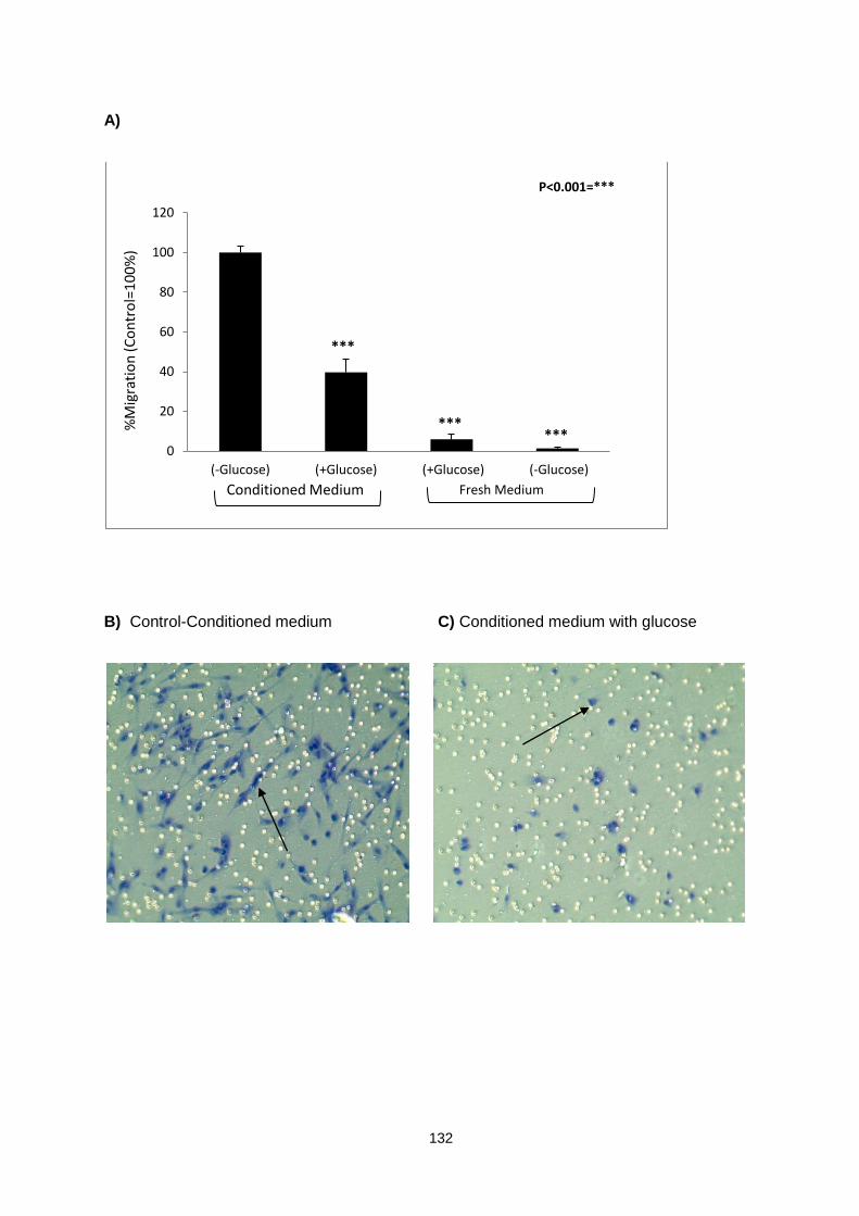
132
A)
B) Control-Conditioned medium C) Conditioned medium with glucose
0
20
40
60
80
100
120
(-Glucose) (+Glucose) (+Glucose) (-Glucose)
%M
igra
tio
n (C
on
tro
l=1
00
%)
P<0.001=***
***
*** ***
Fresh Medium Conditioned Medium

133
D) Serum free fresh medium with glucose E) Serum free medium without glucose
Figure 5.1: CCD18 myofibroblast cells migration assay in response to HT29 conditioned medium. A) The migration assay carried out using HT29 conditioned medium as chemoattractant. The HT29 conditioned medium (-glucose) served as control. The conditioned medium with glucose and fresh serum free medium with and without glucose were compared to the control (100%). The assay was carried out three times with 2 inserts per condition per occasion (5 random fields of vision counted per insert). Student’s t-test was used to determine statistical significance of the results. SEMs were displayed as error bars.
Typical microscope images captured showing the underside of the membrane after completion of the migration assay. Cells that had migrated though the 8µm pores of the membrane were stained blue using the ReaStain Quick-Diff staining method. The white dots are 8µm pores through which cells have not migrated. B) Representative images from migration assays showing cells migrated in response to conditioned medium without glucose (Control=100%), C) cells migrated in response to the conditioned medium with glucose, D) cells migrated in response to fresh serum free medium with glucose and E) cells migrated in response to fresh glucose free and serum free medium. Cells that had migrated on these and other images were counted and the results were presented in A) of this figure. The photographs were taken under x20 magnification.

134
5.4.2 HMGb1 released from HT29 colon adenocarcinoma cells triggers
migration in CCD18 myofibroblasts
The results discussed in chapter 3 suggested that recombinant HMGb1 triggers proliferation in
CCD18 myofibroblast cells in a dose dependent manner. In addition, results from western
blotting suggested that HMGb1 is released from HT29 and other cancer cells including MCF-7
and A549 cells under glucose deprivation normoxic conditions. To investigate whether HMGb1
present in the culture medium of HT29 cells can trigger migration in myofibroblasts, HT29
conditioned medium was used as chemoattractant. The glucose starving-conditioned medium
worked as a strong chemoattractant in our migration assay. However, to validate HMGb1
mediated signalling in the migration of myofibroblasts cells, anti-HMGb1 antibody (5µg/ml) was
added to the conditioned medium at the bottom chamber to neutralise the effect of HMGb1. If
the HMGb1 was involved in the migration of myofibroblast cells, a decrease in the migration of
cells was expected.
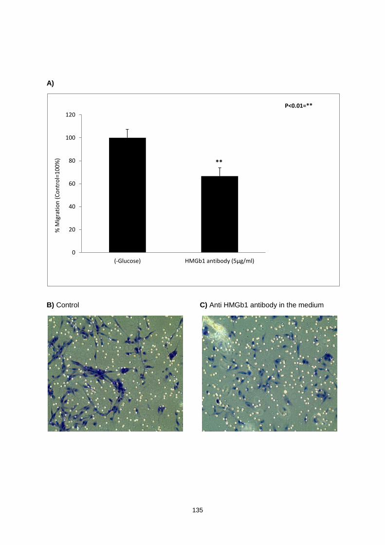
135
A)
B) Control C) Anti HMGb1 antibody in the medium
0
20
40
60
80
100
120
(-Glucose) HMGb1 antibody (5µg/ml)
% M
igra
tio
n (
Co
ntr
ol=
10
0%
)
P<0.01=**
**
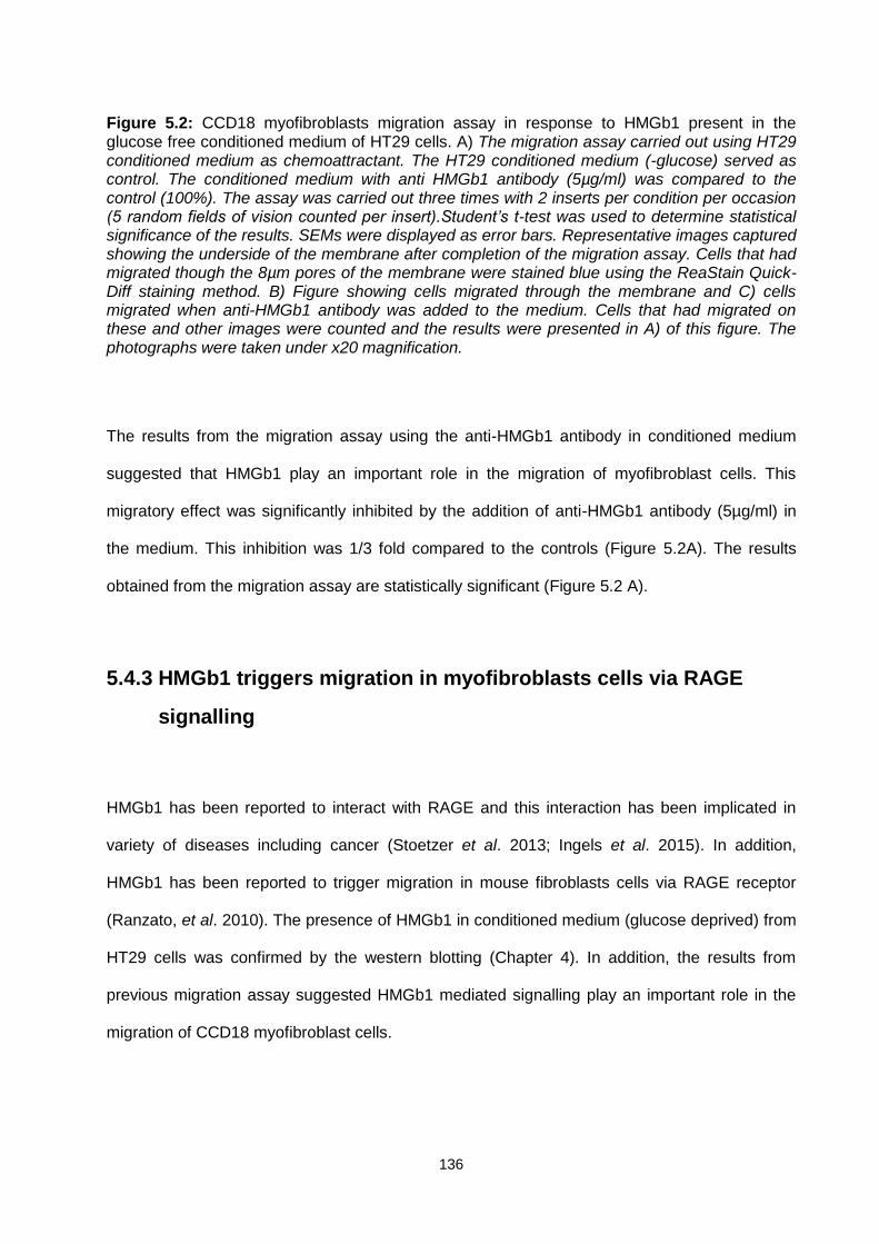
136
Figure 5.2: CCD18 myofibroblasts migration assay in response to HMGb1 present in the glucose free conditioned medium of HT29 cells. A) The migration assay carried out using HT29 conditioned medium as chemoattractant. The HT29 conditioned medium (-glucose) served as control. The conditioned medium with anti HMGb1 antibody (5µg/ml) was compared to the control (100%). The assay was carried out three times with 2 inserts per condition per occasion (5 random fields of vision counted per insert).Student’s t-test was used to determine statistical significance of the results. SEMs were displayed as error bars. Representative images captured showing the underside of the membrane after completion of the migration assay. Cells that had migrated though the 8µm pores of the membrane were stained blue using the ReaStain Quick-Diff staining method. B) Figure showing cells migrated through the membrane and C) cells migrated when anti-HMGb1 antibody was added to the medium. Cells that had migrated on these and other images were counted and the results were presented in A) of this figure. The photographs were taken under x20 magnification.
The results from the migration assay using the anti-HMGb1 antibody in conditioned medium
suggested that HMGb1 play an important role in the migration of myofibroblast cells. This
migratory effect was significantly inhibited by the addition of anti-HMGb1 antibody (5µg/ml) in
the medium. This inhibition was 1/3 fold compared to the controls (Figure 5.2A). The results
obtained from the migration assay are statistically significant (Figure 5.2 A).
5.4.3 HMGb1 triggers migration in myofibroblasts cells via RAGE
signalling
HMGb1 has been reported to interact with RAGE and this interaction has been implicated in
variety of diseases including cancer (Stoetzer et al. 2013; Ingels et al. 2015). In addition,
HMGb1 has been reported to trigger migration in mouse fibroblasts cells via RAGE receptor
(Ranzato, et al. 2010). The presence of HMGb1 in conditioned medium (glucose deprived) from
HT29 cells was confirmed by the western blotting (Chapter 4). In addition, the results from
previous migration assay suggested HMGb1 mediated signalling play an important role in the
migration of CCD18 myofibroblast cells.

137
One of the aims of this migration assay was to determine the involvement of RAGE in the
HMGb1 induced migration of CCD18 myofibroblast cells. RAGE is the main receptor for HMGb1
however; HMGb1 does interact with other receptors including TLR-4. The expression of these
receptors by CCD18 myofibroblast cells was confirmed by western blotting (Chapter 4).
Therefore, it is likely that HMGb1 mediated migration of CCD18 myofibroblast cells involves
activation of HMGb1-RAGE complex. This interaction may trigger downstream signalling
cascade, which may facilitate the migration of myofibroblast cells.
A)
0
20
40
60
80
100
120
(-Glucose) RAGE antibody (8µg/ml) RAGE (8µg/ml) + HMGb1 (5µg/ml) antibody
% M
igra
tio
n (
Co
ntr
ol=
10
0%
)
P<0.01=** P<0.001=***
***
**
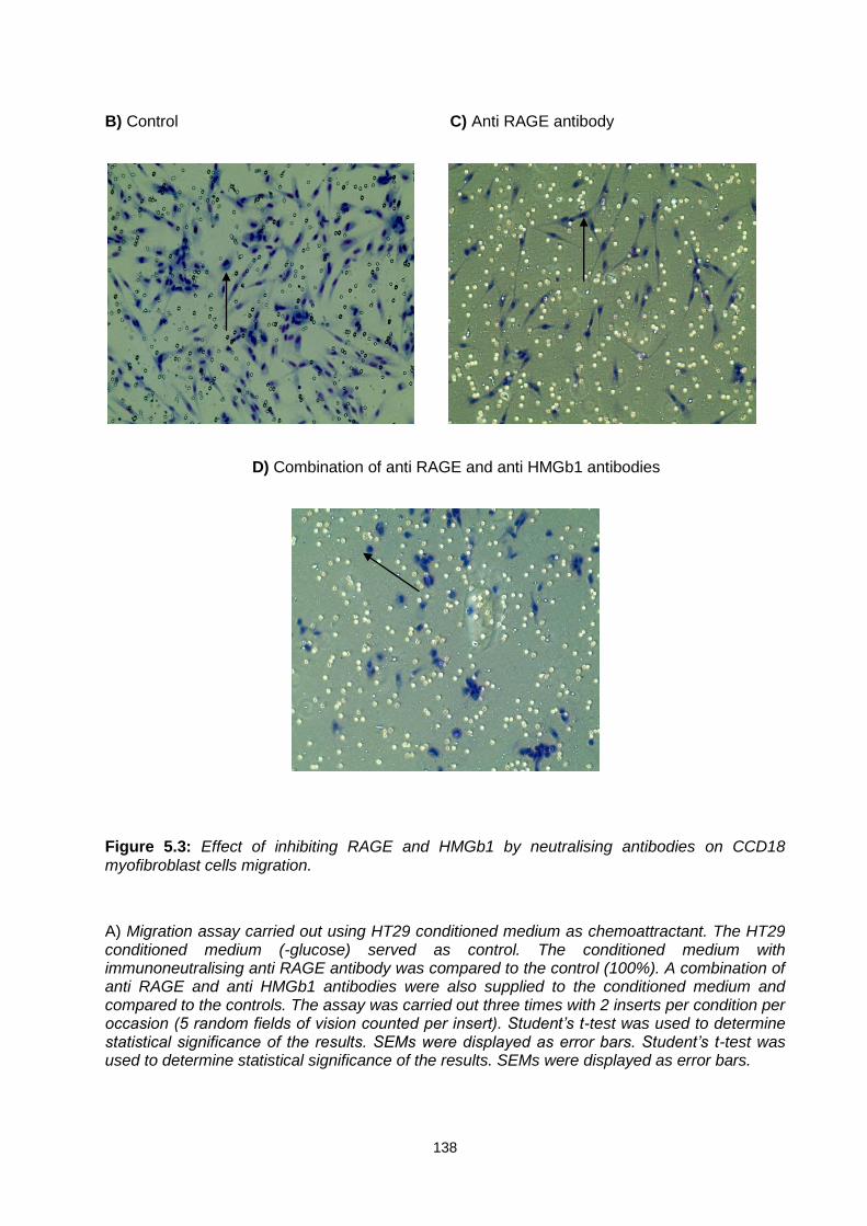
138
B) Control C) Anti RAGE antibody
D) Combination of anti RAGE and anti HMGb1 antibodies
Figure 5.3: Effect of inhibiting RAGE and HMGb1 by neutralising antibodies on CCD18 myofibroblast cells migration.
A) Migration assay carried out using HT29 conditioned medium as chemoattractant. The HT29 conditioned medium (-glucose) served as control. The conditioned medium with immunoneutralising anti RAGE antibody was compared to the control (100%). A combination of anti RAGE and anti HMGb1 antibodies were also supplied to the conditioned medium and compared to the controls. The assay was carried out three times with 2 inserts per condition per occasion (5 random fields of vision counted per insert). Student’s t-test was used to determine statistical significance of the results. SEMs were displayed as error bars. Student’s t-test was used to determine statistical significance of the results. SEMs were displayed as error bars.

139
Representative images captured showing the underside of the membrane after completion of the migration assay. Cells that had migrated though the 8µm pores of the membrane were stained blue using the ReaStain Quick-Diff staining method. The white dots are 8µm pores through which cells have not migrated. B) Cells migrated in response to conditioned medium without glucose, C) cells migrated in response to immunoneutralising RAGE antibody in the conditioned medium without glucose and in the cell suspension in the inserts and D) cells migrated in response to the combination of immunoneutralising RAGE and HMGb1 antibodies in the medium and RAGE antibody in the cell suspension. Cells that had migrated on these and other images were counted and the results were presented in A) of this figure. The photographs were taken under x20 magnification.
The effect of conditioned medium (control) on myofibroblast migration was compared to
myofibroblast migration in response to two other treatments. In the first treatment,
immunoneutralising anti-RAGE antibody (8µg/ml) was added to the conditioned medium and
myofibroblast migration compared to that of the controls. In addition, another treatment,
conditioned medium with a combination of anti-RAGE (8µg/ml) and anti-HMGb1 (5µg/ml)
antibodies were used and the resulting myofibroblast migration was compared to the RAGE
antibodies only treatments and controls. The results obtained from migration assay suggested
that RAGE is involved in the migration of myofibroblasts cells as anti-RAGE antibodies
significantly inhibited (approximately 38%) the migration of myofibroblasts. This inhibition was
further increased by 10% when a combination of anti HMGb1 antibody and anti-RAGE
antibodies were added to the chemoattractant (conditioned medium), confirming the
involvement of HMGb1-RAGE complex in HT29 conditioned medium triggered migration of
CCD18 myofibroblast cells. All results were statistically significant (Figure 5.3 A).

140
5.4.4 HMGb1 present in the conditioned medium triggers migration
in CCD18 myofibroblast cells via TLR-4 signalling
The HMGb1 has been reported to interact with many receptors including RAGE and TLRs
previously (TLR-2 and TLR-4) (Yu et al. 2006; Tian et al. 2007). The glucose free conditioned
medium obtained from HT29 cells at 48h served as chemoattractant for myofibroblast migration
as evident from this study. In addition, this study has also suggested the fact that conditioned
medium obtained from HT29 contains significant amount of HMGb1 (Chapter 4). The results
from migration assays suggested that both HMGb1 and RAGE are involved in the migration of
CCD18 myofibroblast cells. In addition, inhibiting both (HMGb1 and RAGE) resulted in additive
inhibition of migration in CCD18 myofibroblast cells. Therefore, it was important to investigate
the involvement of another receptor TLR-4, which has been implicated in migration of other cell
types previously (Fan and Malik 2003; Liu et al. 2014).

141
A)
B) Conditioned medium (control) C) Anti-TLR-4 antibody
0
20
40
60
80
100
120
(-Glucose) TLR4 antibody (2µg/ml) TLR4 (2µg/ml) + HMGb1 (5µg/ml) antibody
% M
igra
tio
n (
Co
ntr
ol=
10
0%
)
P<0.001=***
*** ***

142
D) Combination of anti HMGb1 and anti TLR-4 antibodies
Figure 5.4: Comparative analysis of CCD18 myofibroblast cells migration assay in response to HT29 conditioned medium and blocking TLR-4 or HMGb1/TLR-4 complex using immunoneutralising anti-HMGb1 or a combination of anti-HMGb1 and anti-TLR-4 antibodies.
A) Migration assay carried out using HT29 conditioned medium as chemoattractant. The HT29 conditioned medium (-glucose) served as control (control=100%). Student’s t-test was used to determine statistical significance of the results. SEMs were displayed as error bars.
Representative images captured showing the underside of the membrane after completion of the migration assay. Cells that had migrated though the 8µm pores of the membrane were stained blue using the ReaStain Quick-Diff staining method. The white dots are 8µm pores through which cells have not migrated. B) Cells migrated in response to conditioned medium without glucose (Control=100%), C) cells migrated in response to immunoneutralising anti-TLR-4 antibody in the conditioned medium without glucose and in the cell suspension in the inserts and C) cells migrated in response to the combination of immunoneutralising anti-TLR-4 and anti-HMGb1 antibodies in the conditioned medium and in the cell suspension. Cells that had migrated on these and other images were counted and the results were presented in A) of this figure. The assay was carried out three times with 2 inserts per condition per occasion (5 random fields of vision counted per insert). The photographs were taken under x20 magnification.
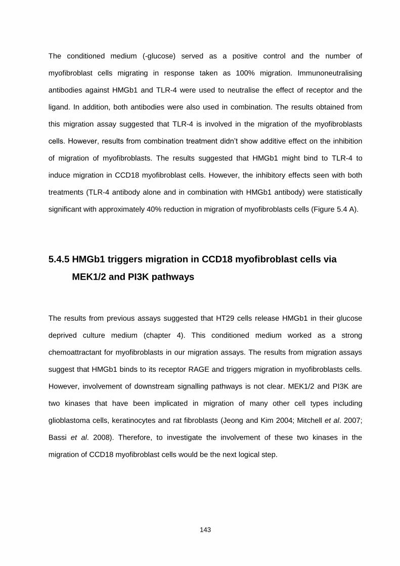
143
The conditioned medium (-glucose) served as a positive control and the number of
myofibroblast cells migrating in response taken as 100% migration. Immunoneutralising
antibodies against HMGb1 and TLR-4 were used to neutralise the effect of receptor and the
ligand. In addition, both antibodies were also used in combination. The results obtained from
this migration assay suggested that TLR-4 is involved in the migration of the myofibroblasts
cells. However, results from combination treatment didn’t show additive effect on the inhibition
of migration of myofibroblasts. The results suggested that HMGb1 might bind to TLR-4 to
induce migration in CCD18 myofibroblast cells. However, the inhibitory effects seen with both
treatments (TLR-4 antibody alone and in combination with HMGb1 antibody) were statistically
significant with approximately 40% reduction in migration of myofibroblasts cells (Figure 5.4 A).
5.4.5 HMGb1 triggers migration in CCD18 myofibroblast cells via
MEK1/2 and PI3K pathways
The results from previous assays suggested that HT29 cells release HMGb1 in their glucose
deprived culture medium (chapter 4). This conditioned medium worked as a strong
chemoattractant for myofibroblasts in our migration assays. The results from migration assays
suggest that HMGb1 binds to its receptor RAGE and triggers migration in myofibroblasts cells.
However, involvement of downstream signalling pathways is not clear. MEK1/2 and PI3K are
two kinases that have been implicated in migration of many other cell types including
glioblastoma cells, keratinocytes and rat fibroblasts (Jeong and Kim 2004; Mitchell et al. 2007;
Bassi et al. 2008). Therefore, to investigate the involvement of these two kinases in the
migration of CCD18 myofibroblast cells would be the next logical step.
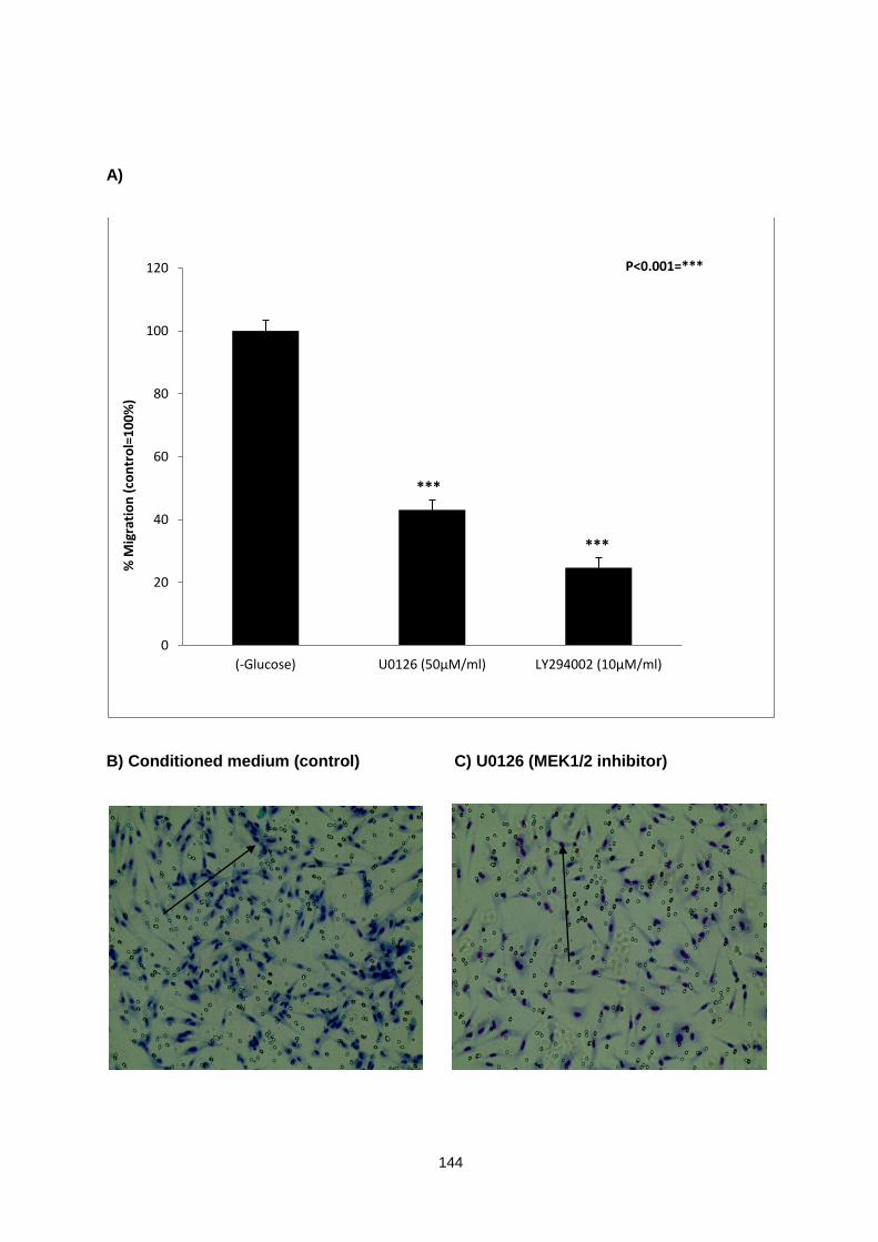
144
A)
B) Conditioned medium (control) C) U0126 (MEK1/2 inhibitor)
0
20
40
60
80
100
120
(-Glucose) U0126 (50µM/ml) LY294002 (10µM/ml)
% M
igra
tio
n (
con
tro
l=1
00
%)
P<0.001=***
***
***

145
D) LY294002 (PI3K inhibitor)
Figure 5.5: Comparative analysis of CCD18 migration assay in response to the treatment with HT29 conditioned medium and MEK1/2 and PI3K inhibitors in the medium.
A) Migration assay carried out using HT29 conditioned medium as chemoattractant. The HT29 conditioned medium (-glucose) served as control (100%). The assay was carried out three times with 2 inserts per condition per occasion (5 random fields of vision counted per insert). Student’s t-test was used to determine statistical significance of the results. SEMs were displayed as error bars. The conditioned medium with MEK1/2 inhibitor (U0126) and PI3K inhibitor (LY294002) were compared to the control (100%). Representative images captured showing the underside of the membrane after completion of the migration assay. Cells that had migrated though the 8µm pores of the membrane were stained blue using the ReaStain Quick-Diff staining method. The white dots are 8µm pores through which cells have not migrated. B) Conditioned media without glucose showing cell migration (control=100%), C) migration of cells in response to U0126 MEK1/2 inhibitor and D) LY294002, PI3K inhibitor. Cells that had migrated on these and other images were counted and the results were presented in A) of this figure. The photographs were taken under x20 magnification.
The involvement of MEK1/2 and PI3K was evaluated by comparing myofibroblast migration in
conditioned medium without inhibitors (controls) with migration in the presence of inhibitors. The
inhibitors U0126, a potent, selective and uncompetitive MEK 1/2 inhibitor was added to the
conditioned medium used in the migration assay and LY294002, a selective inhibitor of PI3K
was also added to the conditioned medium also used in the migration assay. The results from
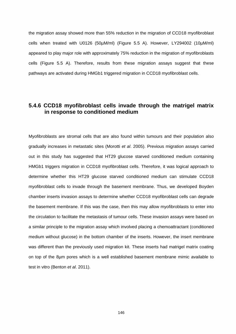
146
the migration assay showed more than 55% reduction in the migration of CCD18 myofibroblast
cells when treated with U0126 (50µM/ml) (Figure 5.5 A). However, LY294002 (10µM/ml)
appeared to play major role with approximately 75% reduction in the migration of myofibroblasts
cells (Figure 5.5 A). Therefore, results from these migration assays suggest that these
pathways are activated during HMGb1 triggered migration in CCD18 myofibroblast cells.
5.4.6 CCD18 myofibroblast cells invade through the matrigel matrix in response to conditioned medium
Myofibroblasts are stromal cells that are also found within tumours and their population also
gradually increases in metastatic sites (Morotti et al. 2005). Previous migration assays carried
out in this study has suggested that HT29 glucose starved conditioned medium containing
HMGb1 triggers migration in CCD18 myofibroblast cells. Therefore, it was logical approach to
determine whether this HT29 glucose starved conditioned medium can stimulate CCD18
myofibroblast cells to invade through the basement membrane. Thus, we developed Boyden
chamber inserts invasion assays to determine whether CCD18 myofibroblast cells can degrade
the basement membrane. If this was the case, then this may allow myofibroblasts to enter into
the circulation to facilitate the metastasis of tumour cells. These invasion assays were based on
a similar principle to the migration assay which involved placing a chemoattractant (conditioned
medium without glucose) in the bottom chamber of the inserts. However, the insert membrane
was different than the previously used migration kit. These inserts had matrigel matrix coating
on top of the 8µm pores which is a well established basement membrane mimic available to
test in vitro (Benton et al. 2011).

147
A)
B) Conditioned medium (Control) C) Conditioned medium (with glucose)
0
20
40
60
80
100
120
(-Glucose) (+Glucose) (-Glucose)
%In
vasi
on
th
rou
gh m
atri
gel m
atri
x (C
on
tro
l=1
00
%)
P<0.001***
***
***
Fresh Medium Conditioned Medium
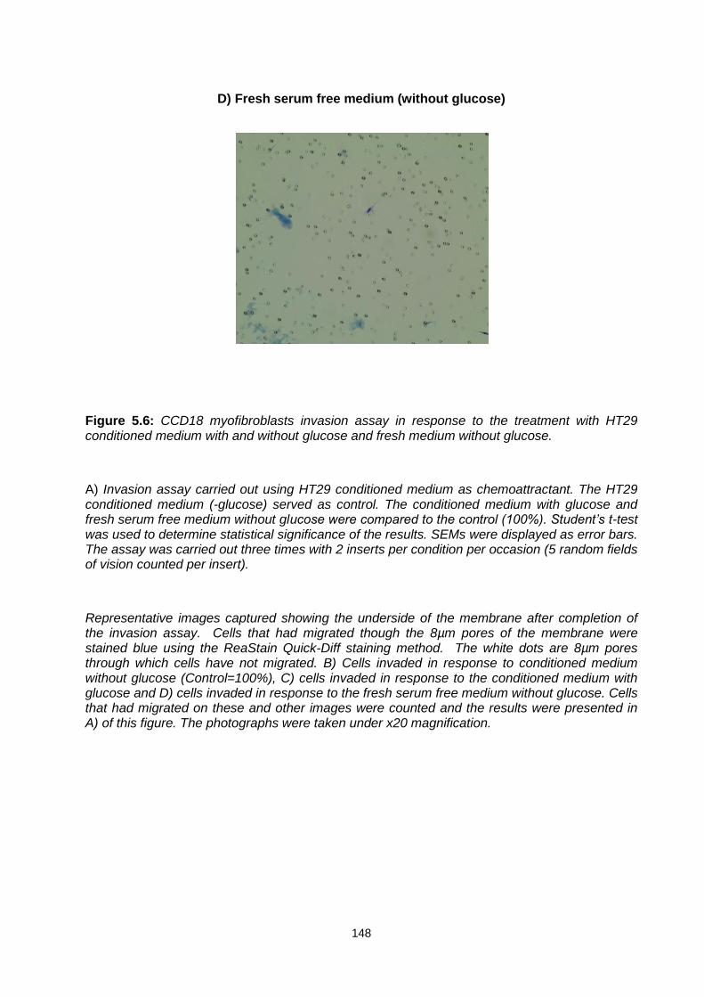
148
D) Fresh serum free medium (without glucose)
Figure 5.6: CCD18 myofibroblasts invasion assay in response to the treatment with HT29 conditioned medium with and without glucose and fresh medium without glucose.
A) Invasion assay carried out using HT29 conditioned medium as chemoattractant. The HT29 conditioned medium (-glucose) served as control. The conditioned medium with glucose and fresh serum free medium without glucose were compared to the control (100%). Student’s t-test was used to determine statistical significance of the results. SEMs were displayed as error bars. The assay was carried out three times with 2 inserts per condition per occasion (5 random fields of vision counted per insert).
Representative images captured showing the underside of the membrane after completion of the invasion assay. Cells that had migrated though the 8µm pores of the membrane were stained blue using the ReaStain Quick-Diff staining method. The white dots are 8µm pores through which cells have not migrated. B) Cells invaded in response to conditioned medium without glucose (Control=100%), C) cells invaded in response to the conditioned medium with glucose and D) cells invaded in response to the fresh serum free medium without glucose. Cells that had migrated on these and other images were counted and the results were presented in A) of this figure. The photographs were taken under x20 magnification.
149
The conditioned medium (without glucose) from HT29 cells served as control (100%). The
conditioned medium (with glucose) and fresh serum free medium (without glucose) were
compared to the conditioned medium (without glucose). The results suggested that the
conditioned medium (with glucose) although able to trigger invasion in myofibroblasts cells with
approximately 30% invasion as compared to the conditioned medium (without glucose) at 100%
(Figure 5.6 A). In addition, fresh serum medium (without glucose) was investigated against
conditioned medium (without glucose). It was observed that cells did not invade through the
matrigel membrane when exposed to glucose free fresh serum free medium (Figure 5.6 D). The
results suggested that glucose deprivation on its own is not a trigger for migration. However,
HT29 cells exposed to glucose deprivation appear to release of certain factors and/or
chemokines which trigger myofibroblasts to invade. The results from this invasion assay support
the results obtained from migration assays carried out using myofibroblasts cells.
5.4.7 HMGb1 in conditioned medium triggers invasion in CCD18
myofibroblast cells
Glucose deprivation triggers the release of HMGb1 from HT29 cells in normoxia. The
chemoattractive properties of HMGb1 have been validated by migration assays carried out in
our lab previously where anti-HMGb1 antibodies significantly reduced the migratory potential of
myofibroblasts cells (Figure 5.2). The conditioned medium (without glucose) has shown positive
results in our invasion assays and therefore investigating the involvement of HMGb1 in glucose
free conditioned medium-induced invasion of myofibroblasts was important.

150
The immunoneutralising anti HMGb1 antibodies (5µg/ml) were added to the conditioned
medium (without glucose) in the bottom chambers of the assay and the results compared to the
control (conditioned medium without glucose). The results suggested that HMGb1 present in
the conditioned medium stimulated CCD18 myofibroblast cells to invade through the matrigel
matrix. A significant reduction in the number of cells that had invaded though the matrigel matrix
was seen with the addition of the HMGb1 immunoneutralising polyclonal anti-HMGb1 antibodies
into the conditioned medium (without glucose) at 22h. This reduction was more than 50% and
statistically significant (Figure 5.7A).
A)
0
20
40
60
80
100
120
Conditioned medium (-Glucose) Anti HMGb1 antibody (5µg/ml)
%In
vasi
on
th
rou
gh m
atri
gel m
atri
x (C
on
tro
l=1
00
%)
P<0.001***
***
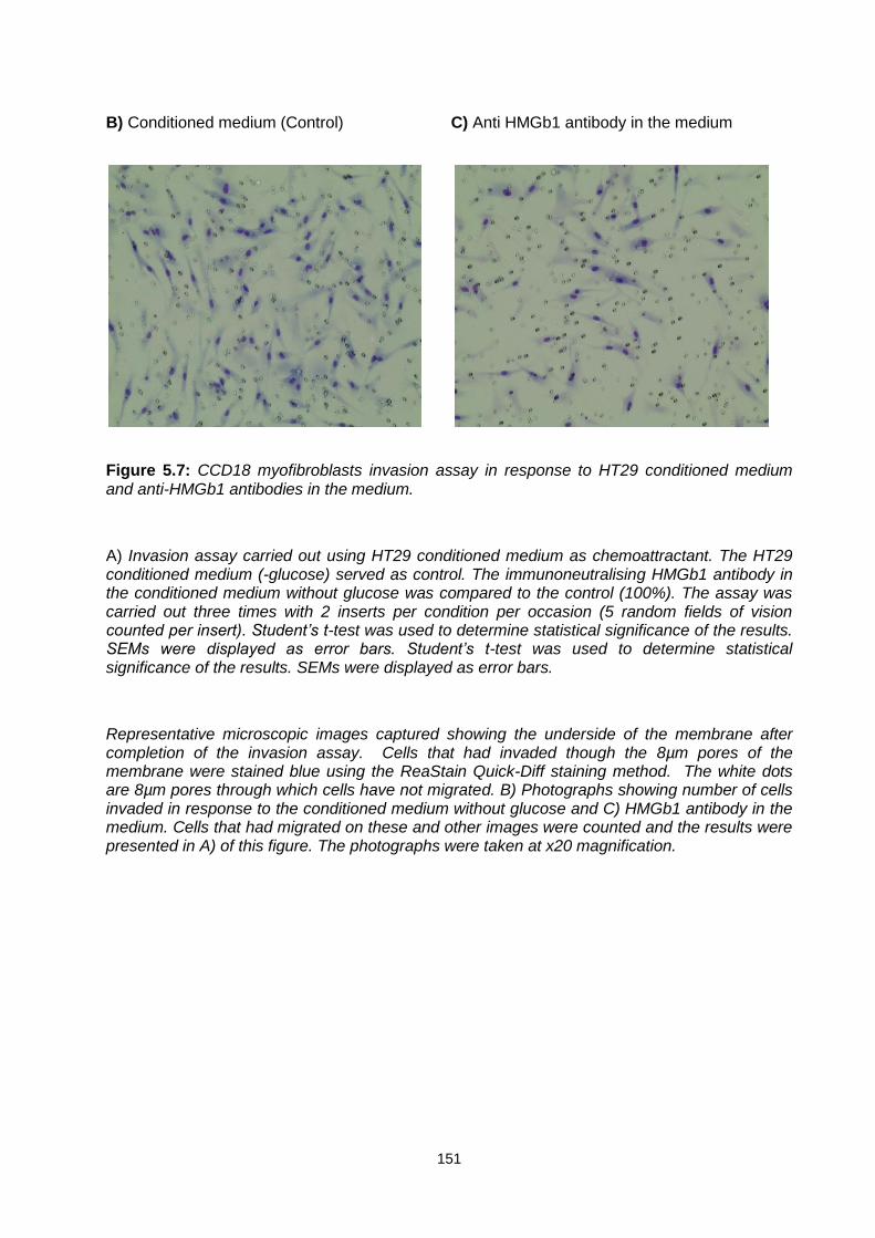
151
B) Conditioned medium (Control) C) Anti HMGb1 antibody in the medium
Figure 5.7: CCD18 myofibroblasts invasion assay in response to HT29 conditioned medium and anti-HMGb1 antibodies in the medium.
A) Invasion assay carried out using HT29 conditioned medium as chemoattractant. The HT29 conditioned medium (-glucose) served as control. The immunoneutralising HMGb1 antibody in the conditioned medium without glucose was compared to the control (100%). The assay was carried out three times with 2 inserts per condition per occasion (5 random fields of vision counted per insert). Student’s t-test was used to determine statistical significance of the results. SEMs were displayed as error bars. Student’s t-test was used to determine statistical significance of the results. SEMs were displayed as error bars.
Representative microscopic images captured showing the underside of the membrane after completion of the invasion assay. Cells that had invaded though the 8µm pores of the membrane were stained blue using the ReaStain Quick-Diff staining method. The white dots are 8µm pores through which cells have not migrated. B) Photographs showing number of cells invaded in response to the conditioned medium without glucose and C) HMGb1 antibody in the medium. Cells that had migrated on these and other images were counted and the results were presented in A) of this figure. The photographs were taken at x20 magnification.
152
5.4.8 RAGE and TLR-4 facilitate CCD18 myofibroblast cells
to invade through matrigel matrix
HMGb1 has been shown to interact with RAGE and TLR-4 and this interaction has been
implicated in various malignancies including cancer (Wang et al. 2012; Zhang et al. 2012;
Stoetzer et al. 2013; Agalave et al. 2014). Our western blot analysis has confirmed the
presence of both RAGE and TLR-4 receptors expressed by CCD18 myofibroblast cells. In
addition, previous migration assay carried out in our lab suggested that RAGE and TLR-4 are
involved in the migration of CCD18 myofibroblast cells (Figure 5.3 and 5.4) therefore it is
possible that these two receptors might also be involved invasion. Thus, investigating the
involvement of RAGE and TLR-4 in the invasion of myofibroblasts cells would be a logical
approach.

153
A)
B) Conditioned medium (Control) C) Anti RAGE antibody in the medium
0
20
40
60
80
100
120
Conditioned medium (-Glucose)
Anti RAGE (8µg/ml) Anti-TLR4 (2µg/ml)
%In
vasi
on
th
rou
gh m
atri
gel m
atri
x (C
on
tro
l=1
00
%)
P<0.001***
***
***

154
D) Anti TLR-4 antibody in the medium
Figure 5.8: CCD18 myofibroblasts invasion assay in response to the treatment with HT29 conditioned medium and anti-RAGE and anti-TLR-4 antibodies in the medium.
A) Invasion assay carried out using HT29 conditioned medium as chemoattractant. The HT29 conditioned medium (-glucose) served as control. The conditioned medium with glucose and RAGE and TLR-4 antibodies in the medium and inserts were compared to the control (100%). The assay was carried out three times with 2 inserts per condition per occasion (5 random fields of vision counted per insert). Student’s t-test was used to determine statistical significance of the results. SEMs were displayed as error bars. Student’s t-test was used to determine statistical significance of the results. SEMs were displayed as error bars.
Representative images captured showing the underside of the membrane after completion of the invasion assay. Cells that had migrated though the 8µm pores of the membrane were stained blue using the ReaStain Quick-Diff staining method. The white dots are 8µm pores through which cells have not migrated. B) Cells invaded in response to the conditioned medium without glucose (Control=100%), C) immunoneutralising RAGE antibody in the conditioned medium without glucose and D) in response to immunoneutralising TLR-4 antibody in the conditioned medium without glucose. Cells that had migrated on these and other images were counted and the results were presented in A) of this figure. The photographs were taken at x20 magnification.
155
The invasion assay carried out using immunoneutralising anti RAGE antibodies (8µg/ml) and
anti TLR-4 antibodies (2µg/ml). The antibodies were added to conditioned medium in the wells
and in CCD18 myofibroblast cell suspension in the inserts. The results after 22h of incubation
suggested that RAGE plays an important role in the HMGb1 mediated invasion of
myofibroblasts as immunoneutralising anti-RAGE antibodies significantly reduced the number
cells invaded through the matrigel matrix. The addition of immunoneutralising anti-TLR-4
antibodies also resulted in significant reduction in number of cells invaded through the matrigel
matrix. However, immunoneutralising RAGE appeared to be the most effective with marked
reduction in invasion of more than 50% whereas immunoneutralising TLR-4 it was
approximately 40% (Figure 5.8 A). These findings are in line with previous findings from the
migration assays where inhibition of RAGE gave greater reduction in migration than inhibition of
TLR-4 (Figure 5.3 and 5.4)
156
5.4.9 The invasion in CCD18 myofibroblast cells take place via
activation of the MEK1/2 and PI3K signalling pathways
The activation of MEK1/2 and PI3K has been implicated in migration of many cells types
including cancer cells (Yao et al. 2004; Ptak et al. 2014; Sobolik et al. 2014). Phosphorylation of
these kinases activates many downstream signalling pathways including NF-kB. The migration
assay carried out in our lab showed that MEK1/2 and PI3K are activated and involved in
migration of CCD18 myofibroblast cells and inhibiting these pathways with selective inhibitors
significantly reduced migration of myofibroblasts cells. Thus, it is possible that these pathways
are involved HMGb1 induced invasion of CCD18 myofibroblast cells.
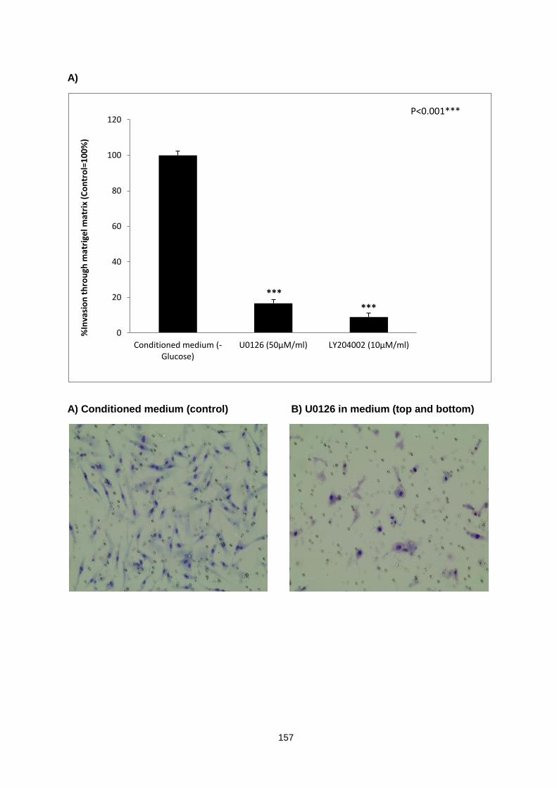
157
A)
A) Conditioned medium (control) B) U0126 in medium (top and bottom)
0
20
40
60
80
100
120
Conditioned medium (-Glucose)
U0126 (50µM/ml) LY204002 (10µM/ml)
%In
vasi
on
th
rou
gh m
atri
gel m
atri
x (C
on
tro
l=1
00
%)
P<0.001***
***
***

158
C) LY294002 in medium (top and bottom)
Figure 5.9: A) Effect of MEK1/2 and PI3K inhibitors on CCD18 myofibroblasts invasion in response to HT29 conditioned medium as chemoattractant.
The HT29 conditioned medium (-glucose) served as control. The MEK1/2 and PI3K inhibitors in the conditioned medium without glucose and in the inserts were compared to the control (100%). The assay was carried out three times with 2 inserts per condition per occasion (5 random fields of vision counted per insert). Student’s t-test was used to determine statistical significance of the results. SEMs were displayed as error bars. Student’s t-test was used to determine statistical significance of the results. SEMs were displayed as error bars.
Representative images captured showing the underside of the membrane after completion of the invasion assay. Cells that had migrated though the 8µm pores of the membrane were stained blue using the ReaStain Quick-Diff staining method. The white dots are 8µm pores through which cells have not migrated. B) Cells invaded in response to the conditioned medium without glucose (Control=100%), C) MEK1/2 inhibitor (U0126) in the conditioned medium without glucose and D) in response to PI3K inhibitor (LY294002) in the conditioned medium without glucose. Cells that had migrated on these and other images were counted and the results were presented in A) of this figure. The photographs were taken under x20 magnification.
The invasion assay was carried out using selective inhibitors of MEK1/2 and PI3K. These
inhibitors were added to medium in the bottom chambers and in the CCD18 cell suspension in
the upper inserts. The data suggested that inhibiting MEK1/2 by its selective inhibitor U0126

159
(50µM/ml) significantly reduced the number of cells that invaded through the matrigel matrix.
There was approximately an 80% reduction in the number of myofibroblasts invading through
compared to the controls (100%). In addition, LY294002 (10µM/ml), a selective inhibitor of
PI3K, resulted in a significant reduction (90%) in number of cells invading through the matrigel
matrix (Figure 5.9A). The results suggested that both MEK1/2 and PI3K are involved in the
invasion of myofibroblasts cells. However, based on the data from migration and invasion
assays, it appears that PI3K-AKT pathways play an important role in CCD18 myofibroblast cells
migration and invasion. Though, the involvement of MAPK-ERK pathway is also significant.
5.4.10 MMP-2 but not MMP-9 is produced and secreted by CCD18
Myofibroblast cells
The other aim was based on the novel findings from the invasion assays. The results from
invasion assays suggested that CCD18 myofibroblasts are able invade through the matrigel
matrix in response to glucose deprived conditioned medium taken from HT29 cells. Many
MMPs including MMP-1,-2,-3,-7,-8, and MMP-9 have been shown to cleave the substrate
during the invasion assay in vitro (Lutolf et al. 2003) and myofibroblasts have been shown to
secrete MMP-2 and MMP-9 previously (Lewis et al. 2004; Turner et al. 2007). Thus we decided
to investigate release of MMP-2 and MMP-9 from CCD18 cells.
Here, we investigate the release of MMP-2 and MMP-9 as these forms of MMPs have shown to
degrade basement membrane previously (Lee et al. 2015; Shen et al. 2015). The CCD18
myofibroblast cells were treated with glucose free and glucose containing medium. In addition,
the myofibroblasts cells were also treated with same conditions as those of migration assays

160
i.e., treatment with serum free medium for 24h followed by treatment with HT29 conditioned
medium.
A)
B)
Figure 5.10: (A) Western blot showing the release of MMP-2 from CCD18 cells. The CCD18 cells were also treated with HT29 conditioned medium (medium containing glucose and glucose free medium collected at 48h) for 22h.
A= Recombinant MMP-2, B=CCD18 (+Glucose), C=CCD18 (-Glucose, D=CCD18 treatment with HT29 conditioned medium (+Glucose), E=CCD18 treatment with HT29 conditioned medium (–Glucose)
B) Western blot analysis of the release of MMP-9 from HT29 cells and CCD18 cells. The cells were treated with presence and absence of glucose for 24h. The CCD18 cells were also treated with HT29 conditioned medium for 22h. The protein concentration was equalised in both conditions and compared.
(+glucose = with glucose*) (-glucose = without glucose*). A= Recombinant MMP-9, B=HT29 (+ Glucose), C=HT29 (–Glucose), D=CCD18 (+ Glucose), E=CCD18 (–Glucose), F=CCD18 treatment with HT29 conditioned medium (+ Glucose), G=CCD18 treatment with HT29 conditioned medium (–Glucose)
72kDa
A B C D E F G
92kDa
A B C D E
MMP-2
MMP-9
161
The results from western blot suggest that CCD18 myofibroblast cells express and release
MMP2 but not MMP-9. The protein bands for MMP-2 were detected at 74kDa (predicted
molecular weight) resembling the molecular weight of recombinant MMP-2. A polyclonal primary
antibody specific to MMP-2 was used to detect MMP-2. The recombinant MMP-2 served as
positive control. The protein concentration of the medium from the treated CCD18 myofibroblast
cells used in the western blot was equalised using BioRad protein assay. There was no
difference observed in protein bands observed in treatment with glucose free fresh serum free
medium and with glucose containing serum free medium at 24h. However, with conditions
mimicking migration and invasion assay conditions, it appeared that MMP-2 is downregulated in
response to glucose free conditioned medium taken from HT29 cells that had previously been
cultured for 48h (Figure 5.10A).
Similar assay conditions were implemented to investigate the presence or upregulation or
downregulation of MMP-9. The protein concentration of the medium from the CCD-18 cells was
again equalised using the BioRad protein assay. A polyclonal primary anti-MMP-9 antibody was
used to detect MMP-9 in the conditioned media samples. The results from western blot
suggested that MMP-9 is not released from CCD18 cells in any of the conditions investigated
i.e., first treatment being the treatment with fresh glucose free or glucose containing treatment
for 24h and second treatment with glucose free condition treatment for 24h plus another 22h
treatment with HT29 conditioned medium which was obtained at 48h (glucose free and glucose
rich). Recombinant MMP-9 was detected at predicted molecular weight of 92kDa (Figure 6B).
Therefore, both western blots were validated with positive controls being detected at correct
molecular weight. These findings are indicative of possibilities for the involvement of other
proteases in facilitating the HMGb1 induced migration and invasion of myofibroblasts.

162
5.5 Discussion
The tumour stromal cells such as fibroblasts and myofibroblasts play an important role in
predicting the outcome of cancer chemotherapy (Yamashita et al. 2012). Cancer metastasis is a
major hurdle in the treatment of cancer (Davidson et al. 1999; Spaderna et al. 2006; Li et al.
2011). It has been established that tumour stromal cells such as CAFs or myofibroblasts
promote tumour proliferation and angiogenesis (Coussens and Werb 2002; Allinen et al. 2004).
In addition, it has been shown that the myofibroblast population gradually increases with the
invasive stages of many cancers (Nakayama et al. 1998). The degradation of ECM is one of the
major events that occur during the migration of cells from one organ to another.
Myofibroblasts are known to play important roles in the formation and repair of the ECM
(Vedrenne et al. 2012; Mia et al. 2014). The ECM is made up of various matrix proteins
including collagen and other glycoproteins and proteoglycans (Robert 2015). Most of these
proteins are produced by myofibroblasts and play important roles in epithelial cell migration and
differentiation (Hinz and Gabbiani 2010). In addition, myofibroblasts have been shown to
secrete MMPs (1-3). These MMPs are major degraders of basement membrane. However, the
action of MMPs is inhibited by TIMPs during the ECM remodelling. Thus, a balance between
MMPs and TIMPs is important for ECM remodelling in the tissue (Benyon et al. 1996).
Most solid tumours are characterised by hypoxic and glucose deprived areas in their central
core within the microenvironment (Laderoute et al. 2006; Jamieson et al. 2015). These

163
conditions have been shown to trigger the release various growth factors such as VEGF, which
then can facilitate angiogenesis and metastasis. The results from our western blot analysis
suggest that various cancer cell lines including HT29 (colon adenocarcinoma), MCF-7 (breast
cancer), EJ138 (Bladder cancer) and A549 (Lung cancer) cells release HMGb1 under glucose
deprivation in normoxia. HMGb1 is an inflammatory cytokine that is released as a danger signal
during inflammation (Hreggvidsdottir et al. 2009; Lu et al. 2014). We have explored
myofibroblast proliferation and found that HMGb1 appeared to drive proliferation in CCD18
myofibroblast cells. Other studies have shown that HMGb1 can trigger migration in many other
cells including glioblastoma and neutrophils (Bassi et al. 2008; Berthelot, et al. 2011). However,
its role in myofibroblasts migration and invasion had previously not been explored. Therefore,
considering the cytokine-like activity of HMGb1, it was important to investigate its role in
signalling migration and invasion of myofibroblasts cells.
The HMGb1 activates various signalling pathways such as MAPK-ERK and PI3K-AKT. These
pathways play an important role in the proliferation and migration of tumour cells. Supporting
evidence includes HMGb1 activated PI3K pathway in neutrophils and colon cancer cells
(Kuniyasu et al. 2003). In addition, HMGb1 has been shown to induce TLR-4 mediated
activation of MyD88-IRAK4-p38 and Myd88-IRAK4-AKT pathways. Also, HMGb1-RAGE
complex has been shown to activate ERK1/2 pathways in rat smooth muscle cells (Degryse et
al. 2001).
The presence of HMGb1 released into the culture medium from HT29 colon adenocarcinoma
cells might contribute to advancing our knowledge of the cellular cross talk within tumours in

164
response to HMGb1 and subsequent impacts within the tumour microenvironment. One main
question that has been answered by this work is that HMGb1 triggers proliferation in
myofibroblasts and plays an important role in the activation of PI3K and MAPK-ERK pathways.
In addition, presence of RAGE and TLR-4 on the membrane of myofibroblasts further indicates
a possible interaction between HMGb1 and its receptors RAGE and TLR-4. Therefore,
validating the involvement of these proteins by using immunoneutralising antibodies was a
logical approach in our migration assays. The migration assays were carried using Boyden
chamber inserts with 8µm pores membrane. This system has been used widely by many other
researchers in vitro (Chen 2005).
The preliminary data from the CCD18 myofibroblast migration assays using the conditioned
medium without glucose as a chemoattractant (conditioned by growing HT29 colon
adenocarcinoma cells in it for 48h) showed significant migration of the myofibroblast cells during
a 20h incubation period (Figure 5.1). Indeed, there was a reasonable chance for the existence
of chemoattractants in the culture medium before exposure to the HT29 cells. However, the
results from our western blots have confirmed the presence of HMGb1 in the culture medium of
HT29 cells but not in the fresh medium (Chapter 4, Figure 4.1). A number of studies have
shown the chemoattractant properties of HMGb1 in relation to a number of cells including
mesoangioblasts and fibroblasts in vitro (Palumbo et al. 2007; Schiraldi et al. 2012).
Since our results suggested that HMGb1 present in the conditioned medium can trigger
migration in myofibroblast cells, a selective immunoneutralising antibody to HMGb1 (5 µg/ml)
was used to block HMGb1 in the conditioned medium to back up our findings. The antibody was
allowed to interact with HMGb1 present in the conditioned medium for 20h. The results from this
165
migration assay revealed that HMGb1 present in the conditioned medium triggers migration in
myofibroblasts cells. The addition of immunoneutralising antibody to HMGb1 (5µg/ml) to
conditioned medium at the bottom chamber significantly reduced the number of myofibroblasts
cells migrated through the membrane. This inhibition was approximately 40% when compared
to the controls (100%) (Figure 5.2).
The interaction of HMGb1 with RAGE and TLR-4 has been established previously. For
example, a recent study showed that HMGb1 is released from necrotic and severely stressed
inflammatory cells and bind to TLR-4 and triggers migration in NIH/3T3 fibroblasts to the site of
necrosis (Schiraldi et al. 2012). However, whilst this was shown in fibroblasts, whether the
migration of myofibroblasts is facilitated by HMGb1 interaction with either RAGE or TLR-4
receptor interaction remained questionable. Therefore, it was logical to explore this by using
immunoneutralising antibodies to RAGE and TLR-4 to specifically block these receptors.
In addition, HMGb1 antibodies were also used in combination with RAGE and TLR-4 antibodies
to investigate synergistic effect. The RAGE and TLR-4 antibodies were added to both chambers
in the migration assay (top chamber containing myofibroblast cell suspension and bottom
chamber containing conditioned medium). The HMGb1 antibodies were added to medium at the
bottom chambers. RAGE and TLR-4, both are multiligand receptors and known to activate
various downstream signalling pathways (Palumbo et al. 2004). The blockade of RAGE by the
use of immunoneutralising antibodies significantly reduced the number of myofibroblast cells
that had migrated through the membrane (Figure 5.3). Interestingly, similar results were seen
with the blockade of TLR-4 with immunoneutralising antibodies (Figure 5.4). This suggested
that both receptors are present on the myofibroblasts cells and take part in signal transduction

166
necessary for the cells to migrate. In addition, an increased inhibition of migration was seen
when both RAGE and HMGb1 were blocked by antibodies (Figure 5.3). This strengthens our
evidence for the involvement of RAGE receptor interaction with HMGb1 and that HMGb1
primarily binds to RAGE to signal migration in CCD18 myofibroblast cells. However, blocking
TLR-4 and HMGb1 together did not show any better inhibition than blocking TLR-4 alone. This
suggests that TLR-4 might take part in signalling pathways in myofibroblasts cells but not
primarily with HMGb1. Therefore it is possible that TLR-4 might interact with other proteins in
the conditioned medium in addition to HMGb1.
The signalling pathways involved in the migration potential of the myofibroblast cells were also
investigated. Our evidence suggests that CCD18 myofibroblast cells migrate in response to
HMGb1 found at relatively high levels in the glucose-deprived medium conditioned by use on
HT29 colon adenocarcinoma in the migration assay. Two key pathways MAPK-ERK and PI3K-
AKT have been implicated widely in the study of migration of a variety of cells including
endothelial, glioblastoma and colon cancer cells (Crean et al. 2002). Selective inhibitors of
these pathways (U126 and LY294002) have been used by many researchers (Hayashi et al.
2008; Ghobrial 2009; Kobayashi et al. 2009) investigating the roles of MAPK-ERK or PI3K-AKT
pathways. In this work, the inhibitors were added to the cell suspension and the medium to
block the action of MEK1/2 and PI3K in CCD18 myofibroblast cells.
The results from the migration assays using these inhibitors revealed that both pathways are
involved in HMGb1 triggered migration of CCD18 myofibroblast cells (Figure 5.5). However,
inhibiting PI3K with LY294002 significantly blocked the migration by 80%, which suggested that
migration of CCD18 myofibroblast cells involves PI3K and associated downstream signalling

167
which may include NF-kB. In addition MAPK-ERK pathway also plays an important role in the
migration of CCD18 myofibroblast cells. The inhibition of MEK1/2 by U0126 significantly
reduced the number of cells (55%) in our migration assay. Thus, according the results from
these migration assays, it is likely that MEK1/2 is activated as a result of the interaction of
HMGb1 and RAGE to trigger the migration of CCD18 myofibroblast cells.
Ability to invade plays a central role in metastasis. This occurs by the degradation of basement
membrane followed by penetration of cells into the circulation or lymphatic system. The results
from this work using western blotting analysis of CCD18 culture medium suggest that CCD18
myofibroblast cells produce and release MMP-2 (Figure 5.10 A). The MMP-2 has been
implicated in the degradation of collagen type I, III and IV (Benyon et al. 1996).
The results from the invasion assays show that CCD18 myofibroblast cells invade through the
matrigel matrix to reach to the bottom part of the membrane (Figure 5.6). This further suggests
that CCD18 myofibroblast cells might release some MMPs which degrade the matrigel matrix
that mimics the basement membrane in the invasion assay setup. However, treatment of
myofibroblast with glucose free conditioned medium obtained from HT29 cells showed that
there was a downregulation in the release of MMP-2 by CCD18 myofibroblast cells (Figure 5.10
A). However, there was a downregulation in MMP-2 levels in the culture medium of CCD18
myofibroblast cells when treated with conditioned medium with glucose (conditioned on HT29
colon adenocarcinoma cells) (Figure 5.10 A). In addition, MMP-9 another potential degrader of
basement membrane was invested and our western blot results suggested that CCD18 cells do
not produce or release MMP-9 when treated with either with or without glucose conditioned
medium treatment (Figure 5.10 B). This suggested that the invasion which occurred in response

168
to the HT29 cell conditioned medium without glucose might upregulate the release of proteases
other than MMP-2 or MMP-9, which might play important roles in degradation of basement
membrane and facilitate invasion by myofibroblasts cells.
Interestingly, when HT29 cell conditioned medium with glucose was analysed for its
chemoattractive properties, it was found that only 30% cells could invade through in comparison
to the controls (100%) (HT29 conditioned medium without glucose) (Figure 5.6). This further
raised a possibility for the release of additional proteases other than MMP-2 and MMP-9 that
might have been released from the colon adenocarcinoma cell line during the glucose
starvation. In addition, the results from invasion assays suggested that glucose deprivation on
its own does not influence CCD18 cells to invade across the matrigel matrix because no cells
invaded across the membrane when fresh medium without glucose was placed at the bottom
chamber (Figure 5.6). Therefore, it was concluded that it’s not the lack of glucose that is
responsible for the invasive properties of CCD18 myofibroblast cells, rather it is the factors that
are being released from the colon adenocarcinoma cells in response to glucose deprivation that
give the myofibroblasts invasive properties.
Like migration, involvement of HMGb1 present in the conditioned medium of HT29 colon
adenocarcinoma was analysed for invasion in myofibroblasts cells. It has been shown
previously in chapter 4 that HMGb1 is present in the conditioned medium of HT29 cells
especially in the glucose free conditioned medium. It was found that addition of anti-HMGb1
antibodies into the conditioned medium (without glucose) at the bottom chamber significantly
reduced the number of cells invading through (Figure 5.7). In addition, RAGE and TLR-4 also
appeared to be involved in initiating the signalling cascade that lead the degradation of the

169
matrigel matrix membrane and invasion of myofibroblast cells. Interestingly, the results from
invasion assays were in line with those of migration assays when comparing the involvement of
RAGE and TLR-4. The blockade of RAGE resulted in greater inhibition than TLR-4 (Figure 5.8).
Though, a combination treatment with anti-HMGb1 antibodies was not investigated but would
be logical approach to see if there is any synergistic effect.
The activation of MAPK-ERK and PI3K-AKT pathways have been implicated in the invasion of
hepatocellular carcinoma and prostate cancer (Chen et al. 2005) The inhibitors of PI3K and
MEK1/2 were used to selectively to inhibit both kinases in our invasion assay. The inhibition of
both kinases resulted in marked reduction in invasion of CCD18 cells (Figure 5.9). However,
PI3K inhibition by LY294002 was more effective than the U0126 like the migration assay. It has
been shown that inhibition of PI3K and MAPKs results in the downregulation of MMP-2 and u-
PA (urokinase-type plasminogen activator) which might suggest that why inhibiting PI3K and
MEK1/2 result in the inhibition of invasion (Chen et al. 2005). However, in our invasion setup,
glucose free HT29 cell conditioned medium triggered myofibroblast invasion and migration but
our western blotting analysis revealed a downregulation of MMP-2 in these conditions.
Therefore, it is likely that other MMPs or factors are involved and should be the subject of future
work.

170
Chapter 6
6. Conclusions and future work
6.1 Major Findings
The HMGb1 is a nuclear protein has been shown to take part in the inflammatory process when
released from immune cells (Scaffidi et al. 2002). In addition, it has recently been shown that
the necrotic cells and the cells undergoing autophagy also release HMGb1 in the extracellular
environment (Fiuza et al. 2003; Thorburn et al. 2009). Although, the release of HMGb1 from
cancer cells has been shown previously (Ito et al. 2007) however, the conditions that promote
HMGb1 release were unclear. The data presented in this thesis suggest that HMGb1 is
released from HT29 colon adenocarcinoma cells grown under anoxic conditions and also from
these cells grown under glucose deprivation particularly in presence of oxygen. In fact, the
results from western blots show that glucose deprivation rather than anoxia is a major stimulus
which triggers the release of HMGb1 from 3 out of 4 cancer cells tested (HT29 colon
adenocarcimona), A549 lung cancer cells and MCF-7 breast cancer cells but not EJ138 bladder
cancer cells) (Chapter 4, Figure 4.1 and 4.2). Therefore, it is possible that glucose deprivation
induced HMGb1 release from cancer cells could be a common phenomenon in the tumour
microenvironment of many tumour types.
In the tumour microenvironment, cancer cells and neighbouring tumour associated cells often
undergo harsh ischemic conditions have alternate strategies to try to survive. For example,
cancer cells are often exposed to an environment where there is not enough glucose and/or
oxygen, especially in the core of tumour and so switch over to aerobic glycolysis (Warburg
171
effect). With anaerobic glycolysis, the tumour cells rapidly consume glucose and oxygen from
the blood supply in order to get energy to survive (Vander Heiden et al. 2009). According to the
data from this work, it may be likely that most cancer cells may release HMGb1 in within areas
of the tumour microenvironment particularly where there is the shortage of glucose.
Glutamine deprivation has previously been shown to induce autophagy in cells (Sakiyama et al.
2009). The autophagy could result in translocation of HMGb1 from the nucleus followed by
exocytosis in cancer and normal cells (Tang et al. 2010). The data presented here suggested
that glucose deprivation should not be confused with glutamine deprivation that may be
responsible for HMGb1 release from cancer cells via autophagy. The glutamine deprivation was
also compared with glucose deprivation of the HT29 adenocarcinoma cell line and it was found
that glucose alone serves as stimulating factor for HMGb1 release not glutamine. There was no
evidence that glutamine deprivation resulted in additional HMGb1 being released into the
medium when compared to normal glutamine and glucose conditions (control).
The HMGb1 has previously been shown to trigger proliferation in many cells including T
lymphocytes, mesoangioblasts and smooth muscle cells (Palumbo et al. 2004; Porto et al.
2006; Sundberg et al. 2009). However, the role of HMGb1 interaction with myofibroblasts, a
type of major mucosal but also tumour stromal cell is still unclear. The results from the
proliferation assays suggest that recombinant HMGb1 triggers proliferation in myofibroblasts in
a dose spectrum of 0.1 to 50ng/ml (Chapter 3, Figure 3.1B). Two major signalling pathways
PI3K-AKT and MAPK were investigated for their role in HMGb1 induced proliferation in
myofibroblasts. Both pathways have previously been reported to play active roles in HMGb1
induced proliferation and migration in glioblastoma and mesoangioblast cells (Feng et al. 2014).
Thus, it was important to investigate the involvement of these pathways in HMGb1 induced
172
proliferation of myofibroblasts. The Inhibition of both pathways by selective inhibitors
significantly reduced HMGb1 induced proliferation in myofibroblasts (Chapter 3, Figure 3.2 A
and 3.2 B). This suggested that PI3K and MAPK both play important roles in the proliferation of
myofibroblasts. Therefore, the data presented here in this thesis suggests that HMGb1 triggers
proliferation in myofibroblasts via PI3K-AKT and MAPK-ERK pathways.
The results from western blot suggested that HMGb1 is present in the conditioned medium
taken from HT29 colon adenocarcinoma cells after 48hrs. Therefore, HMGb1 containing
conditioned media was chosen for use to investigate the effect of HMGb1 on myofibroblasts
migration and invasion. After all, in the actual colon tumour environment, one would expect
tumours to release factors (including HMGb1), that influence the behaviour of tumour-
associated cells such as myofibroblasts. The HMGb1 released into the HT29 cell conditioned
(glucose free) media triggered a chemotactic response in myofibroblasts in our transwell
membrane migration assay setup (Chapter 5, Figure 5.2). The direct involvement of HMGb1
was demonstrated during migration assay by using immunoneutralising antibody against
HMGb1. Neutralising the effect of HMGb1 by antibody significantly reduced migration response
of myofibroblasts cells to the HT29 glucose free conditioned media (Chapter 5, Figure 5.2).
Once the involvement of HMGb1 in migration of myofibroblasts was validated, it was important
to investigate which HMGb1 associated receptors that are being activated during the migration
response in myofibroblasts. Thus, two well-known receptors, RAGE and TLR-4, were
investigated for involvement in HMGb1 induced migration response in myofibroblasts. The
presence of both receptors on myofibroblasts cell membrane was confirmed by western blotting
(Chapter 4, Figure 4.4 and 4.5). The immunoneutralising antibodies against RAGE and TLR-4

173
were added to the myofibroblasts cells suspension in an attempt to inhibit the activation of both
receptors present on the cell membrane of myofibroblasts. The blockade of RAGE or TLR-4
receptors individually by immunoneutralising antibodies significantly reduced migration in
myofibroblasts cells in response to HT29 glucose free conditioned media (Chapter 5, Figure 5.3
and 5.4). In addition, combined inhibition of HMGb1 with RAGE showed some additive inhibition
of migration of myofibroblasts cells (Chapter 5, Figure 5.3). However, no additive effect was
seen with combined inhibition of HMGb1 and TLR-4 using immunoneutralising antibodies
(Chapter 5, Figure 5.4). This further suggested the preferred route of HMGb1 induced migration
of myofibroblasts may act via the activation of HMGb1-RAGE complex and subsequent
downstream signalling leading to migration of myofibroblasts cells.
Indeed, PI3K and MAPK-ERK pathways are most widely studied pathways for migration in
many cell types. The use of selective inhibitors of these pathways validated the involvement of
these pathways in our migration assays. Blocking these pathways with selective inhibitors
significantly reduced HMGb1 induced migration of myofibroblasts cells (Chapter 5, Figure 5.5).
However, the inhibitor for PI3K-AKT pathways appeared to more greatly inhibit myofibroblasts
migration when compared to the inhibitor for MEK1/2 pathways at non-toxic doses. Thus, based
on this finding, it appeared that PI3K-AKT might be the main pathway involved in HMGb1
induced migration of myofibroblasts. However, whilst to a lesser extent, the inhibitor of MEK1/2
significantly reduced migration in myofibroblasts suggesting MEK1/2 is also an important
signalling pathway in HMGb1 induced myofibroblasts migration.
Invasion assays were also carried out to investigate the invasive behaviour of myofibroblasts
cells and the role of HMGb1 in promoting invasiveness of myofibroblasts. The results from

174
invasion assays suggested that HMGb1 present in the conditioned medium of HT29 cells
triggers myofibroblasts cells to invade (Chapter 5, Figure 5.7). It was logical to investigate the
role of the receptors that were reported to be involved in our migration assay. The resulting data
suggested that HMGb1 mediated invasion of myofibroblasts cells again involved the activation
of RAGE and TLR-4. The involvement of RAGE and TLR-4 was validated by
immunoneutralising antibodies against RAGE and TLR-4 which significantly inhibited invasion
of myofibroblasts cells through ECM mimicking matrigel membrane. In addition both pathways
PI3K-AKT and MAPK-ERK which showed positive results in our migration assay were also
investigated. Inhibiting both pathways by using selective inhibitors in the cell suspension
significantly reduced HMGb1 induced invasion in myofibroblasts cells (Chapter 5, Figure 5.9).
Thus, based on the findings, it appeared that HMGb1 mediated invasion take place via both the
PI3K-AKT and MAPK-ERK pathways.
The results from the invasion assays suggest that myofibroblasts can invade through the
basement membrane. To achieve that, basement membrane must be digested by proteases
released from the cells that are responsible for ECM remodelling to facilitate the cells to move
through the matrigel membrane. Hence, it was empirical to look for most prominent MMPs that
might be released from myofibroblasts following the HMGb1 containing HT29 glucose free
conditioned media treatment. Our western blot analysis suggested that MMP-2 is being
released in the culture medium of myofibroblasts cells following the treatment with normal
glucose containing conditioned medium from HT29 cells (Chapter 5, Figure 5.10). However, the
levels of MMP-2 appeared to be reduced when myofibroblasts were treated with low glucose
conditioned medium from HT29 cells (Chapter 5, Figure 5.10 A); a medium that we have
previously shown to have strong chemo-attractive properties for myofibroblasts. In addition,
MMP-9 another major degrader of basement membrane was also investigated. Our western

175
blot data suggested that MMP-9 is not being released from myofibroblasts following the
treatment with glucose free or normal glucose conditioned medium of HT29 cells (Chapter 5,
Figure 5.10 B). This further suggests that myofibroblasts are secreting MMPs other than MMP-2
and MMP-9 or other proteases that are actively taking part in the basement degradation and
facilitating invasion of myofibroblasts cells.
On the basis of this work, we postulate that HMGb1 is released from the areas of low or no
glucose rather than hypoxia in cancer cells. This areas typically found in the central core of
most solid tumours which is also acidic (Schlappack et al. 1991; Rajendran et al. 2004). Once
released from these areas, HMGb1 can reach to the myofibroblasts (may be due to interstitial
fluid pressure) which are present in the stroma of most solid tumours. HMGb1 then can
stimulate myofibroblasts to proliferate on site. The stromal myofibroblasts has been shown to
release proteases to degrade basement membrane and have been related to poor prognosis in
breast cancer (De Wever et al. 2008; Yamashita et al. 2012). Once the ECM has been
digested, myofibroblasts along with other cells can make their way out of the tumour to the
distant sites (Nielsen et al. 1996; Malik et al. 2015). The data presented in this thesis suggest
that increasing number of myofibroblasts after getting signal from HMGb1 would likely to
remodel basement membrane rapidly which would have then facilitate the migration of other
cells too. In addition, levels of glucose in myofibroblastic tumours can become predictive tools
for measuring the release of HMGb1 and its effects on the spreading of tumours. Increasing
population of myofibroblasts on the tumour periphery will increase the tumour mass. It is also
likely that larger tumour mass would have more glucose deprived areas and hence more
HMGb1 being released. This HMGb1 then can further influence myofibroblasts and other
cancer cells to grow, migrate and invade to the distant sites. Although, there is a clear need to
identify other factors that may influence myofibroblasts and cancer cells migration and invasion
176
but the importance of HMGb1 and its crucial role in proliferation, migration and invasion of
myofibroblasts can not be neglected.
6.2 Conceptual Advances
The tumour stroma in many solid tumour including colon cancer and breast cancer comprises a
major part of the tumour mass (Peña et al. 2013). The myofibroblasts are predominant cell
types in most carcinomas found on the periphery of most solid tumours (Desmoulière et al.
2004; Tripathi et al. 2012). The myofibroblasts have been shown to take part in tumour
proliferation by secreting a number of growth factors including IGF-1, IGF-II and HGF (Hinz et
al. 2007). These growth factors may play important roles in initiating certain pathways which are
necessary for tumour cell survival and proliferation (Vanamala et al. 2010). However, contrary
to this, the role of cancer cells in proliferation of myofibroblasts has not been explored,
especially in the microenvironmental stress conditions.
The microenvironment of a tumour represents a number of conditions which may be unique to
certain types of tumour such as hypoxic tumours. The hypoxic tumour may be characterised by
a specific microenvironmental condition of glucose and oxygen deprivation in some parts within
the tumour mass (Rajendran et al. 2004). In addition, the hypoxic tumours have been thought to
be more aggressive in nature due to the adaptation of alternate survival strategy and are more
prone to migrate and invade to the distant organs (Joseph et al. 2015). Although glucose
deprivation has been considered as major hallmark of solid tumour microenvironment, yet this
area remained unexplored with no major developments having been reported in the last decade

177
(Schlappack et al. 1991; Jang and Hill 1997). It has been established that hypoxic tumours are
more likely to spread to distant sites because of the adaptation to adverse conditions and
altered survival strategy (Jögi 2015) However, little is known about glucose deprivation related
upregulation or downregulation of certain factors including ligands, receptors and proteases
which may play important roles in aiding tumour survival, growth and tumour spread or
metastasis in these conditions.
The data presented in this thesis shows that HMGb1, which is known to have role in bending of
DNA in the nucleus of cells, is released from cancer cells that are deprived of glucose. The
results of this work also yield evidence that HMGb1, a non-histone nuclear protein, is actively
secreted by cancer cells that are not undergoing necrosis however may be under the state of
reversible quiescence. This secreted HMGb1 may reach myofibroblasts present within the
tumour stroma and attract myofibroblasts from elsewhere. Previous work has shown that
necrotic cells are able to release HMGb1 in the extracellular environment (Vogel et al. 2015).
Also, the cytoplasmic HMGb1 has been reported to regulate autophagy and may promote
cancer cell survival (Zhang et al. 2015). The proliferative properties of HMGb1 has been
explored recently and it has been shown that HMGb1 once released from damaged cells
triggers proliferation in gingival fibroblasts (Chitanuwat et al. 2013). However, the stimulus for
the release of HMGb1 from tumour cells and the interaction between HMGb1 and
myofibroblasts has not previously been explored. The results from this work suggest that
recombinant HMGb1 at 10ng/ml significantly stimulates proliferation, migration and invasion in
myofibroblasts (Chapter 3, Figure 3.1, 3.2 and 3.3).

178
The tumour microenvironmental stress plays an important role in the outcome to chemotherapy.
Within this microenvironment, it is likely that most solid tumours may have interstitial fluid
pressure building up within which helps cell debris and some live cells to come out from internal
core (Mori et al. 2015). It has been established that the central core of tumours is often hypoxic,
acidic and may lack nutrients to feed the cells within (Sun et al. 2015). As a result, few cells
may die but a few can become adaptive to the microenvironmental stress and continue to grow
via altered route for survival (HIF pathway triggered by hypoxia and other physiological
adaptations). Thus, one of the reasons for treatment failure while treating such tumours may be
linked to interstitial fluid pressure led transfer of cells from the core to the periphery resulting in
the expansion of tumour mass. Therefore, there is a strong possibility that interstitial fluid
pressure inside the tumours may facilitate the transfer of HMGb1 towards the myofibroblasts
from the glucose starving regions of solid tumour (Discussed in Chapter 1 – The interstitial fluid
pressure). Once HMGb1 has reached to the myofibroblasts, it may trigger proliferation in
myofibroblasts which then can result in further expansion of tumour mass.
Previously, it has been shown that the cells undergoing autophagy or necrosis release HMGb1
in the microenvironment (Dong et al. 2007; Ito et al. 2007; Thorburn et al. 2009; Beyer et al.
2012). Autophagy is induced by starvation, which may be caused by a lack of glucose,
glutamine, pyruvate, oxygen and serum (Janji et al. 2013). However, it is not clear that which of
these factors trigger the release of HMGb1 in microenvironment. The data presented in this
thesis suggests that glucose deprivation is the trigger for the release of HMGb1 from HT29 cells
in normoxic conditions. However, it is also released from anoxic cells. These cells (anoxic cells)
represent typical oxidative stress conditions and therefore could undergo autophagy in order to
fuel themselves. Surprisingly, the amount of HMGb1 released under glucose deprivation in
normoxic conditions was much higher than any other conditions such as hypoxic or anoxic
179
conditions. In addition, cells exposed to low glucose but not anoxia were able to proliferate
when treated with normal conditions i.e. with culture medium containing serum, glutamine and
glucose after 48h. Therefore, this release of HMGb1 in glucose deprived normoxic conditions in
our experimental design was an active release instead of passive released caused by necrosis.
The tumour metabolism is fundamental to cancer cell survival, growth and behaviour. It has
been shown that tumour cells have enhanced demand for nutrients to provide energy to sustain
their proliferative status (Brahimi-Horn and Pouysségur 2006). Increased glutamine intake is
one of the key traits that has been reported in wide range of cancers (Márquez et al. 2015). In
addition, glutamine deprivation has been considered as a potential inducer of autophagy and
therefore may facilitate the release of HMGb1 out of the cells even though when cell membrane
is intact (Thorburn et al. 2009; Zhang et al. 2015). Thus, the effect of glutamine deprivation was
also investigated in this work. The data suggests that HMGb1 is released from glutamine-
deprived conditions in normoxia (Chapter 4, Figure 4.3 A). However, when this release was
compared to the release of HMGb1 in glucose deprivation conditions, a negligible amount of
HMGb1 was detected in glutamine deprivation conditions (Chapter 4, Figure 4.3 B). This further
suggests that the release of HMGb1 in glucose starving conditions may not be autophagic. The
next question about this release could be if the cells were undergoing necrosis because HMGb1
has been reported to be released by necrotic cells previously (Scaffidi et al. 2002). In our
experimental setup, the cancer cells investigated were not dead under glucose deprivation and
were able to grow again when treated with fresh medium supplied with appropriate nutrients.
Therefore, this release was suggested to be independent of necrosis and possibly involving a
novel release mechanism. However, the underlying mechanism for this glucose dependent
release of HMGb1 from cancer cells is not clear. Though, it is possible that low glucose may
actively stimulate the release of HMGb1 demonstrates the likelihood that intracellular signalling

180
pathways maybe involved, which may facilitate the release of HMGb1 from the nucleus to the
cytosol followed by translocation into the extracellular environment of the cells.
HMGb1 release has been linked with a number of cells including apoptotic jurkat cells (human T
cell leukaemia) and U937 cells (human promonocytic) previously (Bell et al. 2006; Liu et al.
2011). However, release under glucose depravation has not been reported previously. It was
important to check whether this release of HMGb1 was not cancer cell type specific, few other
cancer cell lines (MCF-7 (breast cancer), A549 (lung cancer) and EJ138 (bladder cancer) were
examined for their response to glucose depravation and compared to the response in the HT29
(colon adenocarcinoma) cell line. It was found that out of the four cell lines, three cell lines
(HT29, MCF-7 and A549) followed the same pattern for HMGb1 release in response to low
glucose. Although EJ138 cells did not appear to follow the same pattern, with increased HMGb1
release under glucose depravation, but showed an equal amount of HMGb1 was being
released under normal and glucose deprived conditions (Chapter 4, Figure 4.2). With three out
of the four cell lines investigated showing an increase in the HMGb1 release in response to low
glucose, suggested that majority of cancer cells might release HMGb1 under glucose
deprivation. Therefore, based on the results presented in this thesis, it is also likely that a large
population of cancer cells will exhibit elevated levels of HMGb1 released in the tumour
microenvironment. In addition, the stromal cells that are present on the periphery of tumour are
potential targets for HMGb1. The data presented in this study suggest that HMGb1 can
stimulate these stromal myofibroblasts, which might in turn facilitate tumour spread, invasion
and metastasis.

181
The data from migration and invasion assays suggest that HMGb1 present in the glucose free
culture medium of used on HT29 cells and collected at 48h triggers level of migration and
invasion in myofibroblasts cells (Chapter 5, Figure 5.1 and 5.6). The presence of HMGb1 in the
culture medium was supported by western blot data (Chapter 4, Figure 4.1). HMGb1 has
previously been observed in culture medium of other cell types, macrophages and monocytes.
However, this release was not due any microenvironmental stress conditions (Tang et al. 2007).
The involvement of HMGb1 in migration and invasion of myofibroblasts was validated by
immunoneutralising antibodies against HMGb1 (Chapter 5, Figure 5.2 and 5.7). The addition of
immunoneutralising antibodies significantly reduced the migration of myofibroblasts cells,
suggesting a key role of HMGb1 in stimulating myofibroblasts to migrate. Indeed, there is a
potential chance that other factors are in the glucose and serum free culture medium taken from
HT29 cells that could promote migration of myofibroblasts cells. However, the data from
western blot confirmed that HMGb1 is present and at relatively high levels in the used glucose
free medium than any other microenvironmental stress condition investigated. In addition,
proliferation, migration and invasion are also relatively high in myofibroblasts exposed to this
glucose free used medium. Furthermore, the fact that myofibroblast proliferation, migration and
invasion were significantly reduced when HMGb1 immunoneutralising antibodies were used
gives strong evidence that it is the HMGb1 that is interacting with the myofibroblasts.
The stimulation of myofibroblasts following the treatment with conditioned medium containing
HMGb1 must involve activation of various receptors for HMGb1. Thus, known receptors (RAGE
and TLR-4) for HMGb1 were investigated for their involvement. The western blot data indicates
that myofibroblasts express both RAGE and TLR-4 (Chapter 4, Figure 4.4. and 4.5). The
activation of TLR-4 has previously been shown to trigger migration in vascular smooth muscle
cells (Yang et al. 2012). However, the activation of TLR-4 by HMGb1 has also been shown to
182
inhibit migration of enterocytes and endothelial cells (Bauer et al. 2013; Dai et al. 2010);
Whereas, activation of RAGE by HMGb1 has been shown to modulate migratory properties in
dendritic cells (DCs) and myoblasts (Dumitriu et al. 2007; Riuzzi, et al. 2006). Thus, it appears
that the role of HMGb1 and its interaction with receptors might be cell-type specific. Here, in our
migration assay setup, combined blockade of RAGE and HMGb1 resulted in additive inhibition
in migration of CCD18 myofibroblasts (Chapter 5, Figure 5.4). This suggested that HMGb1
binds to RAGE which then may activate some downstream signalling pathways. However,
combined blockade of TLR-4 and HMGb1 did not show any additive inhibitory effect on the
migration of myofibroblasts (Chapter 5, Figure 5.5).
Though, it is possible that the effect of the antibodies used to block each of these pathways
might have different efficiencies to each other or at completely different concentrations from
each other. However, the concentration used for antibodies was enough to block more than
50% of the target protein according to the manufacturer data sheet (BioRad Laboratories UK).
Therefore, the concentration of antibodies used was sufficient to block the target protein by at
least 50%. The data suggests that HMGb1 binds to RAGE to trigger migration. However, it is
also possible that some HMGb1 may also bind to TLR-4 and activate some downstream
signalling. Another possibility behind signalling cascade following the interaction of HMGb1 with
TLR-4 could be the involvement of TIRAP and Myd88 which are adaptor proteins for TLR-4.
These downstream signalling molecules have been shown to play important roles in migration
and invasion following the interaction with HMGb1 activated RAGE previously (Sakaguchi et al.
2011). Therefore, it is possible that both adaptor proteins may bind to HMGb1 activated RAGE
and transduce signals to downstream molecules such as PI3K and MEK1/2.

183
The HMGb1 has been shown to interact with many receptors including RAGE and TLR-4
(Apetoh et al. 2007). The interaction of HMGb1 with TLR-4 has been implicated in proliferation
of hepatic stellate cells (HSC) via PI3K-AKT pathway (Wang et al. 2013). Indeed, HMGb1
induced proliferation in myofibroblasts must involve receptor-ligand interaction. Therefore,
future work should be to further investigate ligand-receptor interaction using the ligand-receptor
binding assays (BiaCore) and co-immunoprecipitation followed by selective purification using
affinity chromatography. This approach would further validate the activation and involvement of
the HMGb1-RAGE complex. In addition, the potential involvement of other receptors such as
CXCR4 (chemokine receptor 4) would be interesting because the role of CXCR4 in the
recruitment of inflammatory cells has been explored recently where HMGb1 appeared to form a
complex with CXCL12 (chemokine ligand 12) and triggered signalling via the activation of
CXCR4 (Schiraldi et al. 2012). In addition, chemokine receptor CCR7 has been shown to play
an important role in migration of DCs following the release of HMGb1 (Saïdi et al. 2008).
Therefore, it is possible that both receptors (CCR7 and CXCR4) could play individual or
combined roles in HMGb1 induced migration and invasion of myofibroblasts cells alongside of
RAGE. Based on the data presented in this thesis, we propose a possible mechanism for
tumour spread involving proliferation, migration and invasion of myofibroblasts (Figure 6.1)
Phosphorylation of ERK1/2 has been reported in HMGb1 induced migration of 3T3 fibroblasts
and mesoangioblasts (Palumbo et al. 2007). The migration and invasion reported in this thesis
also validated the involvement of PI3K and MAPK-ERK pathways. The role of proteases
especially metalloproteases in degradation of basement membrane to facilitate migration was
first established in 1983 (Kalebic et al. 1983). Therefore, Two major degraders (MMP-2 and
MMP-9) of basement membrane were investigated and found not be involved in HMGb1
induced myofibroblasts migration and invasion. Recently, both proteases (MMP-2 and MMP-9)

184
have been shown to play important roles in promoting migration and invasion of prostate cancer
cells (Ding et al. 2015). However, they do not play a significant role in HMGb1 induced
migration and invasion of myofibroblasts in response to HMGb1 stimulation according to our
western blot results (Chapter 5, Figure 5.10). Therefore, the next logical step for future work
would be further investigation of all other potential proteases in the culture medium of
myofibroblasts which have been treated with glucose free conditioned medium of HT29 cells
collected at 48h.
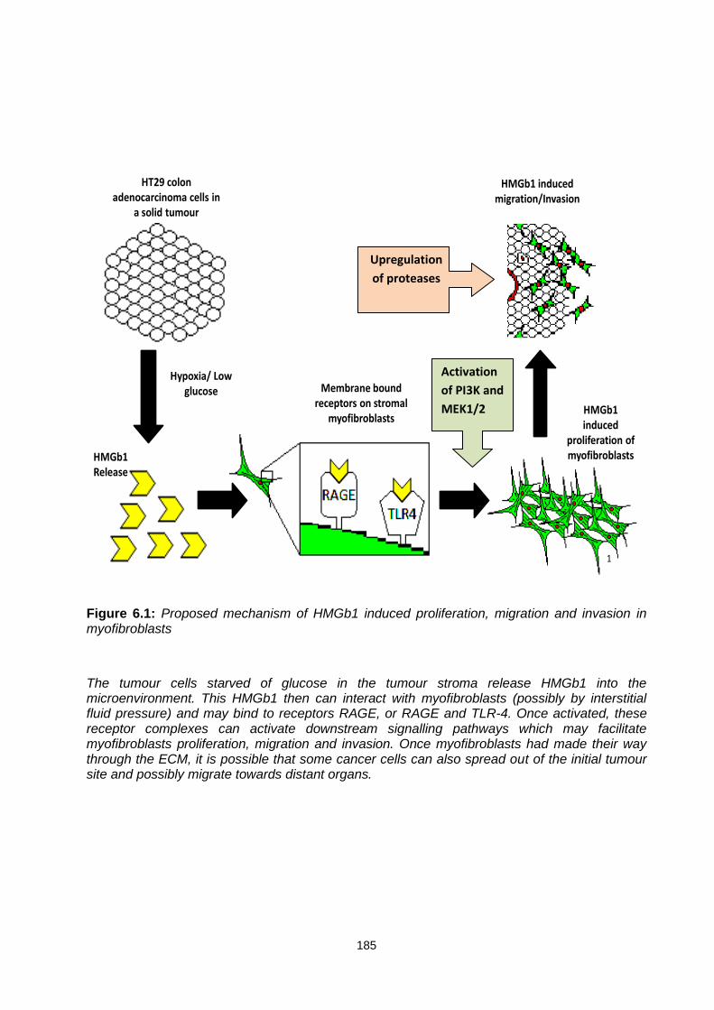
185
1
HT29 colon adenocarcinoma cells in
a solid tumour
Hypoxia/ Low glucose
HMGb1 Release
Membrane bound receptors on stromal
myofibroblastsHMGb1 induced
proliferation of myofibroblasts
HMGb1 induced migration/Invasion
Figure 6.1: Proposed mechanism of HMGb1 induced proliferation, migration and invasion in myofibroblasts
The tumour cells starved of glucose in the tumour stroma release HMGb1 into the microenvironment. This HMGb1 then can interact with myofibroblasts (possibly by interstitial fluid pressure) and may bind to receptors RAGE, or RAGE and TLR-4. Once activated, these receptor complexes can activate downstream signalling pathways which may facilitate myofibroblasts proliferation, migration and invasion. Once myofibroblasts had made their way through the ECM, it is possible that some cancer cells can also spread out of the initial tumour site and possibly migrate towards distant organs.
Activation
of PI3K and
MEK1/2
Upregulation
of proteases

186
Other researchers have reported these pathways (PI3K and MAPK) in both migration and
proliferation in cells such as human melanoma cells, glioblastoma cells and renal cell carcinoma
cells (Chen et al. 2015; Ji et al. 2015; Sun et al. 2015) However, involvement of these pathways
in HMGb1 induced stimulation of myofibroblasts has not been reported. The data presented in
this thesis suggests that HMGb1 induced proliferation in myofibroblasts involves activation of
PI3K and MEK1/2 pathways. Although, PI3K appeared to be a preferential pathway in both
settings (migration and invasion), however, it is possible that the effect of the antibodies or
inhibitors used to block each of these pathways might have different efficiencies to each other
or at completely different concentrations from each other. For example, the IC50 value for PI3K
inhibitor could be less than the MAPK-ERK inhibitor. Therefore, it is difficult to estimate to what
extent both pathways could have been inhibited using these antibodies. It was possible that an
effective dose of these inhibitors might have been toxic to myofibroblasts. Therefore, we
determined a non-toxic dose for both inhibitors that was able to inhibit the activation of these
pathways.
Considering all evidence presented in this thesis, it is clear that both pathways and receptor
RAGE and TLR-4 play important roles in HMGb1 induced proliferation, migration and invasion
of myofibroblasts cells. Therefore, based on the findings presented in this thesis, the
involvement of both pathways and receptors and a possible signalling flow path in HMGb1
induced proliferation, migration and invasion of myofibroblasts have been proposed using a
schematic diagram (Figure 6.2).

187
Figure 6.2: Proposed activation of the pathways involved in proliferation, migration and invasion of myofibroblasts cells.
PIP2
RAS
RAF
AKT
MEK1/2
MDM2 ERK
P53 Myocin
NF-κβ
Myofibroblasts proliferation, migration and
invasion
PI3K
FAK
Membrane protrusion
HMGb1
Cell Membrane

188
References
Aboseif, S., A. El‐Sakka, et al. (1999). "Mesenchymal reprogramming of adult human epithelial differentiation." Differentiation 65(2): 113-118. Agalave, N. M., M. Larsson, et al. (2014). "Spinal HMGB1 induces TLR-4-mediated long-lasting hypersensitivity and glial activation and regulates pain-like behavior in experimental arthritis." PAIN® 155(9): 1802-1813. Alcaraz, J. and P. Roca-Cusachs (2015). "Shape and Mechanical Cues Underlying Cellular Homeostasis in Soft Organs." Cells, Forces, and the Microenvironment: 177. Allinen, M., R. Beroukhim, et al. (2004). "Molecular characterization of the tumor microenvironment in breast cancer." Cancer cell 6(1): 17-32. Amara, N., D. Goven, et al. (2010). "NOX4/NADPH oxidase expression is increased in pulmonary fibroblasts from patients with idiopathic pulmonary fibrosis and mediates TGFβ1-induced fibroblast differentiation into myofibroblasts." Thorax 65(8): 733-738. An, W. G., M. Kanekal, et al. (1998). "Stabilization of wild-type p53 by hypoxia-inducible factor 1α." Nature 392(6674): 405-408. Anborgh, P. H., J. C. Mutrie, et al. (2010). "Role of the metastasis‐promoting protein osteopontin in the tumour microenvironment." Journal of cellular and molecular medicine 14(8): 2037-2044. Andersson, U., H. Erlandsson-Harris, et al. (2002). "HMGB1 as a DNA-binding cytokine." Journal of leukocyte biology 72(6): 1084. Andersson, U. and H. Erlandsson‐Harris (2004). "HMGB1 is a potent trigger of arthritis." Journal of internal medicine 255(3): 344-350. Apetoh, L., F. Ghiringhelli, et al. (2007). "The interaction between HMGB1 and TLR-4 dictates the outcome of anticancer chemotherapy and radiotherapy." Immunological reviews 220(1): 47-59. Arbibe, L., J.-P. Mira, et al. (2000). "Toll-like receptor 2–mediated NF-κB activation requires a Rac1-dependent pathway." Nature immunology 1(6): 533-540. Baek, J. H., J.-E. Jang, et al. (2000). "Hypoxia-induced VEGF enhances tumor survivability via suppression of serum deprivation-induced apoptosis." Oncogene 19(40): 4621. Baghdadi, M., H. Nagao, et al. (2013). "Combined blockade of TIM-3 and TIM-4 augments cancer vaccine efficacy against established melanomas." Cancer Immunology, Immunotherapy: 1-9. Bailey, J. M., B. J. Swanson, et al. (2008). "Sonic hedgehog promotes desmoplasia in pancreatic cancer." Clinical Cancer Research 14(19): 5995-6004.
189
Bain, J., L. Plater, et al. (2007). "The selectivity of protein kinase inhibitors: a further update." Biochem. J 408: 297-315. Bajaj, J., B. Zimdahl, et al. (2015). "Fearful symmetry: subversion of asymmetric division in cancer development and progression." Cancer research 75(5): 792-797. Balmain, A. and K. Brown (1988). "Oncogene activation in chemical carcinogenesis." Adv. Cancer Res 51: 147-182. Bamba, S., A. Andoh, et al. (2003). "Regulation of IL-11 expression in intestinal myofibroblasts: role of c-Jun AP-1-and MAPK-dependent pathways." American Journal of Physiology-Gastrointestinal and Liver Physiology 285(3): G529-G538. Barcellos-de-Souza, P., V. Gori, et al. (2013). "Tumor microenvironment: Bone marrow-mesenchymal stem cells as key players." Biochimica et Biophysica Acta (BBA)-Reviews on Cancer. Barkauskaite, V., M. Ek, et al. (2007). "Translocation of the novel cytokine HMGB1 to the cytoplasm and extracellular space coincides with the peak of clinical activity in experimentally UV-induced lesions of cutaneous lupus erythematosus." Lupus 16(10): 794. Bartling, B., H.-S. Hofmann, et al. (2005). "Down-regulation of the receptor for advanced glycation end-products (RAGE) supports non-small cell lung carcinoma." Carcinogenesis 26(2): 293-301. Bassi, R., P. Giussani, et al. (2008). "HMGB1 as an autocrine stimulus in human T98G glioblastoma cells: role in cell growth and migration." Journal of Neuro-Oncology 87(1): 23-33. Bauer, E. M., R. Shapiro, et al. (2013). "High mobility group box 1 inhibits human pulmonary artery endothelial cell migration via a toll-like receptor 4-and interferon response factor 3-dependent mechanism (s)." Journal of Biological Chemistry 288(2): 1365-1373. Bell, C. W., W. Jiang, et al. (2006). "The extracellular release of HMGB1 during apoptotic cell death." American Journal of Physiology-Cell Physiology 291(6): C1318-C1325. Benton, G., H. K. Kleinman, et al. (2011). "Multiple uses of basement membrane‐like matrix (BME/Matrigel) in vitro and in vivo with cancer cells." International Journal of Cancer 128(8): 1751-1757. Benyon, R., J. Iredale, et al. (1996). "Expression of tissue inhibitor of metalloproteinases 1 and 2 is increased in fibrotic human liver." Gastroenterology 110(3): 821-831. Berg, D. J., N. Davidson, et al. (1996). "Enterocolitis and colon cancer in interleukin-10-deficient mice are associated with aberrant cytokine production and CD4 (+) TH1-like responses." Journal of Clinical Investigation 98(4): 1010. Berthelot, F., L. Fattoum, et al. (2011). "The effect of HMGB1, a damage-associated molecular pattern molecule, on polymorphonuclear neutrophil migration depends on its concentration." Journal of innate immunity 4(1): 41-58.
190
Beyer, C., N. A. Stearns, et al. (2012). "The extracellular release of DNA and HMGB1 from Jurkat T cells during in vitro necrotic cell death." Innate immunity 18(5): 727-737. Bhatia, S., U. Balis, et al. (1999). "Effect of cell–cell interactions in preservation of cellular phenotype: cocultivation of hepatocytes and nonparenchymal cells." The FASEB Journal 13(14): 1883-1900. Bianchi, M. (2004). "Significant (re) location: how to use chromatin and/or abundant proteins as messages of life and death." Trends in cell biology 14(6): 287-293. Bianchi, M. E. and A. A. Manfredi (2014). "How macrophages ring the inflammation alarm." Proceedings of the National Academy of Sciences 111(8): 2866-2867. Bierhaus, A., P. Humpert, et al. (2005). "Understanding RAGE, the receptor for advanced glycation end products." Journal of Molecular Medicine 83(11): 876-886. Bierhaus, A., P. M. Humpert, et al. (2005). "Understanding RAGE, the receptor for advanced glycation end products." Journal of Molecular Medicine 83(11): 876-886. Bissell, M. J. and W. C. Hines (2011). "Why don't we get more cancer? A proposed role of the microenvironment in restraining cancer progression." Nature medicine 17(3): 320-329. Biswas, C., Y. Zhang, et al. (1995). "The human tumor cell-derived collagenase stimulatory factor (renamed EMMPRIN) is a member of the immunoglobulin superfamily." Cancer research 55(2): 434-439. Bonaldi, T., F. Talamo, et al. (2003). "Monocytic cells hyperacetylate chromatin protein HMGB1 to redirect it towards secretion." Science Signaling 22(20): 5551. Bonaldi, T., F. Talamo, et al. (2003). "Monocytic cells hyperacetylate chromatin protein HMGB1 to redirect it towards secretion." The EMBO journal 22(20): 5551-5560. Borenfreund, E. and J. Puerner (1986). "Cytotoxicity of metals, metal-metal and metal-chelator combinations assayed in vitro." Toxicology 39(2): 121-134. Borenfreund, E. and J. A. Puerner (1985). "Toxicity determined in vitro by morphological alterations and neutral red absorption." Toxicology letters 24(2): 119-124. Bosman, F. T. and I. Stamenkovic (2003). "Functional structure and composition of the extracellular matrix." The Journal of pathology 200(4): 423-428. Brahimi-Horn, C. and J. Pouysségur (2006). "The role of the hypoxia-inducible factor in tumor metabolism growth and invasion." Bulletin du cancer 93(8): 10073-10080. Brahimi-Horn, M. C. and J. Pouysségur (2007). "Oxygen, a source of life and stress." FEBS letters 581(19): 3582-3591. Brennecke, P., P. Allavena, et al. (2015). Inflammatory and Innate Immune Cells in Cancer Microenvironment and Progression. Cancer Immunology, Springer: 9-28.

191
Bustin, M. (2001). "Revised nomenclature for high mobility group (HMG) chromosomal proteins." Trends in biochemical sciences 26(3): 152-152. Bustin, M., D. Lehn, et al. (1990). "Structural features of the HMG chromosomal proteins and their genes." Biochimica et biophysica acta 1049(3): 231-243. Califano, R., A. Abidin, et al. (2015). "Beyond EGFR and ALK inhibition: Unravelling and exploiting novel genetic alterations in advanced non small-cell lung cancer." Cancer treatment reviews 41(5): 401-411. Campana, L., L. Bosurgi, et al. (2008). "HMGB1: a two-headed signal regulating tumor progression and immunity." Current opinion in immunology 20(5): 518-523. Carmeliet, P., Y. Dor, et al. (1998). "Role of HIF-1α in hypoxia-mediated apoptosis, cell proliferation and tumour angiogenesis." Nature 394(6692): 485-490. Carmeliet, P. and R. K. Jain (2000). "Angiogenesis in cancer and other diseases." Nature 407(6801): 249-257. Casey, T. M., J. Eneman, et al. (2008). "Cancer associated fibroblasts stimulated by transforming growth factor beta1 (TGF-β1) increase invasion rate of tumor cells: a population study." Breast cancer research and treatment 110(1): 39-49. Castella, L. F., L. Buscemi, et al. (2010). "A new lock-step mechanism of matrix remodelling based on subcellular contractile events." Journal of Cell Science 123(10): 1751-1760. Chambers, A. F., A. C. Groom, et al. (2002). "Metastasis: dissemination and growth of cancer cells in metastatic sites." Nature Reviews Cancer 2(8): 563-572. Chavakis, E., A. Hain, et al. (2007). "High-mobility group box 1 activates integrin-dependent homing of endothelial progenitor cells." Circulation research 100(2): 204-212. Chedid, M., J. Rubin, et al. (1994). "Regulation of keratinocyte growth factor gene expression by interleukin 1." Journal of Biological Chemistry 269(14): 10753. Chen, G., M. Ward, et al. (2004). "Extracellular HMGB1 as a proinflammatory cytokine." Journal of Interferon & Cytokine Research 24(6): 329-333. Chen, H.-C. (2005). Boyden chamber assay. Cell Migration, Springer: 15-22. Chen, P.-N., Y.-S. Hsieh, et al. (2005). "Silibinin inhibits cell invasion through inactivation of both PI3K-Akt and MAPK signaling pathways." Chemico-biological interactions 156(2): 141-150. Chen, W.-L., E. Turlova, et al. (2015). "Xyloketal B Suppresses Glioblastoma Cell Proliferation and Migration in Vitro through Inhibiting TRPM7-Regulated PI3K/Akt and MEK/ERK Signaling Pathways." Marine drugs 13(4): 2505-2525. Cheng, C., K. Tsuneyama, et al. (2005). "Expression profiling of endogenous secretory receptor for advanced glycation end products in human organs." Modern pathology 18(10): 1385-1396.
192
Cheng, S. and D. H. Lovett (2003). "Gelatinase A (MMP-2) is necessary and sufficient for renal tubular cell epithelial-mesenchymal transformation." The American Journal of Pathology 162(6): 1937-1949. Chitanuwat, A., N. Laosrisin, et al. (2013). "Role of HMGB1 in proliferation and migration of human gingival and periodontal ligament fibroblasts." Journal of oral science 55(1): 45-50. Cho, J. A., H. Park, et al. (2012). "Exosomes from breast cancer cells can convert adipose tissue-derived mesenchymal stem cells into myofibroblast-like cells." International journal of oncology 40(1): 130-138. Chung, A. S., J. Lee, et al. (2010). "Targeting the tumour vasculature: insights from physiological angiogenesis." Nature Reviews Cancer 10(7): 505-514. Clericuzio, M., B. Burlando, et al. (2013). "Keratinocyte wound healing activity of galactoglycerolipids from the fern Ophioglossum vulgatum L." Journal of natural medicines: 1-7. Coleman, R. E. (2006). "Clinical features of metastatic bone disease and risk of skeletal morbidity." Clinical Cancer Research 12(20): 6243s-6249s. Courtnay, R., D. C. Ngo, et al. (2015). "Cancer metabolism and the Warburg effect: the role of HIF-1 and PI3K." Molecular biology reports 42(4): 841-851. Coussens, L. M. and Z. Werb (2002). "Inflammation and cancer." Nature 420(6917): 860-867. Crean, J. K., D. Finlay, et al. (2002). "The role of p42/44 MAPK and protein kinase B in connective tissue growth factor induced extracellular matrix protein production, cell migration, and actin cytoskeletal rearrangement in human mesangial cells." Journal of Biological Chemistry 277(46): 44187-44194. Curtin, J. F., N. Liu, et al. (2009). "HMGB1 mediates endogenous TLR-2 activation and brain tumor regression." PLoS medicine 6(1): e1000010. Cuvier, C., A. Jang, et al. (1997). "Exposure to hypoxia, glucose starvation and acidosis: effect on invasive capacity of murine tumor cells and correlation with cathepsin (L+ B) secretion." Clinical & experimental metastasis 15(1): 19-25. Dai, S., C. Sodhi, et al. (2010). "Extracellular high mobility group box-1 (HMGB1) inhibits enterocyte migration via activation of Toll-like receptor-4 and increased cell-matrix adhesiveness." Journal of Biological Chemistry 285(7): 4995-5002. Davidson, B., I. Goldberg, et al. (1999). "High levels of MMP-2, MMP-9, MT1-MMP and TIMP-2 mRNA correlate with poor survival in ovarian carcinoma." Clinical & experimental metastasis 17(10): 799-808. de Claro, R. A., K. McGinn, et al. (2012). "US Food and Drug Administration approval summary: brentuximab vedotin for the treatment of relapsed Hodgkin lymphoma or relapsed systemic anaplastic large-cell lymphoma." Clinical Cancer Research 18(21): 5845-5849. De Wever, O., P. Demetter, et al. (2008). "Stromal myofibroblasts are drivers of invasive cancer growth." International Journal of Cancer 123(10): 2229-2238.

193
De Wever, O. and M. Mareel (2003). "Role of tissue stroma in cancer cell invasion." The Journal of pathology 200(4): 429-447. De Wever, O., W. Westbroek, et al. (2004). "Critical role of N-cadherin in myofibroblast invasion and migration in vitro stimulated by colon-cancer-cell-derived TGF-β or wounding." Journal of cell science 117(20): 4691-4703. Degryse, B., T. Bonaldi, et al. (2001). "The high mobility group (HMG) boxes of the nuclear protein HMG1 induce chemotaxis and cytoskeleton reorganization in rat smooth muscle cells." Science Signaling 152(6): 1197. Derynck, R., R. J. Akhurst, et al. (2001). "TGF-β signaling in tumor suppression and cancer progression." Nature genetics 29(2): 117-129. Desbaillets, I., A.-C. Diserens, et al. (1997). "Upregulation of interleukin 8 by oxygen-deprived cells in glioblastoma suggests a role in leukocyte activation, chemotaxis, and angiogenesis." The Journal of experimental medicine 186(8): 1201-1212. DeSilva, D. R., E. A. Jones, et al. (1998). "Inhibition of mitogen-activated protein kinase kinase blocks T cell proliferation but does not induce or prevent anergy." The Journal of Immunology 160(9): 4175-4181. Desmoulière, A., C. Guyot, et al. (2004). "The stroma reaction myofibroblast: a key player in the control of tumor cell behavior." International Journal of Developmental Biology 48: 509-518. Dhani, N., A. Fyles, et al. (2015). The Clinical Significance of Hypoxia in Human Cancers. Seminars in nuclear medicine, Elsevier. Diederichs, S., E. Bulk, et al. (2004). "S100 family members and trypsinogens are predictors of distant metastasis and survival in early-stage non-small cell lung cancer." Cancer research 64(16): 5564-5569. Ding, X., D.-R. Yang, et al. (2015). "Targeting TR4 nuclear receptor suppresses prostate cancer invasion via reduction of infiltrating macrophages with alteration of the TIMP-1/MMP2/MMP9 signals." Molecular cancer 14(1): 16. Dolci, S., M. Pellegrini, et al. (2001). "Signaling through extracellular signal-regulated kinase is required for spermatogonial proliferative response to stem cell factor." Journal of Biological Chemistry 276(43): 40225-40233. Dong, X. D. E., N. Ito, et al. (2007). "High mobility group box I (HMGB1) release from tumor cells after treatment: implications for development of targeted chemoimmunotherapy." Journal of Immunotherapy 30(6): 596-606. Dragan, Y. P. and H. C. Pitot (1992). "The role of the stages of initiation and promotion in phenotypic diversity during hepatocarcinogenesis in the rat." Carcinogenesis 13(5): 739.

194
Du, L., P. Smolewski, et al. (2001). "Selective protection of mitogenically stimulated human lymphocytes but not leukemic cells from cytosine arabinoside-induced apoptosis by LY294002, a phosphoinositol-3 kinase inhibitor." International journal of oncology 19(4): 811-820. Duda, D. G., A. M. Duyverman, et al. (2010). "Malignant cells facilitate lung metastasis by bringing their own soil." Proceedings of the National Academy of Sciences 107(50): 21677-21682. Dumitriu, I. E., P. Baruah, et al. (2005). "Release of high mobility group box 1 by dendritic cells controls T cell activation via the receptor for advanced glycation end products." The Journal of Immunology 174(12): 7506-7515. Dumitriu, I. E., M. E. Bianchi, et al. (2007). "The secretion of HMGB1 is required for the migration of maturing dendritic cells." Journal of leukocyte biology 81(1): 84-91. Dzaman, K., M. Zagor, et al. (2015). "High motility group box 1 (HMGB1) protein and its receptor for advanced glycation end products (RAGE) expression in chronic rhinosinusitis without nasal polyps." Folia Histochemica et Cytobiologica 53(1): 70-78. Elenbaas, B. and R. A. Weinberg (2001). "Heterotypic signaling between epithelial tumor cells and fibroblasts in carcinoma formation." Experimental cell research 264(1): 169. Elkabets, M., A. M. Gifford, et al. (2011). "Human tumors instigate granulin-expressing hematopoietic cells that promote malignancy by activating stromal fibroblasts in mice." The Journal of clinical investigation 121(2): 784. Ellerman, J. E., C. K. Brown, et al. (2007). "Masquerader: high mobility group box-1 and cancer." Clinical Cancer Research 13(10): 2836-2848. Elliott, R. L. and G. C. Blobe (2005). "Role of transforming growth factor Beta in human cancer." Journal of Clinical Oncology 23(9): 2078-2093. Erez, N., M. Truitt, et al. (2010). "Cancer-associated fibroblasts are activated in incipient neoplasia to orchestrate tumor-promoting inflammation in an NF-κB-dependent manner." Cancer cell 17(2): 135-147. Fan, J., Y. Li, et al. (2007). "Hemorrhagic shock induces NAD (P) H oxidase activation in neutrophils: role of HMGB1-TLR-4 signaling." The Journal of Immunology 178(10): 6573-6580. Fan, J. and A. B. Malik (2003). "Toll-like receptor-4 (TLR-4) signaling augments chemokine-induced neutrophil migration by modulating cell surface expression of chemokine receptors." Nature medicine 9(3): 315-321. Farber, E. and H. Rubin (1991). "Cellular adaptation in the origin and development of cancer." Cancer research 51(11): 2751-2761. Fashena, S. J. and S. M. Thomas (2000). "Signalling by adhesion receptors." Nature cell biology 2(12): E225-E229.

195
Fearon, E. R. and B. Vogelstein (1990). "A genetic model for colorectal tumorigenesis." Cell 61(5): 759-767. Feng, X.-J., S.-X. Liu, et al. (2014). "The PTEN/PI3K/Akt signaling pathway mediates HMGB1-induced cell proliferation by regulating the NF-κB/cyclin D1 pathway in mouse mesangial cells." American Journal of Physiology-Cell Physiology 306(12): C1119-C1128. Ferlay, J., I. Soerjomataram, et al. (2015). "Cancer incidence and mortality worldwide: sources, methods and major patterns in GLOBOCAN 2012." International Journal of Cancer 136(5): E359-E386. Fidler, I. J. (2003). "The pathogenesis of cancer metastasis: the'seed and soil'hypothesis revisited." Nature Reviews Cancer 3(6): 453-458. Fiuza, C., M. Bustin, et al. (2003). "Inflammation-promoting activity of HMGB1 on human microvascular endothelial cells." Blood 101(7): 2652-2660. Fokas, E., R. Engenhart-Cabillic, et al. (2007). "Metastasis: the seed and soil theory gains identity." Cancer and Metastasis Reviews 26(3-4): 705-715. Fokas, E., R. Engenhart-Cabillic, et al. (2007). "Metastasis: the seed and soil theory gains identity." Cancer and Metastasis Reviews 26(3): 705-715. Folkman, J. (1990). "What is the evidence that tumors are angiogenesis dependent?" Journal of the National Cancer Institute 82(1): 4-7. Follonier Castella, L., G. Gabbiani, et al. (2010). "Regulation of myofibroblast activities: calcium pulls some strings behind the scene." Experimental cell research 316(15): 2390-2401. Foukas, L. C., I. M. Berenjeno, et al. (2010). "Activity of any class IA PI3K isoform can sustain cell proliferation and survival." Proceedings of the National Academy of Sciences 107(25): 11381-11386. Frank, M. G., M. D. Weber, et al. (2015). "Stress sounds the alarmin: The role of the danger-associated molecular pattern HMGB1 in stress-induced neuroinflammatory priming." Brain, behavior, and immunity. Frisch, S. M., K. Vuori, et al. (1996). "Control of adhesion-dependent cell survival by focal adhesion kinase." The Journal of cell biology 134(3): 793-799. Fritz, G. (2011). “A single receptor fits multiple ligands.” Trends in biochemical sciences 36(12):625-632. Fukami, K., K. Taguchi, et al. (2015). "Receptor for advanced glycation endproducts and progressive kidney disease." Current opinion in nephrology and hypertension 24(1): 54-60. Fullár, A., J. Dudás, et al. (2015). "Remodeling of extracellular matrix by normal and tumor-associated fibroblasts promotes cervical cancer progression." BMC cancer 15(1): 256. Fynan, T. and M. Reiss (1993). "Resistance to inhibition of cell growth by transforming growth factor-beta and its role in oncogenesis." Critical reviews in oncogenesis 4(5): 493.
196
Gabbiani, G. (1996). "The cellular derivation and the life span of the myofibroblast." Pathology Research and Practice 192(7): 708-711. Gaggioli, C., S. Hooper, et al. (2007). "Fibroblast-led collective invasion of carcinoma cells with differing roles for RhoGTPases in leading and following cells." Nature cell biology 9(12): 1392-1400. Gangoda, L., S. Keerthikumar, et al. (2015). "Inhibition of cathepsin proteases attenuates migration and sensitizes aggressive N-Myc amplified human neuroblastoma cells to doxorubicin." Oncotarget. Ganz, M., T. N. Bukong, et al. (2015). "Progression of non-alcoholic steatosis to steatohepatitis and fibrosis parallels cumulative accumulation of danger signals that promote inflammation and liver tumors in a high fat–cholesterol–sugar diet model in mice." Journal of translational medicine 13(1): 193. Gardella, S., C. Andrei, et al. (2002). "The nuclear protein HMGB1 is secreted by monocytes via a non-classical, vesicle-mediated secretory pathway." EMBO reports 3(10): 995-1001. Gebhardt, C., P. H. Angel, Jochen, et al. (2006). "S100A8 and S100A9 in inflammation and cancer." Biochemical pharmacology 72(11): 1622. Georges, P. C., J.-J. Hui, et al. (2007). "Increased stiffness of the rat liver precedes matrix deposition: implications for fibrosis." American Journal of Physiology-Gastrointestinal and Liver Physiology 293(6): G1147-G1154. Gerald, D., E. Berra, et al. (2004). "JunD reduces tumor angiogenesis by protecting cells from oxidative stress." Cell 118(6): 781-794. Ghobrial, I. M. (2009). "Dual targeting of the PI3K/Akt/mTOR pathway as an anti-tumor strategy in Waldenstrom’s macroglobulinemia." Gnanasekar, M., S. Thirugnanam, et al. (2009). "Short hairpin RNA (shRNA) constructs targeting high mobility group box-1 (HMGB1) expression leads to inhibition of prostate cancer cell survival and apoptosis." International journal of oncology 34(2): 425-431. Gomez, D., D. Alonso, et al. (1997). "Tissue inhibitors of metalloproteinases: structure, regulation and biological functions." European journal of cell biology 74(2): 111-122. Goodman, J. I., J. M. Ward, et al. (1991). "Mouse liver carcinogenesis: mechanisms and relevance." Toxicological Sciences 17(4): 651-665. Gottschalk, A. R., A. Doan, et al. (2005). "Inhibition of phosphatidylinositol-3-kinase causes increased sensitivity to radiation through a PKB-dependent mechanism." International Journal of Radiation Oncology* Biology* Physics 63(4): 1221-1227. Graff, R. (2014). Glucose Regulation of Novel Protein Complexes with Potential Roles in the Metabolic Reprogramming of Human Leukemia Cells, Wesleyan University. Greenhough, A., H. A. Patsos, et al. (2007). "The cannabinoid δ9‐tetrahydrocannabinol inhibits RAS‐MAPK and PI3K‐AKT survival signalling and induces BAD‐mediated apoptosis in colorectal cancer cells." International Journal of Cancer 121(10): 2172-2180.

197
Gullberg, D. and R. K. Reed (2011). Tumor-Stroma Interactions: Focus on Fibroblasts. Mouse as a Model Organism, Springer: 117-130. Gullino, P. M., S. H. Clark, et al. (1964). "The interstitial fluid of solid tumors." Cancer research 24(5): 780-797. Gupta, G. P. and J. Massagué (2006). "Cancer metastasis: building a framework." Cell 127(4): 679-695. Han, S.-H., Y. H. Kim, et al. (2011). "RAGE: the beneficial and deleterious effects by diverse mechanisms of actions." Molecules and cells 31(2): 91-97. Hanahan, D. and R. A. Weinberg (2000). "The hallmarks of cancer." Cell 100(1): 57-70. Hanahan, D. and R. A. Weinberg (2011). "Hallmarks of cancer: the next generation." Cell 144(5): 646-674. Harokopakis, E., M. H. Albzreh, et al. (2006). "TLR-2 transmodulates monocyte adhesion and transmigration via Rac1-and PI3K-mediated inside-out signaling in response to Porphyromonas gingivalis fimbriae." The Journal of Immunology 176(12): 7645-7656. Hart, I. R. and I. J. Fidler (1980). "Role of organ selectivity in the determination of metastatic patterns of B16 melanoma." Cancer research 40(7): 2281-2287. Hasegawa, T., A. Kosaki, et al. (2003). "The regulation of EN-RAGE (S100A12) gene expression in human THP-1 macrophages." Atherosclerosis 171(2): 211-218. Hayakawa, K., A. C.-L. Liang, et al. (2015). In Vitro Angiogenesis Assay: Endothelial Migration, Proliferation, and Tube Formation. Handbook of Vascular Biology Techniques, Springer: 3-12. Hayashi, H., Y. Tsuchiya, et al. (2008). "Down‐regulation of the PI3‐kinase/Akt pathway by ERK MAP kinase in growth factor signaling." Genes to Cells 13(9): 941-947. He, Q., H. You, et al. (2012). "HMGB1 promotes the synthesis of pro-IL-1β and pro-IL-18 by activation of p38 MAPK and NF-κB through receptors for advanced glycation end-products in macrophages." Asian Pacific Journal of Cancer Prevention 13(4): 1365-1370. Hemers, E., C. Duval, et al. (2005). "Insulin-like growth factor binding protein-5 is a target of matrix metalloproteinase-7: implications for epithelial-mesenchymal signaling." Cancer research 65(16): 7363-7369. Hinz, B. and G. Gabbiani (2010). "Fibrosis: recent advances in myofibroblast biology and new therapeutic perspectives." F1000 Biology Reports 2. Hinz, B., S. Phan, et al. (2007). "The myofibroblast: one function, multiple origins." American journal of pathology 170(6): 1807. Hinz, B., S. H. Phan, et al. (2007). "The myofibroblast: one function, multiple origins." The American journal of pathology 170(6): 1807-1816.

198
Hinz, B., S. H. Phan, et al. (2012). "Recent developments in myofibroblast biology: paradigms for connective tissue remodeling." The American Journal of Pathology 180(4): 1340-1355. Hirsilä, M., P. Koivunen, et al. (2003). "Characterization of the human prolyl 4-hydroxylases that modify the hypoxia-inducible factor." Journal of Biological Chemistry 278(33): 30772-30780. Hofmann, M., S. Drury, et al. (2002). "RAGE and arthritis: the G82S polymorphism amplifies the inflammatory response." Genes and immunity 3(3): 123-135. Hofmann, M. A., S. Drury, et al. (1999). "RAGE mediates a novel proinflammatory axis: a central cell surface receptor for S100/calgranulin polypeptides." Cell 97(7): 889-901. Honda, R. and H. Yasuda (1999). "Association of p19ARF with Mdm2 inhibits ubiquitin ligase activity of Mdm2 for tumor suppressor p53." The EMBO journal 18(1): 22-27. Hotary, K., E. Allen, et al. (2000). "Regulation of cell invasion and morphogenesis in a three-dimensional type I collagen matrix by membrane-type matrix metalloproteinases 1, 2, and 3." The Journal of Cell Biology 149(6): 1309-1323. Hreggvidsdottir, H. S., T. Östberg, et al. (2009). "The alarmin HMGB1 acts in synergy with endogenous and exogenous danger signals to promote inflammation." Journal of leukocyte biology 86(3): 655-662. Hu, M., J. Yao, et al. (2008). "Regulation of in situ to invasive breast carcinoma transition." Cancer cell 13(5): 394-406. Huttunen, H. and H. Rauvala (2004). "Amphoterin as an extracellular regulator of cell motility: from discovery to disease." Journal of internal medicine 255(3): 351-366. Huttunen, H. J., C. Fages, et al. (1999). "Receptor for advanced glycation end products (RAGE)-mediated neurite outgrowth and activation of NF-κB require the cytoplasmic domain of the receptor but different downstream signaling pathways." Journal of Biological Chemistry 274(28): 19919-19924. Hwang, R. F., T. Moore, et al. (2008). "Cancer-associated stromal fibroblasts promote pancreatic tumor progression." Cancer research 68(3): 918-926. Hyakushima, N., H. Mitsuzawa, et al. (2004). "Interaction of soluble form of recombinant extracellular TLR-4 domain with MD-2 enables lipopolysaccharide binding and attenuates TLR-4-mediated signaling." The Journal of Immunology 173(11): 6949-6954. Ingels, C., I. Derese, et al. (2015). "Soluble RAGE and the RAGE ligands HMGB1 and S100A12 in critical illness: impact of glycemic control with insulin and relation with clinical outcome." Shock 43(2): 109-116. Inoue, K., K.-i. Kawahara, et al. (2007). "HMGB1 expression by activated vascular smooth muscle cells in advanced human atherosclerosis plaques." Cardiovascular Pathology 16(3): 136-143. Inoue, T., H. Okada, et al. (2009). "A case report suggesting the occurrence of epithelial–mesenchymal transition in obstructive nephropathy." Clinical and experimental nephrology 13(4): 385-388.

199
Ishiguro, H., N. Nakaigawa, et al. (2005). "Receptor for advanced glycation end products (RAGE) and its ligand, amphoterin are overexpressed and associated with prostate cancer development." The Prostate 64(1): 92-100. Ito, N., R. A. DeMarco, et al. (2007). "Cytolytic cells induce HMGB1 release from melanoma cell lines." Journal of leukocyte biology 81(1): 75-83. Ivan, M., K. Kondo, et al. (2001). "HIFalpha targeted for VHL-mediated destruction by proline hydroxylation: implications for O2 sensing." Science Signaling 292(5516): 464. Iwasaki, A. and R. Medzhitov (2004). "Toll-like receptor control of the adaptive immune responses." Nature immunology 5(10): 987-995. Jain, R. K. (1987). "Transport of molecules across tumor vasculature." Cancer and Metastasis Reviews 6(4): 559-593. Jamieson, L., D. Harrison, et al. (2015). "Chemical analysis of multicellular tumour spheroids." Analyst. Jang, A. and R. Hill (1997). "An examination of the effects of hypoxia, acidosis, and glucose starvation on the expression of metastasis-associated genes in murine tumor cells." Clinical & experimental metastasis 15(5): 469-483. Janji, B., E. Viry, et al. (2013). "Role of Autophagy in Cancer and Tumor Progression." Jass, J. (2007). "Classification of colorectal cancer based on correlation of clinical, morphological and molecular features." Histopathology 50(1): 113-130. Jass, J. R. (2004). "Hyperplastic polyps and colorectal cancer: is there a link?" Clinical Gastroenterology and Hepatology 2(1): 1-8. Jeong, H. W. and I. S. Kim (2004). "TGF‐β1 enhances βig‐h3‐mediated keratinocyte cell migration through the α3β1 integrin and PI3K." Journal of cellular biochemistry 92(4): 770-780. Ji, B.-C., Y.-P. Hsiao, et al. (2015). "Cantharidin Impairs Cell Migration and Invasion of A375. S2 Human Melanoma Cells by Suppressing MMP-2 and-9 Through PI3K/NF-κB Signaling Pathways." Anticancer research 35(2): 729-738. Jiao, Y. (2007). "Growth suppression and radiosensitivity increase by HMGB1 in breast cancer1." Acta Pharmacologica Sinica 28(12): 1957-1967. Jögi, A. (2015). Tumour Hypoxia and the Hypoxia-Inducible Transcription Factors: Key Players in Cancer Progression and Metastasis. Tumor Cell Metabolism, Springer: 65-98. Joseph, J. V., S. Conroy, et al. (2015). "Hypoxia enhances migration and invasion in glioblastoma by promoting a mesenchymal shift mediated by the HIF1α-ZEB1 axis." Cancer letters. Kaboord, B. and M. Perr (2008). Isolation of proteins and protein complexes by immunoprecipitation. 2D PAGE: sample preparation and fractionation, Springer: 349-364.

200
Kalebic, T., S. Garbisa, et al. (1983). "Basement membrane collagen: degradation by migrating endothelial cells." Science 221(4607): 281-283. Kalluri, R. (2003). "Basement membranes: structure, assembly and role in tumour angiogenesis." Nature Reviews Cancer 3(6): 422-433. Kamangar, F., G. M. Dores, et al. (2006). "Patterns of cancer incidence, mortality, and prevalence across five continents: defining priorities to reduce cancer disparities in different geographic regions of the world." Journal of Clinical Oncology 24(14): 2137-2150. Kang, R., D. Tang, et al. (2013). "The HMGB1/RAGE inflammatory pathway promotes pancreatic tumor growth by regulating mitochondrial bioenergetics." Oncogene. Kang, R., D. Tang, et al. (2014). "The HMGB1/RAGE inflammatory pathway promotes pancreatic tumor growth by regulating mitochondrial bioenergetics." Oncogene 33(5): 567-577. Kang, R., Q. Zhang, et al. (2013). "HMGB1 in cancer: good, bad, or both?" Clinical Cancer Research 19(15): 4046-4057. Keyel, P. A. (2014). "How is inflammation initiated? Individual influences of IL-1, IL-18 and HMGB1." Cytokine. Khanna, C., R. G. Wells, et al. (2015). "Type III Collagen Directs Stromal Organization and Limits Metastasis in a Murine Model of Breast Cancer." The American Journal of Pathology 185(5): 1. Khozin, S., G. M. Blumenthal, et al. (2015). "FDA Approval: Ceritinib for the Treatment of Metastatic Anaplastic Lymphoma Kinase–Positive Non–Small Cell Lung Cancer." Clinical Cancer Research. Kim, D. E., K.-j. Min, et al. (2012). "High-mobility group box-1 protein induces mucin 8 expression through the activation of the JNK and PI3K/Akt signal pathways in human airway epithelial cells." Biochemical and biophysical research communications 421(3): 436-441. Kim, S., S. Y. Kim, et al. (2013). "Signaling of High Mobility Group Box 1 (HMGB1) through Toll-like Receptor 4 in Macrophages Requires CD14." Molecular Medicine 19(1): 88. Klingberg, F., B. Hinz, et al. (2013). "The myofibroblast matrix: implications for tissue repair and fibrosis." The Journal of Pathology 229(2): 298-309. Kobayashi, S., F. Kimura, et al. (2009). "BCR–ABL promotes neutrophil differentiation in the chronic phase of chronic myeloid leukemia by downregulating c-Jun expression." Leukemia 23(9): 1622-1627. Koh, M. Y. and G. Powis (2012). "Passing the baton: the HIF switch." Trends in biochemical sciences 37(9): 364-372. Kokkola, R., Å. Andersson, et al. (2005). "RAGE is the major receptor for the proinflammatory activity of HMGB1 in rodent macrophages." Scandinavian journal of immunology 61(1): 1-9.
201
Kostova, N., S. Zlateva, et al. (2010). "The expression of HMGB1 protein and its receptor RAGE in human malignant tumors." Molecular and cellular biochemistry 337(1-2): 251-258. Kovacs, E. and L. DiPietro (1994). "Fibrogenic cytokines and connective tissue production." The FASEB Journal 8(11): 854. Kuniyasu, H., Y. Chihara, et al. (2003). "Differential effects between amphoterin and advanced glycation end products on colon cancer cells." International journal of cancer 104(6): 722-727. Kuniyasu, H., Y. Chihara, et al. (2003). "Amphoterin induction in prostatic stromal cells by androgen deprivation is associated with metastatic prostate cancer." Oncology reports 10(6): 1863. Laderoute, K. R., K. Amin, et al. (2006). "5′-AMP-activated protein kinase (AMPK) is induced by low-oxygen and glucose deprivation conditions found in solid-tumor microenvironments." Molecular and cellular biology 26(14): 5336-5347. Lai, C.-H., G.-Y. Shi, et al. (2013). "Recombinant human thrombomodulin suppresses experimental abdominal aortic aneurysms induced by calcium chloride in mice." Annals of surgery. Lai, K.-C., A.-C. Huang, et al. (2010). "Benzyl isothiocyanate (BITC) inhibits migration and invasion of human colon cancer HT29 cells by inhibiting matrix metalloproteinase-2/-9 and urokinase plasminogen (uPA) through PKC and MAPK signaling pathway." Journal of agricultural and food chemistry 58(5): 2935-2942. Lane, D., I. Matte, et al. (2011). "Prognostic significance of IL-6 and IL-8 ascites levels in ovarian cancer patients." BMC cancer 11(1): 210. Lange, S. S., D. L. Mitchell, et al. (2008). "High mobility group protein B1 enhances DNA repair and chromatin modification after DNA damage." Proceedings of the National Academy of Sciences 105(30): 10320. Lee, E., N. B. Pandey, et al. (2015). "Crosstalk between cancer cells and blood endothelial and lymphatic endothelial cells in tumour and organ microenvironment." Expert reviews in molecular medicine 17: e3. Lee, W.-T., T.-H. Lee, et al. (2015). "Antroquinonol from antrodia camphorata suppresses breast tumor migration/invasion through inhibiting ERK-AP-1-and AKT-NF-κB-dependent MMP-9 and epithelial-mesenchymal transition expressions." Food and Chemical Toxicology. Lee, Y.-C., H.-H. Lin, et al. (2010). "Inhibitory effects of andrographolide on migration and invasion in human non-small cell lung cancer A549 cells via down-regulation of PI3K/Akt signaling pathway." European journal of pharmacology 632(1): 23-32. Leivonen, S.-K., K. Lazaridis, et al. (2013). "TGF-β-Elicited Induction of Tissue Inhibitor of Metalloproteinases (TIMP)-3 Expression in Fibroblasts Involves Complex Interplay between Smad3, p38α, and ERK1/2." PLoS ONE 8(2): e57474. Lenga, Y., A. Koh, et al. (2008). "Osteopontin expression is required for myofibroblast differentiation." Circulation research 102(3): 319-327.
202
Levy, A. P., N. S. Levy, et al. (1996). "Post-transcriptional regulation of vascular endothelial growth factor by hypoxia." Journal of Biological Chemistry 271(5): 2746-2753. Lewis, M., K. Lygoe, et al. (2004). "Tumour-derived TGF-β1 modulates myofibroblast differentiation and promotes HGF/SF-dependent invasion of squamous carcinoma cells." British journal of cancer 90(4): 822-832. Li, A., S. Dubey, et al. (2003). "IL-8 directly enhanced endothelial cell survival, proliferation, and matrix metalloproteinases production and regulated angiogenesis." The Journal of Immunology 170(6): 3369-3376. Li, C., D.-R. Liu, et al. (2015). "CD97 promotes gastric cancer cell proliferation and invasion through exosome-mediated MAPK signaling pathway." World J Gastroenterol 21(20): 6215-6228. Li, J., C. Zhang, et al. (2015). "andrographolide inhibits hypoxia-inducible factor-1 through phosphatidylinositol 3-kinase/aKT pathway and suppresses breast cancer growth." OncoTargets and therapy 8: 427. Li, S.-h., H. Tian, et al. (2011). "Overexpression of metastasis-associated protein 1 is significantly correlated with tumor angiogenesis and poor survival in patients with early-stage non-small cell lung cancer." Annals of surgical oncology 18(7): 2048-2056. Liang, X., Y. H. So, et al. (2011). "The low-dose ionizing radiation stimulates cell proliferation via activation of the MAPK/ERK pathway in rat cultured mesenchymal stem cells." Journal of radiation research 52(3): 380-386. Lin, L., S. Park, et al. (2009). "RAGE signaling in inflammation and arterial aging." Frontiers in bioscience: a journal and virtual library 14: 1403. Lin, Y.-T., J.-S. Chen, et al. (2015). "Galectin-1 Accelerates Wound Healing by Regulating the Neuropilin-1/Smad3/NOX4 Pathway and ROS Production in Myofibroblasts." Journal of Investigative Dermatology 135(1): 258-268. Liu, L., M. Yang, et al. (2011). "HMGB1-induced autophagy promotes chemotherapy resistance in leukemia cells." Leukemia 25(1): 23-31. Liu, W., G. Li, et al. (2015). "Neuroprotective effects of geniposide from Alzheimer’s disease pathology." Reviews in the neurosciences. Liu, Y.-W., P.-Y. Zuo, et al. (2015). "Octacosanol Enhances the Proliferation and Migration of Human Umbilical Vein Endothelial Cells via Activation of the PI3K/Akt and MAPK/Erk Pathways." Lipids 50(3): 241-251. Liu, Y. (2006). "Renal fibrosis: new insights into the pathogenesis and therapeutics." Kidney international 69(2): 213-217.

203
Liu, Y., Y. Gao, et al. (2014). "TLR-4 Activation by Lipopolysaccharide and Streptococcus mutans Induces Differential Regulation of Proliferation and Migration in Human Dental Pulp Stem Cells." Journal of endodontics 40(9): 1375-1381. Liu, Y., C. Liang, et al. (2010). "AGEs increased migration and inflammatory responses of adventitial fibroblasts via RAGE, MAPK and NF-κB pathways." Atherosclerosis 208(1): 34-42. Logsdon, C. D., M. K. Fuentes, et al. (2007). "RAGE and RAGE ligands in cancer." Current molecular medicine 7(8): 777-789. Lotze, M. T. and K. J. Tracey (2005). "High-mobility group box 1 protein (HMGB1): nuclear weapon in the immune arsenal." Nature Reviews Immunology 5(4): 331-342. Lovén, J., H. A. Hoke, et al. (2013). "Selective inhibition of tumor oncogenes by disruption of super-enhancers." Cell 153(2): 320-334. Lowry, O. H., N. J. Rosebrough, et al. (1951). "Protein measurement with the Folin phenol reagent." J biol Chem 193(1): 265-275. Lu, B., D. J. Antoine, et al. (2014). "JAK/STAT1 signaling promotes HMGB1 hyperacetylation and nuclear translocation." Proceedings of the National Academy of Sciences 111(8): 3068-3073. Lum, H. and K.-L. Lee (2001). "The human HMGB1 promoter is modulated by a silencer and an enhancer-containing intron." Biochimica et Biophysica Acta (BBA)-Gene Structure and Expression 1520(1): 79-84. Lutolf, M., J. Lauer-Fields, et al. (2003). "Synthetic matrix metalloproteinase-sensitive hydrogels for the conduction of tissue regeneration: engineering cell-invasion characteristics." Proceedings of the National Academy of Sciences 100(9): 5413-5418. Mahajan, N. and V. Dhawan (2013). "Receptor for advanced glycation end products (RAGE) in vascular and inflammatory diseases." International journal of cardiology. Mahoney, B. P., N. Raghunand, et al. (2003). "Tumor acidity, ion trapping and chemotherapeutics: I. Acid pH affects the distribution of chemotherapeutic agents in vitro." Biochemical pharmacology 66(7): 1207-1218. Malik, R., P. I. Lelkes, et al. (2015). "Biomechanical and biochemical remodeling of stromal extracellular matrix in cancer." Trends in biotechnology 33(4): 230-236. Mallat, A. and S. Lotersztajn (2013). "Cellular mechanisms of tissue fibrosis. 5. Novel insights into liver fibrosis." American Journal of Physiology-Cell Physiology 305(8): C789-C799. Manfredi, A. A., A. Capobianco, et al. (2009). "Regulation of dendritic-and T-cell fate by injury-associated endogenous signals." Critical Reviews™ in Immunology 29(1). Mantovani, A. (2008). "Cancer: inflaming metastasis." Nature 457(7225): 36-37.

204
Mantovani, A., P. Allavena, et al. (2008). "Cancer-related inflammation." Nature 454(7203): 436-444. Marangoni, R. G., B. D. Korman, et al. (2015). "Myofibroblasts in Murine Cutaneous Fibrosis Originate From Adiponectin‐Positive Intradermal Progenitors." Arthritis & Rheumatology 67(4): 1062-1073. Mareel, M. and A. Leroy (2003). "Clinical, cellular, and molecular aspects of cancer invasion." Science's STKE 83(2): 337. Mareel, M. and A. Leroy (2003). "Clinical, cellular, and molecular aspects of cancer invasion." Physiological reviews 83(2): 337. Mareel, M. and A. Leroy (2003). "Clinical, cellular, and molecular aspects of cancer invasion." Science Signaling 83(2): 337. Mareel, M., M. J. Oliveira, et al. (2009). "Cancer invasion and metastasis: interacting ecosystems." Virchows Archiv 454(6): 599-622. Markowitz, S. D. and A. B. Roberts (1996). "Tumor suppressor activity of the TGF-β pathway in human cancers." Cytokine & growth factor reviews 7(1): 93-102. Márquez, J., J. M. Matés, et al. (2015). Canceromics Studies Unravel Tumor’s Glutamine Addiction After Metabolic Reprogramming. Tumor Cell Metabolism, Springer: 257-286. Martignone, S., S. Ménard, et al. (1993). "Prognostic significance of the 67-kilodalton laminin receptor expression in human breast carcinomas." Journal of the National Cancer Institute 85(5): 398-402. Martin, M., P. Pujuguet, et al. (1996). "Role of stromal myofibroblasts infiltrating colon cancer in tumor invasion." Pathology-Research and Practice 192(7): 712-717. Matzner, Y., M. Bar-Ner, et al. (1985). "Degradation of heparan sulfate in the subendothelial extracellular matrix by a readily released heparanase from human neutrophils. Possible role in invasion through basement membranes." Journal of Clinical Investigation 76(4): 1306. McAllister, S. S. and R. A. Weinberg (2014). "The tumour-induced systemic environment as a critical regulator of cancer progression and metastasis." Nature cell biology 16(8): 717-727. Mehner, C. and D. C. Radisky (2013). "Triggering The Landslide: The Tumor-Promotional Effects Of Myofibroblasts." Experimental cell research. Mia, M. M., M. Boersema, et al. (2014). "Interleukin-1β attenuates myofibroblast formation and extracellular matrix production in dermal and lung fibroblasts exposed to transforming growth factor-β1." PloS one 9(3): e91559. Michiels, C., E. Minet, et al. (2002). "Regulation of gene expression by oxygen: NF-κB and HIF-1, two extremes." Free Radical Biology and Medicine 33(9): 1231-1242. Micke, P. (2004). "Tumour-stroma interaction: cancer-associated fibroblasts as novel targets in anti-cancer therapy?" Lung cancer 45: S163-S175.
205
Minchinton, A. I. and I. F. Tannock (2006). "Drug penetration in solid tumours." Nature Reviews Cancer 6(8): 583-592. Mitchell, M. D., R. E. Laird, et al. (2007). "IL-1β stimulates rat cardiac fibroblast migration via MAP kinase pathways." American Journal of Physiology-Heart and Circulatory Physiology 292(2): H1139-H1147. Mitola, S., M. Belleri, et al. (2006). "Cutting edge: extracellular high mobility group box-1 protein is a proangiogenic cytokine." The Journal of Immunology 176(1): 12. Mori, T., T. Koga, et al. (2015). "Interstitial Fluid Pressure Correlates Clinicopathological Factors of Lung Cancer." Annals of Thoracic and Cardiovascular Surgery(0). Morotti, R. A., M. D. Legman, et al. (2005). "Pediatric inflammatory myofibroblastic tumor with late metastasis to the lung: case report and review of the literature." Pediatric and Developmental Pathology 8(2): 224-229. Mosevitsky, M. I., V. A. Novitskaya, et al. (1989). "Tissue specificity of nucleo‐cytoplasmic distribution of HMG1 and HMG2 proteins and their probable functions." European Journal of Biochemistry 185(2): 303-310. Mosmann, T. (1983). "Rapid colorimetric assay for cellular growth and survival: application to proliferation and cytotoxicity assays." Journal of immunological methods 65(1): 55-63. Muehlberg, F. L., Y.-H. Song, et al. (2009). "Tissue-resident stem cells promote breast cancer growth and metastasis." Carcinogenesis 30(4): 589-597. Nagasaki, T., M. Hara, et al. (2014). "Interleukin-6 released by colon cancer-associated fibroblasts is critical for tumour angiogenesis: anti-interleukin-6 receptor antibody suppressed angiogenesis and inhibited tumour–stroma interaction." British journal of cancer 110(2): 469-478. Nagase, H., R. Visse, et al. (2006). "Structure and function of matrix metalloproteinases and TIMPs." Cardiovascular research 69(3): 562. Nakayama, H., H. Enzan, et al. (1998). "The role of myofibroblasts at the tumor border of invasive colorectal adenocarcinomas." Japanese Journal of Clinical Oncology 28(10): 615. Nielsen, B. S., M. Sehested, et al. (1996). "Messenger RNA for urokinase plasminogen activator is expressed in myofibroblasts adjacent to cancer cells in human breast cancer." Laboratory investigation; a journal of technical methods and pathology 74(1): 168-177. Nienhuis, H. L., J. Westra, et al. (2009). "AGE and their receptor RAGE in systemic autoimmune diseases: an inflammation propagating factor contributing to accelerated atherosclerosis." Autoimmunity 42(4): 302-304. Nogueira-Machado, J. A., C. M. d. O. Volpe, et al. (2011). "HMGB1, TLR and RAGE: a functional tripod that leads to diabetic inflammation." Expert opinion on therapeutic targets 15(8): 1023-1035. Ogiso, Y., A. Tomida, et al. (1999). "Glucose starvation and hypoxia induce nuclear accumulation of proteasome in cancer cells." Biochemical and biophysical research communications 258(2): 448-452.

206
Ohashi, K., Y. Nagata, et al. (2015). "Zinc promotes proliferation and activation of myogenic cells via the PI3K/Akt and ERK signaling cascade." Experimental cell research 333(2): 228-237. Ohtani, H. (1998). "Stromal reaction in cancer tissue: Pathophysiologic significance of the expression of matrix‐degrading enzymes in relation to matrix turnover and immune/inflammatory reactions." Pathology international 48(1): 1-9. Onyeagucha, B. C., M. E. Mercado-Pimentel, et al. (2013). "S100P/RAGE signaling regulates microRNA-155 expression via AP-1 activation in colon cancer." Experimental cell research. Oren, M., A. Damalas, et al. (2002). "Regulation of p53." Annals of the New York Academy of Sciences 973(1): 374-383. Otrock, Z. K., H. A. Hatoum, et al. (2009). "Hypoxia-inducible factor in cancer angiogenesis: structure, regulation and clinical perspectives." Critical reviews in oncology/hematology 70(2): 93-102. Palumbo, R., B. G. Galvez, et al. (2007). "Cells migrating to sites of tissue damage in response to the danger signal HMGB1 require NF-κB activation." The Journal of cell biology 179(1): 33-40. Palumbo, R., M. Sampaolesi, et al. (2004). "Extracellular HMGB1, a signal of tissue damage, induces mesoangioblast migration and proliferation." The Journal of cell biology 164(3): 441-449. Papetti, M. and I. M. Herman (2002). "Mechanisms of normal and tumor-derived angiogenesis." American Journal of Physiology-Cell Physiology 282(5): C947-C970. Pardal, R., M. F. Clarke, et al. (2003). "Applying the principles of stem-cell biology to cancer." Nature Reviews Cancer 3(12): 895-902. Park, C. C., M. J. Bissell, et al. (2000). "The influence of the microenvironment on the malignant phenotype." Molecular medicine today 6(8): 324-329. Park, J. E., G.-A. Keller, et al. (1993). "The vascular endothelial growth factor (VEGF) isoforms: differential deposition into the subepithelial extracellular matrix and bioactivity of extracellular matrix-bound VEGF." Molecular Biology of the Cell 4(12): 1317. Park, J. S., J. Arcaroli, et al. (2003). "Activation of gene expression in human neutrophils by high mobility group box 1 protein." American Journal of Physiology-Cell Physiology 284(4): C870-C879. Park, J. S., F. Gamboni-Robertson, et al. (2006). "High mobility group box 1 protein interacts with multiple Toll-like receptors." American Journal of Physiology-Cell Physiology 290(3): C917-C924. Park, J. S., D. Svetkauskaite, et al. (2004). "Involvement of toll-like receptors 2 and 4 in cellular activation by high mobility group box 1 protein." Journal of Biological Chemistry 279(9): 7370-7377. Parker, K. H., D. W. Beury, et al. (2015). "Myeloid-Derived Suppressor Cells: Critical Cells Driving Immune Suppression in the Tumor Microenvironment." Advances in Cancer Research.
207
Paulsson, J., T. Sjöblom, et al. (2009). "Prognostic significance of stromal platelet-derived growth factor β-receptor expression in human breast cancer." The American journal of pathology 175(1): 334-341. Pellegrini, P., A. Strambi, et al. (2014). "Acidic extracellular pH neutralizes the autophagy-inhibiting activity of chloroquine: Implications for cancer therapies." Autophagy 10(4): 562-571. Peña, C., M. V. Céspedes, et al. (2013). "STC1 expression by cancer-associated fibroblasts drives metastasis of colorectal cancer." Cancer research 73(4): 1287-1297. Pi, Y., L.-l. Zhang, et al. (2013). "Inhibition of reactive oxygen species generation attenuates TLR-4-mediated proinflammatory and proliferative phenotype of vascular smooth muscle cells." Laboratory Investigation 93(8): 880-887. Pikarsky, E., R. M. Porat, et al. (2004). "NF-κB functions as a tumour promoter in inflammation-associated cancer." Nature 431(7007): 461-466. Pisetsky, D. S. (2011). "Cell death in the pathogenesis of immune-mediated diseases: the role of HMGB1 and DAMP-PAMP complexes." Swiss medical weekly 141: w13256. Pitot, H., H. Campbell, et al. (1989). "Critical parameters in the quantitation of the stages of initiation, promotion, and progression in one model of hepatocarcinogenesis in the rat." Toxicologic pathology 17(4 Pt 1): 594. Pitot, H. C. and Y. Dragan (1991). "Facts and theories concerning the mechanisms of carcinogenesis." The FASEB journal 5(9): 2280-2286. Pitt, J. M., M. Charrier, et al. (2014). "Dendritic Cell–Derived Exosomes as Immunotherapies in the Fight against Cancer." The Journal of Immunology 193(3): 1006-1011. Porto, A., R. Palumbo, et al. (2006). "Smooth muscle cells in human atherosclerotic plaques secrete and proliferate in response to high mobility group box 1 protein." The FASEB Journal 20(14): 2565-2566. Postlethwaite, A., J. Keski-Oja, et al. (1987). "Stimulation of the chemotactic migration of human fibroblasts by transforming growth factor beta." Journal of Experimental Medicine 165(1): 251. Pouysségur, J., F. Dayan, et al. (2006). "Hypoxia signalling in cancer and approaches to enforce tumour regression." Nature 441(7092): 437-443. Powell, D. (2000). "Myofibroblasts: paracrine cells important in health and disease." Transactions of the American Clinical and Climatological Association 111: 271. Powell, D., R. Mifflin, et al. (1999). "Myofibroblasts. I. Paracrine cells important in health and disease." American Journal of Physiology-Cell Physiology 277(1): C1. Provenzano, P. P., D. R. Inman, et al. (2008). "Collagen density promotes mammary tumor initiation and progression." BMC medicine 6(1): 11.
208
Ptak, A., M. Hoffmann, et al. (2014). "Bisphenol A induce ovarian cancer cell migration via the MAPK and PI3K/Akt signalling pathways." Toxicology letters 229(2): 357-365. Pugh, C. W. and P. J. Ratcliffe (2003). "Regulation of angiogenesis by hypoxia: role of the HIF system." Nature medicine 9(6): 677-684. Pullerits, R., M. Jonsson, et al. (2008). "Induction of arthritis by high mobility group box chromosomal protein 1 is independent of tumour necrosis factor signalling." Arthritis Research & Therapy 10(3): R72. Pulskens, W. P., E. Rampanelli, et al. (2010). "TLR-4 promotes fibrosis but attenuates tubular damage in progressive renal injury." Journal of the American Society of Nephrology 21(8): 1299-1308. Pupa, S. M., S. Ménard, et al. (2002). "New insights into the role of extracellular matrix during tumor onset and progression." Journal of cellular physiology 192(3): 259-267. Qin, Z., M. DeFee, et al. (2010). "Extracellular Hsp90 serves as a co-factor for MAPK activation and latent viral gene expression during< i> de novo</i> infection by KSHV." Virology 403(1): 92-102. Quante, M., S. P. Tu, et al. (2011). "Bone marrow-derived myofibroblasts contribute to the mesenchymal stem cell niche and promote tumor growth." Cancer cell 19(2): 257-272. Raghunand, N., R. Gatenby, and, et al. (2014). "Microenvironmental and cellular consequences of altered blood flow in tumours." Rajendran, J. G., D. A. Mankoff, et al. (2004). "Hypoxia and glucose metabolism in malignant tumors evaluation by [18F] fluoromisonidazole and [18F] fluorodeoxyglucose positron emission tomography imaging." Clinical cancer research 10(7): 2245-2252. Ranzato, E., M. Patrone, et al. (2009). "HMGb1 promotes scratch wound closure of HaCaT keratinocytes via ERK1/2 activation." Molecular and cellular biochemistry 332(1-2): 199-205. Ranzato, E., M. Patrone, et al. (2010). "Hmgb1 promotes wound healing of 3T3 mouse fibroblasts via RAGE-dependent ERK1/2 activation." Cell biochemistry and biophysics 57(1): 9-17. Reaves, T. A., A. C. Chin, et al. (2005). "Neutrophil transepithelial migration: role of toll-like receptors in mucosal inflammation." Memórias do Instituto Oswaldo Cruz 100: 191-198. Reinmuth, N., W. Liu, et al. (2001). "Induction of VEGF in perivascular cells defines a potential paracrine mechanism for endothelial cell survival." The FASEB Journal 15(7): 1239-1241. Repetto, G., A. del Peso, et al. (2008). "Neutral red uptake assay for the estimation of cell viability/cytotoxicity." Nature protocols 3(7): 1125-1131. Riuzzi, F., G. Sorci, et al. (2006). "The Amphoterin (HMGB1)/Receptor for Advanced Glycation End Products (RAGE) Pair Modulates Myoblast Proliferation, Apoptosis, Adhesiveness, Migration, and Invasiveness FUNCTIONAL INACTIVATION OF RAGE IN L6 MYOBLASTS RESULTS IN TUMOR FORMATION IN VIVO." Journal of Biological Chemistry 281(12): 8242-8253.

209
Robert, L. (2015). "Biosynthesis of Extracelluar Matrix Components, Glycosaminoglycans, Proteoglycans, Collagens, Elastin and Structural Glycoproteins." Rock, J. R., C. E. Barkauskas, et al. (2011). "Multiple stromal populations contribute to pulmonary fibrosis without evidence for epithelial to mesenchymal transition." Proceedings of the National Academy of Sciences 108(52): E1475-E1483. Rojas, A., H. Figueroa, et al. (2010). "Fueling inflammation at tumor microenvironment: the role of multiligand/RAGE axis." Carcinogenesis 31(3): 334-341. Ronchetti, A., P. Rovere, et al. (1999). "Immunogenicity of apoptotic cells in vivo: role of antigen load, antigen-presenting cells, and cytokines." The Journal of Immunology 163(1): 130. Rönnefarth, V. M., A. I. Erbacher, et al. (2006). "TLR-2/TLR-4-independent neutrophil activation and recruitment upon endocytosis of nucleosomes reveals a new pathway of innate immunity in systemic lupus erythematosus." The Journal of Immunology 177(11): 7740-7749. Rouhiainen, A., J. Kuja-Panula, et al. (2013). RAGE-Mediated Cell Signaling. Calcium-Binding Proteins and RAGE, Springer: 239-263. Rudland, P. S., A. Platt-Higgins, et al. (2000). "Prognostic significance of the metastasis-inducing protein S100A4 (p9Ka) in human breast cancer." Cancer research 60(6): 1595-1603. Rundhaug, J. E. (2005). "Matrix metalloproteinases and angiogenesis." Journal of cellular and molecular medicine 9(2): 267-285. Saïdi, H., M.-T. Melki, et al. (2008). "HMGB1-dependent triggering of HIV-1 replication and persistence in dendritic cells as a consequence of NK-DC cross-talk." PLoS One 3(10): e3601. Sakaguchi, M., H. Murata, et al. (2011). "TIRAP, an adaptor protein for TLR-2/4, transduces a signal from RAGE phosphorylated upon ligand binding." PLoS ONE 6(8): e23132. Sakiyama, T., M. W. Musch, et al. (2009). "Glutamine increases autophagy under basal and stressed conditions in intestinal epithelial cells." Gastroenterology 136(3): 924-932. e922. Sánchez-Martínez, C., L. M. Gelbert, et al. (2015). "Cyclin Dependent Kinase (CDK) inhibitors as anticancer drugs." Bioorganic & Medicinal Chemistry Letters. Sanson‐Fisher, R., A. Girgis, et al. (2000). "The unmet supportive care needs of patients with cancer." Cancer 88(1): 226-237. Sappington, P. L., R. Yang, et al. (2002). "HMGB1 B box increases the permeability of Caco-2 enterocytic monolayers and impairs intestinal barrier function in mice." Gastroenterology 123(3): 790-802. Sasahira, T., Y. Akama, et al. (2005). "Expression of receptor for advanced glycation end products and HMGB1/amphoterin in colorectal adenomas." Virchows Archiv 446(4): 411-415.

210
Sasahira, T., T. Kirita, et al. (2008). "High mobility group box-1-inducible melanoma inhibitory activity is associated with nodal metastasis and lymphangiogenesis in oral squamous cell carcinoma." Cancer science 99(9): 1806-1812. Satoh, T., D. Nakatsuka, et al. (2000). "Neuroprotection by MAPK/ERK kinase inhibition with U0126 against oxidative stress in a mouse neuronal cell line and rat primary cultured cortical neurons." Neuroscience letters 288(2): 163-166. Scaffidi, P., T. Misteli, et al. (2002). "Release of chromatin protein HMGB1 by necrotic cells triggers inflammation." Nature 418(6894): 191-195. Schiraldi, M., A. Raucci, et al. (2012). "HMGB1 promotes recruitment of inflammatory cells to damaged tissues by forming a complex with CXCL12 and signaling via CXCR4." The Journal of experimental medicine 209(3): 551-563. Schlappack, O., A. Zimmermann, et al. (1991). "Glucose starvation and acidosis: effect on experimental metastatic potential, DNA content and MTX resistance of murine tumour cells." British journal of cancer 64(4): 663. Schlueter, C., H. Weber, et al. (2005). "Angiogenetic signaling through hypoxia: HMGB1: an angiogenetic switch molecule." The American journal of pathology 166(4): 1259-1263. Schmaltz, C., P. H. Hardenbergh, et al. (1998). "Regulation of proliferation-survival decisions during tumor cell hypoxia." Molecular and cellular biology 18(5): 2845-2854. Schmidt, A. M., R. Mora, et al. (1994). "The endothelial cell binding site for advanced glycation end products consists of a complex: an integral membrane protein and a lactoferrin-like polypeptide." Journal of Biological Chemistry 269(13): 9882-9888. Schmidt, A. M., S. D. Yan, et al. (2000). "The biology of the receptor for advanced glycation end products and its ligands." Biochimica et biophysica acta 1498(2-3): 99. Schroeder, H. and R. Page (1972). "Lymphocyte-fibroblast interaction in the pathogenesis of inflammatory gingival disease." Cellular and Molecular Life Sciences 28(10): 1228-1230. Semenza, G. L. (2003). "Targeting HIF-1 for cancer therapy." Nature Reviews Cancer 3(10): 721-732. Shaked, Y., A. Ciarrocchi, et al. (2006). "Therapy-induced acute recruitment of circulating endothelial progenitor cells to tumors." Science 313(5794): 1785-1787. Shen, M., J. Lee, et al. (2015). "Divergent Role of Matrix Metalloproteinase 2 in Pathogenesis of Thoracic Aortic Aneurysm." Arteriosclerosis, thrombosis, and vascular biology: ATVBAHA. 114.305115. Shepherd, G. M. (2003). "Hypersensitivity reactions to chemotherapeutic drugs." Clinical reviews in allergy & immunology 24(3): 253-262. Shih, I.-M., W. Zhou, et al. (2001). "Evidence that genetic instability occurs at an early stage of colorectal tumorigenesis." Cancer research 61(3): 818-822.
211
Sims, G. P., D. C. Rowe, et al. (2009). "HMGB1 and RAGE in inflammation and cancer." Annual review of immunology 28: 367-388. Smith, M. F., A. Mitchell, et al. (2003). "Toll-like receptor (TLR) 2 and TLR5, but not TLR-4, are required for Helicobacter pylori-induced NF-κB activation and chemokine expression by epithelial cells." Journal of Biological Chemistry 278(35): 32552-32560. Sobolik, T., Y.-j. Su, et al. (2014). "CXCR4 drives the metastatic phenotype in breast cancer through induction of CXCR2 and activation of MEK and PI3K pathways." Molecular biology of the cell 25(5): 566-582. Song, B., W. g. Song, et al. (2012). "Effect of HMGB1 silencing on cell proliferation, invasion and apoptosis of MGC‐803 gastric cancer cells." Cell biochemistry and function 30(1): 11-17. Song, J., H. Xu, et al. (2012). "Madecassoside suppresses migration of fibroblasts from keloids: involvement of p38 kinase and PI3K signaling pathways." Burns 38(5): 677-684. Song, R., K. Tian, et al. (2015). "P53 suppresses cell proliferation, metastasis, and angiogenesis of osteosarcoma through inhibition of the PI3K/AKT/mTOR pathway." International Journal of Surgery. Spaderna, S., O. Schmalhofer, et al. (2006). "A transient, EMT-linked loss of basement membranes indicates metastasis and poor survival in colorectal cancer." Gastroenterology 131(3): 830-840. Sparvero, L. J., D. Asafu-Adjei, et al. (2009). "RAGE (Receptor for Advanced Glycation Endproducts), RAGE ligands, and their role in cancer and inflammation." J Transl Med 7(17): 949-955. Squires, M. S., E. Hudson, et al. (2003). "Relevance of mitogen activated protein kinase (MAPK) and phosphotidylinositol-3-kinase/protein kinase B (PI3K/PKB) pathways to induction of apoptosis by curcumin in breast cells." Biochemical pharmacology 65(3): 361-376. Stamenkovic, I. (2003). "Extracellular matrix remodelling: the role of matrix metalloproteinases." The Journal of pathology 200(4): 448-464. Stark, G. R. (1986). "DNA amplification in drug resistant cells and in tumours." Cancer surveys 5(1): 1. Steeg, P. S. (2006). "Tumor metastasis: mechanistic insights and clinical challenges." Nature medicine 12(8): 895-904. Stoetzer, O. J., D. M. Fersching, et al. (2013). "Circulating immunogenic cell death biomarkers HMGB1 and RAGE in breast cancer patients during neoadjuvant chemotherapy." Tumor Biology 34(1): 81-90. Streuli, C. (1999). "Extracellular matrix remodelling and cellular differentiation." Current opinion in cell biology 11(5): 634-640. Sugimura, T. and T. Ushijima (2000). "Genetic and epigenetic alterations in carcinogenesis." Mutation Research/Reviews in Mutation Research 462(2): 235-246.

212
Sun, Y., J. M. Stine, et al. (2015). "Structural and Functional Characterization of the Acidic Region from the RIZ Tumor Suppressor." Biochemistry. Sun, Z., B. Cao, et al. (2015). "Protease-activated receptor 2 enhances renal cell carcinoma cell invasion and migration via PI3K/AKT signaling pathway." Experimental and molecular pathology 98(3): 382-389. Sundberg, E., A. E. Fasth, et al. (2009). "High mobility group box chromosomal protein 1 acts as a proliferation signal for activated T lymphocytes." Immunobiology 214(4): 303-309. Tabatabai, G., B. Frank, et al. (2006). "Irradiation and hypoxia promote homing of haematopoietic progenitor cells towards gliomas by TGF-β-dependent HIF-1α-mediated induction of CXCL12." Brain 129(9): 2426-2435. Taboubi, S., J. Milanini, et al. (2007). "Gα (q/11)-coupled P2Y2 nucleotide receptor inhibits human keratinocyte spreading and migration." The FASEB Journal 21(14): 4047-4058. Taguchi, A., D. Blood, et al. (2000). "Blockade of RAGE–amphoterin signalling suppresses tumour growth and metastases." Nature 405(6784): 354-360. Takahra, T., D. E. Smart, et al. (2004). "Induction of myofibroblast MMP-9 transcription in three-dimensional collagen I gel cultures: regulation by NF-κB, AP-1 and Sp1." The international journal of biochemistry & cell biology 36(2): 353-363. Tanaka, K., S. Kohga, et al. (1977). "Tumor metastasis and thrombosis, with special reference to thromboplastic and fibrinolytic activities of tumor cells." Gann 20: 97. Tanaka, N., H. Yonekura, et al. (2000). "The receptor for advanced glycation end products is induced by the glycation products themselves and tumor necrosis factor- through nuclear factor- B, and by 17 -estradiol through Sp-1 in human vascular endothelial cells." Journal of Biological Chemistry 275(33): 25781. Tang, D., R. Kang, et al. (2010). "HMGB1 release and redox regulates autophagy and apoptosis in cancer cells." Oncogene 29(38): 5299-5310. Tang, D., R. Kang, et al. (2010). "Endogenous HMGB1 regulates autophagy." The Journal of cell biology 190(5): 881-892. Tang, D., Y. Shi, et al. (2007). "Hydrogen peroxide stimulates macrophages and monocytes to actively release HMGB1." Journal of leukocyte biology 81(3): 741-747. Tang, Y., P. Kesavan, et al. (2004). "Tumor-stroma interaction: positive feedback regulation of extracellular matrix metalloproteinase inducer (EMMPRIN) expression and matrix metalloproteinase-dependent generation of soluble EMMPRIN." Molecular Cancer Research 2(2): 73-80. Taniguchi, N., K. I. Kawahara, et al. (2003). "High mobility group box chromosomal protein 1 plays a role in the pathogenesis of rheumatoid arthritis as a novel cytokine." Arthritis & Rheumatism 48(4): 971-981.
213
Tannock, I. F. (1972). "Oxygen diffusion and the distribution of cellular radiosensitivity in tumours." The British journal of radiology 45(535): 515-524. Tetsuka, T., D. Daphna-Iken, et al. (1994). "Cross-talk between cyclooxygenase and nitric oxide pathways: prostaglandin E2 negatively modulates induction of nitric oxide synthase by interleukin 1." Proceedings of the National Academy of Sciences 91(25): 12168. Thannickal, V. J., G. B. Toews, et al. (2004). "Mechanisms of pulmonary fibrosis." Annu. Rev. Med. 55: 395-417. Thorburn, J., A. E. Frankel, et al. (2009). "Regulation of HMGB1 release by autophagy." Autophagy 5(2): 247-249. Thorburn, J., H. Horita, et al. (2008). "Autophagy regulates selective HMGB1 release in tumor cells that are destined to die." Cell Death & Differentiation 16(1): 175-183. Thorburn, J., H. Horita, et al. (2009). "Autophagy regulates selective HMGB1 release in tumor cells that are destined to die." Cell Death & Differentiation 16(1): 175-183. Tian, J., A. M. Avalos, et al. (2007). "Toll-like receptor 9–dependent activation by DNA-containing immune complexes is mediated by HMGB1 and RAGE." Nature immunology 8(5): 487-496. Tomasek, J. J., G. Gabbiani, et al. (2002). "Myofibroblasts and mechano-regulation of connective tissue remodelling." Nature Reviews Molecular Cell Biology 3(5): 349-363. Toullec, A., D. Gerald, et al. (2010). "Oxidative stress promotes myofibroblast differentiation and tumour spreading." EMBO molecular medicine 2(6): 211-230. Trimboli, A. J., C. Z. Cantemir-Stone, et al. (2009). "Pten in stromal fibroblasts suppresses mammary epithelial tumours." Nature 461(7267): 1084-1091. Tripathi, M., S. Billet, et al. (2012). "Understanding the role of stromal fibroblasts in cancer progression." Cell adhesion & migration 6(3): 231-235. Tsujino, T., I. Seshimo, et al. (2007). "Stromal myofibroblasts predict disease recurrence for colorectal cancer." Clinical cancer research 13(7): 2082-2090. Tsung, A., J. R. Klune, et al. (2007). "HMGB1 release induced by liver ischemia involves Toll-like receptor 4–dependent reactive oxygen species production and calcium-mediated signaling." The Journal of experimental medicine 204(12): 2913-2923. Tsung, A., R. Sahai, et al. (2005). "The nuclear factor HMGB1 mediates hepatic injury after murine liver ischemia-reperfusion." The Journal of experimental medicine 201(7): 1135-1143. Turner, N. A., P. K. Aley, et al. (2007). "Simvastatin inhibits TNFα-induced invasion of human cardiac myofibroblasts via both MMP-9-dependent and-independent mechanisms." Journal of molecular and cellular cardiology 43(2): 168-176.
214
Tuxhorn, J. A., G. E. Ayala, et al. (2002). "Reactive stroma in human prostate cancer induction of myofibroblast phenotype and extracellular matrix remodeling." Clinical Cancer Research 8(9): 2912-2923. Ueno, H., T. Matsuda, et al. (2004). "Contributions of high mobility group box protein in experimental and clinical acute lung injury." American journal of respiratory and critical care medicine 170(12): 1310-1316. Urbonaviciute, V., B. G. Fürnrohr, et al. (2008). "Induction of inflammatory and immune responses by HMGB1–nucleosome complexes: implications for the pathogenesis of SLE." The Journal of experimental medicine 205(13): 3007-3018. van Beijnum, J. R., W. A. Buurman, et al. (2008). "Convergence and amplification of toll-like receptor (TLR) and receptor for advanced glycation end products (RAGE) signaling pathways via high mobility group B1 (HMGB1)." Angiogenesis 11(1): 91-99. van Beijnum, J. R., R. P. Dings, et al. (2006). "Gene expression of tumor angiogenesis dissected: specific targeting of colon cancer angiogenic vasculature." Blood 108(7): 2339-2348. Van den Steen, P. E., G. Opdenakker, et al. (2001). "Matrix remodelling enzymes, the protease cascade and glycosylation." Biochimica et Biophysica Acta (BBA)-General Subjects 1528(2): 61-73. Van der Loop, F., G. Schaart, et al. (1996). "Smoothelin, a novel cytoskeletal protein specific for smooth muscle cells." Journal of Cell Biology 134(2): 401. Vanamala, J., L. Reddivari, et al. (2010). "Resveratrol suppresses IGF-1 induced human colon cancer cell proliferation and elevates apoptosis via suppression of IGF-1R/Wnt and activation of p53 signaling pathways." BMC cancer 10(1): 238. Vanden, D. T., H. Verschueren, et al. (1990). "Association between MHC class I antigen expression and malignancy of murine T lymphoma variants." Invasion & metastasis 10(2): 65. Vander Heiden, M. G., L. C. Cantley, et al. (2009). "Understanding the Warburg effect: the metabolic requirements of cell proliferation." science 324(5930): 1029-1033. Vaughan, M. B., E. W. Howard, et al. (2000). "Transforming growth factor-[beta] 1 promotes the morphological and functional differentiation of the myofibroblast." Experimental cell research 257(1): 180-189. Vedrenne, N., B. Coulomb, et al. (2012). "The complex dialogue between (myo) fibroblasts and the extracellular matrix during skin repair processes and ageing." Pathologie Biologie 60(1): 20-27. Vi, L., L. Feng, et al. (2009). "Periostin differentially induces proliferation, contraction and apoptosis of primary Dupuytren's disease and adjacent palmar fascia cells." Experimental cell research 315(20): 3574-3586. Vlodavsky, I., O. Goldshmidt, et al. (2002). Mammalian heparanase: involvement in cancer metastasis, angiogenesis and normal development. Seminars in cancer biology, Elsevier.

215
Vogel, S., V. Börger, et al. (2015). "Necrotic cell-derived high mobility group box 1 attracts antigen-presenting cells but inhibits hepatocyte growth factor-mediated tropism of mesenchymal stem cells for apoptotic cell death." Cell Death & Differentiation. Völp, K., M. L. Brezniceanu, et al. (2006). "Increased expression of high mobility group box 1 (HMGB1) is associated with an elevated level of the antiapoptotic c-IAP2 protein in human colon carcinomas." Gut 55(2): 234. Vurusaner, B., G. Poli, et al. (2012). "Tumor suppressor genes and ROS: complex networks of interactions." Free Radical Biology and Medicine 52(1): 7-18. Wallach-Dayan, S. B., R. Golan-Gerstl, et al. (2007). "Evasion of myofibroblasts from immune surveillance: a mechanism for tissue fibrosis." Proceedings of the National Academy of Sciences 104(51): 20460-20465. Wang, C., G. Fei, et al. (2012). "HMGB1 was a pivotal synergistic effecor for CpG oligonucleotide to enhance the progression of human lung cancer cells." Cancer biology & therapy 13(9): 727-736. Wang, F.-p., L. Li, et al. (2013). "High Mobility Group Box-1 Promotes the Proliferation and Migration of Hepatic Stellate Cells via TLR-4-Dependent Signal Pathways of PI3K/Akt and JNK." PLoS ONE 8(5): e64373. Wang, H., H. Liao, et al. (2004). "Cholinergic agonists inhibit HMGB1 release and improve survival in experimental sepsis." Nature medicine 10(11): 1216-1221. Wang, H., H. Yang, et al. (2004). "Extracellular role of HMGB1 in inflammation and sepsis." Journal of internal medicine 255(3): 320-331. Wang, Z., Y. Jiang, et al. (2012). "Advanced glycation end-product N-ɛ-carboxymethyl-Lysine accelerates progression of atherosclerotic calcification in diabetes." Atherosclerosis 221(2): 387-396. Watson, C. J., D. Phelan, et al. (2014). "Extracellular matrix sub-types and mechanical stretch impact human cardiac fibroblast responses to transforming growth factor beta." Connective tissue research 55(3): 248-256. Wei, M., O. Burenkova, et al. (2003). "Cisplatin sensitivity in Hmgb1−/− and Hmgb1+/+ mouse cells." Journal of Biological Chemistry 278(3): 1769-1773. Whatcott, C. J., C. H. Diep, et al. (2015). "Desmoplasia in primary tumors and metastatic lesions of pancreatic cancer." Clinical Cancer Research: clincanres. 1051.2014. Wike-Hooley, J., J. Haveman, et al. (1984). "The relevance of tumour pH to the treatment of malignant disease." Radiotherapy and oncology: journal of the European Society for Therapeutic Radiology and Oncology 2(4): 343. Wilton, R., M. A. Yousef, et al. (2006). "Expression and purification of recombinant human receptor for advanced glycation endproducts in Escherichia coli." Protein expression and purification 47(1): 25-35.

216
Wipff, P.-J., D. B. Rifkin, et al. (2007). "Myofibroblast contraction activates latent TGF-1 from the extracellular matrix." Science Signaling 179(6): 1311. Woodward, G. E. and M. T. Hudson (1954). "The effect of 2-desoxy-D-glucose on glycolysis and respiration of tumor and normal tissues." Cancer research 14(8): 599-605. Wu, S.-H., X.-H. Wu, et al. (2006). "Lipoxin A4 Inhibits Proliferation of Human Lung Fibroblasts Induced by Connective TissueGrowth Factor." American journal of respiratory cell and molecular biology 34(1): 65-72. Wu, Y. and B. Zhou (2010). "TNF-α/NF-κB/Snail pathway in cancer cell migration and invasion." British journal of cancer 102(4): 639-644. Xie, K. and S. Huang (2003). "Regulation of cancer metastasis by stress pathways." Clinical & experimental metastasis 20(1): 31-43. Xue, L., J. H. Murray, et al. (2000). "The Ras/phosphatidylinositol 3-kinase and Ras/ERK pathways function as independent survival modules each of which inhibits a distinct apoptotic signaling pathway in sympathetic neurons." Journal of Biological Chemistry 275(12): 8817-8824. Yamashita, M., T. Ogawa, et al. (2012). "Role of stromal myofibroblasts in invasive breast cancer: stromal expression of alpha-smooth muscle actin correlates with worse clinical outcome." Breast Cancer 19(2): 170-176. Yammani, R. R., C. S. Carlson, et al. (2006). "Increase in production of matrix metalloproteinase 13 by human articular chondrocytes due to stimulation with S100A4: Role of the receptor for advanced glycation end products." Arthritis & Rheumatism 54(9): 2901-2911. Yan, S., M. Sameni, et al. (1998). "Cathepsin B and human tumor progression." Biological chemistry 379(2): 113. Yang, H., H. S. Hreggvidsdottir, et al. (2010). "A critical cysteine is required for HMGB1 binding to Toll-like receptor 4 and activation of macrophage cytokine release." Proceedings of the National Academy of Sciences 107(26): 11942-11947. Yang, H., M. Ochani, et al. (2004). "Reversing established sepsis with antagonists of endogenous high-mobility group box 1." Proceedings of the National Academy of Sciences 101(1): 296-301. Yang, J., L. Chen, et al. (2012). "High mobility group box-1 induces migration of vascular smooth muscle cells via TLR-4-dependent PI3K/Akt pathway activation." Molecular biology reports 39(3): 3361-3367. Yang, J. and Y. Liu (2001). "Dissection of key events in tubular epithelial to myofibroblast transition and its implications in renal interstitial fibrosis." The American journal of pathology 159(4): 1465-1475. Yang, Z., Y. Deng, et al. (2013). "TLR-4 as receptor for HMGB1-mediated acute lung injury after liver ischemia/reperfusion injury." Laboratory Investigation.

217
Yano, S., M. Komine, et al. (2003). "Interleukin 15 induces the signals of epidermal proliferation through ERK and PI 3-kinase in a human epidermal keratinocyte cell line, HaCaT." Biochemical and biophysical research communications 301(4): 841-847. Yao, J. S., Y. Chen, et al. (2004). "Minocycline Exerts Multiple Inhibitory Effects on Vascular Endothelial Growth Factor–Induced Smooth Muscle Cell Migration The Role of ERK1/2, PI3K, and Matrix Metalloproteinases." Circulation research 95(4): 364-371. Ye, J., M. Kumanova, et al. (2010). "The GCN2-ATF4 pathway is critical for tumour cell survival and proliferation in response to nutrient deprivation." The EMBO journal 29(12): 2082-2096. Yonekura, H., Y. Yamamoto, et al. (2003). "Novel splice variants of the receptor for advanced glycation end-products expressed in human vascular endothelial cells and pericytes, and their putative roles in diabetes-induced vascular injury." Biochem. J 370: 1097-1109. Yonekura, H., Y. Yamamoto, et al. (2003). "Novel splice variants of the receptor for advanced glycation end-products expressed in human vascular endothelial cells and pericytes, and their putative roles in diabetes-induced vascular injury." Biochemical Journal 370(Pt 3): 1097. Yoon, S., J. Y. Lee, et al. (2004). "Effects of HMGB-1 overexpression on cell-cycle progression in MCF-7 cells." Journal of Korean medical science 19(3): 321-326. Yu, M., H. Wang, et al. (2006). "HMGB1 signals through toll-like receptor (TLR) 4 and TLR-2." Shock 26(2): 174-179. Zeisberg, E. M., S. Potenta, et al. (2007). "Discovery of endothelial to mesenchymal transition as a source for carcinoma-associated fibroblasts." Cancer research 67(21): 10123-10128. Zeisberg, M. and R. Kalluri (2004). "The role of epithelial-to-mesenchymal transition in renal fibrosis." Journal of Molecular Medicine 82(3): 175-181. Zhang, J., J.-S. Zhu, et al. (2012). "Inhibitory effects of ethyl pyruvate administration on human gastric cancer growth via regulation of the HMGB1-RAGE and Akt pathways in vitro and in vivo." Oncology reports 27(5): 1511-1519. Zhang, Q.-Y., L.-Q. Wu, et al. (2015). "Autophagy-mediated HMGB1 release promotes gastric cancer cell survival via RAGE activation of extracellular signal-regulated kinases 1/2." Oncology reports. Zhang, S. X., D. Gozal, et al. (2003). "Hypoxia induces an autocrine-paracrine survival pathway via platelet-derived growth factor (PDGF)-B/PDGF-β receptor/phosphatidylinositol 3-kinase/Akt signaling in RN46A neuronal cells." The FASEB journal 17(12): 1709-1711. Zhong, H., K. Chiles, et al. (2000). "Modulation of hypoxia-inducible factor 1α expression by the epidermal growth factor/phosphatidylinositol 3-kinase/PTEN/AKT/FRAP pathway in human prostate cancer cells: implications for tumor angiogenesis and therapeutics." Cancer research 60(6): 1541-1545. Zhu, X., J. S. Messer, et al. (2015). "Cytosolic HMGB1 controls the cellular autophagy/apoptosis checkpoint during inflammation." The Journal of clinical investigation 125(125 (3)): 1098-1110.

218
Zong, W.-X., D. Ditsworth, et al. (2004). "Alkylating DNA damage stimulates a regulated form of necrotic cell death." Genes & development 18(11): 1272-1282. Zou, M., K. S. Famulski, et al. (2004). "Microarray analysis of metastasis-associated gene expression profiling in a murine model of thyroid carcinoma pulmonary metastasis: identification of S100A4 (Mts1) gene overexpression as a poor prognostic marker for thyroid carcinoma." Journal of Clinical Endocrinology & Metabolism 89(12): 6146-6154.