Critical Care Created by: Nicole Shafar RN, BSN. Objectives Identify risk factors for shock and...
63
Critical Care Created by: Nicole Shafar RN, BSN
-
Upload
jamil-faunce -
Category
Documents
-
view
221 -
download
0
Transcript of Critical Care Created by: Nicole Shafar RN, BSN. Objectives Identify risk factors for shock and...
Critical Care
Created by: Nicole Shafar RN, BSN
Objectives Identify risk factors for shock and
multiple system organ dysfunction syndrome.
Compare and contrast the effects of sepsis, shock, and multiple organ dysfunction syndrome on the major body parts.
Describe the pathophysiology and clinical manifestations of shock
Objectives continued Compare the collaborative care, drug
therapy, and nursing management of clients with different types of shock.
Describe the nursing management of a client experiencing multiple organ dysfunction syndrome.
Select appropriate nursing interventions to manage common problems and needs of critically ill clients.

Shock Is widespread abnormal metabolism
that occurs when the human need for oxygenation and tissue perfusion is not met to the level needed to maintain cell function.
It is a condition, not a disease and represents the “whole-body” response that occurs when too little oxygen is delivered to the tissues.
Shock Continued All organs are affected by shock. Any problem that impairs oxygen
delivery to tissues and organs can start the syndrome of shock and lead to a life-threatening emergency.

Why does it occur? Cardiovascular problems Infections Patients in acute care settings are at a
higher risk.
Types of Shock Shock is classified by the type of
impairment it causes or by the origin of the problem: Cardiogenic Hypovolemic Neurogenic Anaphylactic Septic

Cardiogenic Shock Occurs when the actual heart muscle is
unhealthy and pumping is directly impaired.
This is sometimes referred to as pump failure.
Myocardial infarction (MI) is the most common cause; affecting nearly 15% of all patients hospitalized with acute MI.
Cardiogenic Shock Other causes:
Myocardial ischemia Papillary muscle dysfunction Cardiomyopathy Chronic or acute heart failure Acidosis
Hypovolemic Shock Occurs when too little circulating blood
volume causes a decrease in mean arterial pressure (MAP), resulting in the body’s need for oxygen being met.
Pathophysiology The reduced MAP slows blood flow,
resulting in decreased tissue perfusion. A decrease in MAP od 5-10mm Hg below
the patient’s baseline is detected by baroreceptors in the aortic arch and carotid sinus.
The brain then responds by moving blood into selected areas and bypassing others. This “shunting” is what causes the manifestations of shock
What Happens if it isn’t Fixed? When shock conditions continue for
periods longer than one to two hours without intervention, the acid-base imbalance, electrolyte imbalance and increased metabolites cause so much damage in vital organs that multiple organ dysfunction syndrome occurs and recovery is no longer possible.
Causes of Hypovolemic Shock Hemorrhage (internal or external)
GI bleeding, or any condition that reduces circulation intravascular volume or other body fluids.
Intestinal obstruction Peritonitis Acute pancreatitis Ascites
Dehydration From excessive perspiration, severe diarrhea or
protracted vomiting, diabetes insipidus, diuresis, or inadequate fluid intake.

Stages of shock Initial stage Nonprogressive stage Progressive stage Refractory stage

Initial Stage of Shock Also known as the early stage; is present when
the patient’s baseline MAP is decreased by less than 10mm Hg.
Compensatory mechanisms return MAP to normal, oxygen flow to all organs is maintained.
Increased anaerobic metabolism with production of lactic acid.
A heart and respiratory rate increased from the patient's baseline level or a slight increase in diastolic blood pressure may be the only objective manifestation of this stage.

Nonprogressive Stage of Shock Also known as compensatory stage;
occurs when MAP decreases by 10-15mm Hg from baseline.
Kidney and hormonal adaptive mechanisms are activated.
Antidiuretic hormone (ADH), aldosterone, epinepherine, and norepinepherine are released.
Nonprogressive Stage Continued Renin causes a decrease in urine output,
sodium reabsorption, and widespread blood vessel constriction.
ADH increases water reabsorption in the kidney, and blood vessel constriction in the skin and other nonvital organs.
Nonprogressive Stage Continued Tissue hypoxia occurs in
nonvital organs, acidosis and hyperkalemia occur.
Comparing these changes with the values and manifestations earlier is critical to identifying this stage of shock.
Manifestations include: Subjective
Thirst sensation Anxiety
Objective Restlessness Tachycardia Increased respiratory rate Decreased urine output Falling systolic blood
pressure Rising diastolic blood
pressure Cool extremities Narrowing pulse pressure Decrease in oxygen
saturation

Progressive Stage Or intermediate stage occurs when
there is a sustained decrease in MAP of more than 20mm Hg from baseline.
Compensatory mechanisms are working but no longer sustaining adequate oxygenation even to the vital organs.

Progressive Stage Continued Vital organs become hypoxic and
nonvital organs become anoxic and ischemic.
Tissues have severe cell damage and die.
This stage is a life threatening emergency. Vital organs can only tolerate this for a short time before becoming permanently damaged. The patient’s life can usually be saved if the conditions creating shock are corrected within an hour or less of the onset of this stage.
Manifestations include: Subjective:
Severe thirst and increased anxiety
Objective: Rapid, weak pulse Low blood pressure Pallor or cyanosis of mucous
membranes and nail beds Cool moist skin Anuria 5-20% decrease in pulse
oximetry Low pH Rising lactic acid and
potassium level

Refractory Stage Or irreversible stage of shock occurs when
too much cell death and tissue damage result from too little oxygen reaching the tissues.
Vital organs have severe damage, and the body can no longer respond to interventions and shock continues.
Therapy is not effective in saving the patient, even if the cause of shock is corrected and the MAP returns to normal.
Refractory Stage Continued Manifestations include:
Rapid loss of consciousness Non-palpable pulse Cold, mottled or dusky extremities Slow shallow respirations Unmeasurable oxygen saturation
Multisystem Organ Dysfunction Assessment and needs identification This is the sequence of cell damage
caused by the massive release of toxic metabolites and enzymes.
This causes more cells to die and break open causing a vicious cycle. Small clots occur.
Occurs first in the liver, heart, brain and kidney.

What to Look ForEarly findings may include: Fever (>101F or 38.3C) Tachycardia Narrowed pulse
pressure Tachypnea Decreased pulmonary
artery pressure and increased cardiac output
Later Findings:
Decreased level of consciousness
Respiratory depression Diminished bowel
sounds Jaundice Oliguria or anuria• Increased pulmonary artery
pressure and decreased cardiac output

How do we fix it? The goals of shock management are to
maintain tissue oxygenation, increase vascular volume to normal range, and support compensatory mechanisms. Oxygen therapy, fluid replacement therapy and drug therapy are all useful for this problem.
Nursing Interventions Oxygen therapy Monitor vital signs
(every 15mins): Blood pressure Pulse pressure Central venous
pressure Respiratory rate Skin and mucosal color Oxygen saturation Mental status Urine output
IV therapy: or fluid resuscitation (crystalloids (normal saline and LR), protein containing colloids (whole blood, PRBCs, plasma, plasma fractions, and plasma expanders).
Inotropics (dobutamine, dopamine)
Adrenergic agonists (epinephrine, norepinephrine, Neo-Synephrine)
Vasopressors/ vasodilators
Pharmacologic Management
Inotropics: Dobutamine (Dobutrex) Dosage: 1.0-20mcg/kg/min IV as a
continuous infusion Purpose: directly stimulates adrenergic
receptor sites on the heart muscle and improves heart muscle contraction
Nursing Intervention: assess for chest pain (increases myocardial oxygen consumption and can cause angina or infarction).

Inotropics: Milrinone (Primacore) Dosage: 50mcg/kg bolus over 10min;
0.3-0.75mcg/kg/min continuous infusion.
Purpose: directly stimulates adrenergic receptor sites on the heart muscle and improves heart muscle contraction.
Assess blood pressure every 15mins (hypertension is a sign of an overdose).
Dopamine (Intropin, Revimine) Dosage: 5-20mcg/kg/min IV Purpose: Improve blood flow by increasing
peripheral resistance, increasing venous return to the heart and improving myocardial contractility.
Nursing Intervention: assess the patient for chest pain (drug increases myocardial oxygen consumption), monitor urine output hourly (higher doses increase renal perfusion and urine output).
Adrenergic Agonists: Epinephrine (Adrenalin) Dosage: 0.1-0.25mg every 5-15min; may be
followed by 1-4mcg/min continuous infusion Purpose: Rapidly stimulates alpha- and beta-
adrenergic receptors of autonomic nervous system (alpha: vasoconstriction, beta: bronchodilation)
Side effects: pallor, tachycardia and palpitations, nervousness, muscle twitching, sweating, anxiety, insomnia, hypertension, headache and hyperglycemia.
Nursing Intervention: Assess lung sounds, respiratory pattern BP and HR before administration and at peak.

Adrenergic Agonists: norepinephrine (Levophed) Dosage: initial: 0.5-1mcg/min IV, to maintain
systolic blood pressure between 90-100mm Hg.
Purpose: Improve blood flow by increasing peripheral resistance, increasing venous return to the heart and improving myocardial contractility.
Nursing Intervention: assess blood pressure every 15min (hypertension is a sign of an overdose), assess the patient for headache (is an early symptom of drug excess).
Adrenergic Agonists: Phenylephrine (Neo-Synephrine) Dosage: 0.1-0.5mg IV every 15min Purpose: Improve blood flow by increasing
peripheral resistance, increasing venous return to the heart and improving myocardial contractility.
Nursing Intervention: Assess every 30min for extravasation, and check extremities for color and perfusion (if the drug gets into the tissues it can cause severe vasoconstriction, tissue ischemia, and tissue necrosis), Assess for chest pain (can cause rapid onset of vasoconstriction in the myocardium and impair oxygenation).

Vasodilators: Sodium Nitroprusside (Nitro-press) Dosage: 0.25-10mcg/kg/min IV Purpose: improves blood flow to the
myocardium by dilating the coronary arteries. This effect is primary and rapid but short.
Nursing Intervention: Protect drug container from light (light degrades the drug quickly), assess blood pressure at least every 15mins (the vasodilating effect can cause systemic vasodilation and hypotension, especially in older adults).
Surgical Intervention Oxygen, IV fluids and medications are all
used to stabilize the patient’s hemodynamic status.
Once the causative factor has been identified surgical intervention may be necessary.
Surgical interventions may include: vascular repair or revision, surgical hemostasis of major wounds, closure of bleeding ulcers, and chemical scaring (chemosclerosis) of varicosities.
Neurogenic Shock Results from disruption in the
communication pathways between upper motor neurons and lower motor neurons.
Most commonly in patients with spinal cord injuries above T6.
Usually occurs within 24 hours after a spinal cord injury (SCI).
Neurogenic Shock Continued Is a type of
hypovolemic shock causing: Severe bradycardia Warm, dry skin Severe hypotension Orthostatic
hypotension and inability to sweat below the level of injury are also symptoms.
Notify the physician immediately if these symptoms occur, because this problem is an emergency!
Treatment for Neurogenic Shock This is done symptomatically by
restoring fluids to the circulating blood volume.
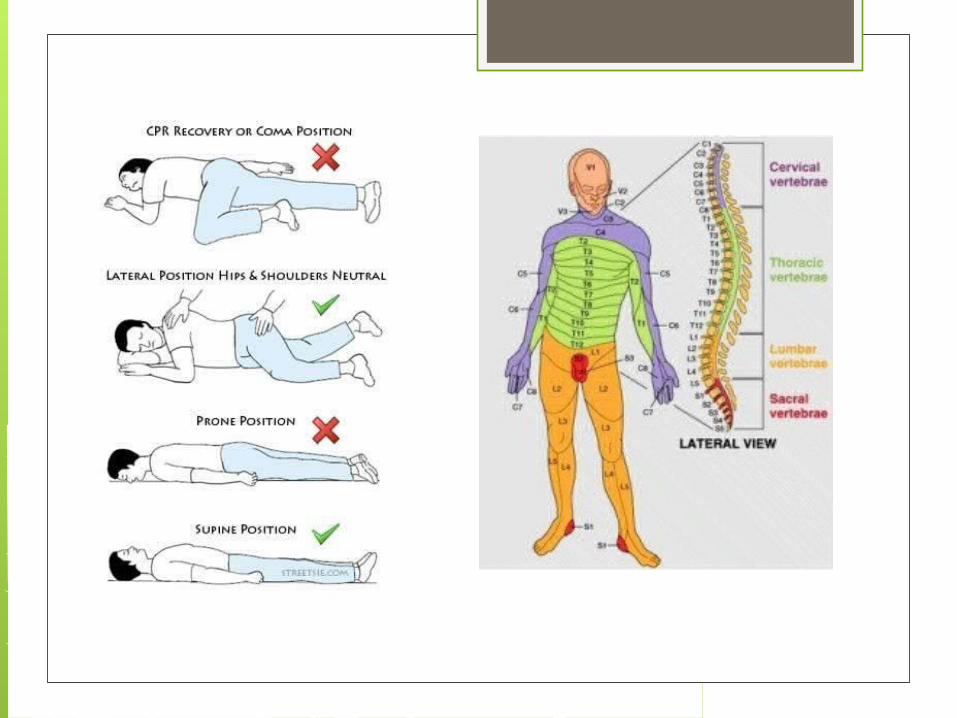
Positioning: Regardless of the level of SCI, keep the patient in proper body alignment to prevent further injury or irritability.
Devices such as traction, orthoses, or collars may be used.
Positioning Immobilize or support the affected body
part, as appropriate. Place in the designated therapeutic
position. Maintain proper body alignment. Position with head and neck in
alignment. Turn using the log roll technique Apply an orthosis collar
Positioning Continued Instruct on orthosis care as needed. Apply and maintain a splinting or bracing
device Monitor skin integrity under bracing
device Instruct on pin site care as needed Monitor traction pin insertion site, and
perform care as needed. Monitor traction pin device care
Anaphylactic Shock Anaphylaxis, is the most dramatic and
life-threatening example of a type I hypersensitivity reaction, occurs rapidly and systemically.
Anaphylaxis is not common and episodes can vary in severity.
It can be fatal!
Common causesDRUGS/FOREIGN PROTEINS Antibiotics Adrenocorticotropic hormone Insulin Vasopressin Protamine Allergen extracts Muscle relaxants Hydrocortisone Vaccines Local anesthetics Whole blood Cryoprecipitate Immune serum globulin Radiocontrast media opiates

Common Causes Continued
FOODS Shellfish Eggs Legumes, nuts Grains Berries Preservatives Bananas Peanuts
Common Causes ContinuedINSECTS/ANIMALS Hymenopetra:
bees, wasps, hornets
Fire ants Snake venom
Common Causes Continued
OTHER AGENTS Pollens Exercise Heat/cold Latex other

This has a rapid onset and a potentially fatal outcome.
Teach the patient with a history of allergic reactions to avoid allergens whenever possible, to wear a medical alert bracelet and to alert health care personnel about specific allergies.
Health PromotionPrevention is critical.
Always Be Prepared Some patient’s must carry an emergency
anaphylaxis kit (bee sting kit with injectable epinepherine)
The EpiPen is a spring-loaded injector that delivers 0.3mg of epinepherine per 2mL dose.
Some assembly is required, practice devices are available. Teach the patient how to use it and get them to demonstrate in return.
How do you know it’s really an allergy? A patient’s medical record should obtain
ALL allergens. If the patient has a known allergy be
sure to document a response.
Anaphylaxis Assessment First reaction is the patient usually reports
feelings of uneasiness, apprehension, weakness, and impending doom.
These feelings are usually quickly followed by generalized itching and urticaria (hives).
Erythema and sometimes angioedema (diffuse swelling), of the eyes, lips or tongue occur next.
Anaphylaxis Interventions Assess respiratory function FIRST! Emergency respiratory management is
critical during an anaphylactic reaction, because the severity of the reaction increases with time.
Call rapid response team Keep IV site, but switch out tubing,
obtain a second line if possible.
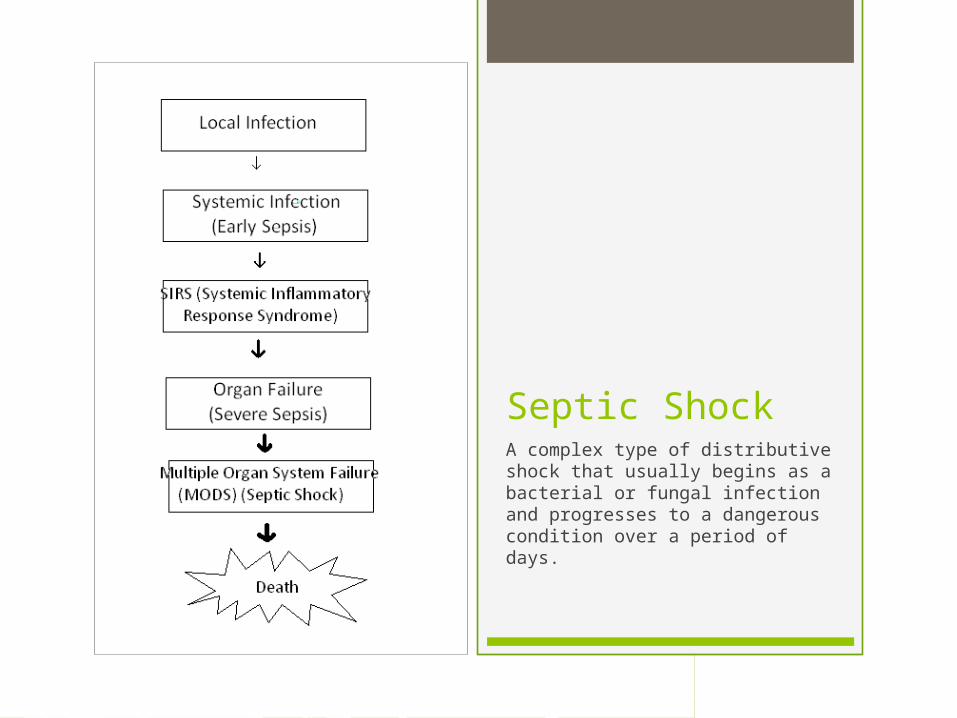
Septic ShockA complex type of distributive shock that usually begins as a bacterial or fungal infection and progresses to a dangerous condition over a period of days.

Sepsis
Is a widespread infection coupled with a more general inflammatory response, known as systemic inflammatory response syndrome (SIRS) that is triggered when an infection escapes local control.
the inflammatory response of the body becomes the enemy leading to extensive tissue and vascular changes that further impair oxygenation and tissue perfusion.

Severe Sepsis Is the progression of sepsis with an
amplified inflammatory response. All tissues are involved and have some
degree of hypoxia, although some organs are experiencing cell death and dysfunction at this time.
Microthrombi formation is widespread, using too much of the available clotting factors. This condition is known as disseminated intravascular coagulation (DIC).
Septic Shock Is the final stage of sepsis and SIRS when
multiple organ failure is evident and uncontrolled bleeding occurs.
Even with the appropriate intervention, the death rate among patients at this stage of sepsis exceeds 60%.
Severe hypovolemic shock is present. this results in an inability of the blood to clot because platelets and clotting factors were consumed earlier.

What causes it? A bacterial infection
that escapes local control.
Fungal infections in immunocompromised patients.
Gram-negative bacteria: Escherichia coli Pseudamonas
aeruginosa Klebsiella
pneumoniae Gram-positive
bacteria: Staphylococcus streptococcus

Prevention is Key Early detection of sepsis before
progression to septic shock is a major nursing responsibility.
Teach patients manifestations of local infections and of early sepsis.

Signs and symptoms Manifestations of sepsis and septic
shock occur over many hours and some change during the progression.
No single lab test confirms the presence of sepsis.
Interventions Focus on identifying the problem as
early as possible and correcting the conditions causing shock, and preventing complications.
Oxygen therapy (same as with hypovolemic)
Drug therapy (to enhance cardiac output and restore vascular volume)
Blood replacement therapy
Communication and support to family members Develop effective strategies to manage
issues related to the families of critically ill clients.
Assist the critically ill client in reducing anxiety by addressing such issues as impaired communication, sensory-perceptual problems, pain, and unfamiliar environment.
Psychosocial Integrity The indicator that the patient may be in
the beginning of severe sepsis is often a change in affect or behavior.
Compare the patient’s current behavior, verbal responses, and general affect with those assessed earlier in the day or the day before. They may just seem slightly different in their reactions.

Cultural considerations for the critically ill client What do you do if a patient refuses
blood products?

Educating the critically ill client and family Protecting patients from infection and
sepsis at home is an important nursing function.
Teach about good hygiene, hand washing, balanced diet, rest, exercise, skin care, and mouth care, how to take a temperature.