
Creation of a Pixel Pipeline and The Need for Image...
52
Creation of a Pixel Pipeline and The Need for Image Analysis To Improve Workflow and Increase Adoption of Digital Pathology for Clinical Use Andrew J. Evans
Transcript of Creation of a Pixel Pipeline and The Need for Image...

Creation of a Pixel Pipeline and The Need for Image Analysis To Improve Workflow and Increase Adoption of Digital Pathology for Clinical UseAndrew J. Evans

Image Analysis and Adoption of Digital Pathology
Disclosure of Relevant Financial Relationships
Dr. Evans declares he has no conflict(s) of interest to disclose.

Image Analysis and Adoption of Digital Pathology
Overview• WSI telepathology at University Health Network
how we started how we have expanded our use of the technology - based on whole slide imaging (WSI)enabling sub-specialty pathology
• How image analysis would benefit a department in which WSI has been used for diagnostic work since 2006.
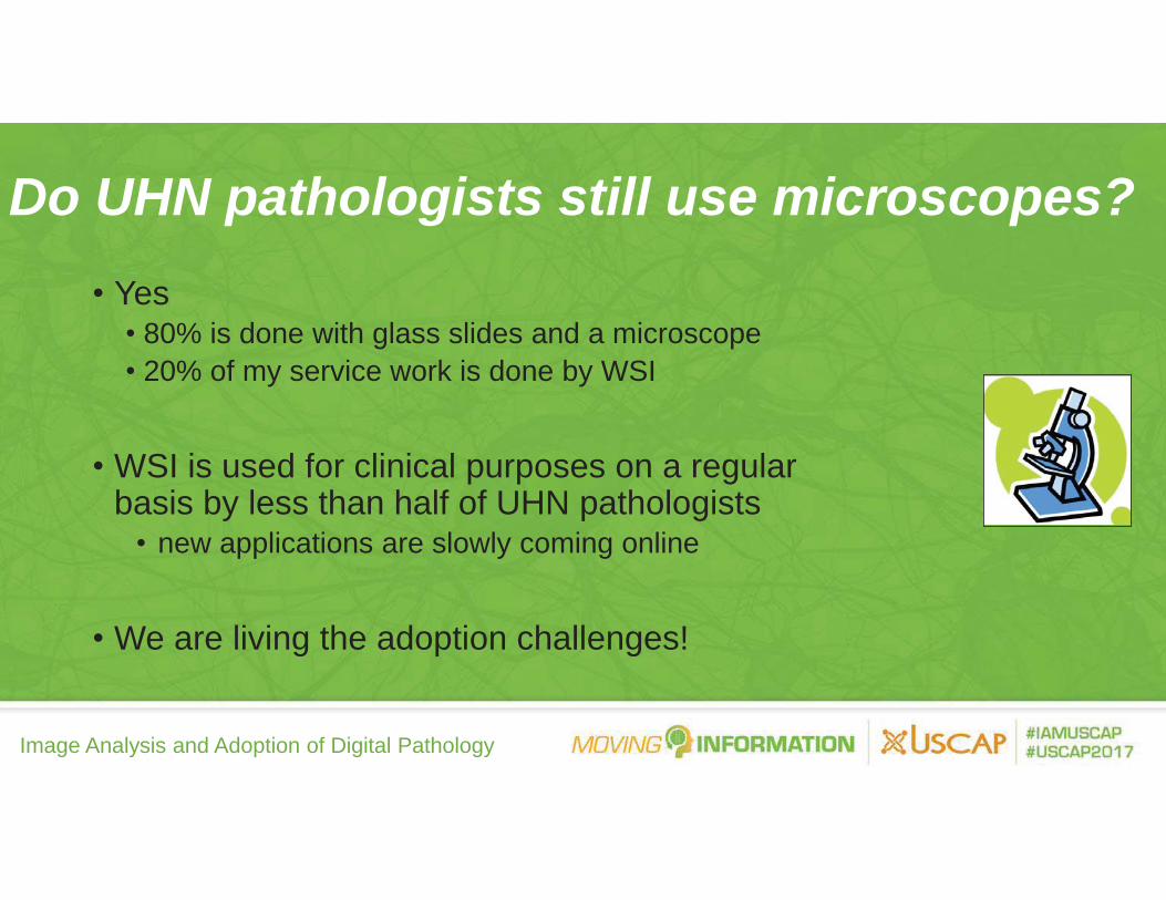
Image Analysis and Adoption of Digital Pathology
• Yes• 80% is done with glass slides and a microscope• 20% of my service work is done by WSI
• WSI is used for clinical purposes on a regular basis by less than half of UHN pathologists
• new applications are slowly coming online
• We are living the adoption challenges!
Do UHN pathologists still use microscopes?

Image Analysis and Adoption of Digital Pathology

Image Analysis and Adoption of Digital Pathology
Value Proposition of Telepathology at UHN
• Full departmental consolidation at TGH in early 2006• No regular on-site pathologist at TWH as of 2004
TWH Frozen Sections: Challenges• Single pathologist traveling from TGH to TWH
• Inefficient - traveling and waiting• Disruptive to regular workflow at TGH
• delays in regular sign-out affecting other UHN patients• No consultation on difficult cases
• potential to affect TWH surgical patients.

Image Analysis and Adoption of Digital Pathology

Image Analysis and Adoption of Digital Pathology
• Move slides?• Move pathologists?• Telepathology?
Expanding list of clinical applications at UHN:o Frozen sections (2004-present)o Consultation - local and internationalo Supporting transplant pathology programso Quality assuranceo Primary diagnosis (2012 - present)
Facilitating Multi-Site Sub-Specialty Pathology

Image Analysis and Adoption of Digital Pathology
TWH Robotic System: November 2004-October 2006
• 350 frozen sections • slow (~ 10 minutes/slide)

Image Analysis and Adoption of Digital Pathology
TWH Whole-Slide Imaging: October 2006-Present
• > 4000 frozen sections/3500 patients
• > 90% from neurosurgery
• 0-2% discrepancy rate
• 14-16 minute total turnaround time
• < 1-5% deferral rate
- 2 pathologists review all deferrals

Image Analysis and Adoption of Digital Pathology
10-12 minutes
1-3 minutes
Intra-Operative Consultations: Work Flow for Single Block Frozen Sections

Image Analysis and Adoption of Digital Pathology

Image Analysis and Adoption of Digital Pathology
Why Has This Worked at UHN?
• Started with a single clearly-defined applicationneurosurgical frozen sections
• Uncomplicated frozen section work flow• Long development period with due diligence
18 months from initial meetings to go-livetime to build confidence and trust
• Implementation team• Standard Operating Procedure (SOP)

Image Analysis and Adoption of Digital Pathology
Image Quality: The Importance of Good Histology
Poor slides = Poor image quality
20x scans – ask for 40x when necessary

Image Analysis and Adoption of Digital Pathology
• 11 episodes (0.2% of cases to date) requiring a pathologist to go to TWH
• Small pale pieces of tissue (x2)• Excess mounting media (x1)• Burned out light bulb (x1)• Calibration errors (x5)
faded H&E test slidesaging light bulb
• 11 episodes (0 2% o
Episodes of Midof
-f cases to date) requirinf casesf case
Case System g a
Failure
www.blog.al.com/spotnews/2009

Image Analysis and Adoption of Digital Pathology
System Failure: Plan B• Pathologist informs surgeon and goes to
TWH if issue not resolved in 5 minutes• A second pathologist works with the
TWH histotechnologist in case the issue is resolved.

Image Analysis and Adoption of Digital Pathology
Frozen Section Telepathology: Remote Sites
©Google Maps 2008

Image Analysis and Adoption of Digital Pathology
Timmins and District Hospital (TADH)• General community hospital• > 10,000 surgical pathology accessions/year• UHN assumed medical leadership of TADH labs in 2006• Pathologist staffing
• 1 on-site at any given time• 1 week per month – no on-site pathologist
• 150 frozen sections per year• tissue identification/intra-operative staging

Image Analysis and Adoption of Digital Pathology
Kingston General Hospital (Queen’s University)• Academic pathology department• Neuropathology frozen sections (1-5 per week)• 1 staff neuropathologist to cover all frozen sections• Need for back-up during vacation, CME leave, etc• UHN pathologists given limited consulting privileges
• remote access to EPR/diagnostic imaging• remote access to KGH LIS
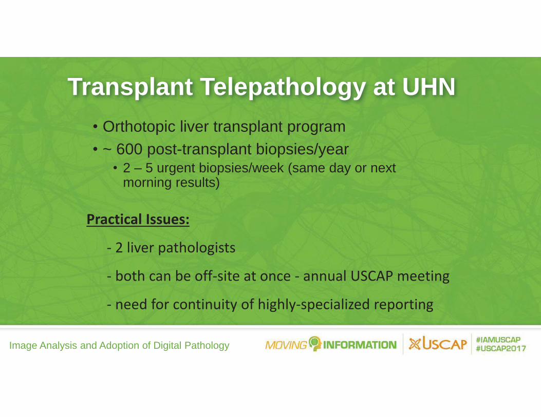
Image Analysis and Adoption of Digital Pathology
Transplant Telepathology at UHN• Orthotopic liver transplant program• ~ 600 post-transplant biopsies/year
• 2 – 5 urgent biopsies/week (same day or next morning results)
Practical Issues:
- 2 liver pathologists
- both can be off-site at once - annual USCAP meeting
- need for continuity of highly-specialized reporting

Image Analysis and Adoption of Digital Pathology
• First diagnosis made on scanned slide images (H&E, special stains/immunohistochemistry)
• Diagnostic information becomes part of the patient record
• Treatment decisions to be made based on this information
Primary Diagnosis By WSI

Image Analysis and Adoption of Digital Pathology
• American Telemedicine Association (2014)• Royal College of Pathologists in Britain (2013)• Canadian Association of Pathologists (2013)• College of American Pathologists WSI Validation (2013)• Others (Japan 2005)
Digital Pathology Guidelines

Image Analysis and Adoption of Digital Pathology
Self-Validation Studies: What is Learned?
• WSI can be used for making accurate and complete diagnoses
• What needs to be optimized in the histopathology laboratory to facilitate digital sign out
• Limitations• cases that require re-scanning• cases to scan at 40X • cases requiring deferral to glass slide review
5%
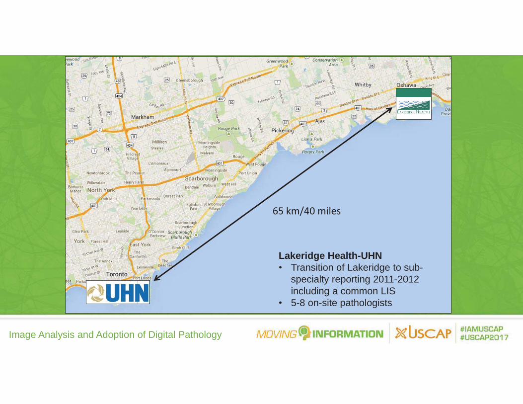
Image Analysis and Adoption of Digital Pathology
65 km/40 miles
Lakeridge Health-UHN• Transition of Lakeridge to sub-
specialty reporting 2011-2012 including a common LIS
• 5-8 on-site pathologists

Image Analysis and Adoption of Digital Pathology
Connie Willis
• regional cancer center
• 25,000 surgical cases per year
• 300-400 slides per day sent to UHN
WSI is an enabler:
1. Cost
2. Delayed TAT
3. Risks
• lost/broken slides
Primary Diagnosis Telepathology (Live as of November 2012)
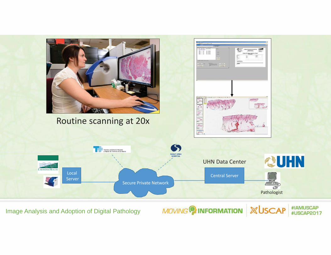
Image Analysis and Adoption of Digital Pathology
Secure Private NetworkCentral ServerLocal
Server
UHN Data Center
Pathologist
Routine scanning at 20x

Image Analysis and Adoption of Digital Pathology
Phased Implementation Strategy• Start with most experienced users
• GU, endocrine, liver, head and neck• placentas, miscellaneous orthopaedic cases
• Attempt to scan all cases for these groups • Review digital slides and sign out
• request glass slides whenever it is required to sign out a given case

Image Analysis and Adoption of Digital Pathology
2012 2013 2014
758
10,430
24,266
35,482
239 2,2114,777
6,682
0
5000
10000
15000
20000
25000
30000
35000
40000O
ctNo
vDe
cJa
nFe
bM
ar Apr
May Jun Jul
Aug
Sep
Oct
Nov
Dec
Jan
Feb
Mar Ap
rM
ay Jun Jul
Aug
Sep
Oct
Nov
Dec
Jan
Feb
Mar Ap
rM
ay Jun Jul
Aug
Sep
Oct
# slides # cases
2015
• October 2012 - present• 9,700 cases (52,000 slides) scanned for primary diagnosis
WSI: Primary Diagnosis

Image Analysis and Adoption of Digital Pathology
Spectrum of GU Cases No. Cases No. Slides2012 2013 2014 Total 2012 2013 2014 Total
Vas Deferens Vasectomy 270 320 590 626 679 1305
Penis
Circumcision 12 21 33 49 39 88Hydrocele Sac 1 1 2 1 1 2Partial penectomy 1 1 12 12Penile biopsy 4 8 12 8 15 23
Bladder
Bladder biopsy 10 11 21 23 26 49Partial cystectomy 2 2 14 14Radical cystoprostatectomy 3 3 81 81TURBT 1 153 239 393 1 448 707 1156
Ureter
Prostatic Urethra 1 1 2 2UPJ 2 2 4 3 5 8Ureter biopsy 5 9 14 5 14 19Ureterectomy 2 2 4 59 16 75
Testes
Appendix testes 1 3 4 1 3 4Bsy for fertility 6 10 16 17 15 32Hydrocele Sac 12 14 26 16 25 41Lipoma Cord 1 2 3 1 9 10Radical orchiectomy 7 13 20 68 130 198Scrotum 1 2 3 7 10 17
ProstateProstate needle biopsy 1 61 126 188 18 999 2335 3352Radical prostatectomy 8 5 13 301 37 338TURP 59 70 129 452 574 1026
Kidney
Kidney biopsy 23 35 58 38 70 108Nephroureterectomy 2 10 12 33 199 232Partial nephrectomy 5 15 20 57 169 226Radical nephrectomy 16 19 35 218 264 482
Hernia Sac 36 53 89 59 71 130Miscellaneous 1 22 45 68 19 46 171 236Grand Total 3 725 1036 1764 38 3632 5596 9266

Image Analysis and Adoption of Digital Pathology
• Difficult or unusual cases especially where there is a high likelihood a case will be sent out for glass slide review by another pathologist.
• “It’s slower than glass and I’m too busy” - if the pathologist has a large volume of cases to report
• Performing diagnostic activities that are currently difficult or cannot be performed using WSI:
counting mitoses per high power field basisidentifying micro calcifications on breast biopsies by polarized light microscopy
• Suboptimal image quality in an area of potential diagnostic importance - a minor reason for deferral
Deferral to Glass Slides: 5-10% of Cases

Image Analysis and Adoption of Digital Pathology
47 year-old male, 6 cm right inguinal mass, “lipoma”
Example of a Case Deferred to Glass

Image Analysis and Adoption of Digital Pathology
Worrisome cellularity with mitoses (some atypical)Spindle cell lipoma/mammary-type myofibroblastomaDe-differentiated liposarcomaRequired IHC and molecular work-up (MDM2 FISH) not available at Lakeridge Health Oshawa

Image Analysis and Adoption of Digital Pathology
Growing Pains So Far
1. IT infrastructure/bandwidth2. Viewer stability and reduced viewing speeds3. Scanner issues 4. WSI-LIS interface and barcode issues
o all cases are primarily accessed via the LIS o no flexibility for organizing electronic worklists
5. Mixed glass slide-WSI workflow

Image Analysis and Adoption of Digital Pathology
Hybrid Glass Slide - WSI Workflow
• Culture change – learning to use the Pathologist’s Console
• need to sort WSI from glass cases within the LIS• Special stains
• when are they ready for review?• glass slides sent when pathologist is looking for
scanned slides

Image Analysis and Adoption of Digital Pathology
Summary of Clinical Use of WSI at UHN• We have used computer screens the same way we use
microscopes.visual interpretation of H&E morphologyvisual interpretation of immunohistochemsitry
• Our WSI system works for those who use it - improvements on several fronts are needed to increase adoption
• Enter the need for a “pixel pipeline” with image analysis!

Image Analysis and Adoption of Digital Pathology
“Image Analysis” To Date
Measurement Tools:Linear extentDistance from marginsDepth of invasionSurface area
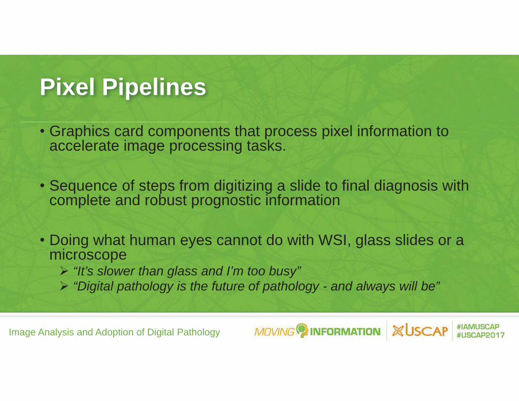
Image Analysis and Adoption of Digital Pathology
Pixel Pipelines• Graphics card components that process pixel information to
accelerate image processing tasks.
• Sequence of steps from digitizing a slide to final diagnosis with complete and robust prognostic information
• Doing what human eyes cannot do with WSI, glass slides or a microscope
“It’s slower than glass and I’m too busy”“Digital pathology is the future of pathology - and always will be”

Image Analysis and Adoption of Digital Pathology
Image Analysis: Software Tools To Help Pathologists (Not Replace Us)
• Intended goals:allow the pathologist to act as the final interpretero not field selection technologist
more robust biomarker quantitationo oncologists and patients like reports with hard numbers
• Use cases we have discussed at UHN
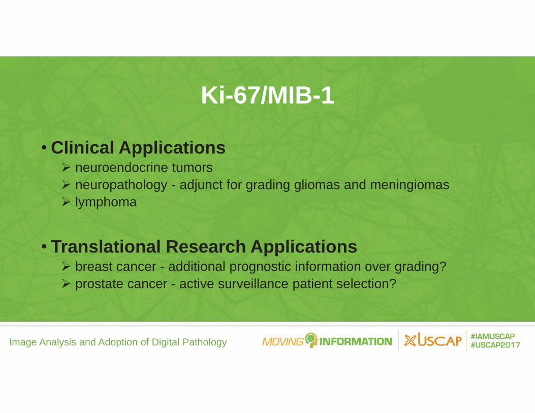
Image Analysis and Adoption of Digital Pathology
Ki-67/MIB-1
• Clinical Applicationsneuroendocrine tumorsneuropathology - adjunct for grading gliomas and meningiomaslymphoma
• Translational Research Applicationsbreast cancer - additional prognostic information over grading?prostate cancer - active surveillance patient selection?

Image Analysis and Adoption of Digital Pathology
Ki-67 Labeling Index• Visual inspection/
hot-spot detectiontime consuming and error prone

Image Analysis and Adoption of Digital Pathology
Mitotic Figure Counting: H&E
Potential confounders for visual counting :
thick sectionsover-stainingapoptotic bodies
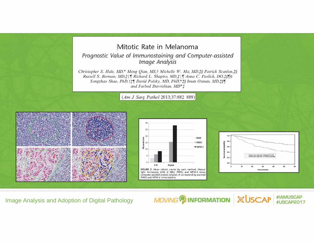
Image Analysis and Adoption of Digital Pathology

Image Analysis and Adoption of Digital Pathology
40 year-old female - needle biopsy of incidentally-found left renal mass
Immunohistochemistry panel: CD10, CD117, CK 7, AMACR, CAIX

Image Analysis and Adoption of Digital Pathology
Multiplex ImmunofluorescenceBiopsy with limited lesional tissueRequires an immunohistochemistry panel (5-10 stains)Lesional tissue is exhausted from the block Can the panel be run on one single paraffin section?
o hyperplexing (60 or more markers)

Image Analysis and Adoption of Digital Pathology
Decision support tools for Gleason
grading in prostate cancer

Image Analysis and Adoption of Digital Pathology
Slide Review36.0%
Other16.0%
Reporting34.6%
Organizing Cases 24.1% (0:10:25)Querying for Cases 18.5% (0:07:59)Waiting for Delivery 11.2% (0:04:49)Searching for Cases 9.4% (0:04:04)Transporting Cases 9.2% (0:03:58)Other 17.0% (0:07:21)
Workflow Opportunities
100% (0:43:09)
13.4%
Pathologist Time & Motion Study:Glass Slide Review (Stratman et al)

Image Analysis and Adoption of Digital Pathology
Concept of “pCAD”Automated, systematic slide review, pre-annotated slidesConstruct a report as slides are reviewedReduce the time spent on non-diagnostic work
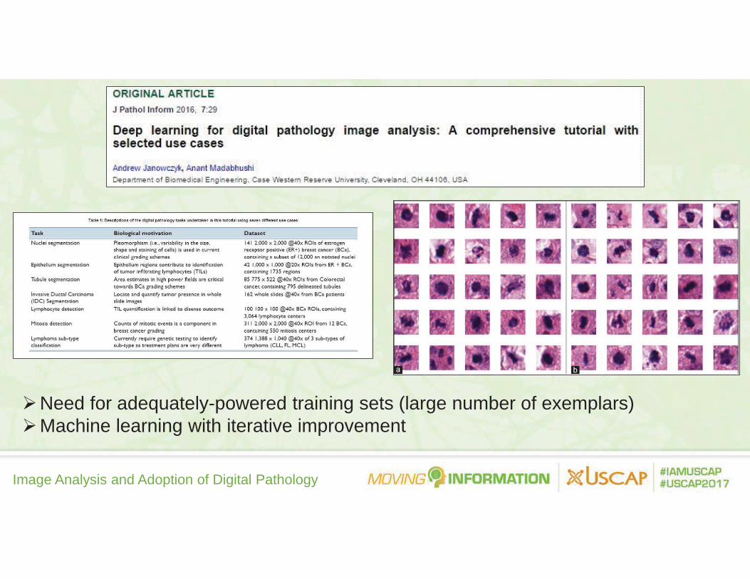
Image Analysis and Adoption of Digital Pathology
Need for adequately-powered training sets (large number of exemplars)Machine learning with iterative improvement

Image Analysis and Adoption of Digital Pathology
How To Make The Pixel Pipeline a Reality
• Do it yourself (?)
• Partner with companies who focus on:streamlining workflow in digital pathology - complete digitizationdependable IT support - continuous system monitoringmanaging large volumes of digital datadevelopment of clinically relevant algorithmscreative business models

Image Analysis and Adoption of Digital Pathology
Summary• WSI telepathology at University Health Network
enabling sub-specialty pathology how we started how we have expanded our use of the
technology
• How image analysis would help pathologists who have been using WSI for diagnostic work since 2006.

Image Analysis and Adoption of Digital Pathology
Acknowledgements• Pathologists
Dr. Sylvia Asa - vision of bringing telepathology to UHNColleagues who worked to move digital pathology forward
• UHN Histotechnologists, Lab Management, LIS and IT Support
Michele HenryPeter Woo Dr. Zoya VolynskayaCelcilia Lagmay-Traya
• Lakeridge Health Oshawa StaffAlan Wolff

Image Analysis and Adoption of Digital Pathology