Cortical areas of brain
43
DR PRATIK MISTRY
-
Upload
drpratik-mistry -
Category
Health & Medicine
-
view
106 -
download
0
Transcript of Cortical areas of brain
DR PRATIK MISTRY

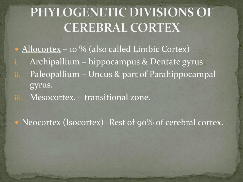
Allocortex – 10 % (also called Limbic Cortex)
i. Archipallium – hippocampus & Dentate gyrus.
ii. Paleopallium – Uncus & part of Parahippocampal gyrus.
iii. Mesocortex. – transitional zone.
Neocortex (Isocortex) -Rest of 90% of cerebral cortex.
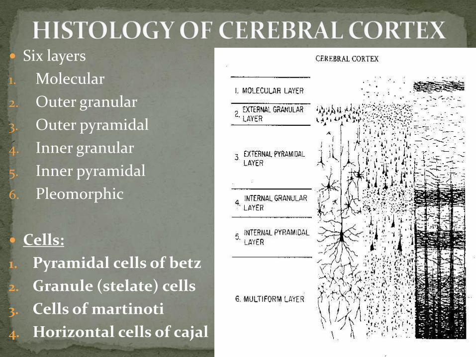
Six layers
1. Molecular
2. Outer granular
3. Outer pyramidal
4. Inner granular
5. Inner pyramidal
6. Pleomorphic
Cells:
1. Pyramidal cells of betz
2. Granule (stelate) cells
3. Cells of martinoti
4. Horizontal cells of cajal
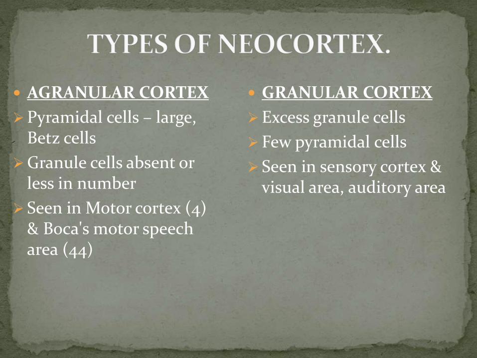
AGRANULAR CORTEX
Pyramidal cells – large, Betz cells
Granule cells absent or less in number
Seen in Motor cortex (4) & Boca's motor speech area (44)
GRANULAR CORTEX
Excess granule cells
Few pyramidal cells
Seen in sensory cortex & visual area, auditory area

FRONTAL CORTEX
Small and medium pyramidal cells
Few stellate cells
Pre-frontal cortex of frontal lobe
PARIETAL CORTEX
More stellate cells
Seen in most of parietal lobe & junction of parietal, temporal & occipital lobes
POLAR CORTEX
Thinnest of all
All layers reduced depth.
Seen in frontal & occipital pole
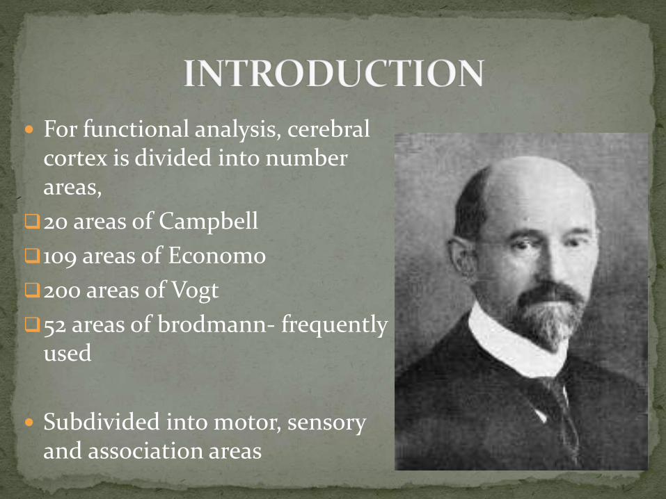
For functional analysis, cerebral cortex is divided into number areas,
20 areas of Campbell
109 areas of Economo
200 areas of Vogt
52 areas of brodmann- frequently used
Subdivided into motor, sensory and association areas

Subdivided into
Primary motor area (area 4)
Pre-motor area (area 6)
Frontal eye field (area 8)
Supplementary motor area
Pre-frontal area (areas 9 to 12)

Location:
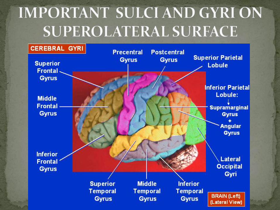
Precentral gyrus (area 4)
Extends to the ant. part of paracentral lobule
Agranular cortex
Afferents :
Premotor area (Area 6)
Somesthetic or somatosensory cortex
Ant. Part ventral nucleus of thalamus(which receives info. from cerebellum
Basal ganglia
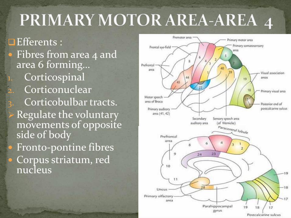
Efferents : Fibres from area 4 and
area 6 forming… 1. Corticospinal 2. Corticonuclear3. Corticobulbar tracts. Regulate the voluntary
movements of opposite side of body
Fronto-pontine fibres Corpus striatum, red
nucleus

Control movements of voluntary muscles of opposite side
Movements represented with head end below and leg end up (INVERTED MOTOR HOMUNCULUS)
Centres from below are: lips, tongue, larynx, pharynx, face, head & neck, upper limb with large area for fingers and hand, trunk, lower limb above knee.
Ant. Part of paracentral lobule
Extent of area depend on skill of movement and not on the bulk of muscle
Somewhat sensory. Receive some sensations like tingling and numbness
Known as MSI
Muscles of forehead, tongue, mastication, larynx, pharynx, extra ocular bilaterally represented
Only movements not muscles
LESION: initially flaccid paralysis
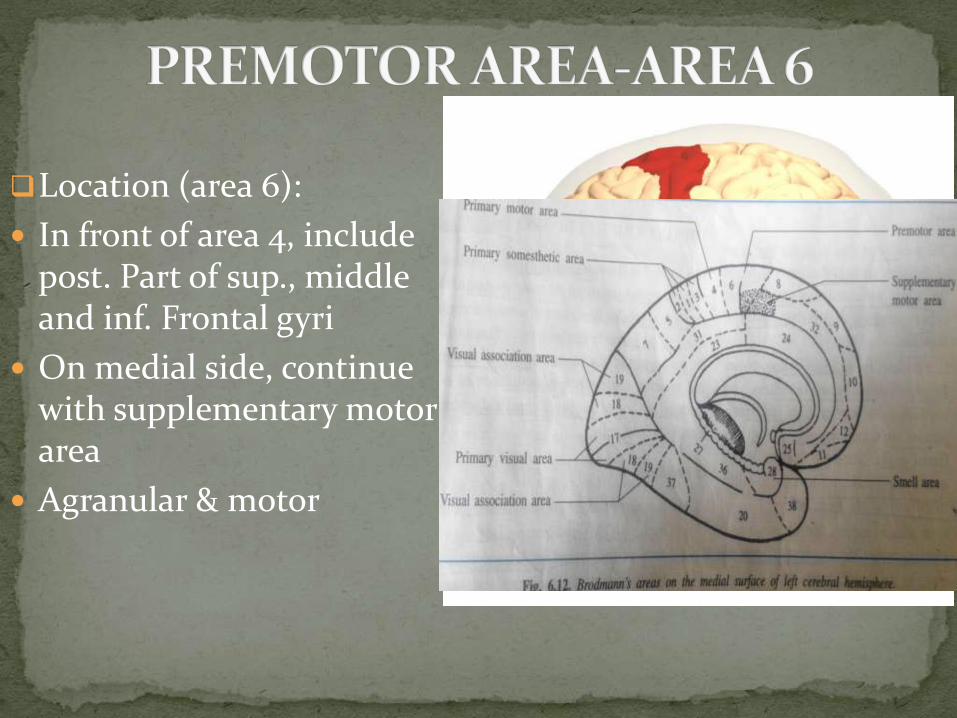
Location (area 6):
In front of area 4, include post. Part of sup., middle and inf. Frontal gyri
On medial side, continue with supplementary motor area
Agranular & motor
Integrates voluntary movements to perform skilful act.
Writing centre
Concerned with programming which is executed by area 4
LESION:
Produce difficulty in the performance of skilled movements.
Apraxia: loss of the ability to do simple or routine acts in the absence of paralysis.
Agraphia: when writing is also involved.
PRIMARY SOMATOMOTOR AREA (MSI)
=
PRIMARY MOTOR AREA
PREMOTOR AREA
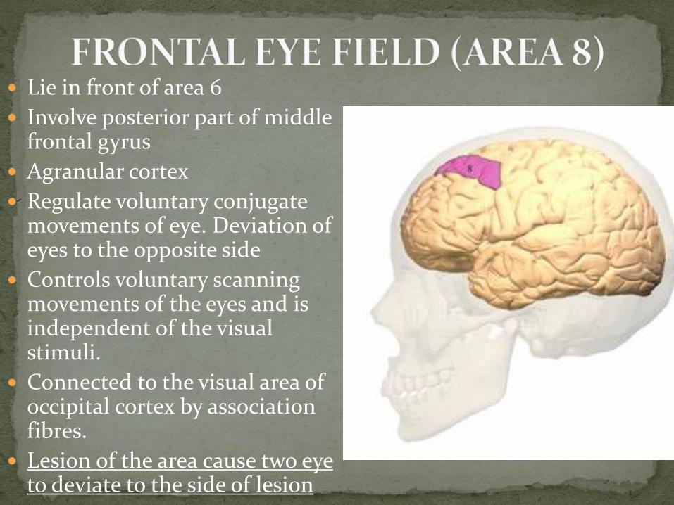
Lie in front of area 6
Involve posterior part of middle frontal gyrus
Agranular cortex
Regulate voluntary conjugate movements of eye. Deviation of eyes to the opposite side
Controls voluntary scanning movements of the eyes and is independent of the visual stimuli.
Connected to the visual area of occipital cortex by association fibres.
Lesion of the area cause two eye to deviate to the side of lesion

Located on medial surface of cerebrum in the post. part of medial frontal gyrus anterior to the paracentral lobule
Afferents from VA and VL of thalamus
Efferents to area 4
Function is to control complex movements. Produce sensation of “URGE TO MOVE’
Receive some senses (MSII)
Lesion of area produce AKINESIA

Rest of frontal lobe ant. to pre-motor area which include orbital surface also
Fibres from thalamus, hypothalamus, limbic system, all areas of cortex
Concerned with individual’s personality
Regulate depth of feeling, thinking, mature judgement, orientation, concentration, pleasure and displeasure, right or wrong.
Bilateral damage due to trauma or tumour: change in personality, loss of concentration, judgement, inappropriate social behaviour like vulgarity of speech, improper clothing

Primary Secondary Sensory Association
Somesthetic (sensory) Visual Auditory
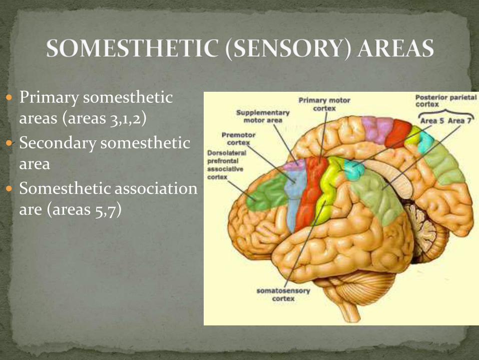
Primary somesthetic areas (areas 3,1,2)
Secondary somesthetic area
Somesthetic association are (areas 5,7)

Located in the post-central gyrus and extends into the posterior part of the paracentral lobule on the medial surface.
Granular cortex
Afferents from VPL and VPM of thalamus and other areas of cortex
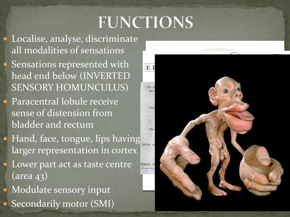
Localise, analyse, discriminate all modalities of sensations
Sensations represented with head end below (INVERTED SENSORY HOMUNCULUS)
Paracentral lobule receive sense of distension from bladder and rectum
Hand, face, tongue, lips having larger representation in cortex
Lower part act as taste centre (area 43)
Modulate sensory input
Secondarily motor (SMI)

Located on the posterior part of posterior ramus of lateral sulcus
Involve lower part of pre and post-central gyri
Receive mainly pain sensation
Somewhat motor in function (SMII)
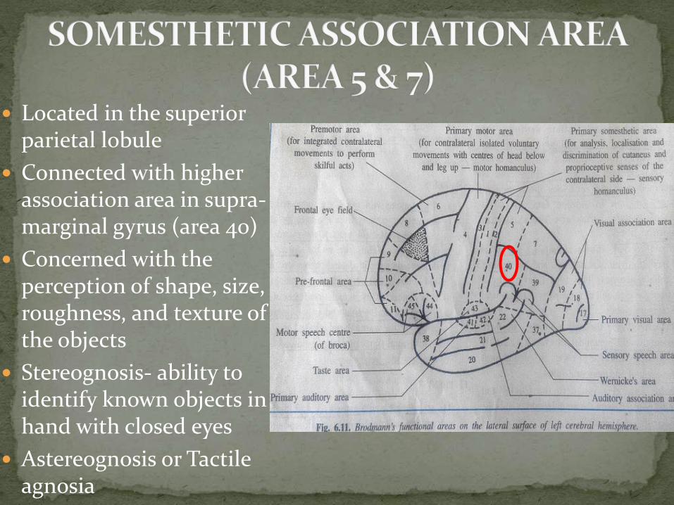
Located in the superior parietal lobule
Connected with higher association area in supra-marginal gyrus (area 40)
Concerned with the perception of shape, size, roughness, and texture of the objects
Stereognosis- ability to identify known objects in hand with closed eyes
Astereognosis or Tactile agnosia
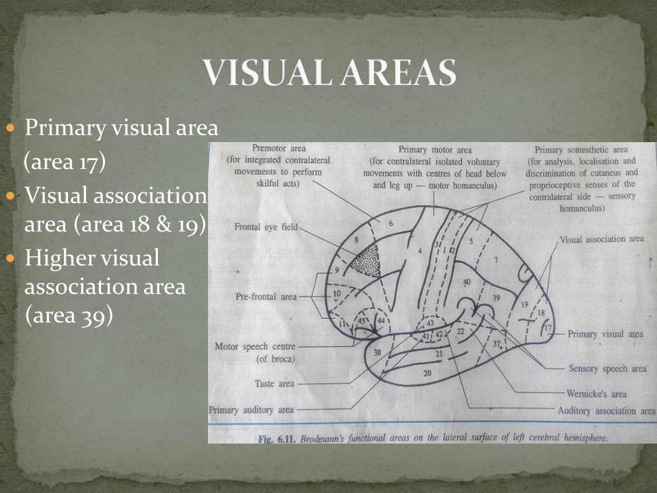
Primary visual area
(area 17)
Visual association area (area 18 & 19)
Higher visual association area (area 39)
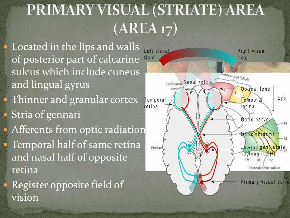
Located in the lips and walls of posterior part of calcarine sulcus which include cuneus and lingual gyrus
Thinner and granular cortex
Stria of gennari
Afferents from optic radiation
Temporal half of same retina and nasal half of opposite retina
Register opposite field of vision
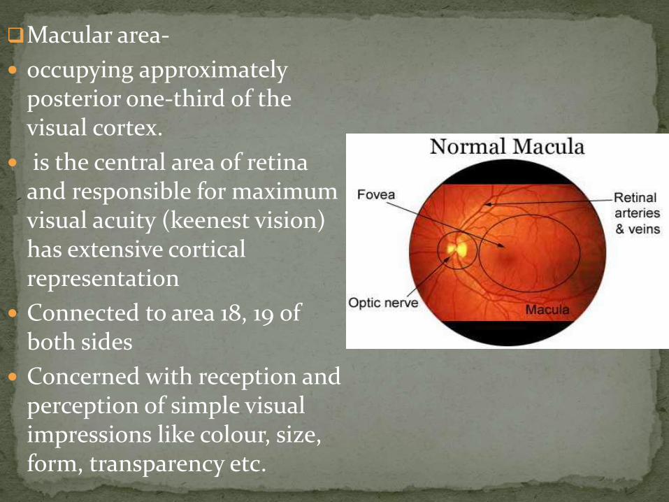
Macular area-
occupying approximately posterior one-third of the visual cortex.
is the central area of retina and responsible for maximum visual acuity (keenest vision) has extensive cortical representation
Connected to area 18, 19 of both sides
Concerned with reception and perception of simple visual impressions like colour, size, form, transparency etc.
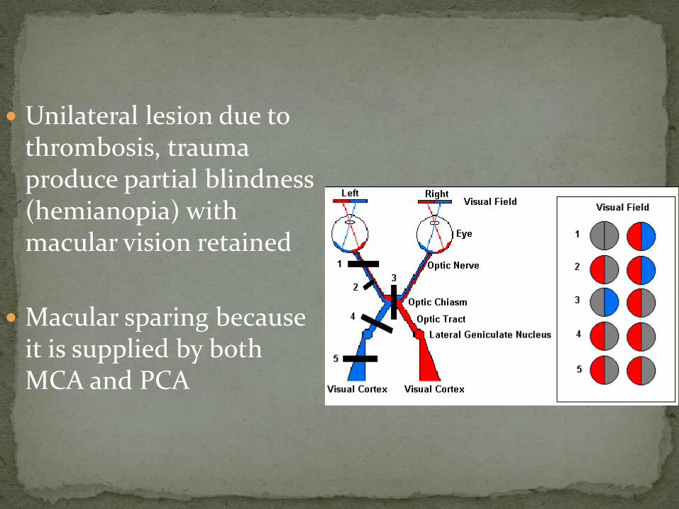
Unilateral lesion due to thrombosis, trauma produce partial blindness (hemianopia) with macular vision retained
Macular sparing because it is supplied by both MCA and PCA
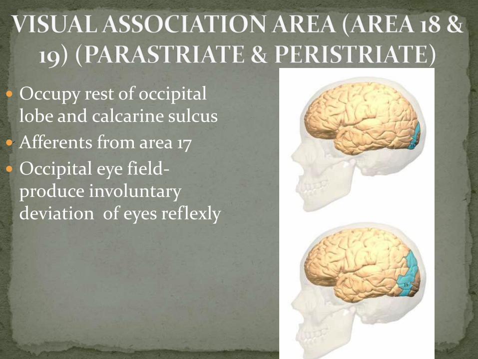
Occupy rest of occipital lobe and calcarine sulcus
Afferents from area 17
Occipital eye field-produce involuntary deviation of eyes reflexly
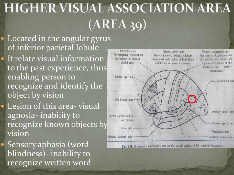
Located in the angular gyrus of inferior parietal lobule
It relate visual information to the past experience, thus enabling person to recognize and identify the object by vision
Lesion of this area- visual agnosia- inability to recognize known objects by vision
Sensory aphasia (word blindness)- inability to recognize written word
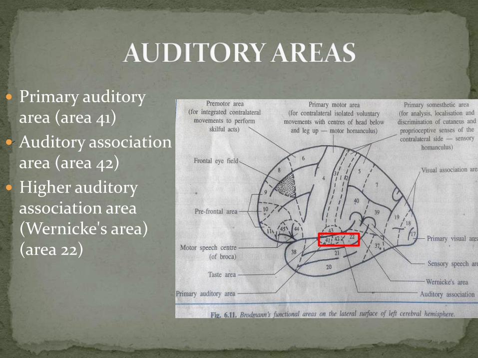
Primary auditory area (area 41)
Auditory association area (area 42)
Higher auditory association area (Wernicke's area) (area 22)
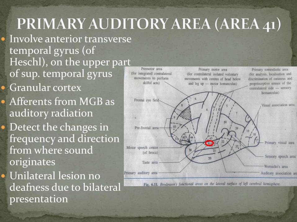
Involve anterior transverse temporal gyrus (of Heschl), on the upper part of sup. temporal gyrus
Granular cortex
Afferents from MGB as auditory radiation
Detect the changes in frequency and direction from where sound originates
Unilateral lesion no deafness due to bilateral presentation

Lie behind area 41
Involve posterior transverse temporal gyrus of superior temporal gyrus
Granular cortex
Same function
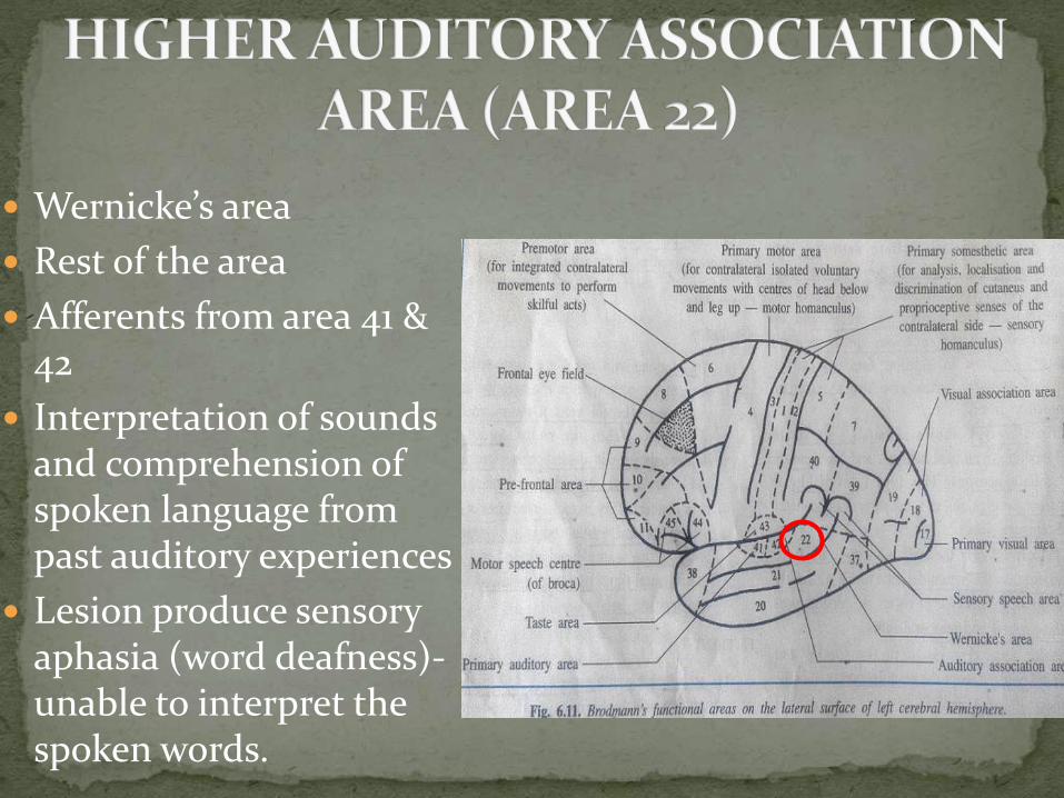
Wernicke’s area
Rest of the area
Afferents from area 41 & 42
Interpretation of sounds and comprehension of spoken language from past auditory experiences
Lesion produce sensory aphasia (word deafness)-unable to interpret the spoken words.
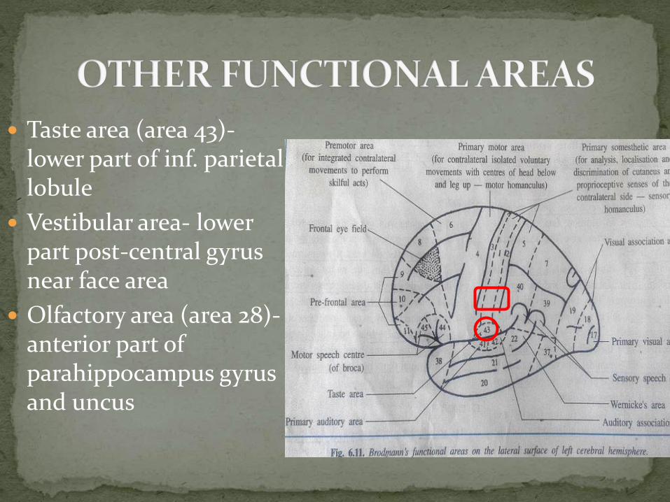
Taste area (area 43)-lower part of inf. parietal lobule
Vestibular area- lower part post-central gyrus near face area
Olfactory area (area 28)-anterior part of parahippocampus gyrus and uncus

Speech- highly complex function
Speech function performed by dominant hemisphere
In 90%, left one, DOMINANT (TALKING BRAIN) & right one, NON DOMINANT (MUTE BRAIN)
FOUR SPEECH CENTRES: 3 sensory & 1 motor
Sensory speech areas:
1. Area 22 (Wernicke's area)
2. Area 39
3. Area 40
Broca’s motor speech area (area 44 & 45)

Area 22 (Wernicke’s area)
Interpret spoken language & recognize familiar words
Congenital deaf child-dumb
Area 39 of angular gyrus-store visual images and recognize them by sight
Area 40 of supramarginal gyrus- recognize familiar objects by touch and proprioception
All these 3 areas receive input from hearing, vision, touch and process it in the area 22 &
Then project it to Broca’s area through ARCUATE FASCICULUS
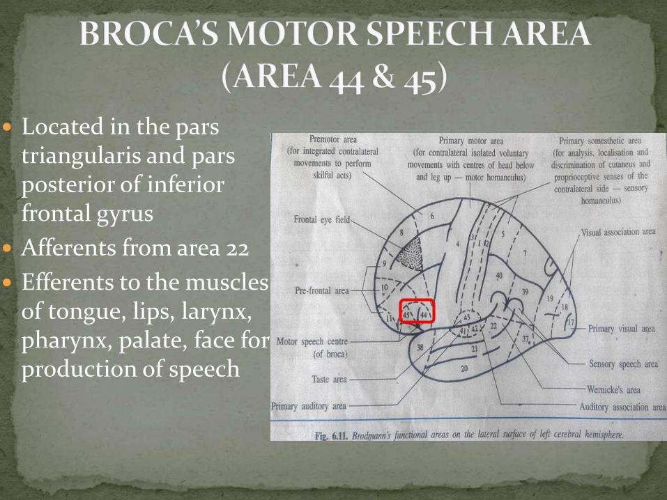
Located in the pars triangularis and pars posterior of inferior frontal gyrus
Afferents from area 22
Efferents to the muscles of tongue, lips, larynx, pharynx, palate, face for production of speech
Area 22- word deafness- unable to interpret spoken words. Speak fluently with incorrect and useless words
Area 39- word blindness- inability to recognize written words even written by self
Alexia, Agraphia
Area 40- Astereognosis
Area 44 & 45- motor aphasia- cannot speak properly although he understand everything. Slow speech with many grammatical mistakes
Conduction aphasia- arcuate fasciculus damage