![[XLS] Qtr 2011 Seniority Tie Breaker List.xls · Web viewGEORGE STUDGEON GERALD SHAW GLORIA SCOTT GLENN SELLARS GARY SADDLER GREGORY SAMPSON GABRIEL SARINANA GEORGE SCHEFFERINE GLENDA](https://static.fdocuments.us/doc/165x107/5ad472807f8b9a1a028be0db/xls-qtr-2011-seniority-tie-breaker-listxlsweb-viewgeorge-studgeon-gerald-shaw.jpg)
Copyright by Gregory John Gabriel 2004
214
Copyright by Gregory John Gabriel 2004
Transcript of Copyright by Gregory John Gabriel 2004

Copyright
by
Gregory John Gabriel
2004

The Dissertation Committee for Gregory John Gabriel certifies that this is
the approved version of the following dissertation:
Exploiting Aromatic Donor-Acceptor Recognition in the Folding and Binding of Naphthyl Oligomers
Committee:
Brent L. Iverson, Supervisor
Eric V. Anslyn
Michael J. Krische
Paul F. Barbara
Venkat Ganesan

Exploiting Aromatic Donor-Acceptor Recognition in the Folding and Binding of Naphthyl Oligomers
by
Gregory John Gabriel, B.S.
Dissertation
Presented to the Faculty of the Graduate School of
The University of Texas at Austin
in Partial Fulfillment
of the Requirements
for the Degree of
Doctor of Philosophy
The University of Texas at Austin
August 2004

Dedication
To my family

Acknowledgments
I am indebted to my advisor, Professor Brent Iverson. His example was a
constant motivation for me to conduct research meticulously and to improve as a
teacher. I can recall many situations when his fairness, creativity, tact, business
sense, etc, have shown me that opportunities to advance our ideas and science can
be extracted from even the most frustrating of situations. I leave the Iverson group
with this belief a permanent part of me.
I thank my defense committee members Professors Eric Anslyn, Michael
Krische, Paul Barbara, and Venkat Ganesan for providing a refreshing new point
of view of my work. I thank the wonderful people at NYU including Professor
Jim Canary, Yu-Hung Chiu, and Lei Zhu. I deeply appreciate all the friendly
people at UT and the talented Iverson lab members. “This crew is good.”
I want to thank several people in particular. Andy Zych and Mark Olsen for
early lessons in constructive skepticism and for their continued friendship. Steve
Sorey for his abundant patience. Yeonsuk Roh for our coffee breaks and six
months of phone calls to get my behind moving. Joe Reczek for timely research
suggestions and getting the aforementioned behind out of trouble (My apologies.
You guys really weren’t hiding my final). Karl Griswold for his impressions
(Prof. Mickey Mouse and Det. Whatman) and providing a nurturing environment
for ridiculous conversations and uncomfortable situations. Jeeyeon Lee for being
a great labmate and classmate. She supported my ideas at group meeting,
challenged me in lab, gladly fed me humble pie when I deserved it, and always
gave me a thoughtful, honest opinion. I was very fortunate.
I thank my brothers whose talks of eschermers, coelenterates, perpetual
motion, etc, have inspired their older brother. Most of all, I thank my parents for
their love and advice in all matters truly important to me.
v

Don't worry about people stealing your ideas.
If your ideas are any good, you'll have to ram them down people's throats.
Howard Aiken
vi

Exploiting Aromatic Donor-Acceptor Recognition in the Folding and Binding of Naphthyl Oligomers
Publication No._____________
Gregory John Gabriel, Ph.D.
The University of Texas at Austin, 2004
Supervisor: Brent L. Iverson
Biomolecules, for example, DNA and enzymes, perform nearly all the
chemical processes essential for life. Their functions are dependent though on
their ability to fold and bind into precise three-dimensional conformations and
assemblies. A variety of oligomers that adopt compact conformations in solution,
termed foldamers, have been synthesized to elucidate strategies to control folding
and binding akin to biomolecules.
The Iverson group has been developing a class of foldamers, called
aedamers, which employ the aromatic-aromatic complexation between electron-
rich 1,8-dialkoxy-naphthalene (Dan) and electron-deficient 1,4,5,8-naphthalene-
vii

tetracarboxylic diimide (Ndi) “building blocks”. It is expected that further work
with these naphthyl oligomers will help establish aromatic interactions as a
reliable tool for the construction of water-stable assemblies with tunable and
predictable properties not found in nature.
Overall, this dissertation describes the group’s first attempts to test the
structural “designability” of naphthyl oligomers of previously unexplored
sequences. Bottomline is that these studies have utilized the Dan:Ndi interaction
to dictate intra- and inter- molecular associations to afford distinct folding
topologies and achieve selective binding, respectively.
Chapter 2 reports the observation that a previously studied amphiphilic
aedamer happens to be an effective refolding inhibitor of RNase thus introducing
the prospect of aedamer-protein interactions, a long-standing aim for these
molecules. Chapter 3 presents the “shuffling” of the aedamer sequence (DanNdi)n
to afford naphthyl oligomers, of the form Dann+1Ndin, that adopt turn structures.
The results here demonstrate the ability of foldamers to access various secondary
structures through changes to their primary sequence analogous to proteins.
Chapter 4 details the first hetero-duplex system to operate via aromatic
interactions in aqueous solutions. Dann and Ndin complementary strands exhibit
high binding affinities and chain discrimination. The ability of the Dan:Ndi
association to direct binding is expected to be extensively used by the laboratory
to create discrete assemblies.
As a whole, these projects probe the folding and binding of naphthyl
oligomers in a variety of situations to demonstrate the wide reach of directed
aromatic interactions to create various architectures. With this level of control
established, surface patterning for microarrays, functional artificial proteins,
biomolecule-aedamer ensembles, and other application-driven pursuits using
naphthyl oligomers are possible in the near future.
viii

Table of Contents
Acknowledgments .................................................................................................. v
Abstract ................................................................................................................vii
List of Figures ......................................................................................................xii
List of Tables ......................................................................................................xxii
Chapter 1
From Aedamers to Aromatic Interactions to Naphthyl Oligomers ................. 1 1.1 Foldamers as Models of Higher-Order Structure .................................... 1
1.1.1 β-Peptides .................................................................................... 7
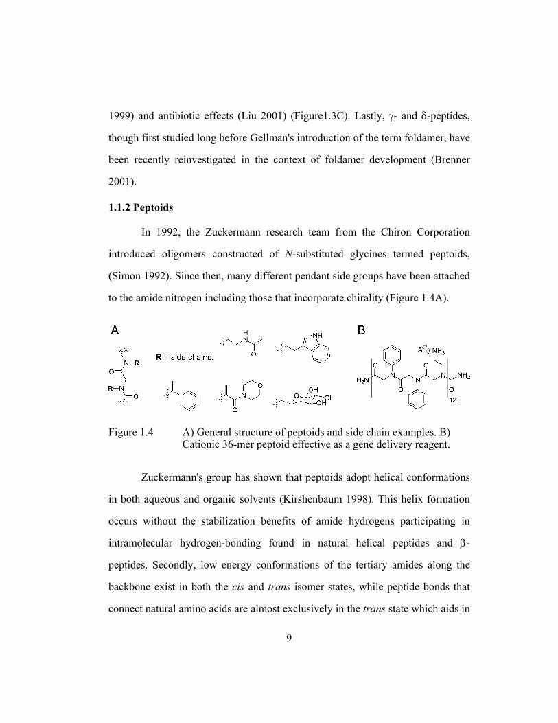
1.1.2 Peptoids ....................................................................................... 9
1.1.3 Oligo(meta-Phenylene Ethynylene)s ......................................... 10
1.1.4 Aedamers ................................................................................... 11
1.2 Update: Foldamers with Aedamer-Like Designs .................................. 15
1.3 Closer Examination of Aedamer Aromatic Interactions ....................... 17
1.3.1 Forces that Dictate Folding ....................................................... 17
1.3.2 Detailed Picture of Folding ....................................................... 22
1.4 Overview of Naphthyl Oligomer Projects ............................................. 26
Chapter 2
Intriguing Heat-Triggered Behavior of an Amphiphilic Aedamer ............... 33
2.1 Chapter Summary .................................................................................. 33
2.2 Background: A Foldamer that Forms Stable Gels in Water .................. 35
2.3 Results and Discussion .......................................................................... 40
2.3.1 Synthesis of Aedamer Derivatives ............................................ 40
2.3.2 UV-Vis, NMR, and LS Comparisons at 25o C .......................... 42
2.3.3 Evaluation of UV-Vis, NMR, and LS Data .............................. 48
ix
2.3.4 Concentration Dependent Gelling Studies ................................ 49
2.3.5 Discussion of Thermal Gelling Data ......................................... 51
2.3.6 Exploring Aedamer-Protein Interactions .................................. 51
2.3.7 Fluorometric Assay of RNase Activity ..................................... 55
2.3.8 Discussion of Refolding Inhibition Data ................................... 59
2.4 Chapter Conclusions ............................................................................. 61
2.5 Ideas for Future Investigations .............................................................. 62
2.6 Experimental Section ............................................................................ 64
Chapter 3
Altering the Folding Patterns of Naphthyl Oligomers .................................... 73
3.1 Chapter Summary .................................................................................. 73
3.2 Background: Mimics of Biological Hairpin Structures ......................... 75
3.3 Results and Discussion .......................................................................... 79
3.3.1 Synthesis of Non-Alternating Naphthyl Trimers ...................... 79
3.3.2 UV-Vis Spectroscopy ................................................................ 82
3.3.3 Evaluation of UV-Vis Data ....................................................... 83
3.3.4 Unfolding Studies by UV Spectroscopy ................................... 90
3.3.5 NMR Techniques for Proton Assignment ................................. 92
3.3.6 NOESY Spectroscopy ............................................................... 95
3.3.7 Computer Modeling .................................................................. 98
3.3.8 Evaluation of NMR Data and Computer Modeling ................ 101
3.3.9 Preliminary Studies of a Potential Hairpin Pentamer .............. 105
3.4 Chapter Conclusions ........................................................................... 107
3.5 Ideas for Future Investigations ............................................................ 107
3.6 Experimental Section .......................................................................... 109
x

Chapter 4
Naphthyl Oligomers that Form Hetero-Duplexes ......................................... 122 4.1 Chapter Summary ................................................................................ 122
4.2 Background: Self-Assembly of Molecular Strands ............................. 124
4.3 Results and Discussion ........................................................................ 131
4.3.1 Synthesis of Homo-Naphthyl Oligomers ................................ 131
4.3.2 NMR Job Plots and Titrations ................................................. 132
4.3.3 Evaluation of One-to-One Binding ......................................... 134
4.3.4 Isothermal Titration Calorimetry (ITC) .................................. 135
4.3.5 Summary of Binding Data ....................................................... 141
4.3.6 Examination of Heat Capacity ................................................ 144
4.3.7 Size Exclusion Chromatography (SEC) .................................. 147
4.3.8 Polyacrylamide Gel Electrophoresis (PAGE) ......................... 150
4.3.9 Discussion of SEC and PAGE Results .................................... 152
4.4 Chapter Conclusions ........................................................................... 154
4.5 Ideas for Future Investigations ............................................................ 154
4.6 Experimental Section .......................................................................... 156
Chapter 5
Final (Personal) Remarks ................................................................................ 176
References .......................................................................................................... 178
Vita ................................................................................................................... 192
xi

List of Figures
Chapter 1 Figure 1.1 A ribbon representation of a hemoglobin complex and a diagram
of the unwinding of a chromosome (adapted from http://www.psc.edu/MetaCenter/MetaScience/Articles/Ho/Ho-hemoglobin.html, Copyright 1993 University of Pittsburgh Supercomputing Department and http://www.accessexcellence .org/AB/GG/chromosome.html, Copyright 2004 the National Human Genome Research Institute. .................................................. 2
Figure 1.2 Examples of the different types of foldamer architectures. Boxed
are the most prevalent natural secondary structures (adapted from Hill 2001). ......................................................................................... 6
Figure 1.3 A) A sampling of various nonnatural amino acids. B) Helical
conformation of a representative β-peptide from crystallography data (from Appella 1996). C) β-peptide studied for its biological activity. .............................................................................................. 8
Figure 1.4 A) General structure of peptoids and side chain examples. B)
Cationic 36-mer peptoid effective as a gene delivery reagent. ......... 9 Figure 1.5 A) Example of an oligo(m-PE). B) Proposed helical folding
(from Hill 2001). C) Terpene examples used in molecular recognition studies. ......................................................................... 11
Figure 1.6 A) Chemical structure of an aedamer hexamer designed by
Lokey and Iverson (Lokey 1995). B) Idealized model of the pleated folding pattern and absorption signatures (in the UV and visible range) that support face-centered stacking of aromatics. Dotted arrows indicate the change in the spectrum upon the denaturing of the aedamer hexamer caused by CTAB addition. .... 13
xii

Figure 1.7 Cartoon representations of recent foldamers reminiscent of the aedamer design. A) Stacked structure using water-solubilizing guanidine linkers. (Tanatani 1998) B = m- or p-substituted benzene. B) System using hexasubstituted benzenes (Zhang 2003). CA = crowded aromatic. C) Zipper-type system (Zhao 2004). D = donor, A = acceptor. D) Aromatic stacking polymer with cation binding (Ghosh 2004). .................................................. 16
Figure 1.8 Compounds used in the monomer study and their associations
(M−1) in a few of the solvents reported (Cubberley 2001b). ........... 18 Figure 1.9 Calculated electrostatic surface potentials and chemical
structures of compounds used to obtain X-ray quality crystals (adapted from Cubberley 2001b). From inspection the expected desolvation driving force is strongest in an equimolar mixture of Dan and Ndi. ................................................................................... 20
Figure 1.10 Early model systems to study aromatic interactions. A) Bicyclo-
cyclophane host/pyrene guest (from Smithrud 1991). B) Diarylnaphthalene system. X = various electron withdrawing and donating groups (Cozzi 1992). C) Diarylcarboxylate models. R = naphthalene or adenine (Newcomb 1994). ...................................... 21
Figure 1.11 Aromatic region of the NMR spectra for a few of the compounds
used for conformational analysis (Zych 2000). ............................... 23 Figure 1.12 Illustrated concept of conformational modularity and chart
showing the high accuracy of chemical shift prediction when this concept is applied to larger aedamers (Zych 2001, 2002). ............. 25
Figure 1.13 Projects in the Iverson laboratory designed to explore aromatic
interactions. All work can trace its beginnings to Lokey’s aedamer paper. Results detailed in chapter 2, 3, and especially 4 have led to several new research directions. ................................... 28
Figure 1.14 Cartoon representation of the naphthyl oligomers studied and the
main question each project aimed to answer. Arrows indicate direction of growth for longer oligomers. ....................................... 30
xiii

Chapter 2 Figure 2.1 Cartoon representation of the modular aedamer design showing
the potential to decorate the periphery of the structure with chosen residues. X = amino acid side chain. SPPS = solid phase peptide synthesis. ............................................................................ 34
Figure 2.2 Chemical structure of the original aedamer hexamer (Lokey
1995) and an amphiphilic derivative (Nguyen 1999). .................... 36 Figure 2.3 Cartoon representation of the folded conformation. Graph of the
kinetics of the conformational transition (dotted line) of the amphiphilic aedamer upon heating at 80° C, as monitored at the charge-transfer band absorbance of 526 nm (from Nguyen 1999). Solid line shows the kinetics of an identical solution except 10% of pregelled material was added before heating. ............................. 37
Figure 2.4 Proposed scheme for the thermal conversion of an aedamer
solution to the tangled aggregate state (adapted from Nguyen 1999). .............................................................................................. 38
Figure 2.5 Compounds synthesized for the gelling study. A.A. 1-6
represents the amino acid linker positions along the backbone. ..... 40 Figure 2.6 Dan and Ndi amino acid adducts synthesized and used in the
standard solid phase oligo synthesis cycle. Full structures can be found in the Experimental Section. Fmoc = 9-fluoronylmethyloxycarbonyl. SPPS = solid phase peptide synthesis. LC = liquid chromatography. ......................................... 42
Figure 2.7 Aromatic region of 1H-NMR spectra. Concentrations were 1.5 in
mM D2O. Even dilute solutions (0.1 mM) of 2.1 failed to give resolved spectra. .............................................................................. 45
Figure 2.8 Scattering ratio, S90, as a function of molality for penicillin in
water. Dotted line below 0.04 m indicates the theoretical line for unassociated monomers and arrows denote the critical concentrations (Varela 1999). ......................................................... 46
xiv

Figure 2.9 Scattered light intensity as a function of concentration. The failure to describe data from solutions of 2.1 with a linear fit is normally indicative of a shift in solute size but at this point the discontinuity cannot be interpreted as a critical concentration. ...... 47
Figure 2.10 Kinetics of the conformational transition of 2.1 and 2.2 at
different concentrations. The loss of the CT band absorbances for 2.1 was a consequence of aedamer unfolding and "entangling" as a gel formed. For 0.1 mM solutions of 2.1, a 1 cm cuvette was necessary to increase the CT signal above baseline noise while a 0.1 cm cuvette was used for the other solutions. 5.0 mM solutions of 2.2 had a CT corrected absorbance of 0.23 ± 0.04 at all times (not shown). Photographs of 2.1 and 2.2 after 150 min of heating are also included. ............................................................ 50
Figure 2.11 Primary structure of ribonuclease (RNase) indicating polar
(green), hydrophobic (orange), basic (blue), and acidic (red) residues. Schematic of fluorometric assay adapted from the manual of the RNaseAlert kit sold by Ambion. .............................. 54
Figure 2.12 Compounds tested in the RNase inhibition study. .......................... 55 Figure 2.13 Fluorescence data of 2.1 at 120-0.15 µM concentrations
measured at 30 minute time intervals. Runs with 2.1 afforded moderate refolding inhibition. Neg = negative control. Secure = Ambion’s commercially available RNase inhibitor, RNAsecure. Pos = positive control. ..................................................................... 57
Figure 2.14 Fluorometric assay results for 2.1, 2.4, and SDS with and without
preheat treatment measured at 105 minutes. Only the 2.1 heat trial gave dramatically different results (~75% inhibition for 30 µM solutions) than the other compounds and conditions. .............. 58
Figure 2.15 Conditions to determine if a preformed tangled aggregate of 2.1
can enhance inhibition in the same manner that gelled 2.1 enhances gelation through a product promoted mechanism. .......... 60
xv

Chapter 3 Figure 3.1 Cartoon representation of two different folding patterns. Arrows
indicate direction of growth with longer oligomers. ....................... 74 Figure 3.2 Hairpin peptides synthesized by Waters and co-workers (Tatko
2002, 2004). X = various sidechains to explore aromatic-aromatic and C-H···π interactions. .................................................. 77
Figure 3.3 Donor-acceptor δ-peptides synthesized by Li and co-workers and
a cartoon representation of their asserted “zipper” structure (Zhao 2004). .................................................................................... 78
Figure 3.4 Design of a proposed hairpin pentamer. ......................................... 80 Figure 3.5 Compounds synthesized for folding studies. .................................. 81 Figure 3.6 Cartoon representation showing A) the likelihood of some type
of intermolecular stacking for 3.2 driven by the Dan:Ndi association and B) that intermolecular Dan:Ndi associations are not available if 3.1, 3.3, and 3.4 fold in the asserted manner. Double-headed arrows represent repulsion of the electron-rich π-clouds of two Dan units. C) Possible explanation of how trimers 3.5 and 3.6 could afford UV spectra similar to dimers. .................. 86
Figure 3.7 Compounds analyzed by 2D NMR and idealized cartoon
representations of possible solution conformations. Letter designation for NMR peak assignments for the Dan and Dan* units are given. Also shown is a design of a hairpin structure incorporating an intercalative fold (dotted boxed). ......................... 89
Figure 3.8 Unfolding curves displayed in two different forms. ....................... 90 Figure 3.9 Proton NMR spectra of the aromatic region representing 16
hydrogens for each spectrum. Spectra were taken at 1 mM concentrations in 50 mM Na phosphate D2O. ................................. 93
xvi

Figure 3.10 Method of “walking” from the terminus of 3.4 in order to assign the Dan and Dan* hydrogen chemical shifts. Examples of key through-space (solid line) and through-bond (dotted line) H-H correlations are marked. .................................................................. 94
Figure 3.11 Expansion of NOESY spectrum for 3.1. ......................................... 96 Figure 3.12 Expansion of NOESY spectrum for 3.4. ......................................... 97 Figure 3.13 Expansion of NOESY spectrum for 3.6. ......................................... 98 Figure 3.14 Side view of the lowest energy conformers of 3.1 and 3.4 and
axis view of the same conformers showing the general topology of the linkers. Arrows indicate the Dan/Dan* linkage. Hydrogens omitted (except for the aromatic of the side views) for clarity. .... 100
Figure 3.15 The assignment of the AA'BB' Ndi protons shown in a partial
spectrum and chemical structure of 3.4. ........................................ 104 Figure 3.16 Pentamers used for preliminary studies on hairpin structures. ..... 105 Figure 3.17 Aromatic region of NOESY spectrum. ......................................... 106 Figure 3.18 Proposed oligomers for future study. ............................................ 108 Figure 3.19 “Water” ROESY spectrum of 3.1. ................................................ 115 Figure 3.20 Expansion of “water” ROESY spectrum of 3.1. ........................... 116 Figure 3.21 “Water” TOCSY spectrum of 3.4. ................................................ 117 Figure 3.22 Expansion of “water” TOCSY spectrum of 3.4. ........................... 118 Figure 3.23 NOESY spectrum of 3.1. .............................................................. 119 Figure 3.24 NOESY spectrum of 3.4. .............................................................. 120 Figure 3.25 NOESY spectrum of 3.6. .............................................................. 121
xvii

Chapter 4 Figure 4.1 Cartoon representation of a proposed hetero-duplex formed to
maximize Dan:Ndi associations in an intermolecular fashion and critical questions answered in this project. ................................... 123
Figure 4.2 Aromatic π-π stacking system from Stoddart’s laboratory
(Ashton 1992). ............................................................................... 127 Figure 4.3 X-ray crystal structure of Lehn’s oligopyridinecarboxamide (R1
= R2 = H, R3 = OtBu) showing interstrand aromatic stacking and hydrogen bonding which includes two bridging NH-O hydrogen bonds. Crystals grown from CH3CN/DMSO. (Berl 2000). .......... 128
Figure 4.4 Proposed structure of zipper complex between two different
aromatic amide oligomers showing hydrogen bonding and edge-to-face aromatic interactions (Bisson 2000). ................................. 129
Figure 4.5 A donor-acceptor tetrameric hetero-duplex formed with comb-
type naphthyl oligomers (Zhou 2003). .......................................... 130 Figure 4.6 Compounds synthesized and studied in Chapter 4. 13-atom
linkers between naphthyl moieties were used. .............................. 131 Figure 4.7 Computer generated space-filled model (right structure is rotated
by 90°) of the duplex formed from truncated 4.3 (black) and 4.4 (white). Linker atoms not along backbone path omitted for clarity. ............................................................................................ 132
Figure 4.8 NMR Job plots and titration curves for evaluating the
complexation of monomers (4.1:4.2) and dimers (4.3:4.4) at T = 318 K. ............................................................................................ 134
xviii

Figure 4.9 Binding isotherms for the titration of 4.7 with 4.8 at two different temperatures. Top panels display, the raw isotherm or the total heat evolved (µcal/sec) per injection of 4.8 (40 injections total). Data points in the bottom panels represent the heat evolved (kcal/mol) after correcting for heats of dilution. ITC data collected at T = 298 K did not fit theoretical curves (line in bottom panels). Data collected at T = 318 K on the other hand fitted predicted curves well according to a chi-square error analysis (Wiseman 1989, Microcal, Inc. 1999). ............................ 137
Figure 4.10 Representative binding isotherms at T = 318 K. Three trials were
performed for each system. Fitted line was based on 40 injection data points but only every other point is shown for clarity. .......... 139
Figure 4.11 Graph of the thermodynamic parameters taken from ITC
experiments of 4.3:4.4 binding and table of the corresponding free energies. ................................................................................. 145
Figure 4.12 UV chromatograph (270 nm) showing that solutions of 4.7 are
aggregated (at the SEC conditions) but are effectively broken up by the addition of 4.8 as seen by the trace give the 1:1 molar mixture. Also the 1:1 mixture results in a complex with a distinctly different retention volume than either strand. ............... 149
Figure 4.13 Photograph of gel from PAGE experiments. Arrow indicates
direction of band migration. .......................................................... 151 Figure 4.14 Representative HOSTEST output for (4.1:4.2) binding at T = 318
K. ................................................................................................... 162 Figure 4.15 Representative HOSTEST output for (4.3:4.4) binding at T = 318
K. ................................................................................................... 163 Figure 4.16 Typical buffer into buffer titration that results in extremely small
heats (0.05-0.30 kcal/mole) released per injection. For comparison the first couple of injections (same volume) in a tetramer run released 20 kcal/mole of heat. .................................. 166
xix
Figure 4.17 Typical tetra-Dan injections into buffer. The small heats absorbed were taken into account when processing the tetramer runs where tetra-Dan 4.8 was injected into tetra-Ndi 4.7. For all ITC data reported, heats of dilutions were subtracted from the titration runs to afford the heats associated with binding. ............. 167
Figure 4.18 Unsuccessful reverse titration at T = 298 K where tetra-Ndi 4.7
was injected into tetra-Dan 4.8. Even a two-sites binding model could not accurately describe the experimental data. All reported ITC data used a one-site binding model where the N value was allowed to vary. ............................................................................. 168
Figure 4.19 A representative trimeric system run at T = 298 K. Though the
chi-square value is 55,000 the fit appeared to be fairly accurate. On average the Ka(4.5:4.6) was 1.1 × 10⁵ M-1 for the trimeric system at T = 298 K, more than twice as large as that measured at T = 318 K. ................................................................................. 169
Figure 4.20 A tetrameric system run at T = 298 K. A chi-square value of
250,000 indicated an inability to describe the binding with a one sites model. Only about three-fourths of the curve fit the model possibly indicating a switch in binding stoichiometry at excess titrant (tetra-Dan 4.8) at room temperature. .................................. 170
Figure 4.21 Representative isotherm that afforded excellent results for the
tetrameric system (T = 318 K). Notice the chi-square value was below 50,000 and the N value was within a 10% error of 1.0. ..... 171
Figure 4.22 90 degree light scattering chromatograms of individual
components and 1:1 molar mixture. Note that the intensity of the aggregate peak (1.5 mL) for the 1:1 mixture is less than half that of sample 4A, implying that some of the aggregates are broken during complex formation. ............................................................ 173
xx

Figure 4.23 90 degree light scattering chromatograms of 1:1 molar complex 10 minutes after preparation (15 minute of heating, 60°C) (green), 24 min (cyan), 79 min (red), 93 min (blue). The peak at 1.5 mL is attributed to aggregates; peak at 2.7 mL is from the complex. (Author’s comments – magnitude of 90 degree light scattered is dependent on size of aggregate or complex. See Figure 4.12 for a better quantitative idea of the amount of presumed 1:1 complex to aggregate.) ........................................... 174
xxi

xxii
List of Tables
Table 2.1 Analyses at 25 oC for 2.1-2.4. ......................................................... 43 Table 3.1 UV-Vis data for 3.1-3.6. ................................................................. 83 Table 4.1 Summary of hetero-duplex binding data. ...................................... 141 Table 4.2 NMR data for Job plots and titrations. .......................................... 161 Table 4.3 Complete hetero-duplex binding data. .......................................... 165

CHAPTER 1
From Aedamers to Aromatic Interactions to Naphthyl Oligomers
1.1 FOLDAMERS AS MODELS OF HIGHER-ORDER STRUCTURE
Although biological molecules such as proteins and DNA are inherently
complex due to the wide range of cellular functions they carry out, their primary
architecture is surprisingly not at all that intricate. Most proteins and DNA are
simply long chain molecules synthesized in the cell from a set of monomeric
building blocks, twenty amino acids for proteins and a mere four nucleic acids for
DNA. As shown in Figure 1.1, these string-like molecules can be thought of as
necklaces in a way, with each amino acid residue (R) or nucleic acid base (B)
acting as a bead along a strand. Though the linear (or primary) sequence of beads
is important, these chain molecules are only functional after they adopt a three-
dimensional structure through specific folding and binding. For instance
hemoglobin, a transport protein that delivers oxygen, is made up of four
polypeptide chains that are folded and bound together. In another example,
uncoiling a human chromosome, which is about five micrometers long, results in
a piece of DNA 10,000 times that length. This piece of DNA can then be
recognized as having the familiar double-helix conformation formed by two
complementary strands. It is this higher-order structure (the precise folding and
binding) that positions functional groups properly and endows biomolecules with
their important properties.
1

Figure 1.1 A ribbon representation of a hemoglobin complex and a diagram of the unwinding of a chromosome (adapted from http://www.psc.edu/MetaCenter/MetaScience/Articles/Ho/Ho-hemoglobin.html, Copyright 1993 University of Pittsburgh Supercomputing Department and http://www.accessexcellence .org/AB/GG/chromosome.html, Copyright 2004 the National Human Genome Research Institute.
2

The necessity for specific folding and selective binding in order to arrange
the numerous functional groups of biomolecules into precise and chemically
useful positions can hardly be overstated. To sum up: Structure is crucial for
function in all biopolymers. Without the many types of noncovalent molecular
attractions (hydrogen bonding, ionic interactions, metal-complexation, and
aromatic stacking to name a few), which constitute the driving forces for folding
and binding, a cell would closely resemble a disordered jewelry box of tangled
necklaces.
There have been significant efforts to understand the folding of natural
biomolecules in relationship to its structure along with its function and
malfunction (i.e. disease). More reliable three-dimensional structure prediction
from the primary sequences of proteins may ultimately lead to widespread use of
enzymes and antibodies with tailored activities. Scientists are also motivated to
elucidate the folding mechanisms of particular proteins to provide a better
understanding of illnesses such as Alzheimer's and Creutzfeldt-Jakob’s disease,
both of which are neuro-degenerative diseases involving irreversible protein-
misfolding events (Buxbaum 2000).
Rather than constructing derivatives of known proteins and natural DNA
to create and study molecules with biological complexity, several chemistry
research groups, both in academia and industry, are using a complementary
approach to develop macromolecules possessing well-defined folding and in a
growing number of cases, designed functions. Organic chemists have synthesized
artificial folding chain-like molecules that promise to open up avenues to new
3

types of self-organizing polymers in part because they are not constrained to use
the limited set of natural building blocks mentioned above (Hill 2001, Cubberley
2001a). Also the reliance on reversible noncovalent interactions rather than
covalent bonds to drive assembly allows for exploring many different chain
conformations and could lead to distinct abiotic folding motifs, which may afford
activities not necessarily evolved in nature.
In classifying these folding molecules, Gellman first coined the term
“foldamer” in a 1998 article “to describe any polymer with a strong tendency to
adopt a specific compact conformation” (Gellman 1998). While proteins and
DNA are by definition natural foldamers and their general structures have been
elucidated more than fifty years ago, this account was extremely timely
considering that nonnatural foldamers were emerging as useful tools for
investigating the use of noncovalent interactions to access supramolecular
structures. Since then there has been increased activity in the field and Moore, and
co-workers have published a comprehensive review in 2001 entitled “A Field
Guide to Foldamers” (Hill 2001). Here, they updated the definition of foldamers
to, “any oligomer that folds into a conformationally ordered state in solution, the
structures of which are stabilized by a collection of noncovalent interactions
between nonadjacent monomer units. There are two major classes of foldamers:
single-stranded foldamers that only fold (peptidomimetics and their abiotic
analogues) and multiple-stranded foldamers that both associate and fold
(nucleotidomimetics and their abiotic analogues).” The above definition is
4

appropriate for the purposes of this dissertation and the use of the term
foldamer(s) will explicitly refer only to the synthetic, nonnatural variety.
Research in foldamers has attracted scientists from many different fields.
What engages scientists in organic and computational chemistry, biology, physics,
engineering and many other disciplines is the mix of projects spanning basic
research and applied science and the often serendipitous interplay between these
two motivations. Whether the aims of these research programs are the basic
understanding of interesting phenomena or the application of foldamers such as
for molecular recognition, the foremost goal is to synthesize novel chain
molecules that possess some type of stable secondary structure in solution based
on noncovalent interactions. Natural biomolecules use this organizational strategy
and provide a seemingly infinite number of examples of how to use secondary
structures to tune chemical properties and impart function (Wang 2001,
Venkatraman 2001, Anfinsen 1967). The prevalent secondary structures found in
nature are the α-helices and the β-sheets along with the B-form helix of DNA.
Illustrated in Figure 1.2 are a couple of the topologies (natural and artificial) that
have been achieved by foldamers.
5
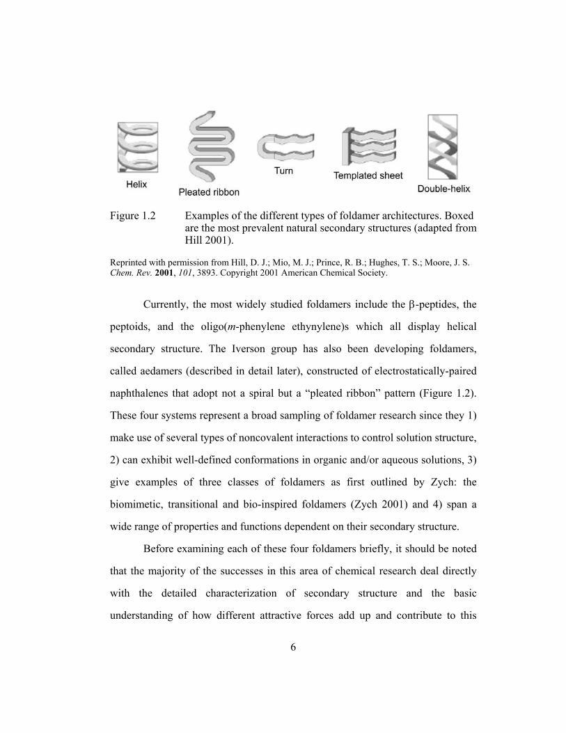
Figure 1.2 Examples of the different types of foldamer architectures. Boxed are the most prevalent natural secondary structures (adapted from Hill 2001).
Reprinted with permission from Hill, D. J.; Mio, M. J.; Prince, R. B.; Hughes, T. S.; Moore, J. S. Chem. Rev. 2001, 101, 3893. Copyright 2001 American Chemical Society.
Currently, the most widely studied foldamers include the β-peptides, the
peptoids, and the oligo(m-phenylene ethynylene)s which all display helical
secondary structure. The Iverson group has also been developing foldamers,
called aedamers (described in detail later), constructed of electrostatically-paired
naphthalenes that adopt not a spiral but a “pleated ribbon” pattern (Figure 1.2).
These four systems represent a broad sampling of foldamer research since they 1)
make use of several types of noncovalent interactions to control solution structure,
2) can exhibit well-defined conformations in organic and/or aqueous solutions, 3)
give examples of three classes of foldamers as first outlined by Zych: the
biomimetic, transitional and bio-inspired foldamers (Zych 2001) and 4) span a
wide range of properties and functions dependent on their secondary structure.
Before examining each of these four foldamers briefly, it should be noted
that the majority of the successes in this area of chemical research deal directly
with the detailed characterization of secondary structure and the basic
understanding of how different attractive forces add up and contribute to this
6

structure. Initial attempts though at tertiary structure (an association of distinct
secondary structures on one strand) are beginning to be reported. By extension,
quaternary structures (an ordered bundle made up of more than one strand like
hemoglobin) are also being pursued. Finally, as this field matures and as chemists
become more adept at fine-tuning folding and binding properties, more uses for
foldamers will be discovered. Examples of these higher-order structures and
published applications will be included in the descriptions of these four foldamers
below.
1.1.1 β-Peptides
The artificial β-amino acids that make up β-peptides are structurally
similar to natural α-amino acids and therefore have the benefit of well-established
characterization techniques provided by decades of protein research.
(Ramachandran 1968, Brandon 1999). The synthesis of many types of β-amino
acids, including those with R groups identical to natural amino acids and those
incorporating rigid cis or trans rings (Figure 1.3A), make it possible to construct a
wide variety of β-peptides. The majority of β-peptide research has come
independently from the Seebach (Seebach 1998) and Gellman (Appella 1996)
groups and an informative review by DeGrado and Gellman entitled "β-Peptides:
From structure to function" was published in 2001 (Cheng 2001).
7
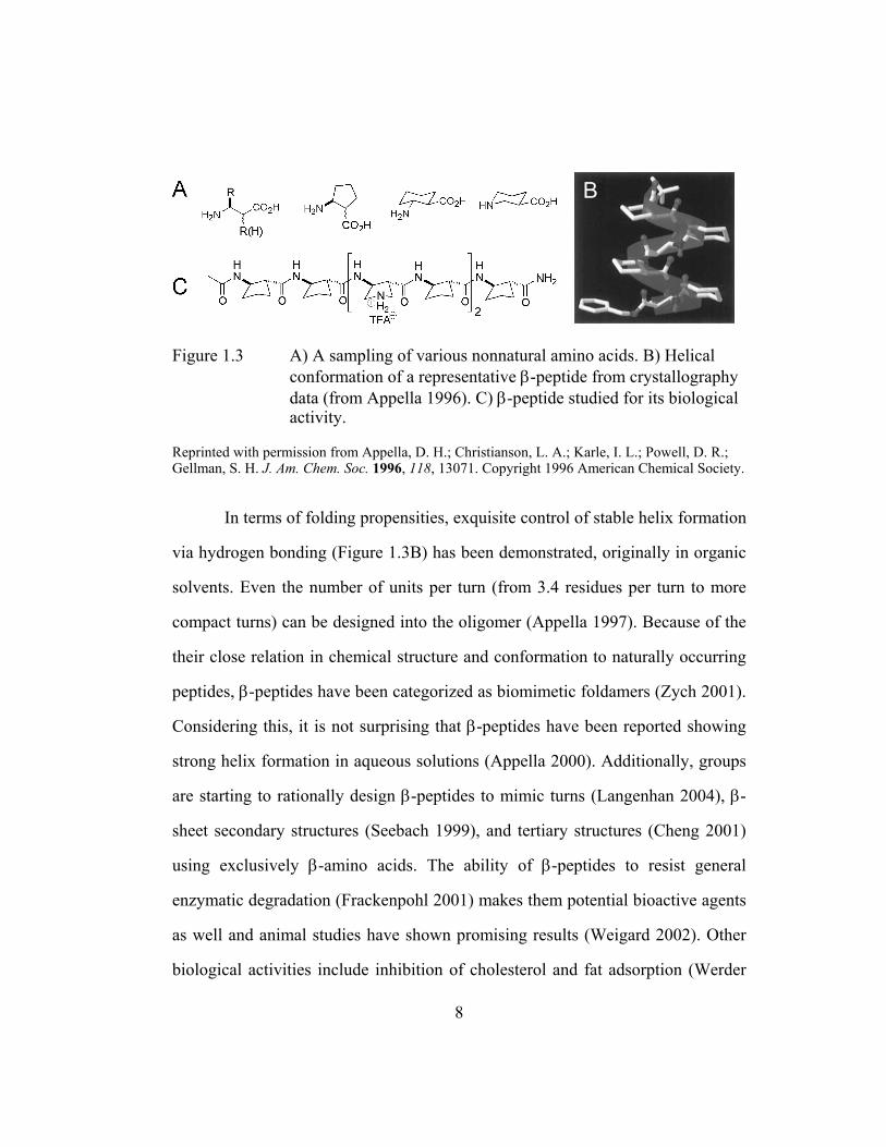
Figure 1.3 A) A sampling of various nonnatural amino acids. B) Helical conformation of a representative β-peptide from crystallography data (from Appella 1996). C) β-peptide studied for its biological activity.
Reprinted with permission from Appella, D. H.; Christianson, L. A.; Karle, I. L.; Powell, D. R.; Gellman, S. H. J. Am. Chem. Soc. 1996, 118, 13071. Copyright 1996 American Chemical Society.
In terms of folding propensities, exquisite control of stable helix formation
via hydrogen bonding (Figure 1.3B) has been demonstrated, originally in organic
solvents. Even the number of units per turn (from 3.4 residues per turn to more
compact turns) can be designed into the oligomer (Appella 1997). Because of the
their close relation in chemical structure and conformation to naturally occurring
peptides, β-peptides have been categorized as biomimetic foldamers (Zych 2001).
Considering this, it is not surprising that β-peptides have been reported showing
strong helix formation in aqueous solutions (Appella 2000). Additionally, groups
are starting to rationally design β-peptides to mimic turns (Langenhan 2004), β-
sheet secondary structures (Seebach 1999), and tertiary structures (Cheng 2001)
using exclusively β-amino acids. The ability of β-peptides to resist general
enzymatic degradation (Frackenpohl 2001) makes them potential bioactive agents
as well and animal studies have shown promising results (Weigard 2002). Other
biological activities include inhibition of cholesterol and fat adsorption (Werder
8
1999) and antibiotic effects (Liu 2001) (Figure1.3C). Lastly, γ- and δ-peptides,
though first studied long before Gellman's introduction of the term foldamer, have
been recently reinvestigated in the context of foldamer development (Brenner
2001).
1.1.2 Peptoids
In 1992, the Zuckermann research team from the Chiron Corporation
introduced oligomers constructed of N-substituted glycines termed peptoids,
(Simon 1992). Since then, many different pendant side groups have been attached
to the amide nitrogen including those that incorporate chirality (Figure 1.4A).
Figure 1.4 A) General structure of peptoids and side chain examples. B) Cationic 36-mer peptoid effective as a gene delivery reagent.
Zuckermann's group has shown that peptoids adopt helical conformations
in both aqueous and organic solvents (Kirshenbaum 1998). This helix formation
occurs without the stabilization benefits of amide hydrogens participating in
intramolecular hydrogen-bonding found in natural helical peptides and β-
peptides. Secondly, low energy conformations of the tertiary amides along the
backbone exist in both the cis and trans isomer states, while peptide bonds that
connect natural amino acids are almost exclusively in the trans state which aids in
9

the structural organization of natural peptides. It has been postulated that the
steric limitations of peptoids with chiral bulky side chains limit the set of
available conformations and general solvophobic interactions also contribute to
the stability of peptoids (Armand 1997, Wu 2001a, b). These key differences from
both α and β-peptides place peptoids in the transitional class of foldamers. Just as
the β-peptides though, peptoids are resistant to proteases and analogues of natural
peptide ligands have shown effective biological activity (Simon 1992). Peptoids
have even been shown to have good transfection (specifically lipofection)
activity, which is useful for gene delivery therapies (Murphy 1998, Figure 1.4B).
1.1.3 Oligo(meta-Phenylene Ethynylene)s
Moore and co-workers have published extensively on oligo(meta-
phenylene ethynylene)s, also known as oligo(m-PE)s (Figure 1.5A). These
foldamers also adopt helical conformations employing solvophobic interactions
and local geometric constraints along the backbone to stabilize folding (Hill 2001)
(Figure 1.5B). These molecules are considered to be in the bio-inspired class of
foldamers in which there is little relation to any natural system and many times
applications lean towards material science rather than protein emulation (Mio
2000). The first generation of oligo(m-PE)s varied in length and pendant groups
and demonstrated the manipulation of the conformational transition via changes in
solvent and temperature (Nelson 1997). Additionally, twist-sense preferences by
means of a chiral perturbation in the side chains (Prince 2000a) or backbone (Gin
1999) have also been reported.
10

Figure 1.5 A) Example of an oligo(m-PE). B) Proposed helical folding (from Hill 2001). C) Terpene examples used in molecular recognition studies.
Reprinted with permission from Hill, D. J.; Mio, M. J.; Prince, R. B.; Hughes, T. S.; Moore, J. S. Chem. Rev. 2001, 101, 3893. Copyright 2001 American Chemical Society.
Numerous analytical studies (UV-Vis, fluorescence, circular dichroism,
and NMR spectroscopy) support the proposed helical conformation and observed
twist-sense. These studies have even led to the design of properly sized cavities
for molecular recognition. 12-mers can function as receptors for various
monoterpenes displaying association strengths in the 103 M-1 range and modest
selectivities in polar solvents (Prince 2000b, Figure 1.5C). Other oligo(m-PE)s
have been derivatized to accept rodlike guests as well, such as
diphenylpiperazines, in order to template the growth of chains of specific length
(Tanatani 2001). Finally, Moore and co-workers have recently published the
synthesis of a new water-soluble oligo(m-PE) (Stone 2004).
1.1.4 Aedamers
In 1995 Lokey and Iverson described the first foldamers to make use of
aromatic stacking interactions in water to direct folding (Lokey 1995). These
molecules utilize the hydrophobically-driven face-to-face complexation between
11

electron-rich 1,5-dialkoxynaphthalene (Dan) and electron-deficient 1,4,5,8-
naphthalenetetracarboxylic diimide (Ndi) units to attain a compact conformation
(Figure 1.6A). When these two moieties were linearly connected with
appropriately flexible linkers, and in an alternating fashion, the resulting oligomer
adopted an entirely abiotic secondary structure, a pleated fold conformation, in
water (Figure 1.6B, Lokey 1995). These molecules were termed aedamers after
the aromatic electron donor-acceptor interactions that direct folding, and just as
the oligo(m-PE)s, these aromatic containing foldamers belong also to the bio-
inspired class of foldamers.
12

Figure 1.6 A) Chemical structure of an aedamer hexamer designed by Lokey and Iverson (Lokey 1995). B) Idealized model of the pleated folding pattern and absorption signatures (in the UV and visible range) that support face-centered stacking of aromatics. Dotted arrows indicate the change in the spectrum upon the denaturing of the aedamer hexamer caused by CTAB addition.
13

A prominent feature of aedmaers is the modular design that allows facile
solid-phase synthesis plus the incorporation of a wide variety of linkers that might
possibly be used to modify folding and binding properties. There are also useful
spectroscopic handles that are consistent with aedamers adopting a folded
conformation in solution. Hypochromism (such as that found with DNA base
stacking) and the existence of a charge transfer band are consistent with face-to-
face ring stacking arrangements (Lokey 1995, 1997, Cantor 1980, Figure 1.6B).
Under denaturing conditions such as with the addition of the cationic detergent
cetyltrimethylammonium bromide (CTAB), these spectroscopic signatures are
significantly altered to reflect approximately the superposition of the spectra of
isolated Dan and Ndi monomer solutions (Figure 1.6B). Also, ring current effects
caused by stacked π-systems, results in characteristic upfield chemical shifts of
the Dan and Ndi aromatic hydrogens relative to the signals given by Ndi and Dan
monomers separately (Zych 2000, 2002).
Finally, the space-fill model in Figure 1.6B suggests that the adopted
conformation creates a hydrophobic column with the potential to display
functional groups along its periphery. Couple this well-defined scaffold and its
ability to be easily derivatized with the fact that these foldamers are most stable in
water, it is envisaged that aedamers that interact with biological systems can be
constructed.
14

1.2 UPDATE: FOLDAMERS WITH AEDAMER-LIKE DESIGNS
Since the original publication of aedamers appeared in 1995, several
foldamers based on aromatic interactions have been reported. The use of aromatic
interactions, in particular donor-acceptor type interactions, is emerging as a
reliable strategy to build molecular scaffolds complementary to other non-
covalent forces.
Figure 1.7 shows schematic representations of the most recent of these
systems, all of which are reminiscent of the aedamer design. The N-methylated
phenylguanidines developed by the Kagechika group is the only system of the
four presented that operates in aqueous solutions (Tanatani 1998, Figure 1.7A).
Impressively, x-ray crystal structures were obtained for both pentamers whose
benzene rings were either attached at the meta or para positions, plus several
derivatives afforded chiral crystals. The folded columnar superstructures of the
Nuckolls group were an extension of their work on discotic-like liquid crystals
(Zhang 2003, Figure 1.7.B). Their pleated-ribbon secondary structure is driven by
a combination hydrogen bonding and π-stacking. A δ-peptide foldamer was
developed using Dan and pyromellitic diimide (Pdi) units (Zhao 2004, Figure
1.7C). Further description on this system from the Li group can be found in
Chapter 3 in the context of peptide-turn mimics. Lastly, the Ramakrishnan group
was able to polymerize Dan and Pdi units in an alternating fashion with
hexa(ethylene oxide) linkages (Ghosh 2004, Figure 1.7D). They assert that
charge-transfer, solvophobic, and metal binding effects leads to a strongly folded
15
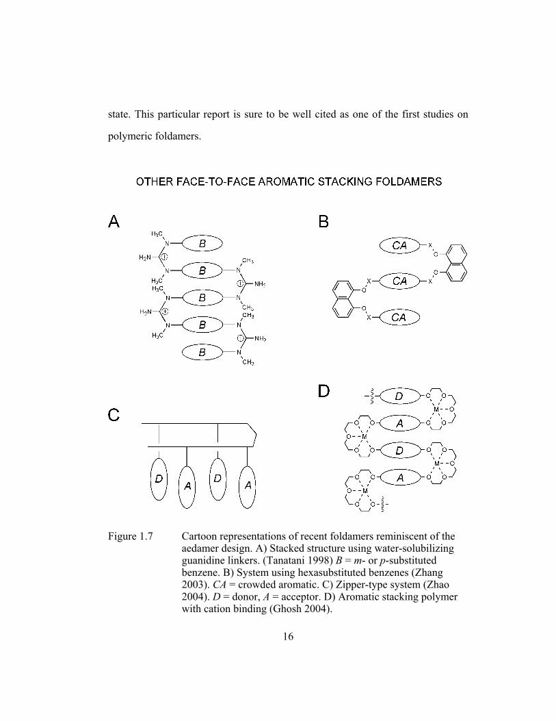
state. This particular report is sure to be well cited as one of the first studies on
polymeric foldamers.
Figure 1.7 Cartoon representations of recent foldamers reminiscent of the aedamer design. A) Stacked structure using water-solubilizing guanidine linkers. (Tanatani 1998) B = m- or p-substituted benzene. B) System using hexasubstituted benzenes (Zhang 2003). CA = crowded aromatic. C) Zipper-type system (Zhao 2004). D = donor, A = acceptor. D) Aromatic stacking polymer with cation binding (Ghosh 2004).
16

1.3 CLOSER EXAMINATION OF AEDAMER AROMATIC INTERACTIONS
1.3.1 Forces that Dictate Folding
After reporting on a hexameric aedamer as a novel abiotic folding
molecule (Lokey 1995), the Iverson group found it beneficial to study models at
the monomer (Cubberley 2001) and dimer (Zych 2000) level to expound a much
better description of aedamer folding. These investigations aimed to answer two
important questions: 1) What are the fundamental forces responsible for Dan:Ndi
association in aedamer folding? and 2) Can methods be developed to better
describe the inter-ring orientations to give a more comprehensive picture of
aedamer folding? Ultimately, these necessary fundamental studies paved the way
for much of the work described in this dissertation.
To elucidate the driving forces of aromatic stacking, Cubberley performed 1H-NMR binding titrations to measure association constants of the complexation
of Dan and Ndi neutral monomers as well as Ndi:Ndi and Dan:Dan associations
(Cubberley 2001b, Figure 1.8). A sampling of the calculated association constants
(M-1) in deuterated solvents covering a broad polarity range is shown in Figure
1.8. The data in the solid-line box pointed to a strong desolvation (in particular a
hydrophobic) effect for Dan:Ndi complexation since the strength of this
association is 2-3 orders of magnitude greater in D2O than in organic solvents.
This result is not surprising for the stacking of flat molecules with relatively
nonpolar faces that self-organize to minimize contact with polar solvents.
17

Figure 1.8 Compounds used in the monomer study and their associations (M−1) in a few of the solvents reported (Cubberley 2001b).
Unanticipated though was the data (in the dotted box, Figure 1.8) that
revealed Ndi:Ndi and Dan:Dan association was 10 and 100 times less stable than
Dan:Ndi complexation, respectively. If desolvation were the lone important
factor for association then it would be predicted that Dan:Ndi, Ndi:Ndi, and
Dan:Dan associations would all be comparable due to the similar sizes of the
hydrophobic aromatic surfaces of the Dan and Ndi rings. Therefore, this trend
argued for an electrostatic driving force in which matching the electrostatic
surface potentials would predict Dan:Ndi complexation to be the strongest
scenario.
Inspection of the X-ray crystal structures (Figure 1.9) nicely explained
how hydrophobics and electrostatics act in concert. It appeared that the low
18

stability of Dan self-association stemmed form its inability to stack in a face-
centered manner due to putative repulsion of the electron-rich π-cloud of one Dan
face with another. The resulting herringbone orientation thus tempers the extent of
the desolvation driving force for Dan self-complexation. To explain the
intermediate strengths of Ndi self-association, Cubberley and Iverson postulated
that the preferred stacking geometry of Ndi also tempers desolvation effects. Ndi
crystal data displayed a face-to-face but off-set stacking likely due to the electron
repulsion of the electron-rich oxygen atoms of the carbonyl groups. Interestingly,
the Hunter and Sanders model for aromatic stacking had previously described,
albeit in a different context, edge-to-face geometries for electron-rich aromatics
and slipped face-to-face stacking for electron-deficient aromatics that contain
electron-rich heteroatoms (Hunter 1990, 2001). In summary, the dominant driving
force was found to be the desolvation of the aromatic faces. However Cubberley
and Iverson emphasized that “the magnitude of desolvation is modulated
significantly by stacking geometry, which, in turn, is dictated by the electrostatic
complementarity in predictable fashion (Cubberley 2001b).”
19

Figure 1.9 Calculated electrostatic surface potentials and chemical structures of compounds used to obtain X-ray quality crystals (adapted from Cubberley 2001b). From inspection the expected desolvation driving force is strongest in an equimolar mixture of Dan and Ndi.
Reprinted with permission from Cubberley, M. S.; Iverson, B. L. J. Am. Chem. Soc. 2001, 123, 7560. Copyright 2001 American Chemical Society.
20

In general, many factors can contribute to aromatic interactions including
Van der Waal’s, charge-transfer, desolvation and electrostatic forces (Hunter
2001). The findings of Cubberley and Iverson, coupled with seminal work from
several other groups studying synthetic models, have added greatly to the
understanding of aromatic interactions, which also includes π-π stacking. Figure
1.10 briefly highlights three examples that have influenced how scientists view
the fundamental basis of aromatic interactions.
Figure 1.10 Early model systems to study aromatic interactions. A) Bicyclo-cyclophane host/pyrene guest (from Smithrud 1991). B) Diarylnaphthalene system. X = various electron withdrawing and donating groups (Cozzi 1992). C) Diarylcarboxylate models. R = naphthalene or adenine (Newcomb 1994).
Reprinted with permission from Smithrud, D. B.; Wyman, T. B.; Diederich, F. J. Am. Chem. Soc. 1991, 113, 5420.Copyright 1991 American Chemical Society.
Diederich’s group examined the stability of pyrene-cyclophane complexes
in water and organic solvents by NMR and calorimetry (Smithrud 1990, 1991,
Figure 1.10A). A model of solvation effects on apolar binding resulted from their
work and interestingly they found that the strength of the complexation for their
system could be predicted and controlled by solvent polarity. In an ingenious
21
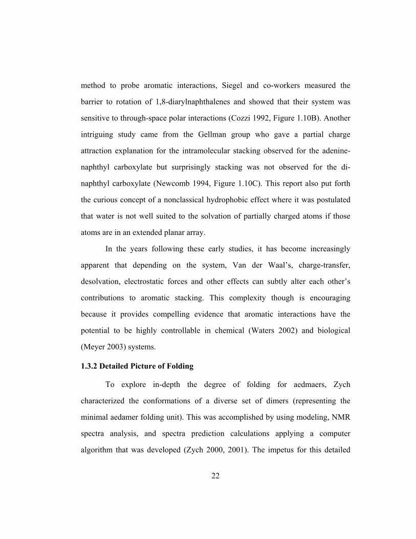
method to probe aromatic interactions, Siegel and co-workers measured the
barrier to rotation of 1,8-diarylnaphthalenes and showed that their system was
sensitive to through-space polar interactions (Cozzi 1992, Figure 1.10B). Another
intriguing study came from the Gellman group who gave a partial charge
attraction explanation for the intramolecular stacking observed for the adenine-
naphthyl carboxylate but surprisingly stacking was not observed for the di-
naphthyl carboxylate (Newcomb 1994, Figure 1.10C). This report also put forth
the curious concept of a nonclassical hydrophobic effect where it was postulated
that water is not well suited to the solvation of partially charged atoms if those
atoms are in an extended planar array.
In the years following these early studies, it has become increasingly
apparent that depending on the system, Van der Waal’s, charge-transfer,
desolvation, electrostatic forces and other effects can subtly alter each other’s
contributions to aromatic stacking. This complexity though is encouraging
because it provides compelling evidence that aromatic interactions have the
potential to be highly controllable in chemical (Waters 2002) and biological
(Meyer 2003) systems.
1.3.2 Detailed Picture of Folding
To explore in-depth the degree of folding for aedmaers, Zych
characterized the conformations of a diverse set of dimers (representing the
minimal aedamer folding unit). This was accomplished by using modeling, NMR
spectra analysis, and spectra prediction calculations applying a computer
algorithm that was developed (Zych 2000, 2001). The impetus for this detailed
22
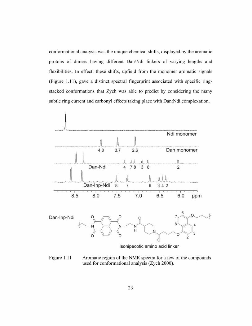
conformational analysis was the unique chemical shifts, displayed by the aromatic
protons of dimers having different Dan/Ndi linkers of varying lengths and
flexibilities. In effect, these shifts, upfield from the monomer aromatic signals
(Figure 1.11), gave a distinct spectral fingerprint associated with specific ring-
stacked conformations that Zych was able to predict by considering the many
subtle ring current and carbonyl effects taking place with Dan:Ndi complexation.
Figure 1.11 Aromatic region of the NMR spectra for a few of the compounds used for conformational analysis (Zych 2000).
23

A total of eleven dimers were investigated and for each, 100 computer-
model conformations were generated from molecular dynamics simulation and
geometry optimization. After 1) constructing conformational “maps” for all
conformers and 2) applying equations to predict their chemical shifts, Zych found
that, in most instances, an ensemble of predicted low-energy structures as
opposed to one definitive conformer best described the experimentally acquired
spectra. Thus, Zych and Iverson concluded that “folding” does not appear to
follow a two-state unfolded/folded model with a rigid, unique conformation but
follows a more dynamic model in which all the different folded conformers are
related by having a face-to-face stacking arrangement. (Zych 2000).
This study also revealed that the aedamer pleated structure was tolerant of
a wide range of linkers. On the other hand, it has been proven difficult to
substitute amino acids without inadvertently disrupting the structure of proteins
(LaBrenz 1995, Quinn 1994). Thus, this may prove beneficial for the
development of functional aedamers since ideally one could decouple properties
from the overall architecture of the molecule (Zych 2001).
Ultimately, larger aedmaers will have to be used to approach the size and
functionality found in proteins but the type of detailed conformational analysis
performed with dimers becomes prohibitive with increasing size. Therefore, in
follow-up work, Zych and Iverson looked at the possibility that the conformations
of larger aedamers are actually the “sum” of their component dimers (Zych 2002).
Figure 1.12 illustrates the concept of this behavior that was designated as
conformational modularity. For instance the Dan region of the NMR spectrum of
24

a Dan-Inp-Ndi-Dan trimer could be closely predicted by the chemical shifts found
for its component dimers, Dan-Inp-Ndi and Ndi-Dan (Figure 1.12). In another
situation, if a Dan unit was sandwiched in between two Ndi units in an assumed
pleated fold stack, conformational modularity still held fairly true but only when
ring current effects were considered to be additive. For instance, the delta
chemical shifts found for the Dan unit in the tetramer, Dan-Asp-Ndi-Dan-Asp-
Ndi, nearly matched the sum of the delta chemical shifts for the Dan unit of Ndi-
Dan and Dan-Asp-Ndi.
Figure 1.12 Illustrated concept of conformational modularity and chart showing the high accuracy of chemical shift prediction when this concept is applied to larger aedamers (Zych 2001, 2002).
Certain trimers and tetramers were found though that did not follow the
conformational behaviors of their component dimers. It was proposed that for
25
some aedamers steric clashes between the linkers shifted the expected orientations
of the rings. Zych and Iverson pointed out that this insight would have been
dramatically more difficult to obtain if a de novo conformational analysis of the
larger structures were attempted. Nonetheless, most of the trimers and tetramers
studied displayed this highly desirable feature of conformational modularity.
Further refinement of this feature could greatly aid in the design of larger folding
systems with predictable structure and function.
As a whole, the meticulous studies into the fundamental basis of folding,
done independently by Cubberley and Zych, laid a foundation for future aedamer
work and their findings will probably increase in importance as aedamer research
moves towards applications. In fact, only after these basic science studies were
performed did this dissertation research begin to significantly move forward;
where the reported strengths and directionality of the “directed” interactions of
Dan and Ndi moieties could be utilized in previously unexplored ways.
1.4 OVERVIEW OF NAPHTHYL OLIGOMER PROJECTS
The projects in the Iverson laboratory dealing with aromatic interactions
can all trace their beginnings to the original aedamer paper (Lokey 1995). Since
then, research utilizing Dan and Ndi moieties has diversified to investigate varied
aromatic interactions phenomena in areas spanning from molecular biology to
materials science. Figure 1.13 organizes these projects roughly based on the size
of the Dan/Ndi compounds synthesized and the general focus of the study. As
presented above, several significant results have come from exploring monomer
associations and dimer conformations. Currently the Iverson group is well
26

involved with studying systems at the oligomeric size regime and has even
recently started investigating polymeric materials containing Dan and Ndi
building blocks.
27

Figure 1.13 Projects in the Iverson laboratory designed to explore aromatic interactions. All work can trace its beginnings to Lokey’s aedamer paper. Results detailed in chapter 2, 3, and especially 4 have led to several new research directions.
28

The focus of this dissertation is to illustrate the ability to exploit the
Dan:Ndi interaction for creating novel folding and binding properties not
previously accessed with naphthyl oligomers of the original “alternating” (Dan-
Ndi)n aedamer design. Quite simply, this dissertation documents the group’s
initial attempts to extend the Dan:Ndi interaction to controlling other behaviors
beyond driving a pleated fold conformation. Figure 1.14 is a cartoon illustrating
this basic premise using “non-alternating” naphthyl oligomers, for example,
compounds of such sequences as, Dann+1-Ndin, Dann and Ndin.
29

Figure 1.14 Cartoon representation of the naphthyl oligomers studied and the main question each project aimed to answer. Arrows indicate direction of growth for longer oligomers.
The project described in Chapter 2: Intriguing Heat-Triggered
Behavior of an Amphiphilic Aedamer involves work initiated by Nguyen, who
discovered that amphiphilic aedamers could be thermally triggered to irreversibly
30

form stable aqueous gels (Nguyen 1999). Studies were done with a derivatized
aedamer set to probe whether residue substitutions could modulate gelling
properties. The amphiphilic aedamer (ungelled) was also shown to act as an
effective refolding inhibitor of RNase. Inhibition studies testing several
compounds and conditions were thus tested. Of interest to the Iverson group are
the potentially selective interactions between the aromatic moieties of aedamers
and the aromatic amino acids of proteins. This work represents the group’s first
steps towards identifying aedamer-protein interactions.
Many groups working with foldamers have shown that the secondary
structures of their synthetic strands remain unchanged and are thus remarkably
stable to a wide array of residue substitutions. The project described in Chapter
3: Switching the Folding Patterns of Naphthyl Oligomers reports the first
successful demonstration of a foldamer accessing significantly different abiotic
secondary structures in water simply by rearranging their primary sequence
analogous to proteins. Here synthetic strands of the new type (Dann+1-Ndin) were
synthesized and found to adopt hairpin-type turn structures. The results presented
in this chapter represent another checkmark on a list of properties that are
possessed by natural biomolecules that foldamers can now emulate. The
designability of these naphthyl oligomers is not only useful to create different
folding topologies but is expected to provide a facile route to the development of
larger, extremely stable hairpin structures with useful properties.
Lastly, this dissertation will account the group’s entrance into the growing
field of designed, synthetic duplexes. The project described in Chapter 4:
31

Naphthyl Oligomers that Form Hetero-Duplexes details several analytical
methods that provided evidence of the first hetero-duplex system to form via
aromatic interactions and operate in aqueous solutions. This system uses
complementary Dann and Ndin strands to form robust artificial duplexes that
exhibited high chain discrimination. Noteworthy was that even though these
chains each possess substantial negative charge, they still associate with
substantial affinities regardless of potential charge repulsion. This work, which
was also the first to demonstrate the strength and directionality of the Dan:Ndi
association in an intermolecular format, has opened up several research paths
dealing with orthogonal self-assembly of complementary species (Figure 1.13).
32

CHAPTER 2
Intriguing Heat-Triggered Behavior of an Amphiphilic Aedamer
2.1 CHAPTER SUMMARY
Introduction. Due to the modular design of aedamers (Figure 2.1),
synthesizing an amphiphilic aedamer with three aspartate and three leucines
residues, instead of with six aspartates as in the original aedamer (Chapter 1), was
straightforward. While studying this new aedamer, Nguyen and Iverson
discovered unusual gelling properties in which an aqueous solution of this
compound is irreversibly converted thermally to a viscous gel accompanied by a
color change from purple to pale pink (Nguyen 1999). The original aedamer on
the other hand was found to be resistant to this change in physical state. This
chapter will describe aggregation studies on a set of different aspartate/leucine
containing aedamers evaluated for their gelling propensities. It also appeared that
heat-induced aggregation related to aqueous gel formation plays a role in the
inhibition of the enzyme RNase (folding, not active-site, inhibition). It is proposed
that RNase cannot refold properly after heat denaturation in the presence of the
amphiphilic aedamer. When the original aedamer was tested, RNase recovered its
full catalytic activity. This proof-of-concept study evaluating the possibility of
strong aedamer-protein interactions will be discussed in the second half of this
chapter.
33
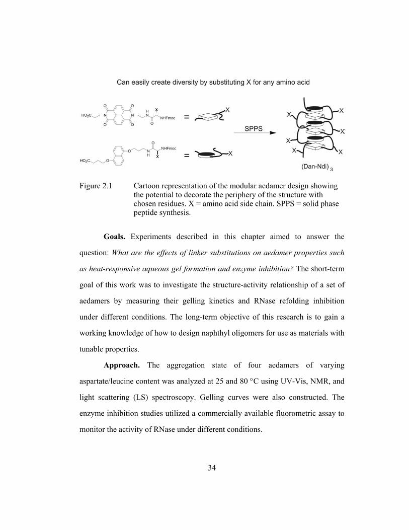
Figure 2.1 Cartoon representation of the modular aedamer design showing the potential to decorate the periphery of the structure with chosen residues. X = amino acid side chain. SPPS = solid phase peptide synthesis.
Goals. Experiments described in this chapter aimed to answer the
question: What are the effects of linker substitutions on aedamer properties such
as heat-responsive aqueous gel formation and enzyme inhibition? The short-term
goal of this work was to investigate the structure-activity relationship of a set of
aedamers by measuring their gelling kinetics and RNase refolding inhibition
under different conditions. The long-term objective of this research is to gain a
working knowledge of how to design naphthyl oligomers for use as materials with
tunable properties.
Approach. The aggregation state of four aedamers of varying
aspartate/leucine content was analyzed at 25 and 80 °C using UV-Vis, NMR, and
light scattering (LS) spectroscopy. Gelling curves were also constructed. The
enzyme inhibition studies utilized a commercially available fluorometric assay to
monitor the activity of RNase under different conditions.
34

Results. It was found that only a solution of the amphiphilic aedamer was
able to undergo a conformational transition to form aqueous gels, while the
aedamers with less than three leucine residues were unchanged when heated. At
room temperature the amphiphilic aedamer exhibited a highly aggregated, yet
soluble, state (by NMR and LS) but still displayed characteristics of being well-
folded in the expected pleated ribbon fashion (by UV-Vis). When heated,
insoluble gels formed after an initial slow period followed by a rapid gelling
phase. In the RNase studies, again the amphiphilic aedamer was found to exhibit
unique properties. Only when RNase was heated in the presence of the
amphiphilic aedamer did refolding inhibition occur (Gabriel 2004a). The original
aedamer and even detergent had little effect. This result is encouraging for long-
term studies of selective aedamer-protein interactions. Mechanisms were thus
proposed to explain both the unique gelling and enzyme inhibition properties of
the amphiphilic aedamer.
2.2 BACKGROUND: A FOLDAMER THAT FORMS STABLE GELS IN WATER
Nguyen and Iverson previously reported on an aedamer hexamer in which
three of the aspartate linkers in the original aedamer developed by Lokey and
Iverson were replaced with hydrophobic leucine residues (Nguyen 1999, Lokey
1995) (Figure 2.2). This facially amphiphilic aedamer, once folded, presents one
side with hydrophobic side chains and the other with hydrophilic aspartate side
chains (Figure 2.3). Characterization of this compound gave unresolved NMR
spectra and an apparent molecular weight of 400,000 g·mol-1 by dynamic light
scattering indicating extensive aggregation in solution. When heated, in an
35

attempt to break up aggregates, the NMR sample, initially a purple aqueous
solution, surprisingly became a pale pink gel.
Figure 2.2 Chemical structure of the original aedamer hexamer (Lokey 1995) and an amphiphilic derivative (Nguyen 1999).
The resulting gel was quite stable in the sense that upon cooling and
standing at room temperature for weeks it did not revert back to the properties of
the original solution. Mass spectrometry and reverse phase liquid chromatography
ascribed these properties to conformational changes and not covalent
transformations of the chemical structure. The kinetics of the conformational
transition is shown in Figure 2.3 showing the loss of the charge-transfer (CT)
absorbance at 526 nm and loss of its intense purple color as the solution became
more viscous. Interestingly, there was a lag period followed by a relatively rapid
gelation phase suggesting that this transition is product promoted. This behavior
was confirmed when 10% of pregelled aedamer was added to a solution before
heating which resulted in complete transformation of the solution within 15
minutes rather than 70 minutes (Figure 2.3).
36

Figure 2.3 Cartoon representation of the folded conformation. Graph of the kinetics of the conformational transition (dotted line) of the amphiphilic aedamer upon heating at 80° C, as monitored at the charge-transfer band absorbance of 526 nm (from Nguyen 1999). Solid line shows the kinetics of an identical solution except 10% of pregelled material was added before heating.
Reprinted with permission from Nguyen, J. Q.; Iverson, B. L. J. Am. Chem. Soc. 1999, 121, 2639. Copyright 1999 American Chemical Society.
A proposed mechanism of this behavior is shown in Figure 2.4. In brief,
heating causes partial unfolding of the aedamer that promotes a new type of
hydrophobics-driven interaction where the aromatics screen their relatively non-
polar surfaces from water by forming tangled aggregates instead of refolding.
Most importantly, this type of aggregation proceeds slowly at first because the
concentration of unstacked molecules is initially low and collisions between these
unstacked molecules are rare. It was proposed that the tangled aggregate becomes
more and more efficient at capturing unstacked molecules as it increases in size,
hence the relatively rapid gel phase after a perceived lag period. It was assumed
37

that the final viscous material is composed of a randomly tangled aggregate
absent of the ordered face-to-face stacking of chromophore units responsible for
the CT absorbance, hence the loss of color. It was also pointed out that the
irreversibility of this transition stems from the near impossibility of untangling
this aggregate, at least through temperature changes or dilution (Nguyen 1999).
Figure 2.4 Proposed scheme for the thermal conversion of an aedamer solution to the tangled aggregate state (adapted from Nguyen 1999).
Reprinted with permission from Nguyen, J. Q.; Iverson, B. L. J. Am. Chem. Soc. 1999, 121, 2639. Copyright 1999 American Chemical Society.
The data emphasized that an initial folded state at ambient temperatures
should be assumed since any substantial population of significantly unfolded
aedamer would lead to gel formation at room temperature. Also Nguyen and
Iverson drew comparisons between the behavior of this aedamer and collagen, a
biological polymer that forms gelatin when heated but is more than 40 times
larger than the amphiphilic aedamer. As Gellman commented, “[this] behavior
establishes a link between synthetic folding systems and materials science (Rouhi
1999),” thus presenting a new motivation for foldamers which were previously
38

studied mainly with emulating proteins in mind. Ironically, the initial aim of
Nguyen’s amphiphilic aedamer was to design a mimic of leucine zipper motifs for
biological and not materials investigations.
The Iverson group has been intent on understanding and hopefully
controlling conformation-dependent behavior of these foldamers. In broader
terms, the general field of hydrogels is finding applications in tissue engineering
(for instance as biocompatible scaffolds), controlled drug delivery, bioadhesives
(for wound healing), glucose sensors, and other biomaterials such as soft contact
lenses and kidney dialyzers (Langer 2003). Hydrogels have been constructed from
molecules of varying sizes from short oligopeptides (Pochan 2003) to polymers
(Aamer 2003). Also there is a report of a thermally coaxed gel system developed
by Tirrell and co-workers that interestingly employs two short leucine zipper
domains (Petka 1998). Most recently, a coordination gelator that exhibits a
chromatic and sol-gel transition for both thermal and chemical stimuli, has been
reported as well (Kawano 2004). Even with these numerous examples, there is
still a need for better modulation of hydrogel properties, including texture,
strength, and water content. These issues were addressed in a comprehensive
review published by Langer and Peppas (Langer 2003).
With such activity currently in the hydrogel field it seemed logical to
explore the possibility of tunable gels with the aedamer system. Specifically, the
kinetic curves for gel formation in Figure 2.3 piqued the Iverson group’s interest
in modulating such gelling parameters as the onset temperature, speed of
transition, and overall strength of the resulting gel.
39

2.3 RESULTS AND DISCUSSION
2.3.1 Synthesis of Aedamer Derivatives
New aedamer hexamers, 2.2 and 2.3, were synthesized, as well as 2.1 and
2.4 from previous studies, in order to probe how varying the number of aspartate
and leucine side chains and thus changing the hydrophilic/phobic content, affects
aqueous gel formation (Figure 2.5).
Figure 2.5 Compounds synthesized for the gelling study. A.A. 1-6 represents the amino acid linker positions along the backbone.
Fmoc-based solid-phase peptide protocols (Chan 2000) were followed
(Figure 2.6) to synthesize 2.1-2.4. Also shown in Figure 2.6 are the three amino
acid adducts 2.5-2.7 synthesized in solution phase following analogous
procedures previously described (Zych 2000 - 2.5, [2.6 by an analogous
40

procedure], Guelev 2001c - 2.7). These amino adducts were coupled sequentially
onto Wang resin using PyBOP (Benzotriazole-1-yl-oxy-tris-pyrrolidino-
phosphonium-hexafluorophosphate) with Fmoc deprotections using
piperidine/DMA (N,N-Dimethyacetamide) between each coupling step. After the
last Fmoc deprotection, the oligomer was then capped, in this case using acetic
anhydride, to provide a N-terminal acetyl group. Aedamers were then fully
deprotected and cleaved off the resin with 95/5 w/w TFA (Trifluoroactic acid)
/phenol. Purification by FPLC (Fast protein liquid chromatography) using an
aqueous ammonium acetate and CH3CN gradient afforded homogeneous samples
in 40-50% yield based on the mmol/mg loading capacity of the resin. All
compounds were soluble in 50 mM Na phosphate buffer, pH = 7 at up to 2.0 mM
for 2.1 and up to 5.0 mM concentrations for 2.2-2.4 before a precipitate formed.
Each compound gave satisfactory HPLC (High performance liquid
chromatography) and mass spectrometry analysis.
41

Figure 2.6 Dan and Ndi amino acid adducts synthesized and used in the standard solid phase oligo synthesis cycle. Full structures can be found in the Experimental Section. Fmoc = 9-fluoronylmethyloxycarbonyl. SPPS = solid phase peptide synthesis. LC = liquid chromatography.
2.3.2 UV-Vis, NMR, and LS Comparisons at 25o C
UV-Vis, NMR, and light scattering (LS) experiments were carried out at
room temperature (25o C) to possibly gain insight into the aggregation state of
2.1-2.4 prior to heat-triggered gelation. Nguyen’s report had already asserted that
even though 2.1 is extensively aggregated, it still formed a stack of aromatics in
an intramolecular fashion at room temperature with UV-Vis characteristics
42

indicative of the pleated folded structure attributed to aedamers (Nguyen 1999).
Table 2.1 shows that solutions of all four compounds exhibited marked
hypochromism (48-54%) that is fully consistent with substantial face-centered π-
stacking (Lokey 1995, Cantor 1980). The CT band absorbances (526-540 nm) for
each compound that resulted in the solutions being purple to reddish purple in
color indicated donor-acceptor aromatic stacking and were also seen previously
with folded aedamers (Zych 2002, Lokey 1995).
Table 2.1 Analyses at 25 oC for 2.1-2.4.
UV data Vis data NMR data Compound Hypo (%) a λCT (nm) b εCT (M-1·cm-1) c Linewidth (Hz) d
2.1 54 540 1400 NS 2.2 51 538 1500 10.8 2.3 48 536 1500 5.3 2.4 51 526 1500 2.8
a Solutions at 40 µM concentrations. Hypo(chromism) = [1 - (absorbance at 382 nm without CTAB ÷ with excess CTAB)] × 100%. b λCT = wavelength of charge-transfer band. Concentrations at 1 mM c ε = extinction coefficient. d Solutions at 1.5 mM concentrations. Linewidth of singlet of the Gly methylene at the C-terminus. NS = no discernable signal.
It could not be ruled out rigorously that the hypochromism and CT bands
are derived entirely from intramolecular interactions because of aggregation. It is
reasonable to believe though that these UV-Vis spectroscopic signatures were
predominantly due to intramolecular interactions since the CT bands exhibited
linear Beer’s law behavior below 2.0 mM. Likewise, Nguyen and Iverson
contended that the spectral similarities between 2.1 and 2.4 argue against
43

intermolecular stacking effects playing a major role in the UV-Vis spectra for 2.1
(Nguyen 1999).
While the UV-Vis data suggested that the intramolecular interactions for
2.1-2.4 are comparable, it was clear from NMR data that 2.1 was significantly
aggregated compared to 2.2-2.4 as evidenced by the NMR spectra in Figure 2.7
and the linewidth data in Table 2.1. Figure 2.7 shows that a solution of 2.1 failed
to give resolved spectra diagnostic of a highly aggregated state (albeit a soluble
aggregated state). On the other hand, although line broadening was observed,
spectra of 2.2-2.4 gave distinguishable signals. Linewidths of NMR resonances
have been shown to be sensitive to aggregate size (Whitford 1991). For instance,
the methylene linewidths from spectra of phospholipid vesicles increase in a
roughly linearly fashion with vesicle radius (Liao 1980). In spectra of 1.5 mM
solutions of 2.2-2.4 the linewidths of the singlet assigned to the C-terminal
methylene roughly doubles with the sequential replacement of aspartates on 2.4
with leucines (Table 2.1). Thus, the effect of increasing leucine content on
aggregation appeared to be somewhat additive for the series 2.4-2.2 with a
dramatic jump in signal broadening for aedamer 2.1.
44

Figure 2.7 Aromatic region of 1H-NMR spectra. Concentrations were 1.5 in mM D2O. Even dilute solutions (0.1 mM) of 2.1 failed to give resolved spectra.
Light scattering (LS) has been shown to be an effective tool for studying
the aggregation properties of self-associating molecules including polymers
(Nolan 1997), detergents (Perez-Rodriguez 1998), and bioactive molecules
(Varela 1999). The use of LS is sure to expand as pharmaceutical companies
examine more closely how aggregation enhances or decreases the effectiveness of
certain drug formulations (Seidler 2003). A classic LS experiment is to probe for
discontinuities in the amount of light scattered as a function of concentration to
identify critical aggregation concentrations (CACs). An example is shown in
Figure 2.8 from a study of the self-association of penicillin. For penicillin, 0.04
and 0.25 m (molality) were identified by Mosquera and co-workers as critical
concentrations for an observed monomer/trimer transition and a trimer/12-mer
transition, respectively (Varela 1999).
45

Figure 2.8 Scattering ratio, S90, as a function of molality for penicillin in water. Dotted line below 0.04 m indicates the theoretical line for unassociated monomers and arrows denote the critical concentrations (Varela 1999).
Reprinted with permission from Varela, L. M.; Rega, C.; Suarez-Filloy, M. J.; Ruso, J. M.; Prieto, G.; Attwood, D.; Sarmiento, F.; Mosquera, V. Langmuir 1999, 15, 6285. Copyright 1999 American Chemical Society.
Analogous graphs (Figure 2.9) were created for compounds 2.1 and 2.2,
two compounds that differ by only one residue yet seemingly have drastically
different room temperature aggregation states by NMR. The “discontinuity”
shown by 2.1 was judged to be too slight and not compelling to suggest a CAC
while the data for 2.2 fit a linear model well (R2 = 0.996) for all concentrations
indicating size homogeneity (Figure 2.9). What was quite striking is that the
scattered light intensity of 2.1 was more than an order of magnitude greater than
2.2 corroborating NMR data implying the presence of large aggregates of 2.1
even at low concentrations.
46

Figure 2.9 Scattered light intensity as a function of concentration. The failure to describe data from solutions of 2.1 with a linear fit is normally indicative of a shift in solute size but at this point the discontinuity cannot be interpreted as a critical concentration.
The above graphs measured the time-average intensity of scattered light
also known as static scattering. Dynamic light scattering (DLS) on the other hand
measures the movements of isolated particles. With a large collection of
measurements DLS can afford estimated molecular weights and yield
hydrodynamic radii (Ruso 2000, Pecora 1985). Unfortunately, DLS results varied
too much between separate measurements from trial to trial and even within
single runs to allow for any confidence in the data acquired. This was likely due
to the available fitting models that limit analyses to only systems with well-
defined monomodal or bimodal weight distributions. The aggregated state of 2.1
at room temperature therefore likely spans a wide range of sizes and may be quite
variable over time and sensitive to how solutions are handled. Future studies
47

obtaining sedimentation equilibrium data via analytical ultracentrifugation might
lead to better descriptions of these aggregates of 2.1 (Raguse 2001).
2.3.3 Evaluation of UV-Vis, NMR, and LS Data
UV-Vis spectroscopy fully supported that all compounds had a similar
folding pattern in solution, where the aromatics are stacked face-centered at 25 oC. While the intramolecular folding of 2.1-2.4 appeared to be comparable to each
other, it was clear from NMR and LS that 2.1 was drastically aggregated but in
such a way that the Dan:Ndi intramolecular stacking appeared mostly unaffected.
This suggests that intermolecular interactions were significantly different between
2.1 and 2.2-2.4. Possibly there are enough leucine-leucine intermolecular
interactions to cause aggregation for 2.1. One must also consider charge effects as
an explanation for the apparent differences between 2.1 and 2.2 whose five
carboxylates in solution may have presented adequate intermolecular charge
repulsion to prevent the rapid capture of partially unfolded aedamers. To address
these parameters it would be worthwhile to synthesize hexamers with identical
charges as 2.1 but with less hydrophobic residues, such as glycine, to probe
whether the hydrophobicity of the linker side chains could truly be used to
modulate aggregation properties. Finally, although 2.1 existed as large soluble
aggregates in solution, HPLC and mass spectrometry conditions seemed to break
up these aggregates for measurements of presumably monomeric species during
these analyses. This observation indicates that the intermolecular interactions
responsible for this aggregation state at ambient conditions are fairly weak and
reversible.
48

2.3.4 Concentration Dependent Gelling Studies
While it was important to characterize the aggregation state at room
temperature, the main inquiry of this study is whether compounds 2.2-2.4 gel at
elevated temperatures such as 2.1. Thermally-induced gelling experiments were
performed and the data for 2.1 and 2.2 is shown in Figure 2.10 (compounds 2.3
and 2.4 gave near identical results as 2.2). The gelling of 2.1 (1.5 mM)
reproduced the results found previously (Nguyen 1999) while solutions of 2.2 at
the same concentration did not gel. Attempts to “turn off” the gelling propensity
of 2.1 by dilution were unsuccessful where even 0.1 mM solutions became
viscous and pale pink although the onset of gelling was slightly delayed (Figure
2.10). Likewise, raising the concentration of 2.2 up to 5.0 mM did not “turn on”
the propensity for gelling. Originally it was thought that dilute solutions of 2.1
could limit the intermolecular interactions that caused a highly aggregated state at
room temperature, which in turn would inhibit gel formation. Also it was thought
that 2.2 could be forced to mimic the leucine intermolecular associations
supposedly occurring for 2.1 at room temperature just by increasing its
concentration. At this point, these two compounds, whose aggregation properties
appeared to be dictated by just one residue substitution, are resistant to any
attempts to switch their gelling behaviors.
49
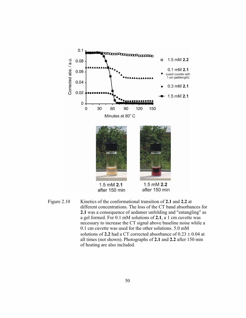
Figure 2.10 Kinetics of the conformational transition of 2.1 and 2.2 at different concentrations. The loss of the CT band absorbances for 2.1 was a consequence of aedamer unfolding and "entangling" as a gel formed. For 0.1 mM solutions of 2.1, a 1 cm cuvette was necessary to increase the CT signal above baseline noise while a 0.1 cm cuvette was used for the other solutions. 5.0 mM solutions of 2.2 had a CT corrected absorbance of 0.23 ± 0.04 at all times (not shown). Photographs of 2.1 and 2.2 after 150 min of heating are also included.
50
2.3.5 Discussion of Thermal Gelling Data
Although the hope was to modulate gelling properties through residue
substitution it appeared that gelation by aedamers was an all-or-none proposition
with no gray area in which to observe intermediate differences in the time course
of the slow and rapid phases of the conformational transition. Encouraging
though, was that a lone residue substitution (Asp→Leu) was identified that in
effect converted 2.2, which did not form aqueous gels, into 2.1, an effective
gelator. A report from Rotello and co-workers may provide clues as to how to
modulate the gelling properties of aedamers (Ilhan 1999). That report illustrated
the use of small molecule intercalators to control the solution structures of their
globular polymers via intra- and intermolecular aromatic stacking under varying
temperatures. A more thorough knowledge of both the intra- and intermolecular
interactions at play in the gel formation of aedamers would be indispensable in
achieving the control of structure demonstrated by Rotello and co-workers. Direct
measurements of the physical properties of the aedamer gels formed at different
conditions would also advance this research. Worthwhile future experiments may
include variable-temperature rheology (to calculate gel strengths and water
loss/recovery while stressed) and transmission electron microscopy (to visualize
gel morphology) (Menger 2003).
2.3.6 Exploring Aedamer-Protein Interactions
The ability of aedamers to adopt well-defined conformations in water
stimulated the group to pursue aedamers that have useful interactions with natural
molecules in biological systems. Recent papers, extending the seminal work of
51

Lokey and Iverson (Lokey 1997), have exquisitely shown that oligo(Ndi)s can
intercalate different DNA sequences with remarkable specificity (Lee 2004,
Guelev 2000, 2001b, 2002). The strength and selectively of binding was attributed
in part to a) the aromatic interactions of the electron-deficient Ndi with the DNA
bases and b) the linkers which were amenable to combinatorial library synthesis
to create a diverse set of intercalators to screen. Aedamers might also have the
same attributes in the context of protein binding especially considering that a)
most proteins contain aromatic amino acids and b) aedamer folding remains
robust with a wide range of linkers (Zych 2000) making combinatorial libraries
possible. Therefore, in light of the group’s success investigating oligo(Ndi)-DNA
interactions, a strong motivation grew to explore the possibility of oligo(Dan-
Ndi)s (i.e. aedamers) interacting with proteins.
The proposed gelling mechanism shown in Figure 2.4 sparked thoughts of
a proof-of-concept experiment in which enzyme capturing (and putative refolding
inhibition) by 2.1 would be a good indicator of aedamer-protein interactions. It
was expected that 2.1 would be most effective as an inhibitor under heating
conditions (80 °C) in which its exposed hydrophobic aromatic units could trap a
thermally denatured enzyme before both could properly refold back when the
solution cooled to room temperature. Any aromatic interactions in proteins would
be between residues with relatively electron-rich aromatics (phenylalanine,
tyrosine, histidine, and tryptophan). It is reasonable to believe that donor-acceptor
interactions between an Ndi moiety of an unfolded aedamer and a natural amino
52
acid would be very competitive with the donor-donor aromatic interactions found
in proteins.
Also proposed was to test aedamer 2.4, which remains highly folded at 80
°C, on the off chance it would be able to capture unfolded enzyme just due to the
availability of it terminal aromatic units or linker carboxylate functionalities.
Lastly, it was not known whether aedamers inherently have elements that might
endow these molecules with active site recognition capabilities in which case
inhibition may not even require a heat-induced denature/trap route.
It was decided that the enzyme RNase would present an interesting
challenge because of its robust structure and its ability to renature efficiently even
after heating to above 60 °C (Figure 2.11). Plus, fluorometric assay kits called
RNaseAlert can be purchased from Ambion (Austin, TX). These kits are used
often by molecular biology laboratories to monitor RNase activity and to detect
RNase contamination in solutions containing susceptible oligonucleotides. The
RNase substrate probe emits a green fluorescence when cleaved (Figure 2.11).
Lastly and most intriguing is that Ambion does indeed provide a storage solution
called RNAsecure that inhibits present RNase only when heated to above 60 °C.
Once cooled this solution does not produce any measurable downstream enzyme
inhibition. The solution contains proprietary ingredients therefore the active
molecules and mode of action cannot be speculated on.
53

Figure 2.11 Primary structure of ribonuclease (RNase) indicating polar (green), hydrophobic (orange), basic (blue), and acidic (red) residues. Schematic of fluorometric assay adapted from the manual of the RNaseAlert kit sold by Ambion.
Compounds 2.1 and 2.4 at different concentrations were tested for their
ability to inhibit RNase with and without a preheat treatment (Figure 2.12).
Sodium dodecyl sulfate (SDS), an anionic detergent, was also tested. The general
procedure for a “heat trial” involved heating the RNase enzyme solution (5 µL,
activity = 0.01 U/mL) in the presence of aedamer or SDS (40 µL, various known
concentrations) in (45 µL Na phosphate buffer) for 1 hour at 80 °C. Once the
solution had cooled, probe (10 µL, 5 mM) was added. Solutions were then
transferred to a 96-well plate and fluorescence measured (Ex/Em 485/516 nm) at
30 minute time intervals. “Non-heat trials” were identical except the compound
and RNase were mixed and left standing at room temperature for 1 hour before
54

the probe was added. High fluorescence readings indicate that RNase activity still
remained and little inhibition had taken place (Figure 2.11). Negative controls
contained only test compound, while positive controls contained only added
RNase before probe was added. Lastly, it was observed that fluorescence
quenching of the fully cleaved probe by Dan (2.8) or Ndi (2.9) monomers is
insignificant.
Figure 2.12 Compounds tested in the RNase refolding inhibition study.
2.3.7 Fluorometric Assay of RNase Activity
Figure 2.13 shows the fluorometric assay data obtained when 2.1 was used
in the heat trial protocol described above. Keeping in mind that fluorescence is
55

proportional to enzyme activity, several trends from the bar graph are noteworthy.
First, RNAsecure was astonishingly effective at RNase inhibition giving readings
near identical to the negative control. Second, while 30 µM concentrations of 2.1
exhibited moderate inhibitory effects, attempts to completely wipe out RNase
activity with higher concentrations of 2.1 surprisingly did not translate into
greater inactivation of RNase. Lastly, since full inhibition was not reached in the
runs with 2.1, it is reasonable to assume that any active RNase remaining would
demonstrate catalytic turnover and continue to cleave the probe leading to the
observed rise in fluorescence with time. The positive control also showed this rise
in fluorescence with time. Measurements after 48 hours gave readings of about
250 emission units (judged to represent complete cleavage of probe) for all plate
wells except for the “Neg” and “Secure” wells. Based on the promising results
from this heat trial using 2.1, similar assays were carried out with the other test
compounds, and without preheat treatments as well (Figure 2.4).
56

Figure 2.13 Fluorescence data of 2.1 at 120-0.15 µM concentrations measured at 30 minute time intervals. Runs with 2.1 afforded moderate refolding inhibition. Neg = negative control. Secure = Ambion’s commercially available RNase inhibitor, RNAsecure. Pos = positive control.
Figure 2.14 exhibits more extensive data; specifically the heat and non-
heat trials using 2.1, 2.4, and SDS at different concentrations. Results at 105
minutes were reported at which time cleavage of the probe was judged to have
reached near completion for the positive control. Three runs were performed for
each of the conditions and results were reproducible (within 10%). Overall, the
results comparing 2.1, 2.4, and SDS can be simply summarized: RNase activity
was significantly decreased only by 2.1 and only when RNase was heated in the
presence of 2.1. Compound data from the other five bar graphs (grayed in Figure
2.14) gave similar emission numbers as their respective positive controls. Data for
monomers 2.8 and 2.9 and a 1:1 mixture at all concentrations studied had limited
inhibition effect as well. This monomer data indicates that there is probably a
57

strong dependence on the structural elements of 2.1 rather than purely aromatic-
aromatic recognition events when 2.1 inhibits RNase.
Figure 2.14 Fluorometric assay results for 2.1, 2.4, and SDS with and without preheat treatment measured at 105 minutes. Only the 2.1 heat trial gave dramatically different results (~75% inhibition for 30 µM solutions) than the other compounds and conditions.
58

2.3.8 Discussion of Refolding Inhibition Data
Results shown in Figure 2.13 indicate that heating RNase in the presence
of 2.1 certainly does decrease its activity but still the fact that a 120 µM solution
of 2.1 performed worse than a 30 µM solution needed to be addressed. A possible
explanation is that at higher concentrations the presumably aggregating aedamer
in effect “gets in its own way” and entangles itself more efficiently than trapping
the unfolding RNase. It follows to argue that 30 µM solutions of 2.1 possibly
allowed better contact between unfolded aedamer and unfolded RNase at elevated
temperatures while at higher concentrations unfolded 2.1 traps itself before
having a chance to capture unfolded RNase. Still one has to remember that a 120
µM solution of 2.1 gave a modest 66% inhibition (a 30 µM solution gave ~80%
inhibition) as compared to the positive control at 45 and 75 minutes (Figure 2.13).
In additional experiments related to the proposed explanation above,
heating RNase with “gelled” 2.1 inhibited RNase but was slightly less effective
than when heating RNase with “ungelled” 2.1 (Figure 2.15). (Aedamer solutions,
at these 30 µM concentrations, were determined as “gelled” when the purple color
of the solution faded after heating. Recall, that to physically confirm a viscous gel
as in experiments described in section 2.3.4 required solutions 100 times more
concentrated.) The results in Figure 2.15 suggested that an aggregated 2.1 surface
might not be required for capturing unfolded RNase. Unlike the Nguyen study
where gel formation was accelerated by the addition of pregelled material
(Ngyuen 1999), it did not seem this was the case for RNase inhibition. In
Nguyen’s study, unfolded 2.1 was trapped much more efficiently when the
59

solution contained initially 10% gel product suggesting a high affinity of unfolded
2.1 for the tangled aggregate (Graph in Figure 2.3 and schematic in Figure 2.4). In
sum, unfolded RNase did not appear to have this same high affinity. Instead it
appeared that when RNase and 2.1 unfold in the presence of each other this led to
greater enzyme inhibition than when RNase unfolds in the presence of pregelled
2.1 (Figure 2.15). Together, the results shown in Figures 2.13 and 2.15 imply that
inhibition by 2.1 proceeded by a different mechanism than RNAsecure. Also it is
clear that more investigation is required to approach the outstanding inhibitory
effects of Ambion’s heat-activated RNase inhibitor solution.
Figure 2.15 Conditions to determine if a preformed tangled aggregate of 2.1 can enhance inhibition in the same manner that gelled 2.1 enhances gelation through a product promoted mechanism.
The results in Figure 2.14 comment quite persuasively on the special
inhibitory properties of 2.1, but only when heated, as compared to 2.4, SDS, and
unheated 2.1. This data bodes well for slight differences in aedamer chemical
structure to modulate its physical properties and functions. A proposed
mechanism of heat-triggered inhibition by 2.1 invokes a general, non-specific
60

association between the hydrophobic residues of the aedamer (in particular the
electron-deficient Ndi moiety) and the RNase (and probably its electron-rich
aromatics). Interestingly Ndi monomer 2.9 had no effect so it may be a
combination of conformational effects and aromatic associations that ultimately
give 2.1 its properties. The ability of 2.1 to aggregate appears to be important
since 2.4, 2.8, and 2.9 (each of which do not form gels) had no significant effect.
Still even with this proposed non-specific mode-of-action, one can envision future
work taking advantage of the selective and opportunistic interactions between
natural aromatic amino acids (phenylalanine, tyrosine, histidine, and tryptophan)
and the naphthyl moieties of aedamers. In related research, screening of a peptide
library has commenced to select sequences that strongly interact with a set of
aedamers to identify high-affinity peptide tags. Hopefully these selection
experiments will yield new insights into specific recognition elements and
naphthyl oligomer-natural protein interactions.
2.4 CHAPTER CONCLUSIONS
Overall, the effects of linker substitutions on aedamer properties such as
heat-responsive gelling and enzyme inhibition can be quite profound. Structure-
activity relationship studies on gelling showed that while 2.1 has essentially the
same intramolecular structure as aedamers 2.2-2.4, apparently its intermolecular
aggregation endows it with a propensity to form gels at elevated temperatures.
This aqueous gel formation appeared to be an all-or-none situation where 2.1 gels
but 2.2, which differed by only a leucine to aspartate residue change, did not, even
at high concentrations. This study also demonstrated that intramolecular folding
61

could be decoupled from bulk properties. This potential attribute could aid in
designing highly derivatized aedamers (to target different properties) but with
folding features that make the placement of functional groups fully predictable in
the final conformation.
The investigation of 2.1 as an enzyme inhibitor further corroborated the
unusual aggregation properties of 2.1 and emphasized that simple residue changes
can have interesting effects. Only when RNase was heated in the presence of 2.1
was enzyme inhibition observed. This inhibition is a strong indication that there
exist considerable interactions between unfolded 2.1 and unfolded RNase, enough
to affect its activity. Lastly, this study is encouraging for future experiments
probing aedamer-protein interactions.
2.5 IDEAS FOR FUTURE INVESTIGATIONS
Future investigations focused on the in-depth characterization of aedamer
aqueous gels would advance both the gelling and the enzyme inhibition project. A
couple of experiments are proposed below with this goal in mind along with the
fact that SPPS makes it easy to synthesize a range of aedamer/peptide hybrids.
Constitutional isomers of the gelling aedamer and engineering
techniques for hydrogel characterization. There is currently strong motivation
to understand how to tune the properties of hydrogels to create biomaterials for
tissue engineering and other applications (Langer 2003). Thus compounds 2.1-2.4
with varied hydrophobicity/philicity were tested. Other hydrophobic residues
besides leucine though could be employed in future analogues. It would also be
worthwhile to take 2.1, which was the only aedamer shown to gel, and scramble
62

its primary sequence to see if leucine/aspartate position along the hydrophobic
column could afford subtle differences in properties. Important for the success of
these future studies though would be precise analytical techniques to monitor
properties of these molecules in a wide range of conditions. The Chemical
Engineering department at the University of Texas at Austin has such instruments
as variable-temperature rheometers, light scatterers designed for large gels rather
than proteins, and specialized microscopes that should prove to be extremely
useful for this proposed study.
Investigation of new aedamer-protein interactions. As stated before
aedamer-peptide affinity studies have been initiated. Another related project that
has been kicked around is to use the Dan:Ndi interaction to create pseudocyclic
peptides. An example would be a tetrapeptide capped on one end with a Dan unit
and the other end with a Ndi unit which might induce a molecular turn via the
Dan:Ndi association. This potentially would create a new set of compounds with
flexibilities in between that of linear and covalently looped cyclic peptides.
Peptide-based enzyme inhibitors have been explored testing how rigidification
affects their binding and it is possible that intermediate flexibilities may be most
effective for some tetrapeptide drugs. With this being the case,
aedamer/tetrapeptide hybrids may act favorably as protein ligands. The feasibility
of this proposed study will become more apparent after the description of
naphthyl oligomer turns (Chapter 3) and artificial hetero-duplexes (Chapter 4),
both of which demonstrate the strength and specificity of the Dan:Ndi interaction
to cause the formation of structures beyond a pleated-ribbon fold.
63

2.6 EXPERIMENTAL SECTION
Solid phase peptide synthesis. A typical synthesis started with 0.10g
(0.060 mmol) Fmoc-Gly-Wang resin (Advanced ChemTech, 100-200 mesh, 0.6
mmol/g) placed in a 5 mL fritted polypropylene syringe (Torviq) and swelled in 4
mL DMA (N,N-dimethyacetamide) using a wrist-action shaker at low speed for
30 min. The DMA was drained and the Fmoc group removed by shaking with 4
mL deprotection solution (25 vol% piperidine in DMA) for 20 min, drained and
repeated. The resin was then rinsed with DMA (3 × 4 mL), iPrOH (3 × 4 mL), and
again with DMA (3 × 4 mL) and this wash was repeated after each step. Coupling
reactions consisted of 3 equivalents (0.18 mmol) of the particular amino acid
adduct dissolved in 3 mL DMA and taken up by the syringe followed by 3
equivalents (0.18 mmol) coupling reagent, PyBOP, (benzotriazole-1-yl-oxy-tris-
pyrrolidino-phosphonium-hexafluorophosphate) (Novabiochem) in 1 mL DMA
and 6 equivalents (0.36 mmol) base (N-methylmorpholine). The resin was shaken
for 60 min for each coupling reaction. Rinsing, deprotection, rinsing, and
coupling were repeated until the desired length was obtained. Either a rotary or
orbital shaker was used for long periods of shaking/agitation but each time new
solvent of solution was added to the resin the reaction syringe was capped and
vortexed for a couple of seconds to break up clumps of resin. This seemed to
improve overall yields by at least 10%.
After the last Fmoc deprotection the N-terminus of the oligomer was
capped with an acetyl group by addition of 15 equivalents (0.9 mmol) DIEA
(N,N-diisopropylethylamine) in 1.5 mL of DMA followed by 15 equivalents (0.9
64

mmol) acetic anhydride in 1.5 mL of DMA and shaken for 45 min. The oligomer
was cleaved off the resin by shaking for 60 min with 4 mL TFA with 0.1g phenol
as a cation scavenger and this solution also removed the t-butyl protecting group
on the aspartate side chains. The TFA solution was collected and the resin was
rinsed with additional TFA and CH2Cl2. The rinses were combined with the TFA
solution and evaporated to an oil under reduced pressure at 60 °C. The oil was
soncicated with heptane for 10 min and once again evaporated. The residue was
then sonicated with diethyl ether for 5 min and cooled to 0 °C resulting in a
granular precipitate that was collected by a glass fritted funnel and washed with
additional cold diethyl ether. Crude solids were then dissolved through the vessel
filter with a minimum amount of 50 mM sodium phosphate buffer, pH = 7.0 and
subjected to fast protein liquid chromatography (FPLC) purification (details
follow). Fractions judged pure by high performance liquid chromatography
(HPLC) (details follow) were lyophilized and afforded 2.1-2.4 as airy purple
solids.
Purification by FPLC. Crude oligomers dissolved in 50 mM sodium
phosphate buffer (pH = 7) were purified on a Pharmacia FPLC system equipped
with LKB P-500 pumps, a GP250 Plus programmer, and 254 nm UV detection
using a Pharmacia column filled with Source 15RPC reverse-phase media from
Amersham Pharmacia. A solvent gradient of 95% 0.1 mM ammonium acetate /
5% CH3CN to 50% 0.1 mM ammonium acetate / 50% CH3CN over 60 min was
used. Solvents were degassed, filtered and were of high purity (double distilled
H2O and spectrograde acetonitrile from EM Science). Purity of the collected
65

fractions was judged by HPLC using a 50 × 4.6 mm “Luna” 5 micron C18 column
from Phenomenex and a Beckman Coulter HPLC system equipped with a
photodiode array detector scanning between 190 nm and 600 nm. A solvent
gradient of 95% 0.1 mM ammonium acetate / 5% CH3CN held for 1 min then
ramped to 50% 0.1 mM ammonium acetate / 50% CH3CN over 14 min was used
and gave good resolution. Appropriate fractions were pooled and lyophilized for
48 hrs under a vacuum of < 10 microns. Pure compound was dissolved in H2O
and desalted by adsorption onto a C18 Sep-Pak Plus cartridge from Waters
followed by a H2O wash and recovery with a H2O/CH3CN mixture and then
lyophilized again for 48 hrs prior to experiments.
The recipe for the 50 mM sodium phosphate buffer, pH = 7.0, used in
most experiments follows: Dissolve 8.16 g Na2HPO4·7H2O and 2.70 g
NaH2PO4·H2O in 1L of dd H2O.
Compound characterization (1D-NMR general methods). Samples
were readily soluble in 50 mM sodium phosphate, pH = 7.0 D2O. Routine spectra
of monomers and precursors were obtained on a Varian UNITY+ 300 MHz
spectrometer. Spectra were recorded on a Varian INOVA 500 MHz spectrometer
at 1 mM concentrations of compound and TSP-d4 (3-trimethylsilyl-propionic-
2,2,3,3-d4 acid, sodium salt) was used as a reference (δ = 0.00 ppm). Chemical
shifts reported in ppm and abbreviations used are singlet (s), doublet (d), doublet
of doublet (dd), triplet (t), quartet (q), multiplet (m) and complex multiplet of non-
equivalent protons (comp). J coupling constants (J) reported in Hz.
66

“AcHN-(LeuDanAspNdi)3Gly-OH” (2.1). (SPPS yield = 40%) 1H NMR (500
MHz, D2O) Spectra gave prohibitively broadened signals. MALDI MS calcd for
C142H151N19NaO39 [MH]+ 2771.8, found 2770.8.
“AcHN-(LeuDanAspNdi)2AspDanAspNdiGly-OH” (2.2). (45% yield) 1H NMR
(500 MHz, D2O) δ = 8.01-7.82 (12H), 6.80-6.42 (12H), 6.31-6.09 (6H), 4.68-4.64
(4H), 4.26-4.05 (13H), 3.83-3.92 (26H), 2.83-2.54 (15H), 2.48-2.34 (6H), 2.09-
1.80 (15H), 1.55-1.46 (4H), 0.83 (d, J = 6.1, 3H), 0.78 (d, J = 6.1, 3H); FAB MS
calcd for C140H146N19O41 [MH]+ 2749.0, found 2749.0.
“AcHN-LeuDanAspNdi(AspDanAspNdi)2Gly-OH” (2.3). (49% yield) 1H NMR
(500 MHz, D2O) δ = 8.05-7.83 (12H), 6.71-5.81 (18H), 4.66-4.64 (3H), 4.34-3.95
(14H), 3.69- 3.30 (26H), 2.82-2.39 (20H), 2.02-1.89 (16H), 1.65-1.40 (8H), 0.92-
0.81 (12H); FAB MS calcd for C138H140N19O43 [MH]+ 2752.7, found 2752.7.
“AcHN-(AspDanAspNdi)3Gly-OH” (2.4). (50% yield) 1H NMR (500 MHz,
D2O) δ = 8.00-7.65 (12H), 6.81-6.59 (18H), 4.70-4.60 (5H), 4.52 (dd, J = 8.8, 5.0,
1H), 4.25-4.40 (12H), 3.87-3.77 (6H), 3.73 (s, 2H), 3.70-3.40 (18H), 2.47-2.37
(6H), 2.07-1.85 (6H), 1.93 (s, 3H); MALDI MS calcd for C136H133N19NaO45
[MH]+ 2775.6, found 2775.7.
“HO-Dan-Asp(OtBu)-NHFmoc” (2.5).
1H NMR (300 MHz, DMSO-d6) δ = 12.17 (s, 1H OH), 8.03 (t, J = 5.4, 1H NH),
7.86 (d, J = 7.5, 2H x), 7.73-7.67 (comp, 2H x, 2H n), 7.6 (d, J = 8.3, 1H NH),
67

7.41-7.28 (comp, 4H x, 2H o), 6.94 (d, J = 7.6, 1H p), 6.91 (d, J = 7.6, 1H p),
4.35-4.28 (comp, 1H t, 1H w), 4.25-4.18 (m, 2H v), 4.15-4.10 (comp 2H m, 2H q),
3.38-3.31 (comp, 2H s, 2H u), 2.65 (dd, J = 10.3, 5.7 1H k), 2.45 (m, 1H k), 2.10-
1.97 (comp, 2H l, 2H r), 1.35 (s, 9H y); 13C NMR (300 MHz, DMSO-d6) δ =
174.1, 170.4, 169.4, 155.7, 153.9, 153.8, 143.8, 143.7, 140.7, 127.6, 127.0, 126.0,
125.9, 125.3, 125.2, 120.1, 113.6, 113.5, 105.7, 80.0, 66.9, 65.7, 65.4, 64.9, 51.6,
46.6, 37.7, 35.8, 30.4, 28.8, 27.6, 24.3, 15.1; CI HRMS calcd for C40H45N2O9
[MH]+ 697.313, found 697.312.
“HO-Dan-Leu-NHFmoc” (2.6).
1H NMR (300 MHz, D2O) δ = 12.15 (s, 1H OH), 8.01 (t, J = 5.5, 1H NH), 7.87
(d, J = 7.5, 2H x), 7.73-7.69 (comp, 2H x, 2H n), 7.4 (d, J = 8.3, 1H NH), 7.41-
7.28 (comp, 4H x, 2H o), 6.95 (d, J = 7.3, 1H p), 6.91 (d, J = 7.3, 1H p), 4.28 (m,
1H w), 4.20 (m, 2H v), 4.15-4.11 (comp 2H m, 2H q), 3.99 (m, 1H t), 3.39-3.29
(comp, 2H s, 2H u), 2.49-2.48 (m, 2H k), 2.10-1.95 (comp, 2H l, 2H r), 1.60-1.55
(m, 1H y), 1.50-1.37 (m, 2H u), 0.84 (dd, J = 13.3, 6.6, 6H z); 13C NMR (300
MHz, DMSO-d6) δ = 174.1, 172.3, 155.9, 153.9, 153.8, 143.9, 143.7, 140.7,
127.6, 127.0, 126.0, 125.9, 125.3, 120.0, 113.6, 113.5, 105.7, 66.9, 65.5, 65.3,
53.1, 46.7, 40.8, 35.6, 30.4, 28.8, 24.3, 24.2, 22.9, 21.5; CI HRMS calcd for
C38H43N2O7 [MH]+ 639.307, found 639.306.
“HO-Ndi-Asp(OtBu)-NHFmoc” (2.7).
68

1H NMR (300 MHz, D2O) δ = 12.39 (s, 1H OH), 8.58 (s, 4H d), 8.05 (t, J = 5.9
1H NH), 7.83 (d, J = 4.2, 1H k), 7.82 (d, J = 4.2, 1H k), 7.61 (d, J = 7.6, 1H k),
7.58 (d, J = 7.6, 1H k), 7.44 (d, J = 8.5, 1H NH), 7.37 (t, J = 7.6, 1H k), 7.36 (t, J
= 7.6, 1H k), 7.25 (t, J = 6.9, 1H k), 7.23 (t, J = 7.6, 1H k), 4.26-4.17 (comp, 1H g,
1H j, 2H i), 4.14-4.03 (conp, 2H c, 2H f), 3.54-3.35 (m, 2H e), 2.61 (t, J = 7.6, 2H
h), 2.55-2.31 (m, 2H b), 1.31 (s, 9H l); 13C NMR (300 MHz, DMSO-d6) δ =
172.3, 170.8, 169.3, 162.7, 162.3, 155.6, 143.7, 143.5, 140.6, 140.5, 130.2, 127.6,
127.5, 127.0, 126.9, 126.4, 126.1, 126.0, 125.9, 125.2,125.1, 120.0, 119.9, 127.6,
127.5, 127.0, 126.9, 126.4, 126.1, 126.0, 125.9, 125.2, 125.1; 80.0, 65.7, 51.4,
46.4, 37.4, 37.3, 36.6, 36.0, 32.0; CI HRMS calcd for C42H39N4O11 [MH]+
775.262, found 775.261.
“Dan monomer” (2.8).
3-acetylamino-N-(3-{5-[3-(carboxymethyl-carbamoyl)-propoxy]-naphthalen-
1-yloxy}-propyl)-succinamic acid (2.8). (SPPS yield = 91%) 1H NMR (300
MHz, D2O) δ = 7.88 (d, J = 8.4, 1H n), 7.84 (d, J = 8.6, 1H n), 7.50 (m, J = 2H o),
7.07 (t, J = 7.6, 2H p), 4.47 (dd, J = 5.2, 3.4, 1H t), 4.29-4.25 (comp, 2H m, 2H q),
3.69 (s, 2H j), 3.50 (t, J = 6.7, 2H s), 2.60 (dd, J = 5.2, 7.8, 1H u), 2.58 (t, J = 7.0,
69
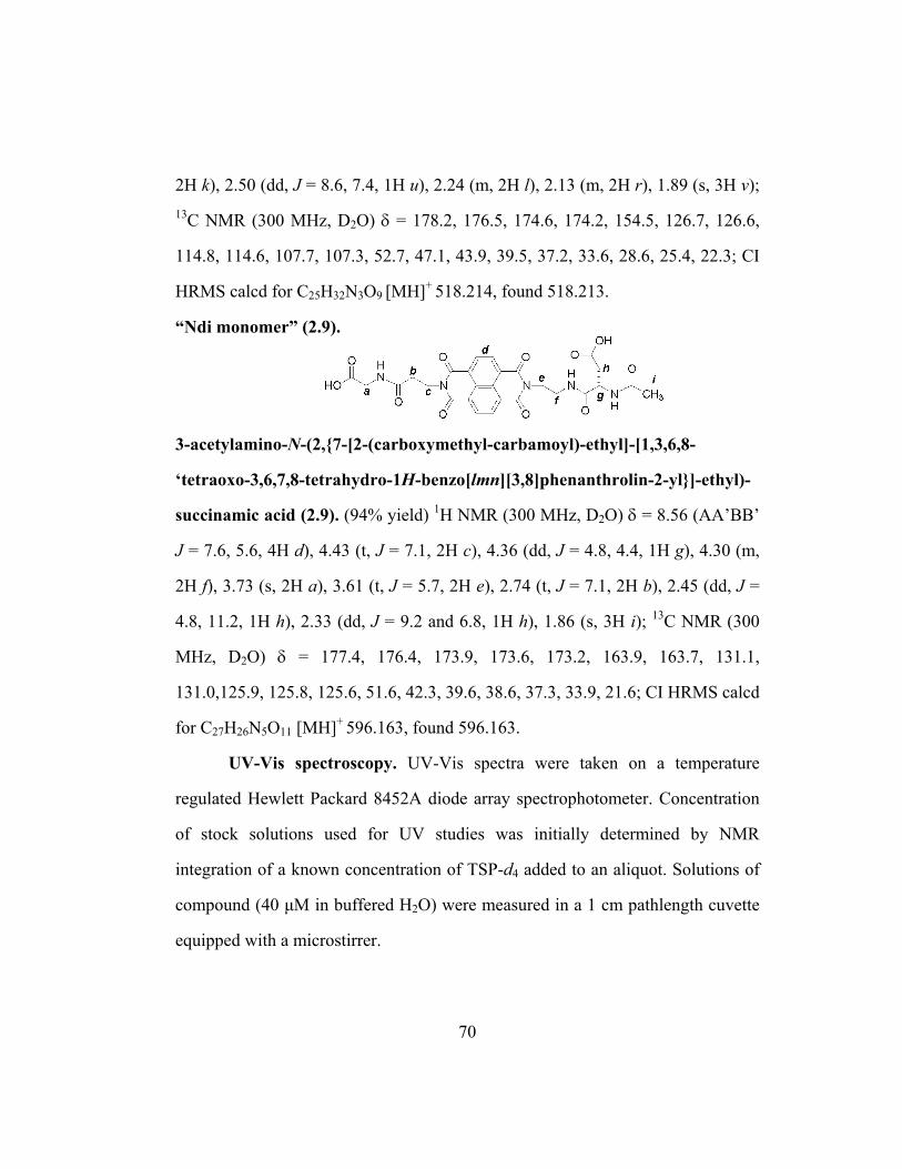
2H k), 2.50 (dd, J = 8.6, 7.4, 1H u), 2.24 (m, 2H l), 2.13 (m, 2H r), 1.89 (s, 3H v); 13C NMR (300 MHz, D2O) δ = 178.2, 176.5, 174.6, 174.2, 154.5, 126.7, 126.6,
114.8, 114.6, 107.7, 107.3, 52.7, 47.1, 43.9, 39.5, 37.2, 33.6, 28.6, 25.4, 22.3; CI
HRMS calcd for C25H32N3O9 [MH]+ 518.214, found 518.213.
“Ndi monomer” (2.9).
3-acetylamino-N-(2,{7-[2-(carboxymethyl-carbamoyl)-ethyl]-[1,3,6,8-
‘tetraoxo-3,6,7,8-tetrahydro-1H-benzo[lmn][3,8]phenanthrolin-2-yl}]-ethyl)-
succinamic acid (2.9). (94% yield) 1H NMR (300 MHz, D2O) δ = 8.56 (AA’BB’
J = 7.6, 5.6, 4H d), 4.43 (t, J = 7.1, 2H c), 4.36 (dd, J = 4.8, 4.4, 1H g), 4.30 (m,
2H f), 3.73 (s, 2H a), 3.61 (t, J = 5.7, 2H e), 2.74 (t, J = 7.1, 2H b), 2.45 (dd, J =
4.8, 11.2, 1H h), 2.33 (dd, J = 9.2 and 6.8, 1H h), 1.86 (s, 3H i); 13C NMR (300
MHz, D2O) δ = 177.4, 176.4, 173.9, 173.6, 173.2, 163.9, 163.7, 131.1,
131.0,125.9, 125.8, 125.6, 51.6, 42.3, 39.6, 38.6, 37.3, 33.9, 21.6; CI HRMS calcd
for C27H26N5O11 [MH]+ 596.163, found 596.163.
UV-Vis spectroscopy. UV-Vis spectra were taken on a temperature
regulated Hewlett Packard 8452A diode array spectrophotometer. Concentration
of stock solutions used for UV studies was initially determined by NMR
integration of a known concentration of TSP-d4 added to an aliquot. Solutions of
compound (40 µM in buffered H2O) were measured in a 1 cm pathlength cuvette
equipped with a microstirrer.
70

Light scattering (LS). A DynaPro-801 light scatterer from Protein
Solutions was used with the accompanying software. All data points represent the
average of > 20 measurements per dilution. Aedamer solutions were made with
50 mM Na phosphate buffer six hours before measurements. Solutions were
filtered through a 0.02 mm pore 13 mm Anodisc membrane filter from Whatman
with no evidence of clogging or adsorbed compound just before filling a 12 µL
cuvette and measurements taken. It usually took an equilibration time of 5
minutes before the average intensity of light scattered remained sufficiently
steady (± 10%). Only data taken after this equilibration were used in the analysis.
Data of the average intensity of light scattered were reproducible from trial to
trial, sizing data though gave high errors and were not considered in this
dissertation.
Thermally-triggered gelling. Solutions were placed in a 0.1 cm
pathlength cuvette and plugged with a sliver of rubber. The cuvette was placed in
the Hewlett Packard 8452A diode array spectrophotometer whose cuvette holder
had been heated to 80 °C by a water bath. After 1 minute to heat the cuvette
through, measurements were taken every five minutes with manual shaking in
between each UV-Vis spectrum taken. The CT band absorbance was monitored
and corrected as previously describe (Figure 2 and 3 from Nguyen 1999). In brief,
as the solution became more viscous the baseline in the visible region of the
spectrum rose indicating increased light scattering of the tangled aggregate. The
absorbance at the CT wavelength of the fully gelled material was subtracted form
CT band absorbance during the heating.
71
Fluorometric assay. Material and supplies were purchased from Ambion
unless otherwise noted (product numbers included). The bench surface, pipettors,
and gloves were treated with RnaseZap (#9780). A bunsen burner flame was left
running as well. Na phosphate buffer was made with nuclease-free water (non
DEPC treated, #9937). Pre-sterilized, 200 µL size pipet tips from ART Molecular
Bioproducts were used. Test compound solutions were made and 40 µL were
pipetted into RNase-free microfuge tubes (#12350). 45 µL of Na phosphate buffer
was added along with 5 µL of RNase (activity = 0.01 U/mL, included in
RNaseAlert kit #1964). For heat trials, the microfuge tube was placed in a 80 °C
oil bath for 1 hour with strong agitation by vortex and then centrifuged every 20
minutes. For non-heat trials the samples stood at room temperature but were stil
agitated. Meanwhile the probe (aliquots were lyophilized in microfuge tubes by
Ambion) was suspended with 10 µL of provided buffer (probe and buffer include
in RNaseAlert kit). The samples were brought to room temperature and pipetted
into the probe, agitated, and transferred to a 96 well assay plate from Corning
(#3610). Large air bubbles were popped using disposable needles and
fluorescence measurements were made at 30-minute time intervals. All samples,
including the negative and positive controls contained a total of 100 µL of
solution and concentrations reported in the data figures represent concentrations
of these 100 µL solutions.
72

CHAPTER 3
Switching the Folding Patterns of Naphthyl Oligomers
3.1 CHAPTER SUMMARY
Introduction. While Chapters 1 and 2 focused on aedamers, which have
an alternating sequence of the form (Dan-Ndi)n, this chapter will describe work
with non-alternating naphthyl oligomers of the sequence Dann+1-Ndin (Figure 3.1).
These naphthyl oligomers are expected to fold with a different topology than the
pleated ribbon folds exhibited by aedamers. Therefore this project represents the
group’s first attempts to diverge from the original aedamer design in order to
explore the strength and specificity of the Dan:Ndi interaction to direct more
challenging folding. While natural α-peptides and nucleotides can form different
secondary structures based on their primary sequence, β-peptides are the only
foldamers that possess this same ability with reported examples of β-peptide
helices and turns (Langenhan 2004) in literature. These systems employ hydrogen
bonding and though helices have been shown to be stable in water, the reported
turn structures are only stable in non-aqueous solvents. The work presented here
with naphthyl oligomers demonstrates a strategy to control distinctly different
folding patterns in water.
73

Figure 3.1 Cartoon representation of two different folding patterns. Arrows indicate direction of growth with longer oligomers.
Goals. Experiments described in this chapter aimed to answer the
question: Can naphthyl oligomers be rationally designed to adopt hairpin
structures based on directed aromatic stacking? The short-term goal of this work
was to demonstrate the ability of non-alternating naphthyl oligomers to adopt
stable folding patterns, in particular turn motifs, as an alternative to the pleated
secondary structures of aedamers. The long-term objective of this research is to
use various folding patterns to possibly increase the stability of the hydrophobic
column and eventually exploit this stability and predictive manipulation of folding
to design complex functional molecules.
Approach. Trimers of the type Dan-(X)-Dan-Ndi, varying in linker length
(X), were probed by UV-Vis spectroscopy for the formation of a turn motif where
the Ndi unit folds back into the trimer and inserts in between the two Dan units. A
structural analysis of the best turn candidate, along with control compounds, was
then accomplished using 2D NMR techniques and computer modeling.
74
Results. Overall, the work described in this chapter demonstrated that two
simple, but discrete, folding patterns can be created just by changing the sequence
of linked Dan and Ndi units (Gabriel 2004b). Data from unfolding investigations,
NOESY spectroscopy and computer modeling supported conformational
interpretations of a pleated fold for a Dan-Ndi-Dan trimer and an “intercalative”
fold for a Dan-Dan-Ndi trimer. Appropriate control compounds were also useful
for this investigation. This intercalative trimer was incorporated into a Dan3-Ndi2
pentamer to test if the turn motif can aid in templating hairpin-type structures.
Preliminary NMR studies were promising and encouraged further use of the
predictable and strong Dan:Ndi association to study different binding patterns
(Chapter 4). Lastly, the creation of different folding patterns via the facile
shuffling of two aromatic monomer types may also provide insights into the
general role of aromatic interactions in stabilizing structures.
3.2 BACKGROUND: MIMICS OF BIOLOGICAL HAIRPIN STRUCTURES
The continued pursuit of controlling molecular topology, whether through
the development of foldamers, peptidomimetics or other chemical models, may
lead to a greater understanding of the folding and binding properties associated
with biomolecules (Brandon 1999). One of the key oligomeric conformations in
biology is the hairpin structure of a single-stranded biomolecule, which
incorporates a U-shaped turn motif. These structures are ubiquitous in
oligonucleotides such as DNA loops, ribozymes (catalytic RNA) (Liley 1999),
and aptamers, (RNA with molecular recognition properties) (Marshall 1997).
Research on hairpin nucleotidomimetics has led to the discovery of extremely
75

compact DNA fragments (Hirao 1994) and a fuller knowledge of the
thermodynamics of DNA folding (Soto 2000).
Concerning peptides, the β-sheet secondary structure found in many
proteins is controlled by a collection of hairpin folds. While researchers
synthesizing peptidomimetics are actively engaged in using artificial turns to
design β-sheet mimics (Kemp 1990, Nowick 2000), researchers designing
foldamers have also created synthetic β-sheets and examples were presented in
Chapter 1. Also, a comprehensive review on peptide models of β-hairpin structure
has been published (Searle 2001). Though there are many noteworthy examples of
models of molecular turns, this introduction will touch briefly upon the work of
two particular groups, Waters and Li that have specifically utilized aromatic
interactions to probe turn conformations.
Waters and co-workers have synthesized structural mimics to investigate
β-hairpin peptide stability and to understand how aromatic interactions contribute
to peptide structure and binding specificity (Tatko 2002, Butterfield 2002). A
substantial data set has been built from which to elucidate the driving forces for
folding and molecular recognition of β-hairpin peptides with specific residue
substitutions (Figure 3.2). They have also developed models to probe “diagonal”
C-H···π interactions that are distinct from the noncovalent interactions driven by
the hydrophobic effect (Tatko 2004). With rigorous NMR and thermal
denaturation experiments they have found interesting geometries of the
interacting residues and have parsed the energy of the folding of a β-hairpin
peptide into its enthalpic and entropic components.
76

Figure 3.2 Hairpin peptides synthesized by Waters and co-workers (Tatko 2002, 2004). X = various sidechains to explore aromatic-aromatic and C-H···π interactions.
Detailed investigations into the energetics of noncovalent interactions are
growing in importance since it has been demonstrated that many proteins are
stable by only 5-10 kcal per mol over misfolded states (Tatko 2002). So, although
isolated noncovalent interactions are weak, the combination of these many subtle
attractive forces and their binding geometries can have a pronounced effect on
stability and structure, respectively. This information will be significant for de
novo protein design. As expected, their meticulous studies into the basic science
of noncovalent interactions have indeed accelerated the development of functional
peptides with tunable properties including ATP recognition (Butterfield 2003) and
flavin redox potential modulation (Butterfield 2004).
Li and coworkers have developed δ-peptide foldamers that adopt a turn-
type conformation using a donor-acceptor interaction between electron-rich 1,5-
dialkoxynaphthalene (identical to the Dan moiety) and an electron-deficient
pyromellitic diimide (Pdi) (Zhao 2004). There are two main differences between
77

δ-peptide foldamers and the naphthyl oligomer foldamers from the Iverson group.
One is that δ-peptides present the aromatic rings as side chains projecting along
the backbone while the aromatic rings in naphthyl oligomers are incorporated
within the backbone. This arrangement is predicted to cause a weaving scenario
for naphthyl oligomers (Figure 3.1) rather than a zipper-type situation for the Li
system (Figure 3.3). Another important distinction is that the δ-peptide system
operates in chloroform and DMF while naphthyl oligomers are water-soluble and
are anticipated to fold most stably in aqueous solutions.
Figure 3.3 Donor-acceptor δ-peptides synthesized by Li and co-workers and a cartoon representation of their asserted “zipper” structure (Zhao 2004).
It is predicted that the Dan:Ndi interaction can be used to design similar
turn structures predicated on the idea that the aromatic units will arrange
themselves to maximize the number of Ndi:Dan face-to-face contacts, even if it
required the chain molecule to fold back on itself. Evidence of this hairpin fold
would represent further control of secondary structures through the fairly simple
means of shuffling the naphthyl primary sequence.
Also, there is a possibility that hairpin structures will stabilize the naphthyl
hydrophobic column further. First, it was postulated that the mechanism of
78
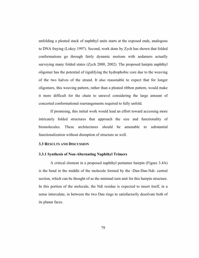
unfolding a pleated stack of naphthyl units starts at the exposed ends, analogous
to DNA fraying (Lokey 1997). Second, work done by Zych has shown that folded
conformations go through fairly dynamic motions with aedamers actually
surveying many folded states (Zych 2000, 2002). The proposed hairpin naphthyl
oligomer has the potential of rigidifying the hydrophobic core due to the weaving
of the two halves of the strand. It also reasonable to expect that for longer
oligomers, this weaving pattern, rather than a pleated ribbon pattern, would make
it more difficult for the chain to unravel considering the large amount of
concerted conformational rearrangements required to fully unfold.
If promising, this initial work would lead an effort toward accessing more
intricately folded structures that approach the size and functionality of
biomolecules. These architectures should be amenable to substantial
functionalization without disruption of structure as well.
3.3 RESULTS AND DISCUSSION
3.3.1 Synthesis of Non-Alternating Naphthyl Trimers
A critical element in a proposed naphthyl pentamer hairpin (Figure 3.4A)
is the bend in the middle of the molecule formed by the -Dan-Dan-Ndi- central
section, which can be thought of as the minimal turn unit for this hairpin structure.
In this portion of the molecule, the Ndi residue is expected to insert itself, in a
sense intercalate, in between the two Dan rings to satisfactorily desolvate both of
its planar faces.
79
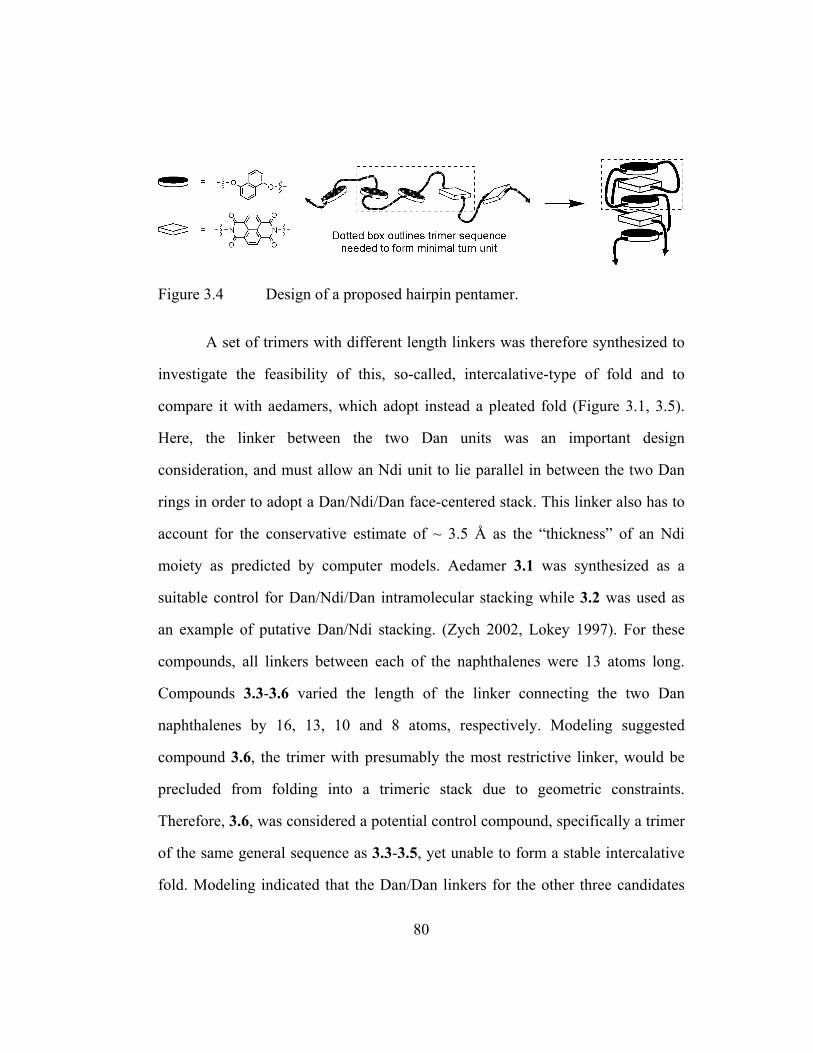
Figure 3.4 Design of a proposed hairpin pentamer.
A set of trimers with different length linkers was therefore synthesized to
investigate the feasibility of this, so-called, intercalative-type of fold and to
compare it with aedamers, which adopt instead a pleated fold (Figure 3.1, 3.5).
Here, the linker between the two Dan units was an important design
consideration, and must allow an Ndi unit to lie parallel in between the two Dan
rings in order to adopt a Dan/Ndi/Dan face-centered stack. This linker also has to
account for the conservative estimate of ~ 3.5 Å as the “thickness” of an Ndi
moiety as predicted by computer models. Aedamer 3.1 was synthesized as a
suitable control for Dan/Ndi/Dan intramolecular stacking while 3.2 was used as
an example of putative Dan/Ndi stacking. (Zych 2002, Lokey 1997). For these
compounds, all linkers between each of the naphthalenes were 13 atoms long.
Compounds 3.3-3.6 varied the length of the linker connecting the two Dan
naphthalenes by 16, 13, 10 and 8 atoms, respectively. Modeling suggested
compound 3.6, the trimer with presumably the most restrictive linker, would be
precluded from folding into a trimeric stack due to geometric constraints.
Therefore, 3.6, was considered a potential control compound, specifically a trimer
of the same general sequence as 3.3-3.5, yet unable to form a stable intercalative
fold. Modeling indicated that the Dan/Dan linkers for the other three candidates
80

for the intercalative fold were theoretically long enough but rigorous energy
calculations were reserved until UV-Vis studies were performed.
Figure 3.5 Compounds synthesized for folding studies.
The synthesis of these compounds proceeded smoothly using monomers
and SPPS protocols previously described in chapter 2. Purification via FPLC
(>98% pure judged by HPLC peak area) followed by desalting via a C18 Sep-Pak
cartridge and freeze-drying afforded soft pale purple samples.
81

3.3.2 UV-Vis Spectroscopy
When aedamers were first being developed, Lokey and Iverson identified
extremely useful UV-Vis spectroscopic handles to monitor the complexation of
the Ndi and Dan moieties in folded oligomers (Lokey 1995). These spectroscopic
signatures for interacting Dan/Ndi chromophores included a charge-transfer (CT)
band, characteristic extinction coefficients (ε) and hypochromism for the naphthyl
moieties, all of which changed dramatically upon unfolding of the oligomer by
addition of CTAB (Chapter 1). It was envisaged that UV-Vis data would provide
a qualitative indication of the types of folding occurring for the previously
unexplored non-alternating trimers, 3.3-3.6. In other words, it was thought that
this data might shed light on whether a) these trimers adopt a Dan/Ndi/Dan stack
such as aedamer 3.1 or b) their Dan-Dan-Ndi sequences force them into
alternative conformations.
UV-Vis data was acquired for solutions of 3.1-3.6 in 50 mM Na phosphate
buffer H2O, pH = 7 (Table 3.1). A CT band appeared for all solutions between
510 and 532 nm, and resulted in a purple color that did not exist with solutions of
Dan monomer or Ndi monomer, both of which were colorless. Table 3.1 reports
the ε of each compound at two wavelengths, at the CT band absorbance and at the
Ndi moiety absorbance at 382 nm. The absorbance of the Ndi unit was the more
reasonable of the two types of naphthyl units to monitor since 3.1-3.6 had one Ndi
moiety each and there was actually very little absorbance overlap between the
Dan and Ndi moiety around the maximum Ndi absorbance of 382 nm. Also an
Ndi ring flanked on one face by a Dan unit was expected to give distinctive
82

spectroscopies compared to an Ndi unit with both of its faces in close contact with
the Dan ring systems. The ε at elevated temperatures (80o C) was measured as
well to detect any possible temperature dependence on the UV spectrum which
would presumably reflect any ring orientation or oligomer conformation
dependence on temperature too. Finally, Table 3.1 reports percent hypochromism
at 382 nm, using unstacked monomeric Ndi values for comparison.
Table 3.1 UV-Vis data for 3.1-3.6. a
Compound b εCT and (λCT) c ε382 nm ε382 nm (80 oC) ∆ ε382 nm d % Hypo e
Alternating trimer and dimer 3.1 (13) 700 (532) 12200 12400 200 57 3.2 (13) 380 (526) 14600 15300 700 45
Non-alternating trimers 3.3 (16) 680 (532) 11300 12700 1400 54 3.4 (13) 670 (532) 11200 11800 600 60 3.5 (10) 500 (512) 13000 15100 2100 49 3.6 (8) 490 (510) 13200 15200 2000 48
a Extinction coefficients (ε) reported in M-1·cm-1, at 25 oC, and using solutions at 40 µM concentrations, unless otherwise noted. b Parentheses indicate the number of atoms between the naphthyl units for 3.1 and 3.2 and between the Dan naphthalenes for 3.3-3.6. c Solutions at 1 mM concentrations. d ∆ ε382 nm
= ε382 nm (80o C) - ε382 nm (25 oC) d % Hypo(chromism) = 100% × [1 - (absorbance at 382nm without CTAB ÷ with excess CTAB)].
3.3.3 Evaluation of UV-Vis Data
Overall, UV-Vis experiments qualitatively showed that all compounds
have interacting π-systems arranged in a face-to-face orientation in water (Lokey
1995, Cantor 1980). First, the percent hypochromism, measured at 382 nm, was a
83

good indication that multiple rings are stacked in a parallel fashion similar to
DNA whose bases, within a double helix, display a hypochromism of about 50%
(Cantor 1980). Second, solutions of 3.3 and 3.4 gave values of 680 and 670 M-
1·cm-1, respectively for the ε of the CT absorbance, both close to the value found
for solutions of 3.1 (700 M-1·cm-1) that was previously designed to adopt a
Dan/Ndi/Dan stack (Lokey 1995, 1997). On the other hand, 3.5 and 3.6 exhibited
significantly lower values implicating a ring arrangement deviated from a
Dan/Ndi/Dan stack. The Dan-Ndi dimer 3.2 gave a much lower εCT value of 380
M-1·cm-1 as well. Third, the 532 nm maximum wavelength of the CT band for 3.3
and 3.4, was identical to 3.1, which also suggested that their rings arrange in a
Dan/Ndi/Dan stack which can reasonably be explained with the proposed
intercalative fold conformation while once again 3.5, and 3.6 deviated from this
wavelength and presumably from this trimeric stack arrangement. Thus far it had
appeared only 3.3 and 3.4 were promising as non-alternating trimers that can
adopt a Dan/Ndi/Dan stacked order analogous to 3.1.
The ε at 382 nm was also useful since for some compounds these
measurements changed significantly when the solution was heated. For instance,
the ε382nm of 3.1 was raised by 200 M-1·cm-1 when heated to 80o C compared to an
unusually large increase of 700 units for 3.2. Besides a likely slight perturbation
of intramolecular folding, it was argued that intermolecular aggregates or
specifically multimeric stacking might possibly be occurring for 3.2 (Figure
3.6A). This intermolecular stacking could be broken up at elevated temperatures
(based on entropy arguments), leaving the presumably stronger intramolecular
84

interactions relatively unchanged. This multimeric stacking was observed
previously with monomers (Cubberley 2001b) and was indeed expected for these
chains of nonpolar residues and especially for chain systems that, once folded,
still presented opportunities for Dan:Ndi complexation albeit in an intermolecular
fashion (Figure 3.6A). The strength of the interaction between separate chains
though is expected to become weaker with longer, more unwieldy oligomers as
evidenced by multimeric stacking appearing with monomers and dimers but not
observed with hexamers (Lokey 1997). Furthermore, it can be argued that trimer
3.1 has a decreased driving force for intermolecular stacking since this situation
would require Dan:Dan face-to-face association which has been proven to be
relatively unfavorable (Chapter 1, Cubberley 2001b, Figure 3.6B).
85

Figure 3.6 Cartoon representation showing A) the likelihood of some type of intermolecular stacking for 3.2 driven by the Dan:Ndi association and B) that intermolecular Dan:Ndi associations are not available if 3.1, 3.3, and 3.4 fold in the asserted manner. Double-headed arrows represent repulsion of the electron-rich π-clouds of two Dan units. C) Possible explanation of how trimers 3.5 and 3.6 could afford UV spectra similar to dimers.
As Figure 3.6B shows (right structures), the general non-alternating trimer
design was fortunate in the sense that, if folded correctly, the whole structure
would be “capped” by two Dan units, just as with the alternating trimer 3.1, with
no exposure of an Ndi surface to propagate intermolecular interactions. Although,
if a prohibitively short linker does indeed prevent a stable Dan/Ndi/Dan stack,
then it would be possible that significant aggregation may occur. This appeared to
be the case for 3.5 and 3.6, whose ε382nm increased by around 2100 and 2000 M-
1·cm-1, respectively when their solutions were heated. Figure 3.6C is a cartoon
representation of a postulated explanation of this change in UV spectra.
Interestingly 3.5, 3.6, and dimer 3.2 displayed surprisingly close UV spectra with
86
ε382nm (80o C) values of 15100, 15200, and 15300 M-1·cm-1, respectively. Upon
heating, it seemed that the UV absorbances of trimers 3.5 and 3.6 become more
attributable to just the intramolecular association between the Ndi unit and only
one Dan residue giving rise to “dimer-like” spectra (Figure 3.6C). This data
provided evidence that the shortened Dan/Dan linker of both 3.5 and 3.6
precluded them from being candidates for intercalative folding but demonstrated
their usefulness as negative controls.
Lastly, it was unexpected that the non-alternating trimer with the longest
Dan/Dan linker 3.3 also had a relatively large shift in ε382nm when heated
compared to that of 3.4. It is possible that solutions of 3.3 are conformationally
heterogeneous (i.e. different aggregated species exist) due to intermolecular
interactions, such as claimed for 3.5 and 3.6. There could also be distinctly
dissimilar intramolecular conformations that are favorable at room and not at
elevated temperatures and vice versa due to a longer linker making it possible to
survey many different structural orientations dependent on temperature.
Examining how heat effects these conformations is a difficult problem and this
supposed discrepancy of conformations might make further structure analysis of
3.3 difficult.
Compound 3.4, on the other hand, displayed comparable values for εCT
(λCT), ε382nm (at 25 and 80º C), and percent hypochromism as 3.1 without the
unusually high, and currently unexplained, ∆ε382nm shown by 3.3. These
similarities suggested that 3.4 did exhibit Dan/Ndi/Dan trimeric stacking, as 3.1,
even though it was not biased by a primary sequence of alternating Dan and Ndi
87

residues. The slight differences in the UV-Vis data between 3.1 and 3.4 likely
stemmed from the inherently dynamic nature of folded naphthyl oligomers and
the fact that these spectroscopic properties are very sensitive to the stacking
orientation of the aromatic rings (Zych 2001, 2002). Also the difference in linker
topologies between a pleated fold and an intercalative fold would be expected to
alter the ring-to-ring geometries enough to give distinct spectra. Although these
UV-Vis measurements by no means gave a crystal clear picture of folding, it did
point to compound 3.4 as a reasonable candidate for more comprehensive
structural analysis to investigate intercalative folding that would afford a turn-type
conformation.
In summary, this initial study determined that trimer 3.4 would be a good
candidate for rigorous 2D NMR analysis that could reveal specific aromatic H-H
contacts. Also compound 3.1 would be an appropriate “positive” control for
Dan/Ndi/Dan stacking. As mentioned previously 3.6 could act as a “negative”
control. It was suggested, based on the UV-Vis studies, that the terminal Dan unit
of 3.6 is likely energetically prevented from completely “swinging around” to
shield the other face of the Ndi ring due to a shortened linker thus preventing a
Dan/Ndi/Dan stack (Figure 3.7).
88

Figure 3.7 Compounds analyzed by 2D NMR and idealized cartoon representations of possible solution conformations. Letter designation for NMR peak assignments for the Dan and Dan* units are given. Also shown is a design of a hairpin structure incorporating an intercalative fold (dotted boxed).
Compounds 3.1, 3.4, and 3.6 were chosen for NOESY and modeling
studies (Figure 3.7). It was hoped that structural analysis would reveal 3.1 and 3.4
are related by a similar Dan/Ndi/Dan ring stacked arrangement even though the
required type of folding to achieve this arrangement would have to be different.
Conversely, even though 3.4 and 3.6 are related by an identical Dan-Dan-Ndi
89

primary sequence, it was anticipated that they would adopt different
conformations. Figure 3.7 illustrates these expected folding patterns.
3.3.4 Unfolding Studies by UV Spectroscopy
As alluded to before, more stable structures may be a possible upshot from
folding manipulation studies. Obtaining unfolding profiles, shown in Figure 3.8,
was a step towards developing a method to analyze the stability of the different
folds that have been, and will be, attained in the future. Unfolding traces could
also possibly reflect the total number of aromatic units in the stacked hydrophobic
core with conformations that have comparable ring arrangements displaying
similar curves.
Figure 3.8 Unfolding curves displayed in two different forms.
The unfolding of 3.1, 3.4, and 3.6 was monitored via the rise of
absorbance with the addition of CTAB, a detergent that was found to denature
aedamers (Lokey 1997). Solutions of trimers were initially at 40 µM before the
titration of a 2.7 µM (0.1 w/v %) solution of CTAB. Absorbances were adjusted
90
for volume changes and the concentrations of CTAB reported in w/v %. As more
CTAB was added the absorbance increased (same as stating that the
hypochromism decreased) as aromatic stacking was disrupted (Figure 3.8, left
graph). Complete unfolding appeared at around 0.03 w/v % CTAB. At excess
CTAB, the Ndi absorbance for each compound still varied slightly from each
other even at identical concentrations. This was expected since different linkers
and residue sidechains still affect, in subtle ways, the Ndi absorbance even in the
absence of π-stacking (Gabriel 2002). Therefore to visualize the percent of
unfolded trimer as CTAB was added, the hypochromism was calculated for each
data point and related to the hypochromism of the initial solution (Figure 3.8,
right graph). As an example, at 0.01 w/v % CTAB the hypochromism of 3.4 was
calculated to be 18% compared to 60% of the initial solution and this translates to
about 70% [1 - (0.18 ÷ 0.60) × 100%] unfolded trimer.
It seemed that the unfolding pathways of the three compounds were not
overwhelmingly different from each other. However, Figure 3.8 (right graph) did
indicate that 3.6 (the trimer that cannot adopt a full Dan/Ndi/Dan stack) was
folded slightly less stably than 3.1 and 3.4 under these denaturing conditions.
Also, this study provided strong evidence that 3.1 and 3.4 share a similar trimeric-
stacked arrangement signified by the near identical stabilities. Indeed, it had been
observed earlier that the absorbance traces from detergent-induced denaturation of
aedamer dimer, tetramers, and hexamers were near identical (Lokey 1997). So,
even as Lokey increased the length of the aedamers, the stability remained
unchanged because these molecules had the same pleated structure conformation
91
and therefore presumably a similar unfolding mechanism. It was encouraging that
oligomers with a previously unexplored naphthyl sequence still gave easily
interpretable curves (for both 3.4 and 3.6). Even though the apparent strength of
folding was comparable for 3.1 and 3.4, a hairpin structure (Figure 3.4) may still
have potential to a give markedly stable conformation due to the anticipated
entwined arrangement of the proposed molecule.
3.3.5 NMR Techniques for Proton Assignment
It was anticipated that the most direct evidence for folding and the specific
aromatic-aromatic contacts that guide this folding would come from 2D-NMR
experiments. When naphthyl oligomers with linkers greater than eight atoms were
modeled in extended conformations, aromatic protons on different rings were at
least 10 Å from each other, much too far apart to expect through-space coupling
with NOESY spectroscopy. A co-crystal X-ray structure indicated the planes
defined by the Ndi and Dan monomer units are separated by approximately 3.5 Å
(Lokey 1995). It was reasonable to expect that hydrophobics-driven aromatic
stacking would orient these rings close enough to each other, excluding water, and
bring particular protons on different aromatic rings within 4-5 Å of each other, a
practical range for NOESY spectroscopy.
This NMR technique though becomes truly valuable for these folding
systems only if the naphthyl protons can be unambiguously assigned.
Unfortunately there was extensive overlap (Figure 3.9) of the two sets of Dan
aromatic protons that would complicate matters in differentiating specific
aromatic-aromatic contacts between the Ndi and Dan and/or Dan* residue
92

(Dan/Dan* notation in Figure 3.7). So while the oligomeric nature of our
molecules allowed for facile synthesis of a set of foldamers with altered
sequences, rigorous assignment of the hydrogens of repeat units (in this case the
Dan hydrogens) became quite challenging.
Figure 3.9 Proton NMR spectra of the aromatic region representing 16 hydrogens for each spectrum. Spectra were taken at 1 mM concentrations in 50 mM Na phosphate D2O.
This general problem is also encountered with the proton assignment of
peptides with repeat sequences and has been solved mostly by NOESY
experiments ran in 90% H2O in D2O, rather than in pure D2O to keep
exchangeable protons observable. These experiments made it possible to “walk”
93

along the backbone chain using the amide hydrogen to form a continuous
correlation across the carbonyls of the amide bond. Naphthyl oligomers though
presented a special challenge not seen with peptides. The Ndi and Dan
naphthalene moieties essentially were isolated spin systems that “act as insulators
along the backbone” (Zych 2001). Fortunately, TOCSY (total correlated
spectroscopy) methods, which afforded long-range through-bond correlations for
Ha-Hd and Ha-Hf, were previously established for aromatic systems (Martin 1994,
Johnston 1989) (Figure 3.10). Zych tested TOCSY methods in the context of our
aedamer systems with much success, the details of which have been reported
(Zych 2001, 2002). Figure 3.10 summarizes the helpful NOE connections and
long-range TOCSY correlations used by this study.
Figure 3.10 Method of “walking” from the terminus of 3.4 in order to assign the Dan and Dan* hydrogen chemical shifts. Examples of key through-space (solid line) and through-bond (dotted line) H-H correlations are marked.
Overall, attaining resonance assignments for the Dan protons of each
compound required NOESY or ROESY (rotational nuclear Overhauser effect
spectroscopy) spectra taken in 90% H2O in D2O and TOCSY spectra taken in
100% D2O. For some cases, several mixing times had to be tried along with other
parameter changes during acquisition and processing performed by Sorey of the
94

NMR facility at the University of Texas at Austin (Gabriel 2004b).
Concentrations were at 1 mM of compound and the solvents were buffered with
50 mM Na phosphate, pH = 7. Sparky v3.85, an online visualization program,
facilitated the imaging and interpretation of 2D NMR spectra (Goddard 2000).
Interpretation of these spectra led to the peak assignments for the Dan and
Dan* aromatic hydrogens. As Figure 3.10 shows, the four Ndi protons are
isolated in that they are more than 5 Å away from the closest methylenes and
NOEs were not observed. Also, TOCSY type couplings between these aromatic
protons were not observed since these protons are not attached to the same π
system as in the case of the Dan residues (Figure 3.10). Therefore the AA'BB'
signals in the Ndi region of the spectra (Figure 3.9) could not be assigned to
specific protons. Regardless, accurate conformational analysis using the Dan
resonances and computer modeling can still be carried out.
3.3.6 NOESY Spectroscopy
Once sufficient assignment of the naphthyl hydrogens was accomplished,
NOESY experiments, for the purposes of identifying specific H-H contacts
between the aromatic rings, were performed. NOE patterns were considerably
different for each compound and NOESY spectra expansions exhibited cross
peaks between the Ndi and one or both Dan units (Figures 3.11-3.13). Dan proton
signals a-f and a*-f* are labeled and the cross peaks that provided strong evidence
of interactions between the Ndi and these Dan aromatic hydrogens are boxed with
a dotted line. Interpretations of the observed cross peaks for each NOESY
spectrum were discussed in Section 3.3.9.
95

Figure 3.11 Expansion of NOESY spectrum for 3.1.
96

Figure 3.12 Expansion of NOESY spectrum for 3.4.
97

Figure 3.13 Expansion of NOESY spectrum for 3.6.
3.3.7 Computer Modeling
Fairly conservative procedures were used for computer modeling, taking
care not to make unreasonable assumptions. To expedite calculations, models of
3.1, 3.4, and 3.6 were used with the side chains removed. It was shown previously
that these modifications did not compromise the modeling of naphthyl oligomers
where the predominant driving force for folding is the aromatic interactions of the
98

Ndi and Dan moiety (Zych 2001). Also important to note is that the goal of these
computations was not an exhaustive survey of conformational space since there
was already strong spectroscopic evidence that the compounds do fold to some
extent. The aim of computer modeling was simply to see if energy minimized
structures corroborate the general aromatic stacking of a new type of intercalative
fold for these naphthyl oligomers.
All modeling simulations and geometry optimizations utilized the
HyperChem software bundle (HyperCube, Inc. 1996) set to employ the MM+
force field. Twenty diverse, unfolded conformers were generated for each initial
model using molecular dynamics. Each of the twenty structures was then allowed
to go through simulated annealing from a temperature of 1000 to 300 K applying
weak distance restraints. Lastly and most importantly, each structure was
subjected to geometry optimization without any restraints to afford low energy
conformers and to probe subtle differences in stacking orientations. This strategy
to obtain families of low-energy structures, rather than using prohibitively long
molecular dynamics simulations to obtain one assumed lowest energy structure,
was proven to be extremely successful for aedamers (Zych 2002).
Since the four Ndi protons of each compound could not be unequivocally
assigned, distance restraints for all models were defined from the center of the
Ndi ring rather than from a particular hydrogen. For models of 3.1, a general
restraint between the Ndi ring and the center of both Dan naphthalenes was
applied that was reasonable based on detailed studies on aedamer trimer structure
previously reported (Lokey 1997, Zych 2002). For models of 3.4, restraints
99

between the Ndi ring and Dan hydrogens c, c*, and a* (these gave medium
strength NOEs with Ndi protons) were used. Finally for models of 3.6, only two
distance restraints were defined, those relating the center of the Ndi ring to
protons f* and to d*, again keeping restraints fairly conservative. Only after
geometry optimization without restraints would the low-energy conformers be
probed for corroboration of the NOE couplings from 2D NMR experiments
(Figure 3.14).
Figure 3.14 Side view of the lowest energy conformers of 3.1 and 3.4 and axis view of the same conformers showing the general topology of the linkers. Arrows indicate the Dan/Dan* linkage. Hydrogens omitted (except for the aromatic of the side views) for clarity.
While most final structures were “folded” with clear face-centered
aromatic stacking, it was also satisfying that this computational protocol led to
other diverse structures (owing to the relatively unbiased nature of this modeling
100

strategy) that revealed folding “defects”. Such conformations that deviated from a
Dan/Ndi/Dan parallel stack included off-set and non-parallel stacking,
Dan/Dan/Ndi stacking and cis amide bond orientations, all of which were between
2 and 10 kcal·mol-1 higher in energy than the lowest energy conformer calculated
for each compound. In this was families of low-energy conformers could be
identified.
3.3.8 Evaluation of NMR Data and Computer Modeling
The spectrum for 3.1 (Figure 3.11) exhibited HDan-HNdi cross peaks along
the entire range of Dan resonances indicative of the Ndi unit making contacts with
both Dan rings. Also, Zych previously showed that when there was only a slight
chemical shift difference between the AA' and BB' signals of the Ndi unit, as was
the case for 3.1, it often signified that the Ndi unit is well centered between two
Dan units. (Zych 2001, 2002). Additionally, aedamer trimers such as 3.1 have
been extensively investigated in several different contexts and all studies
including this one supported a pleated fold interpretation of their solution
structure (Lokey 1997, Zych 2002).
For compound 3.4, several unambiguous H-H correlations were observed
between the Ndi and both Dan groups as well (Figure 3.12). In the spectra for 3.4
there were explicit Ndi contacts with both Dan and Dan* hydrogens (c, c*, e, and
a* - medium intensity; b and f* - weak intensity). These NOEs were consistent
with a structure having the Ndi unit insert in between the Dan rings, implicating
an intercalative fold. Interestingly, several cross peaks could be attributed to a
specific pair of Ndi protons since the AA' and BB' signals were more separated
101
for 3.4 (Figure 3.12) than seen for 3.1 (Figure 3.11). For instance protons c and c*
only had coupling with the Ndi doublet (representing 2 of the 4 Ndi protons) near
8.1 ppm while protons e* and f* showed exclusive coupling with the other
doublet near 8.0 ppm. Interpretation of the modeled conformations (presented
later) did allow assignment of the Ndi protons thus giving an even clearer picture
of 3.4 folding. In sum, the spectrum of 3.4 strongly supported an intercalative fold
that forms a Dan/Ndi/Dan stack just as 3.1 but with a different folding topology.
The NOESY spectrum of 3.6 indicated an interaction between the Ndi unit
with the Dan* residue only, consistent with the expected interrupted folding
pattern of this molecule. It was rather apparent that 3.6 had a significantly
different inter-ring geometry from 3.1 and 3.4 especially when inspecting the
cross peak(s) near 6.5 ppm and lower in each of the dotted boxes of Figures 3.11-
3.13. It can reasonably be assumed based on relative cross peak intensities that
protons a, a*, d, and d* of 3.1 and 3.4 coupled to the Ndi unit. On the other hand,
there were absolutely no clear cross peaks involving any of the protons of the Dan
(non-asterisked) residue of 3.6 and specifically couplings with protons a and d
were noticeably absent (Figure 3.13). This spectral interpretation is consistent
with 3.6 unable to form a trimeric stack corroborating UV-Vis studies.
While the NOESY spectra were consistent with a pleated fold structure for
3.1 and an intercalative fold for 3.4, it was important to consider alternative
interpretations. For example it is possible to imagine a conformation for 3.4 in
which edges of both Dan rings make contacts (and thus NOEs) with the same face
of the Ndi unit, in a roughly triangular geometry. Such a scenario was unlikely,
102

however, in light of the UV-Vis data that strongly supported a face-to-face
stacking geometry of the aromatic units. Moreover such a triangular geometry
would have had significantly different ring current effects and therefore
dramatically different chemical shifts of the aromatic protons for 3.4 compared
with 3.1. Such differences were not observed, and in fact, the chemical shifts for
the aromatic regions of the spectra for 3.1 and 3.4 were comparable.
Computer modeling also corroborated the proposed Dan/Ndi/Dan stacking
for 3.1 and 3.4 (Figure 3.14). In earlier investigations, Zych and Iverson rejected a
rigid two-state unfolded/folded model for aedamers and provided evidence that
there exists families of stably folded structures, but importantly, all of these
conformers were characterized by face-centered stacking (Zych 2000, 2002). The
same was found to be true for trimers 3.1 and 3.4 where all structures within 1
kcal·mol-1 of the most stable conformation assumed a tri-aromatic parallel stack
that maximized Dan:Ndi interactions. Although no lone structure satisfied all the
NOEs found, when considered together, these low energy conformers
representing 3.1 and 3.4 supported the H-H correlations from the NOESY
experiments.
For 3.4, modeling did suggest an assignment of the Ndi protons as
postulated above. Low energy conformers helped identify the Ndi doublet at 7.98
ppm, which coupled explicitly with protons f* and e*, as the pair of protons closer
to the terminus (HB in Figure 3.15). The internal pair of Ndi hydrogens (HA in
Figure 3.15), which coupled unmistakably with protons c and c*, were assigned
as the doublet at 8.08 ppm based on low energy structures (Figure 3.12, 3.15).
103

Figure 3.15 The assignment of the AA'BB' Ndi protons shown in a partial spectrum and chemical structure of 3.4.
Low-energy conformers for 3.6 gave mostly structures having the Ndi and
Dan* residues stacked but with the terminal Dan unit in several different
positions, sometimes in an edge-to-face orientation but none resulting in face-to-
face Dan/Ndi/Dan or even Dan/Dan/Ndi stacking. In fact by inspection of the
structures and relative energies, there seemed to be no obvious consensus for the
most favorable placement of that Dan residue. As expected for this designed
control compound, 2D NMR and modeling indicated that 3.6 did not adopt any
conformation even close to a Dan/Ndi/Dan stack.
Lastly, it initially seemed unusual that a Dan/Dan* linker length of 13
atoms, equal to that of aedamers, worked so well for 3.4 considering it had to
extend over an entire naphthyl ring (Figure 3.7). The axis view of the lowest
energy conformers of 3.1 and 3.4 shown in Figure 3.14 plainly displays the
topologies of the linkers (all of which were 13 atoms long) compatible for a
pleated or an intercalative fold. For 3.1 to stack adjacent Dan and Ndi units, the
linker appeared to have to accommodate a quarter turn. This was observed for
104

both linkers for 3.1 and the Ndi/Dan* linker of 3.4. The Dan/Dan* linker
apparently did not have to provide this turn since the points of attachment were
approximately lined up on the same face of the folded structure (Figure 3.14). As
a result all linkers spanned similar distances in the folded state even if the two
folding patterns of 3.1 and 3.4 were fundamentally different.
3.3.9 Preliminary Studies of a Potential Hairpin Pentamer
Recently, a non-alternating pentamer 3.8 was synthesized incorporating
the newly developed intercalative fold of 3.4 to possibly create stable hairpin
structures in water. Aedamer pentamer 3.7 was synthesized for future 2D spectral
comparisons though the 1D spectrum had comparable shifts with that of 3.8
indicating, at least qualitatively, that 3.8 has significant intramolecular aromatic
stacking (Figure 3.16).
Figure 3.16 Pentamers used for preliminary studies on hairpin structures.
105
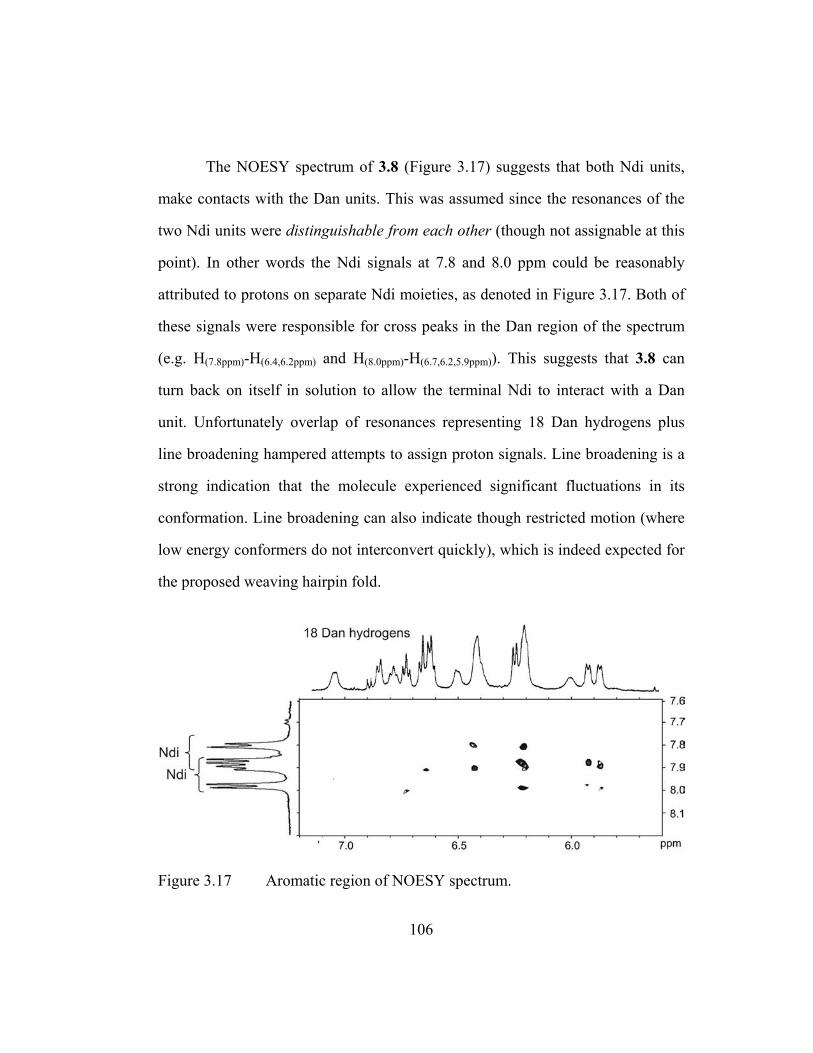
The NOESY spectrum of 3.8 (Figure 3.17) suggests that both Ndi units,
make contacts with the Dan units. This was assumed since the resonances of the
two Ndi units were distinguishable from each other (though not assignable at this
point). In other words the Ndi signals at 7.8 and 8.0 ppm could be reasonably
attributed to protons on separate Ndi moieties, as denoted in Figure 3.17. Both of
these signals were responsible for cross peaks in the Dan region of the spectrum
(e.g. H(7.8ppm)-H(6.4,6.2ppm) and H(8.0ppm)-H(6.7,6.2,5.9ppm)). This suggests that 3.8 can
turn back on itself in solution to allow the terminal Ndi to interact with a Dan
unit. Unfortunately overlap of resonances representing 18 Dan hydrogens plus
line broadening hampered attempts to assign proton signals. Line broadening is a
strong indication that the molecule experienced significant fluctuations in its
conformation. Line broadening can also indicate though restricted motion (where
low energy conformers do not interconvert quickly), which is indeed expected for
the proposed weaving hairpin fold.
Figure 3.17 Aromatic region of NOESY spectrum.
106

3.4 CHAPTER CONCLUSIONS
In summary, UV-Vis spectroscopy, unfolding studies, NMR, and
modeling investigations are fully consistent with the interpretation that both
naphthyl trimers 3.1 and 3.4 adopt a Dan/Ndi/Dan face-to-face stack in aqueous
solution. The consequence of this ring arrangement is a pleated fold for 3.1 and an
intercalative fold for 3.4. It thus appears that these folding patterns were predicted
by the linear sequence of two residue types owing to the strong directionality of
the Dan:Ndi association and the designability of naphthyl oligomers. This work
also demonstrates that foldamers for the first time can emulate natural proteins in
the sense that significantly different abiotic secondary structures can be accessed
with carefully chosen residue substitutions. Lastly, this intercalative fold may
represent an alternative to stabilizing turns or bends in water using aromatic
interactions an aim of many research groups.
3.5 IDEAS FOR FUTURE INVESTIGATIONS
Two research directions are proposed below to address the difficulties in
interpreting NMR spectra of naphthyl oligomers longer than trimers.
Deuterated aromatic units as an indispensable tool for proton
assignment and structure analysis. The hairpin pentamer conformation can be
greatly supported by proving the terminal Dan and Ndi units, which are distant in
an extended conformation, end up adjacent to each other in the folded structure.
By selective deuteration certain cross peaks found in Figure 3.17 can be
eliminated and thus attributed to specific aromatic-aromatic contacts, something
107
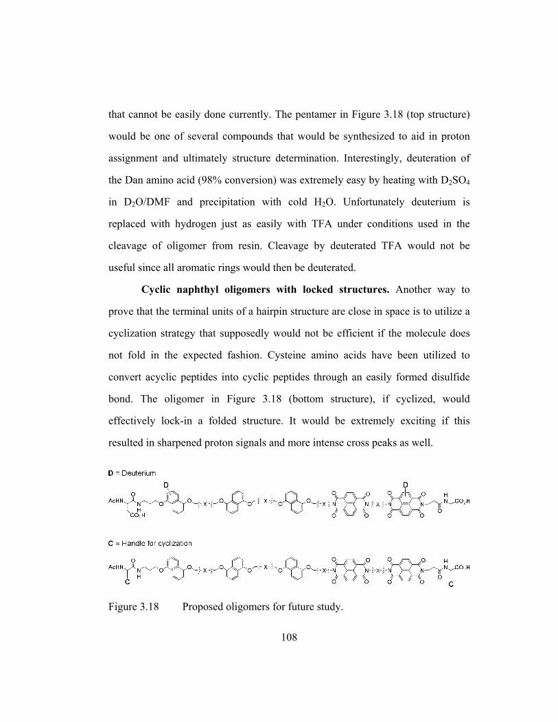
that cannot be easily done currently. The pentamer in Figure 3.18 (top structure)
would be one of several compounds that would be synthesized to aid in proton
assignment and ultimately structure determination. Interestingly, deuteration of
the Dan amino acid (98% conversion) was extremely easy by heating with D2SO4
in D2O/DMF and precipitation with cold H2O. Unfortunately deuterium is
replaced with hydrogen just as easily with TFA under conditions used in the
cleavage of oligomer from resin. Cleavage by deuterated TFA would not be
useful since all aromatic rings would then be deuterated.
Cyclic naphthyl oligomers with locked structures. Another way to
prove that the terminal units of a hairpin structure are close in space is to utilize a
cyclization strategy that supposedly would not be efficient if the molecule does
not fold in the expected fashion. Cysteine amino acids have been utilized to
convert acyclic peptides into cyclic peptides through an easily formed disulfide
bond. The oligomer in Figure 3.18 (bottom structure), if cyclized, would
effectively lock-in a folded structure. It would be extremely exciting if this
resulted in sharpened proton signals and more intense cross peaks as well.
Figure 3.18 Proposed oligomers for future study.
108

3.6 EXPERIMENTAL SECTION
Synthesis. General methods, full protocols for solid phase peptide
synthesis and FPLC/HPLC purification used for oligomers 3.1-3.8 were detailed
in chapter 2. Synthesis, followed by purification, desalting, and freeze-drying
afforded soft pale purple solids for all compounds.
Compound characterization (1D-NMR general methods). Samples
were readily soluble in 50 mM sodium phosphate, pH = 7.0 D2O. Spectra were
recorded on a Varian INOVA 500 MHz spectrometer at 1 mM concentrations of
compound and TSP-d4 (3-trimethylsilyl-propionic-2,2,3,3-d4 acid, sodium salt)
was used as a reference (δ = 0.00 ppm). Chemical shifts reported in ppm and
abbreviations used are singlet (s), doublet (d), doublet of doublet (dd), triplet (t),
quartet (q), multiplet (m) and complex multiplet of non-equivalent protons
(comp). J coupling constants (J) reported in Hz.
“AcHN-AspDanAspNdiAspDan*Gly-OH” (3.1). (SPPS yield = 91%) 1H NMR
(500 MHz, D2O) δ = 8.04 (q, J = 10.0 and 7.6, 4H), 7.22 (d, J = 8.4, 2H), 6.97-
6.94 (comp, 2H), 6.92 (d, J = 8.0, 1H), 6.86 (t, J = 7.6, 1H), 6.82 (t, J = 7.6, 1H),
6.72 (d, J = 8.4, 1H), 6.54-6.53 (comp, 2H), 6.34-6.30 (comp, 2H), 4.72 (dd, J =
5.6 and 2.2, 1H), 4.63 (dd, J = 5.4 and 2.4, 1H), 4.52 (dd, J = 5.0 and 3.8, 1H),
4.37-4.33 (m, 1H), 4.29-4.23 (m, 2H), 4.16-4.13 (m, 1H), 4.02-4.01 (m, 2H),
3.93-3.91 (m, 2H), 3.83-3.52 (comp, 9H), 3.49 (t, J = 6.8, 2H), 3.41-3.38 (m, 1H),
2.78-2.74 (m, 2H), 2.72-2.51 (comp, 6H), 2.45 (t, J = 7.4, 2H), 2.42-2.37 (m, 2H),
2.10-1.96 (comp, 8H), 1.94 (s, 3H); ESI MS calcd for C69H74N9O23 [MH]+ 1397,
found 1395.
109

“AcHN-AspDanAspNdiGly-OH” (3.2). (93% yield) 1H NMR (500 MHz, D2O)
δ = 8.33 (q, J = 10.0 and 7.5, 4H), 7.02 (d, J = 8.4, 1H), 6.98 (t, J = 8.4, 1H), 6.93
(d, J = 8.4, 1H), 6.90 (t, J = 7.7, 1H), 6.59 (d, J = 7.0, 1H), 6.29 (d, J = 7.6, 1H),
4.65 (dd, J = 8.1 and 5.2, 1H), 4.40-4.25 (m. 4H), 4.15 (t, J = 5.7, 1H), 4.01 (m,
2H), 3.80-3.70 (m, 2H), 3.73 (s, 2H), 3.63 (m, 3H), 3.52 (m, 1H), 2.78-2.72 (m,
3H), 2.65 (dd, J = 16.0 and 8.2, 1H), 2.51-2.44 (m, 3H), 2.35 (dd, J = 16.0 and
9.2, 1H), 2.08 (m, 4H), 1.89 (s, 3H); ESI MS calcd for C48H49N7NaO17 [MH]+
1019, found 1019.
“AcHN-AspDanAspAspDan*AspNdiGly-OH” (3.3). (80% yield) 1H NMR (500
MHz, D2O) δ = 8.00 (d, J = 7.6, 2H), 7.91 (d, J = 7.6, 2H), 7.13 (d, J = 8.3, 1H),
6.89 (d, J = 8.1, 1H), 6.84-6.82 (comp, 2H), 6.80-6.67 (comp, 4H), 6.48 (d, J =
7.5, 1H), 6.30 (d, J = 7.9, 1H), 6.27 (d, J = 7.5, 1H), 6.24 (d, J = 6.9, 1H), 4.61
(dd, J = 5.9 and 1.6, 1H), 4.44-4.39 (m, 2H), 4.24-4.13 (comp, 4H), 3.95 (d, J =
2.1, 1H), 3.91 (d, J = 6.6, 2H), 3.83 (s, 2H), 3.74-3.62 (comp, 7H), 3.48-3.45 (m,
1H), 3.40 (t, J = 6.2, 2H), 3.36-3.32 (m, 2H), 3.06 (dd, J = 5.5 and 0.1, 1H), 2.69-
2.63 (m, 3H), 2.58-2.25 (comp 12H), 2.03-2.00 (comp, 4H), 1.97-1.82 (comp
4H); ESI MS calcd for C73H78N10NaO26 [MH]+ 1534, found 1534.
“AcHN-AspDanAspDan*AspNdiGly-OH” (3.4). (86% yield) 1H NMR (500
MHz, D2O) δ = 8.07 (d, J = 7.5, 2H), 7.99 (d, J = 7.5, 2H), 7.18 (d, J = 8.5, 1H),
6.97 (d, J = 8.5, 1H), 6.93 (t, J = 7.9, 1H), 6.85 (t, J = 7.7, 1H), 6.80-6.77 (comp,
2H), 6.75 (t, J = 8.3, 1H), 6.64 (d, J = 8.3, 1H), 6.55 (d, J = 7.5, 1H), 6.36-6.32
(comp, 3H), 4.51 (dd, J = 5.2 and 3.6, 1H), 4.37-4.30 (comp, 3H), 4.27-4.23 (m,
1H), 4.05-4.00 (comp, 3H), 3.91-3.65 (comp, 11H), 3.62-3.58 (m, 1H), 3.47-3.42
110

(comp, 3H), 2.82-2.44 (comp, 11H), 2.40-2.35 (m, 1H), 2.13-1.95 (comp, 8H),
1.92 (s, 3H); ESI MS calcd for C69H73N9NaO23 [MH]+ 1419, found 1420.
“SucHN-AspDanDan*AspNdiGly-OH” (3.5). (83% yield) 1H NMR (500 MHz,
D2O) δ = 7.90 (d, J = 7.5, 2H), 7.78 (d, J = 7.5, 2H), 7.07 (d, J = 8.3, 1H), 6.96 (d,
J = 8.5, 1H), 6.86 (t, J = 8.2, 1H), 6.76-6.65 (comp, 5H), 6.58 (d, J = 7.8, 1H),
6.38 (d, J = 7.5, 1H), 6.12 (d, J = 7.8, 1H), 6.05 (d, J = 7.6, 1H), 4.44 (dd, J = 5.1
and 4.1, 2H), 4.39-4.31 (comp, 2H), 4.24-4.17 (comp 3H), 4.12-4.03 (m, 2H),
3.89 (t, J = 5.2, 2H), 3.74 (m, 1H), 3.63-3.45 (comp, 6H), 3.34 (m, 1H), 3.25 (m,
2H), 2.74-2.65 (comp, 4H), 2.61-2.39 (comp, 6H), 2.34-2.27 (comp, 6H), 2.14-
2.07 (comp, 4H), 1.91-1.82 (comp, 4H); ESI MS calcd for C69H72N8NaO24 [MH]+
1420, found 1420.
“SucHN-AspDan(8atoms)Dan*AspNdiGly-OH” (3.6). (84% yield) 1H NMR (500
MHz, D2O) δ = 8.03 (d, J = 7.6, 2H), 7.77 (d, J = 7.4, 2H), 7.49 (d, J = 8.8, 1H),
7.21-7.19 (comp, 2H), 7.14 (t, J = 8.2, 1H), 7.05-7.02 (comp, 2H), 6.99 (t, J = 8.0,
1H), 6.81-6.78 (comp, 2H), 6.53 (d, J = 7.6, 1H), 6.47 (d, J = 7.8, 1H), 6.33 (d, J
= 7.4, 1H), 4.63 (dd, J = 5.4 and 2.6, 1H), 4.54 (d, J = 2.2, 2H), 4.48-4.45 (m,
2H), 4.24-4.21 (comp, 6H), 3.79-3.70 (comp, 7H), 3.45-3.43 (m, 1H), 3.36-3.32
(m, 2H), 2.73-2.66 (m, 6H), 2.65-2.56 (m, 2H), 2.45-2.34 (comp, 4H), 2.27-2.18
(m, 2H), 2.00-1.91 (comp, 6H); ESI MS calcd for C67H67N8O24 [M]- 1368
(negative mode), found 1368.
“AcHN-AspDan(AspNdiAspDan)2Gly-OH” (3.7). (68% yield) 1H NMR (500
MHz, D2O) δ = 7.81-7.73 (comp, 8H), 6.86-6.81 (comp, 2H), 6.71-6.58 (comp,
7H), 6.52-6.48 (comp, 3H), 6.33 (d, J = 7.8, 1H), 6.24 (d, J = 7.1, 1H), 6.17 (d, J
111

= 7.2, 1H), 6.09 (d, J = 7.8, 1H), 6.05 (d, J = 7.9, 1H), 5.94 (broad d, 1H), 4.59-
4.52 (comp, 3H), 4.42 (dd, J = 4.9 and 4.0, 1H), 4.10-4.04 (comp, 7H) 3.96 (m,
1H), 3.82-3.76 (comp, 4H), 3.64-3.26 (comp, 22H), 2.73-2.40 (comp, 14H), 2.33-
2.28 (comp, 7H), 1.96-1.82 (comp, 15H); ESI MS calcd for C113H115N15NaO37
[MH]+ 2298, found 2298.
“AcHN-(AspDan)3(AspNdi)2Gly-OH” (3.8). (61% yield) 1H NMR (500 MHz,
D2O) δ = 7.88 (d, J = 7.6, 2H), 7.81-7.77 (comp, 2H), 7.70 (d, J = 7.2, 2H), 6.95
(broad, 1H), 6.78-6.63 (comp, 3H), 6.59-6.52 (comp, 3H), 6.44 (broad, 1H), 6.33
(comp, 3H), 6.16-6.11 (comp, 4H), 5.94 (broad, 1H), 5.84 (broad d, 1H), 5.78
(broad d, 1H), 4.62-4.58 (comp, 2H), 4.38 (dd, J = 5.1 and 4.0, 1H), 4.22-3.95
(comp, 8H), 3.85-3.78 (broad, 2H), 3.71-3.42 (comp, 18H), 3.40-3.22 (comp, 4H),
2.70-2.23 (comp, 20H), 1.92-1.78 (comp, 16H); ESI MS calcd for
C113H115N15NaO37 [MH]+ 2298, found 2298.
UV-Vis spectroscopy. UV-Vis spectra were taken on a temperature
regulated Hewlett Packard 8452A diode array spectrophotometer. Concentration
of stock solutions used for UV studies was initially determined by NMR
integration of a known concentration of TSP-d4 added to an aliquot. Unfolding
studies were carried out by monitoring the increase in the UV absorbance at 382
nm with the addition of cetyltrimethylammonium bromide detergent (CTAB).
Solutions of compound (40 µM in buffered H2O) were measured in a 1 cm
pathlength cuvette equipped with a microstirrer as a solution of 0.1 w/v % CTAB
in buffered H2O was added in 0.025-0.050 mL increments. Complete unfolding of
112

the naphthyl trimers was determined to be around 0.03 w/v % CTAB in the
cuvette. When graphed, absorbances were corrected for dilution.
2D-NMR spectroscopy. For proton assignment purposes several 2D-
NMR spectra were taken for 3.1, 3.4, and 3.6, all at 1 mM concentrations of
compound in sodium phosphate buffered solvents as above. Representative
spectra with acquisition parameters are shown in Figures 3.19-3.25. Samples in
90/10 buffered H2O/D2O were prepared for 2D NMR spectra which were
acquired at different mixing times, 200-800 milliseconds (ms) for NOESY spectra
and 50-150 ms for TOCSY spectra. Since the molecular weight of the compounds
fell in between the 800-1500 MW range where the negative/positive transition of
NOEs may occur, a ROESY, (which give always positive ROESY peaks and
negative exchange peaks) was required when NOESY afforded unexpectedly
weak NOEs. Depending on the conditions, this switch resulted sometimes in
clearer (more intense) cross peaks, other times not. Other considerations for
spectra acquisition were solvent suppression methods (1-1 jump-return
suppression sequence for ROESY and presaturation for NOESY and TOCSY
spectra) and filtering parameters, such as “gf” and “gfs”, during processing.
Only NOESY spectra taken in 100% buffered D2O with a mixing time of
800 ms was used for structural analysis. SPARKY, an online visualization
program, facilitated the imaging and interpretation of 2D-NMR spectra (Goddard
2000).
Molecular modeling. Computations were performed with the HyperChem
software using the MM+ force field. A set of twenty random starting
113

conformations of models for 3.1, 3.4, and 3.6 were generated using unrestrained
molecular dynamics at 1000 K. Each of the twenty diverse structures was then
allowed to anneal with a weak distance restraint as the simulation temperature
was lowered from 1000 K to 300 K over 10 ps. After annealing, a final geometry
optimization was performed without restraints using Fletcher-Reeves conjugate
gradient, 0.01 kcal·mol-1. The lowest energy conformer for both 3.1 (relative
energy = -18.97 kcal·mol-1) and its sequence isomer 3.4 (-19.27 kcal·mol-1)
afforded a structure that adopts a parallel Dan/Ndi/Dan stack.
114
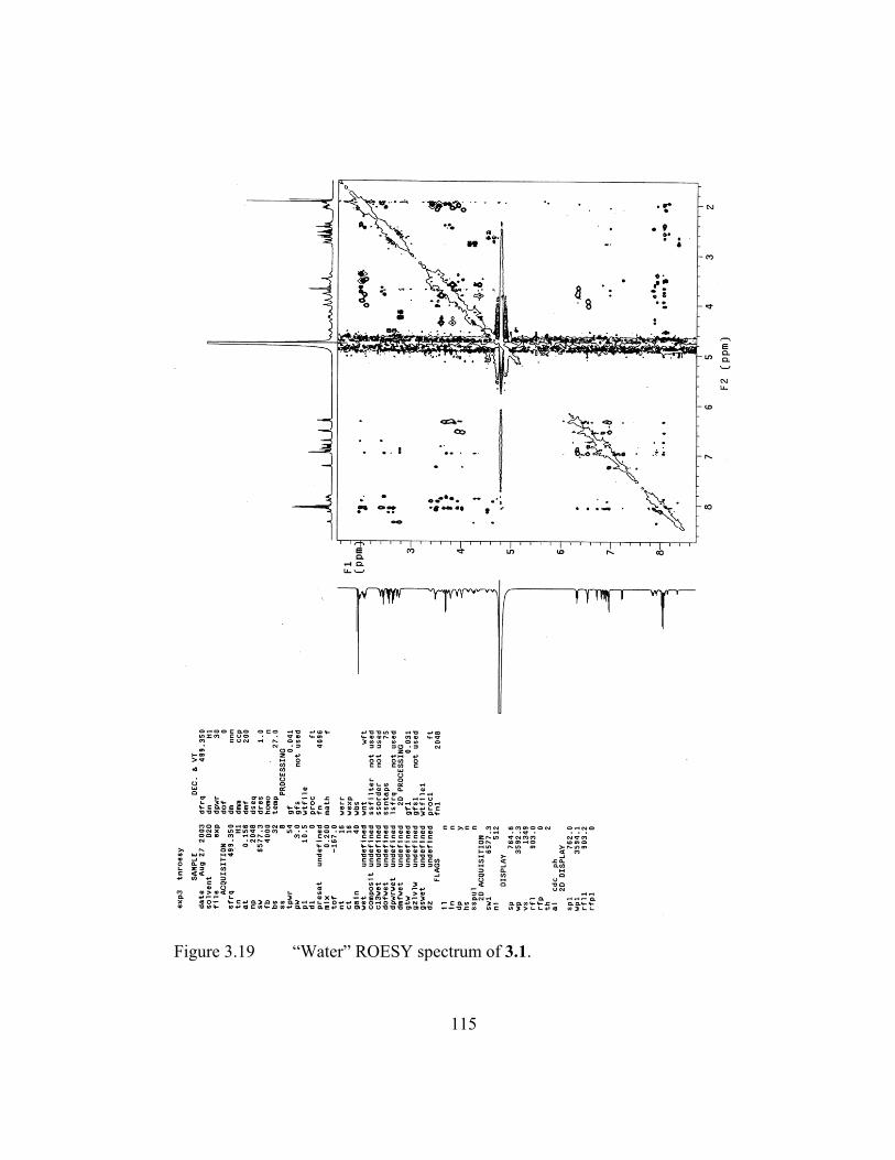
Figure 3.19 “Water” ROESY spectrum of 3.1.
115

Figure 3.20 Expansion of “water” ROESY spectrum of 3.1.
116

Figure 3.21 “Water” TOCSY spectrum of 3.4.
117

Figure 3.22 Expansion of “water” TOCSY spectrum of 3.4.
118
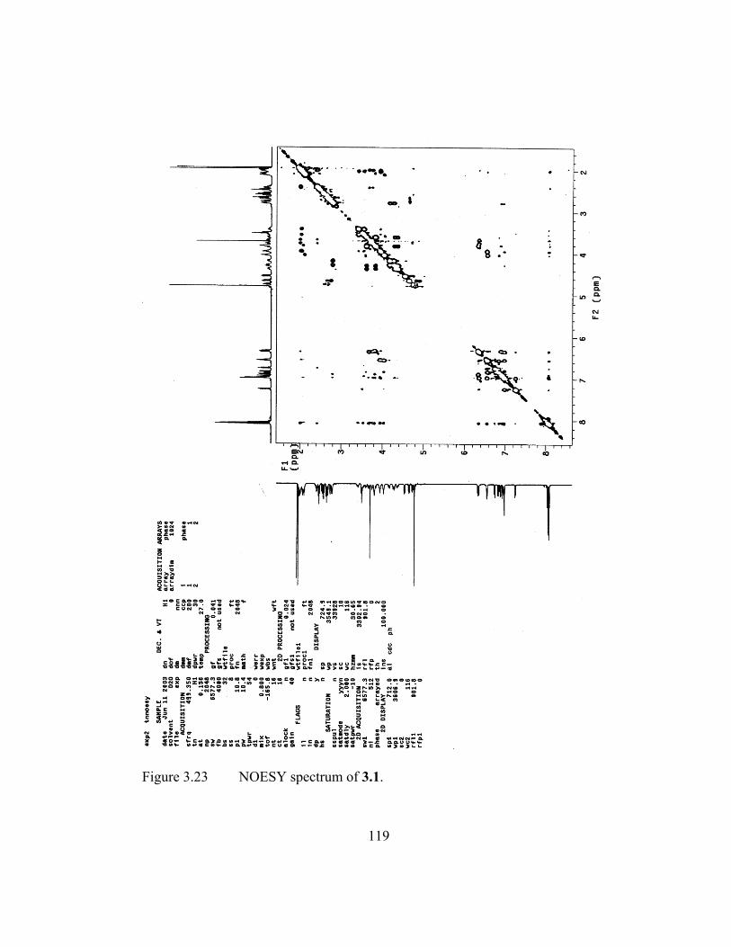
Figure 3.23 NOESY spectrum of 3.1.
119
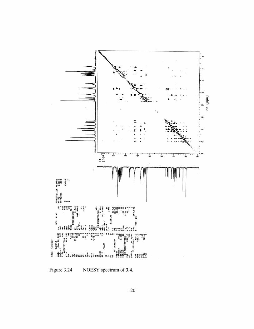
Figure 3.24 NOESY spectrum of 3.4.
120

Figure 3.25 NOESY spectrum of 3.6.
121

CHAPTER 4
Naphthyl Oligomers that Form Hetero-Duplexes
4.1 CHAPTER SUMMARY
Introduction. The previous chapter illustrated that the Dan:Ndi
interaction can dictate intramolecular folding of intercalative turns, a more
challenging topology than the pleated structures exhibited by aedamers. This
chapter will discuss work showing that the Dan:Ndi interaction can also direct
intermolecular binding and bring together Dann and Ndin chains to form hetero-
duplexes (Figure 4.1). Chemists have utilized, quite successfully, hydrogen
bonding and metal-coordination to construct duplex-forming oligomers with
desirable properties such as high binding affinities/stabilities and good chain
recognition. Examples of duplexes that use predominantly aromatic interactions
are few and examples that work in water mainly through aromatic stacking were,
to the best of our knowledge, non-existent prior to the system described in this
chapter. Synthetic hetero-duplexes that can operate in aqueous solutions could
possibly be exploited in a wide range of fields ranging from materials (high
strength fibers processible in water, a fairly benign solvent) to medicine
(diagnostic kits that require robust biomolecule immobilization). Next-generation
duplex systems that can self-sort large chain populations, can exhibit reversible
binding triggered by various stimuli, and can operate in a range of solvents and on
solid phase would also be very attractive.
122

Figure 4.1 Cartoon representation of a proposed hetero-duplex formed to maximize Dan:Ndi associations in an intermolecular fashion and critical questions answered in this project.
Goals. Experiments described in this chapter aimed to answer the
question: Can the Dan:Ndi interaction be exploited in an intermolecular fashion
to form stable hetero-duplexes with high affinities and chain recognition? The
short-term goals of this work were to develop a first-generation water compatible
hetero-duplex system formed from complementary oligo-Dan and oligo-Ndi
molecular strands and to investigate their binding behavior. The long-term
objective of this research is to create hetero-duplex systems with affinities and
binding selectivities sophisticated enough to be used possibly for building well-
defined aedamer assemblies with dramatically larger size domains and for
exploring surface patterning applications.
Approach. Binding stoichiometries and association constants were
determined by NMR and isothermal titration calorimetry (ITC) at different
temperatures. The thermodynamics of binding was also investigated. Size
exclusion chromatography (SEC) and polyacrylamide gel electrophoresis (PAGE)
123

experiments were carried out to further test the robustness of the complexes
formed.
Results. Overall, the work described in this chapter demonstrated the
assembly of oligo-Dan and oligo-Ndi strands into stable hetero-duplexes (Gabriel
2002). NMR and ITC investigations were consistent with a 1:1 binding ratio. ITC
revealed that tetra-Dan and tetra-Ndi chains (tetrameric system) formed hetero-
duplexes with a stability constant of 350,000 M–1 at T = 318 K, three orders of
magnitude higher than the binding constant of the monomeric system.
Additionally, ITC analysis calculated a heat capacity between –50 and –94
cal·mol–1·K–1 depending on the temperature range studied. Furthermore,
duplexation was enthalpically favored and an enthalpy-entropy compensation
effect was seen with the dimeric system. SEC chromatographs showed that a 1:1
molar mixture of tetra-Dan:tetra-Ndi resulted in material that eluted
approximately when a hetero-dimer would be expected to elute relative to
retention times of uncomplexed tetra-Dan and tetra-Ndi chains. Lastly, discrete
hetero-duplex formation for the tetrameric system was observed under PAGE
conditions even when one component was in slight excess over the other further
signifying a strong and discriminating inter-chain interaction.
4.2 BACKGROUND: SELF-ASSEMBLY OF MOLECULAR STRANDS
Natural chain molecules, or biopolymers, possess a range of functions due
largely to the organization of linear precursors into defined assemblies such as
multistranded complexes for proteins and duplexes in the case of DNA
(Venkatraman 2001, Wang 1991). Nearly 10 years after the review “Interlocked
124

and Intertwined Structures and Superstructures" was published (Amabilino 1995),
the field of self-assembly has continued to expand and has led to emerging
research programs in “bottom-up engineering" (Stoddart 2001), dynamic covalent
chemistry (Rowan 2002), and supramolecular protein assemblies (Yeates 2002).
The development of artificial chains that self-assemble into defined
complexes, in particular into duplexes, has drawn active interest recently.
Synthetic assemblies, where binding is based on recognition between two
complementary chains, could potentially afford capabilities for information
storage and transfer, reminiscent of DNA. Strands of protein, which form
biological fibers such as muscle (von Kiedrowski 1994) and spider silk (Vollrath
2000, Kubik 2002), have set high standards for materials considered for their
mechanical properties. Another protein example is the hetero-dimerization of
leucine zipper transcription factors that bind specific sequences of DNA (Glover
1995). Finally, molecular strands with orthogonal binding and self-sorting
capacities would nicely complement existing patterning and immobilization
technologies (Wilchek 1990).
While certain hydrogen bonding patterns are responsible for the secondary
structures of proteins and the base-pairing specificities of DNA, several groups
have reported impressive results using significantly different hydrogen bonding
strategies for their nonnatural duplex oligomers (Gong 2001, Archer 2000). For
instance, Krische and co-workers have synthesized oligomers applying a
“covalent casting” strategy to link amino-dichlorotriazines to design duplex
oligomers with predefined hydrogen bonding patterns (Archer 2002a, b). These
125
oligomers exhibited up to nanomolar binding affinities and a strong cooperative
effect was observed upon extension from monomer to dimer to trimer. Also at the
forefront of hydrogen bonded duplexes, is the work from Gong and co-workers
who have established specific recognition with their “hydrogen bonded tapes”
through the precise placement of hydrogen bonding donors and acceptors (Zeng
2001, 2002).
The wealth of hydrogen bonding donor and acceptor precursors has no
doubt been key for the investigation of a variety of bonding patterns and creative
design strategies. Though the use of hydrogen bonds has afforded synthetic
duplex systems with high binding strengths and selectivities, a discussion of
artificial duplexes would not be complete without referencing complexes based on
metal-coordination (Albrecht 2001). In 1987 Lehn introduced the term “double-
stranded helicates” for the complexes between oligobipyridine and copper(I)
cations that were shown by x-ray crystallography to form double helices (Lehn
1987). Studies with double-stranded helicates continue to this day with recent
examples showing special features, for instance, stereoselective self-assembly
(Telfer 2004) and synergism of metal coordination and hydrogen bonding effects
(Chowdhury 2004).
In close relation to the work presented in this chapter, duplexes having an
aromatic interactions component have also been reported. Probably the earliest
published report of an aromatic donor-acceptor hetero-duplex system came from
Stoddart's group using oligomers of linked hydroquinol and linked bipyridinium
units (Figure 4.2). Structure characterization from this pioneering effort was not
126

conclusive for double-helical conformations, as hoped for by the authors, since
low temperature NMR did not seem to significantly freeze out a unique structure.
Nonetheless, this groundbreaking study put forth the idea of using aromatic
stacking that is, in a sense, less geometrically rigid than hydrogen bonding and
metal coordination.
Figure 4.2 Aromatic π-π stacking system from Stoddart’s laboratory (Ashton 1992).
The Stoddart group has continued to do important work in the field of
supramolecular chemistry (Philp 1996) mainly focusing on the development of
aromatic donor-acceptor based catenanes and rotaxanes (rather than hetero-
duplexes) for use in nanoscale devices (Stoddart 2001, Feynman 1960). This
research program has been tremendously prolific and most recently, in a
milestone achievement in molecular topology, they reported the self-assembly of
the first wholly synthetic Borromean rings (Chichak 2004, Mao 1997).
The next two examples described used a combination of hydrogen
bonding and aromatic interactions. Lehn and co-workers have synthesized a
family of oligo(pyridylamide)s that exhibited dynamic exchange between single
127

and double helices (Berl 2000, 2001). In general, intramolecular hydrogen bonds
were responsible for the helical conformations while aromatic stacking stabilized
dimerizations (Figure 4.3).
Figure 4.3 X-ray crystal structure of Lehn’s oligopyridinecarboxamide (R1 = R2 = H, R3 = OtBu) showing interstrand aromatic stacking and hydrogen bonding which includes two bridging NH-O hydrogen bonds. Crystals grown from CH3CN/DMSO. (Berl 2000).
The “molecular zippers” designed by Hunter and co-workers were
prepared form isophthalic acid and bisanaline derivatives (Bisson 1994, 2000,
Hunter 2003). Hydrogen bonding was determined to be the main driving force for
complexation but interestingly aromatic interactions that had edge-to-face
orientations also existed (Figure 4.4). For this system, recognition properties
between unlike strands seemed to rely simply on the length of the oligomers.
128
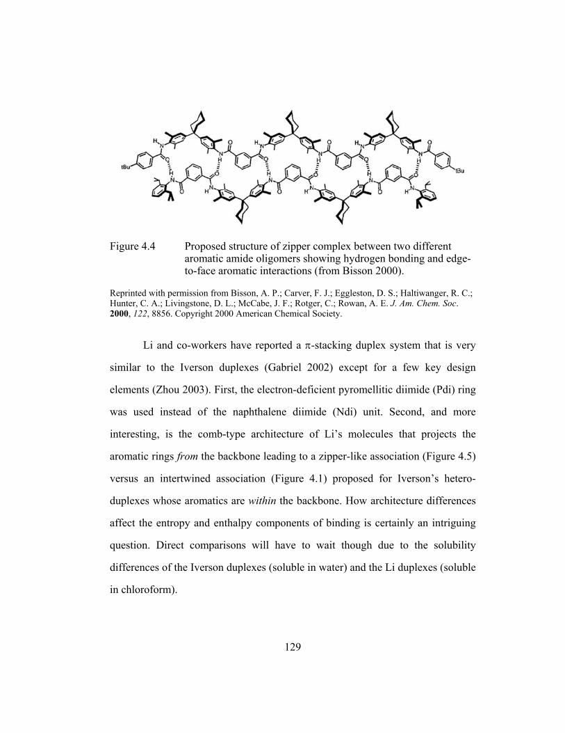
Figure 4.4 Proposed structure of zipper complex between two different aromatic amide oligomers showing hydrogen bonding and edge-to-face aromatic interactions (from Bisson 2000).
Reprinted with permission from Bisson, A. P.; Carver, F. J.; Eggleston, D. S.; Haltiwanger, R. C.; Hunter, C. A.; Livingstone, D. L.; McCabe, J. F.; Rotger, C.; Rowan, A. E. J. Am. Chem. Soc. 2000, 122, 8856. Copyright 2000 American Chemical Society.
Li and co-workers have reported a π-stacking duplex system that is very
similar to the Iverson duplexes (Gabriel 2002) except for a few key design
elements (Zhou 2003). First, the electron-deficient pyromellitic diimide (Pdi) ring
was used instead of the naphthalene diimide (Ndi) unit. Second, and more
interesting, is the comb-type architecture of Li’s molecules that projects the
aromatic rings from the backbone leading to a zipper-like association (Figure 4.5)
versus an intertwined association (Figure 4.1) proposed for Iverson’s hetero-
duplexes whose aromatics are within the backbone. How architecture differences
affect the entropy and enthalpy components of binding is certainly an intriguing
question. Direct comparisons will have to wait though due to the solubility
differences of the Iverson duplexes (soluble in water) and the Li duplexes (soluble
in chloroform).
129

Figure 4.5 A donor-acceptor tetrameric hetero-duplex formed with comb-type naphthyl oligomers (Zhou 2003).
As mentioned above, the hetero-duplexes introduced by the Iverson
laboratory were soluble in aqueous solutions and previous studies have shown
that the Dan:Ndi interaction was overwhelmingly stronger in water than in
organic solvents (Cubberley 2001b). On the other hand, all of the synthetic
duplexes presented above were studied in organic solutions in which extremely
strong associations were typically obtained. In studies that tested the addition of
polar protic solvents such as methanol (Bisson 2000) and water (Berl 2000),
duplex formation was disrupted. Structure characterization of most of these
systems though has benefited greatly from x-ray quality crystals grown from
organic solutions. Attempts to grow crystals of water-soluble naphthyl oligomers
have been unsuccessful. Thus the synthesis of organic-soluble analogues is
another pursuit of the Iverson laboratory (Cubberley 2000). Nonetheless, the
water solubility of the hetero-duplexes presented in the following sections may
confer its own advantages (long-term), most promising of which is the possibility
to construct duplex foldamers compatible with biological systems.
130

4.3 RESULTS AND DISCUSSION
4.3.1 Synthesis of Homo-Naphthyl Oligomers
The synthesis of 4.1-4.8 (Figure 4.6) used monomers and SPPS protocols
previously described in Chapter 2. While the incorporated aspartate residues
provided the much desired solubility in water buffered to pH = 7, consequently,
the desired hetero-duplex formation might experience significant charge repulsion
involved with the assembly of like-charged chains. Purification via FPLC (>98%
pure judged by HPLC peak area) was straightforward for all compounds except
4.7, which required multiple FPLC runs at 50% longer gradients since a small
amount of the 4.5, formed as a deletion side-product, had similar retention times
on the reverse-phase column. Final desalting via a C18 Sep-Pak cartridge and
freeze-drying afforded soft solids (white and pale yellow in color for the oligo-
Dan and oligo-Ndi series, respectively).
Figure 4.6 Compounds synthesized and studied in Chapter 4. 13-atom linkers between naphthyl moieties were used.
131

Figure 4.7 shows an idealized model of the dimeric (4.3:4.4) duplex. As
previously seen with computer modeling of the intercalative fold topology
(Chapter 3, Section 3.3.7), a 13-atom linkage between naphthyl units would allow
“sandwiching” of an aromatic ring for intertwining chains also (Figure 4.7).
Geometry optimization of a 10-atom linker indicated that it was too short to allow
ideal face-centered stacking in which the aromatic rings are parallel and ~ 3.5 Å
apart (approximate inter-ring distance based on van der Waals radii. Modeling of
dimers with a 16-atom linkage resulted in structures with markedly kinked
linkers, which would be expected to lower binding affinities. Although far from
rigorous, modeling still provided a good starting point for the design of
intertwining strands of oligo-Dan and Ndi chains.
Figure 4.7 Computer generated space-filled model (right structure is rotated by 90°) of the duplex formed from truncated 4.3 (black) and 4.4 (white). Linker atoms not along backbone path omitted for clarity.
4.3.2 NMR Job Plots and Titrations
Foremost, a one-to-one complex between oligo-Dan and oligo-Ndi
molecules had to be established. Although detailed complexation studies have
been previously performed for uncharged, but aqueous-soluble monomers (Zych
132

2000, Cubberley 2001b), 4.1 and 4.2 were designed to be multi-charged in water
akin to the longer oligomers of this study and thus required their own NMR
analysis. NMR experiments were also performed for the dimeric duplex complex
4.3:4.4. The chemical shifts of the Ndi unit(s) in 4.1 and 4.3 conveniently allowed
the determination of binding stoichiometry by the Job’s method (Connors 1987,
Gil 1990). One can use Job plots to distinguish between one-to-one from higher-
order binding simply by inspection (Figure 4.8). The Ndi chemical shifts also
permitted calculation of binding constants, Ka(4.1:4.2) and Ka(4.3:4.4), using
titration data (Macomber 1992, Figure 4.8). Quantitative analysis of titration data
was accomplished using the HOSTEST program developed by Wilcox and
Glagovich (Wilcox 1997).
133
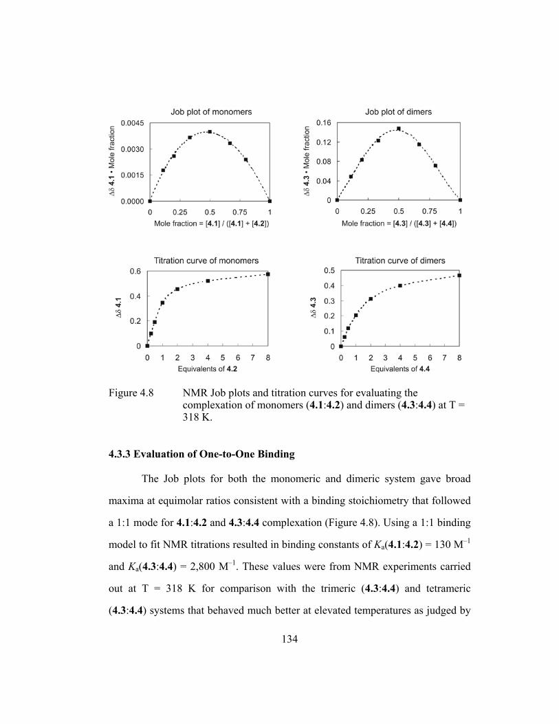
Figure 4.8 NMR Job plots and titration curves for evaluating the complexation of monomers (4.1:4.2) and dimers (4.3:4.4) at T = 318 K.
4.3.3 Evaluation of One-to-One Binding
The Job plots for both the monomeric and dimeric system gave broad
maxima at equimolar ratios consistent with a binding stoichiometry that followed
a 1:1 mode for 4.1:4.2 and 4.3:4.4 complexation (Figure 4.8). Using a 1:1 binding
model to fit NMR titrations resulted in binding constants of Ka(4.1:4.2) = 130 M–1
and Ka(4.3:4.4) = 2,800 M–1. These values were from NMR experiments carried
out at T = 318 K for comparison with the trimeric (4.3:4.4) and tetrameric
(4.3:4.4) systems that behaved much better at elevated temperatures as judged by
134

ITC data (more on this behavior in the following sections). As expected, binding
constants increased when the monomeric and dimeric complexes were analyzed at
T = 298 K affording a Ka(4.1:4.2) = 300 M-1 and Ka(4.3:4.4) = 7,500 M-1 , more
than twice the value measured a t T = 298 K.. Thus the binding stoichiometry and
binding constants of the monomeric and dimeric systems were determined and the
results are summarized in Table 4.1.
4.3.4 Isothermal Titration Calorimetry (ITC)
Reliable binding analysis of NMR titration data is dependent on tracking a
particular proton signal, but this task became increasingly difficult with longer,
unit-repeating oligomers due to the shifting of severely overlapped signals. There
was little confidence placed in data based on following the intractable Ndi signals
for 4.5 and 4.7 in titration experiments. Thus a new method had to be identified to
obtain Ka(4.5:4.6) and Ka(4.7:4.8) for trimeric and tetrameric duplexation,
respectively.
Analysis of titration data collected with ITC experiments is an attractive
alternative on several fronts (Wiseman 1989). ITC measures the heat evolved or
absorbed by a system during a titration run and uses these values as its data set
rather than chemical shifts. If an injection of titrant results in the system absorbing
heat an upward spike is observed indicating the instrument had to add energy to
the system to maintain a constant heat signal. If the injection of titrant results in
heat being evolved (i.e. an exothermic process) then the heat signals spike
downward. ITC provides an efficient means of obtaining Ka, enthalpy (∆H), and a
stoichiometry (N = molar equivalent of titrant) of binding. With equation 1, free
135

energy (∆G) and entropy ∆S of binding can be calculated hence the
thermodynamic parameters associated with the binding event can be determined
in a single titration. For further discussion, a review of the advantages and
limitations of ITC has been published (Wadso 1997).
∆G° = –RT lnKa = ∆H° – T∆S° (1)
Unfortunately, monomeric complexation of 4.1 with 4.2 was below the
sensitivity limit of microcalorimetry (see explanation in Experimental Section,
Wiseman 1989). ITC though was carried out appropriately for the dimeric (whose
Ka values can be compared with those derived from NMR), the trimeric, and
tetrameric systems. A VP-ITC MicroCalorimeter instrument and the analysis
software provided with it were used in these experiments (Microcal, Inc. 1999).
When examining the tetrameric system it could be clearly seen that the
isotherms collected at T = 298 K 1) did not satisfactorily exhibit binding
saturation and 2) did not fit any of the Microcal binding models well (Figure 4.9,
left isotherm). Comparing the experimental data to a best-fit theoretical curve
gave a chi-square fitting analysis of 250,000 (unitless). In personal
communications, ITC technicians recommended using data with chi-square values
closer to 50,000 or lower (Microcal, Inc 1999). Similar problems were
encountered with the trimeric system at this temperature (Isotherm of trimeric
system at T = 298 K in Experimental Section). Although the average chi-square
of 55,000 was better than that calculated for the isotherm of the tetrameric system
at identical conditions, the trimer isotherm did not show sufficient saturation
136

(arbitrary judgment based on the amount of heat still being released at the end of
the titration).
Figure 4.9 Binding isotherms for the titration of 4.7 with 4.8 at two different temperatures. Top panels display, the raw isotherm or the total heat evolved (µcal/sec) per injection of 4.8 (40 injections total). Data points in the bottom panels represent the heat evolved (kcal/mol) after correcting for heats of dilution. ITC data collected at T = 298 K did not fit theoretical curves (line in bottom panels). Data collected at T = 318 K on the other hand fitted predicted curves well according to a chi-square error analysis (Wiseman 1989, Microcal, Inc. 1999).
Faced with these initial difficulties, reverse titrations and sectional
titrations where the whole binding isotherm was pieced together from several ITC
runs were tried. These setups though were unsuccessful. Fortunately, when
137

identical runs were performed at T = 318 K (Figure 4.9, right isotherm), well-
behaved, 1:1 binding isotherms were acquired that fit the predicted curves
accurately (Chi-square = 35,000 for the tetrameric system and 18,000 for the
trimeric system). These results suggested that alternate modes of association, such
as 1:2, 2:1 binding and problematic homomeric aggregation, possibly exist at
ambient conditions but are disfavored at elevated temperatures. Also more telling
is the apparent non-saturation of isotherms obtained at ambient conditions near
the end of the titration run where the oligo-Dan is in molar excess which may
signal possibly “off-register” binding to provide desolvation of added oligo-Dan.
Importantly, results were consistent over multiple trials conducted at T = 318 K
and the fit of the curves, calculated by the analysis software, were entirely
acceptable based on chi-square error analysis (Figure 4.9, see Experimental
Section for supporting isotherms with corresponding thermodynamic parameters
analyses.
With experimental conditions set, isotherms were collected for dimer
(4.3:4.4), trimer (4.5:4.6), and tetramer (4.7:4.8) duplexation. Three trials were
performed for each system. Figure 4.10 shows representative isotherms. The
predicted curves varied the N value and for all cases the best fits were obtained
when N = 1 ± 0.1 molar equivalent of titrant.
138

Figure 4.10 Representative binding isotherms at T = 318 K. Three trials were performed for each system. Fitted line was based on 40 injection data points but only every other point is shown for clarity.
139

Table 4.1 reports the binding constants and provides a thermodynamic
profile for 1:1 binding of each of the pairings. Before a discussion of the binding
constants takes place, several points need to be made about the table. First, NMR
and ITC values at T = 298 K are absent for the trimeric and tetrameric systems for
reasons mentioned before. Second, only the dimeric system had binding constants
within the practical range of both NMR and ITC analysis. These values will be
considered shortly. Third, while the binding thermodynamics were parsed into
their enthalpy (∆H) and entropy (∆S) components for ITC experiments, a
published report on the pitfalls of using the van’t Hoff method of obtaining ∆H
and ∆S from NMR data led to the decision to pass on additional experiments to
calculate these thermodynamic parameters (Naghibi 1995). In this report several
examples were cited where significant discrepancies were found between
enthalpies derived by the van’t Hoff method and enthalpies directly measured by
ITC. The rule of thumb presented was that ∆H and ∆S should not be derived from
NMR data if enthalpy of the system is significantly temperature dependent. This
dependence was shown for the dimeric system 4.3:4.4 with ITC experiments
conducted at four different temperatures (Figure 4.11, this data is presented in the
context of heat capacity discussed in Section 4.3.6).
140

Table 4.1 Summary of hetero-duplex binding data. a
Complex b Ka (T=298 K) ∆G° ∆H° ∆S° N value 4.1:4.2 (Mono-NMR) 3.0 × 10² −3.4 NA NA 1 c
4.3:4.4 (Di-NMR) 7.5 × 10³ −5.3 NA NA 1 c
4.3:4.4 (Di-ITC) 7.6 × 10³ −5.3 −10.4 −17.2 1.01 Ka (T=318 K) 4.1:4.2 (Mono-NMR) 1.3 × 10² −3.1 NA NA 1 c
4.3:4.4 (Di-NMR) 2.8 × 10³ −5.0 NA NA 1 c
4.3:4.4 (Di-ITC) 2.7 × 10³ −5.0 −12.3 −23.0 0.91 4.5:4.6 (Tri-ITC) 4.5 × 10⁴ −6.8 −17.7 −34.2 0.97 4.7:4.8 (Tetra-ITC) 3.5 × 10⁵ −8.1 −19.3 −35.3 0.91 a Ka (M–1), ∆G° (kcal·mol–1), ∆H° (kcal·mol–1), ∆S° (cal·mol–1·K–1). Complete hetero-duplex binding data and experimental errors for the measured values of Ka, ∆H°, and N can be found in the experimental section. ∆G° and ∆S° were calculated from averaged Ka and ∆H° values, respectively. b Size of oligomers used and method of analysis in parentheses. c A binding stoichiometry of 1:1 was determined by inspection of Job plots. NA = Not applicable for NMR analysis (see text for explanation).
4.3.5 Summary of Binding Data
Results in Table 4.1 were gratifying for two main reasons. First, binding
constants from separate analyses for the dimeric system (4.3:4.4) were in
agreement. NMR indicated a Ka(T=298K)
= 7,500 M−1 and Ka(T=318K)
= 2,800 M−1
while ITC corroborated these numbers with a Ka(T=298K)
= 7,600 M−1 and Ka(T=318K)
= 2,700 M−1. Also, fitted curves point to discrete one-to-one binding (N values in
Table 4.1). Raising the temperature 20 degrees K leds to more than a 50%
decrease in association constants for the dimeric system. Even for the trimeric
system at 298 K (whose data is just outside of the chi-square error window) the
fitting analysis pointed to Ka(4.5:4.6)s above 10,000 M−1 versus 45,000 M−1 at
141

318 K. It would not be unreasonable to believe that with the proper design
adjustments longer oligomer would give discrete hetero-duplexes at ambient
conditions with much higher binding constants than those observed at elevated
temperatures. Second, association improved with increasing chain length, with
free energy (∆G°) of duplex formation being roughly additive, with a change of
−1.3 to −1.9 kcal·mol−1 per additional aromatic unit. Though the increasingly
favorable free energy with increasing length is promising, future design
improvements are hoped to lead to “cooperative binding effects” (Ercolani 2003)
with longer oligomers.
ITC clearly revealed that association was enthalpically favored with
∆H°(T=318K) values ranging from −12 to −19 kcal·mol−1. Though this data is
irrefutable, any explanation as to why a process is enthalpically (and/or
entropically) favored has to proceed with caution to avoid confusion.
Typically, favorable changes in solvent-solvent cohesion interactions
(presuamably an increase in hydrogen bonding in the case where water is the
solvent) are ascribed to the favorable enthalpies seen with binding systems
operating on desolvation (or the hydrophobic effect). In other words the solvent
rather than the binding molecules (or solutes) is mainly responsible for the
observed favorable enthalpies (once again in the cases with a strong hydrophobic
driving force). The hydrophobic effect though should not automatically be
synonymous with an “enthalpy-driven” process. Desolvation in fact has been
implemented in entropically favored binding events where there is an entropy
gain by the solvent since supposedly there is less solute surface required to be
142

solvated by highly ordered solvent molecules after an apolar binding event. A
classic example would be micelle formation in water driven by the desolvation of
apolar surfaces. After micelle formation, water is simply more disordered
(solvation of more apolar areas means more ordered water molecules) hence this
particular process is entropically favored. It is this situation most often attributed
to the hydrophobic effect (Tanford 1980).
One could imagine that this scenario would also apply to the Iverson
duplex system predicated on the hydrophobically-driven interaction between the
Dan and Ndi units. ITC though indicated that entropy was unfavorable with
∆S°(T=318K) values ranging from −23 to −35 cal·mol−1. For comparison, Diederich
and co-workers who have studied, in detail, aromatic ring inclusion by
cyclophanes in water, have found similar favorable enthalpy values ranging from
−8 to −13 kcal·mol−1 and unfavorable entropy values ranging from −6 to −22
cal·mol−1·K−1 depending on the nature of the aromatic guests used (Smithrud
1991). Diederich and co-workers characterized these apolar binding processes as
being driven by hydrophobic bonding and proposed that the degrees of freedom
lost by the bound molecules masked any positive entropic benefit experienced by
the solvent due to the hydrophobic effect (Smithrud 1991). Therefore, there is no
contradiction in stating that tight host-guest complexation in water, which is often
observed to be enthalpically favored (∆H° << 0, T∆S° < 0) and “less tight”
micelle formation, which is often entropically favored (∆H° ≈ 0, T∆S° > 0) can
both be considered hydrophobically-driven processes (Smithrud 1991). The larger
entropic cost for the assembly of the Iverson duplexes as compared to the
143

Diederich cyclophane system is thus expected since the ordering of flexible
strands into an entwined duplex likely results in more degrees of freedom lost
than when cyclic hosts bind small molecule guests in general.
Returning to the evaluation of binding affinities, it was most satisfying to
observe that the hetero-duplex 4.7:4.8 displayed an association constant of
350,000 M-1, three orders of magnitude larger than 4.1:4.2, despite presumably
larger charge repulsion between the longer chains. This result emphasizes the
relatively strong driving force for duplex formation present in oligo-naphthyl
systems. In fact, negative charge along the backbone of both oligomers may
confer an important advantage over neutral strands. As is thought for DNA,
intramolecular charge repulsion might keep oligomer chains “spread out” and
more available to interact with a complementary chain (Benner 2002). Secondly,
having like-charged strands, instead of complementary charged chains, kept the
final hetero-duplex water-soluble as was required for NMR and ITC binding
analysis.
4.3.6 Examination of Heat Capacity
To elucidate the underlying thermodynamic parameters of binding, 4.3:4.4
association was reexamined from T = 288 to 318 K (Figure 4.11) to obtain heat
capacities. The change in the heat capacity, ∆Cp, (∆Cp = δ∆H° / δT) associated
with binding is often reported for biological systems in order to determine the
extent of hydrophobic driven recognition (Murphy 1999). There are still questions
though whether heat capacity can be correlated accurately with the change in the
amount of buried surface area for a binding event (Guinto 1996, Gill 1985).
144

Notwithstanding this debate, heat capacity still remains an important
thermodynamic quantity to measure differences in solvent-solute interactions and
can lead to insights into conformational changes of the binding partners (Murphy
1999).
Figure 4.11 Graph of the thermodynamic parameters taken from ITC experiments of 4.3:4.4 binding and table of the corresponding free energies.
A change in heat capacity (∆Cp) is typically reported in cal·mol−1·K−1 and
is defined as the amount of heat required to increase the temperature of a
substance (or solution) by 1 Kelvin. In essence, it represents a change in the way
the solvent solvates the solute. Therefore, ∆Cp is a convenient term to compare
not only other different binding systems, but also folding systems. A simple
example is when a protein unfolds there is a positive change in the heat capacity.
The same effect is seen when hydrophobic solutes are added to water; the
observed ∆Cp is positive. The converse is true as well and protein folding,
145

protein-protein binding, and adding ionic solutes in water almost always afford
negative ∆Cp values.
ITC experiments revealed a significant temperature dependence of
enthalpy ∆H° (Figure 4.11) that is not linear thus precluding determination of a
single ∆Cp value over the whole temperature range investigated (Gill 1985).
Linear-fitting over narrower temperature ranges gives ∆Cp values between −50 (at
lower temperature ranges) and −95 cal·mol−1·K−1 (at higher ranges). Note, that this
does not imply that the overall stability of the duplex is greater at elevated
temperatures. It could indicate that there exists a larger difference in exposed
surface area between unbound single strands and duplexes at higher temperatures
versus lower temperatures. The Diederich studies with cyclophane hosts reported
∆Cp values up to −130 cal·mol−1·K−1 for the binding of relatively polar guests
(nitrophenol) and smaller negative values (−20 cal·mol−1·K−1) for less polar guest
such as p-xylene (Ferguson 1991, Smithrud 1991).
Finally, while evaluating data in the context of heat capacity
determination, an apparent enthalpy-entropy compensation effect (Liu 2001) was
observed with ∆G°(4.3:4.4) values that decrease only slightly with increasing
temperature (Figure 4.11). Another way of stating this is ∆∆G° ≈ 0 while ∆∆H°
and ∆∆S° were significant but compensatory. In Figure 4.11 the compensation
occurred with one constant system (the dimeric system) studied at various
temperatures. Often though the phenomenon of enthalpy-entropy compensation
arises in studies that vary the system slightly, such as in the case of rigidification
of ligands to possibly improve the entropic component of ligand-protein binding.
146

Explanations for this compensation have been debated. Some reports
simply, and quite reasonably, state, “a stronger intermolecular interaction or
bonding (related to enthalpy) will lead to a greater reduction of the
configurational freedom…(Liu 2001)” The review written by Liu and Guo give
other numerous explanations some choosing to focus on the thermodynamic
parameters of the solvent rather than the interlocking molecules (Liu 2001).
Counterpoints though exist in such articles entitled, “Entropy-enthalpy
compensation: fact or artifact?” (Sharp 2001) and “Enthalpy-entropy
compensation: a phantom phenomenon” (Cornish-Bowden 2002). Invariably,
more studies will be reported supporting one explanation over the other, but
hopefully a working knowledge of how to improve ∆G° of binding through
molecular designs that favorably affect both enthalpy and entropy will also come
forth from these investigations.
4.3.7 Size Exclusion Chromatography (SEC)
With binding constants and the thermodynamic profiles of hetero-duplex
formation well in hand the following two analyses (size exclusion
chromatography, SEC, and polyacrylamide gel electrophoresis, PAGE) were
carried out to gain further insight as to the robustness of the complexes under
other experimental conditions.
A 1:1 solution of 4.7:4.8 resulted in two separate signals when passed
through a C18 reverse-phase column using the HPLC conditions that were used to
purify naphthyl oligomers. The two signals corresponded to the retention times of
pure 4.7 and pure 4.8 indicating that the putative tetrameric duplex did not survive
147
these conditions. In brief, the HPLC column separates oligomers by hydrophobic
interactions with the packing material. Samples are usually loaded onto the
column with predominantly water (>9:1 ratio of H2O:CH3CN). As the percentage
of acetonitrile is increased the solvent (mobile phase) competes with the packing
material (stationary phase) for interactions with the compound (duplex) and thus
elutes the sample. Most likely the increase in organic solvent disrupts duplex
formation. In pure water runs the compound did not migrate at all through the
column and in isocratic runs (at a constant H2O:CH3CN composition) still only
separation of chains was observed.
On the other hand, SEC runs (using a proprietary Bio-Sil column, Bio-Rad
Inc., Hercules, CA 94547) afforded a different retention profile for a 1:1 mixture
than both pure 4.7 and pure 4.8 (Figure 4.12). SEC, synonymous with gel
filtration chromatography, separates compounds based on diffusion in and around
highly porous silica beads. Retention is more dependent on the size and shape of
the molecules rather than their hydrophobicity and charge, although these
properties can play an important role. Since large molecules are sterically
excluded and pass around the beads these molecules elute fastest (at lower
retention volumes). SEC columns are usually more amenable than HPLC columns
to isocratic runs ideal for separating proteins and nucleic acids that have
molecular weights of 2,000-1,000,000 g·mol−1.
148

Figure 4.12 UV chromatograph (270 nm) showing that solutions of 4.7 are aggregated (at the SEC conditions) but are effectively broken up by the addition of 4.8 as seen by the trace give the 1:1 molar mixture. Also the 1:1 mixture results in a complex with a distinctly different retention volume than either strand.
Dr. Thomas Mourey from the Eastman Kodak Company (Rochester, NY
14650) performed all the SEC studies discussed with runs using Na phosphate
buffered water. The chromatograph in Figure 4.12 indicated that 4.7 was
aggregated (retention volume 1.4 mL). The 4.7 species at retention volume 2.7
mL was probably in the monomeric form considering that 4.8 showed no sign of
forming large aggregates and its retention volume was similar at 3.0 mL. Most
interestingly was that the addition of 4.8 to 4.7 appeared to significantly break up
the 4.7 aggregates. It is important to remember that ITC analyses at T = 318 K
were not hampered by aggregates. If aggregates under these ITC conditions were
significant then reproducibility of measurements and curve-fitting to afford N
149
values near 1 molar equivalent definitely would have been problematic and
substantial errors reported. With that being the case, aggregation was not
prohibitive to the interpretation of SEC results.
The trace for the 1:1 mixture showed the appearance of one new signal
with a lower retention volume (2.5 mL) than either 4.8 or 4.7. This retention
volume is consistent with a complex, larger than either strands, presumed to be a
tetrameric 4.7:4.8 duplex. These encouraging SEC results will be evaluated along
with the PAGE results presented next.
4.3.8 Polyacrylamide Gel Electrophoresis (PAGE)
Due to the physical similarity of the Iverson duplexes to DNA (both are
mulit-anionic unit-repeating duplexes formed by complementary strands
containing aromatic units), PAGE was used to separate naphthyl oligomer species
of different sizes analogous to DNA. PAGE, in practice, separates DNA pieces
based on size (since charge density is constant regardless of length) with smaller
strands migrating through the gel faster. For the naphthyl duplexes though it could
be expected that the 1:1 complex, if stacked in a relatively compact fashion, might
have a higher charge density than unbound single strands that are in a more
extended conformation absent of Dan:Ndi stacking. Therefore the duplex, though
more massive, may migrate farther than single chains due to a higher charge
density.
Figure 4.13 shows both the tetrameric and trimeric system run on the same
gel and visualized under a UV lamp after a Coomassie Blue stain/destain
treatment (Sambrook 2001). Compounds were loaded in wells atop a vertical gel
150

(19:1 crosslinker ratio [acrylamide:bis], 20% w/v% acrylamide solution). The
photograph in Figure 4.13 is of a 3 cm swath about two thirds of the way down
from the top of the gel (height of gel = 11 cm). Each of the 12 lanes loaded with
compound had visible bands. Concentrations of solutions in each lane are given
(Figure 4.13). For instance Lane 1 was loaded with a solution of 0.4 mM 4.7
while Lane 2 was loaded with a 2:1 4.7:4.8 solution with a final concentration of
0.4 mM 4.7 and 0.2 mM 4.8.
Figure 4.13 Photograph of gel from PAGE experiments. Arrow indicates direction of band migration.
For the trimeric system, Lanes 7-12 in Figure 4.13 indicated good
complex formation when 4.5 and 4.6 were mixed. Lane 8, which contained a 2:1
ratio of 4.5:4.6, had two bands one of which correlated with uncomplexed 4.5
(Lane 7) while the band below it was taken to be some type of complex. Lane 9
suggested that this complex was probably a duplex since the 1:1 molar mixture of
Lane 9 resulted in essentially a single band with the same migration as the faster
151
migrating band in Lane 8. There was a bit of a shadow in Lane 9 indicating a band
of single-stranded 4.5, but it was a relatively tiny amount according to the
intensity of the band. There should be a small amount of uncomplexed 4.6 as
well, but probably this material was not visible since oligo-(Dan)s were observed
to stain less intensely than oligo-(Ndi)s.
For the tetrameric system, Lanes 1-6 in Figure 4.13 indicated even better
formation of a discrete 1:1 complex. Notice that a 1:1 molar mixture of 4.7 and
4.8 resulted in only one band (with no faint second band present). When one
component was in slight excess as in Lane 2 and 4 the duplex still appeared to be
formed with the seemingly complete exclusion of the excess strand that remained
unbound. This exclusion underscored the high degree of chain discrimination with
the tetrameric system and its ability to maintain a hetero-duplex formation in the
presence of unbound naphthyl oligomers that one might think would be driven to
hydrophobic collapse with the complex. Once again the highly negative charge
surrounding the duplex may be fortuitous in this regard.
4.3.9 Discussion of SEC and PAGE Results
SEC data was consistent but not exclusive of a 1:1 complex forming. The
absence of standards that are known to mimic the shapes of these proposed
complexes makes conclusive sizing determination difficult. It is extremely
unlikely though that 2:2, 3:3, and higher complexes with a ratio of 1:1 exists
considering the retentions of pure samples of 4.7 and 4.8.
PAGE studies were also quite persuasive supporting the formation of
discrete hetero-duplexes and were successful on other fronts too. The detection of
152

unbound oligomers in mixtures containing a slight excess of one component was
impressive. Even the band intensities seemed to fully support claims of the
creation of a high-affinity duplex system. Notice that the duplex bands in lanes 3,
4, and 5 had similar intensities and theoretically under perfect 1:1 complexation
the concentration of the duplex should be the same (0.4 mM). Lane 2, which
would have a duplex concentration of 0.2 mM, had a duplex band with an
intensity, judged by eye, of half that observed for Lanes 3, 4, and 5.
One feature of PAGE is the belief that there is a cage effect where once a
complex enters the gel, the gel matrix forms a “cage”. If the complex then
dissociates during the running of the gel the components have a better opportunity
to rebind than if dissociation had taken place in solution. The opposite effect
probably occurs during an HPLC run where once a duplex dissociates, rebinding
actually is less likely as the strong resolving abilities of HPLC separates the
chains even further. A consequence of the cage effect for gels is that the bands
approximate the distribution of bound and unbound species present in solution
just before PAGE is performed. Another advantage in using gels to analyze
species distributions is that a typical experiment requires about a thousand times
less material than NMR, ITC, or SEC.
Based on these results it is expected that both SEC and PAGE analyses
will become standard procedures in the Iverson laboratory for characterizing the
binding and self-assembly properties of naphthyl oligomers.
153

4.4 CHAPTER CONCLUSIONS
In general, this study is another example of the attractive designability of
naphthyl oligomers due to the predictable association of Dan and Ndi moieties.
Specifically, a first-generation duplex system, based on aromatic stacking, has
been designed that forms discrete hetero-duplexes in aqueous solutions from
complementary oligo-Dan and oligo-Ndi strands. The 1:1 association occurs with
strong binding affinities and high chain discrimination according to ITC, SEC,
and PAGE analyses. ITC indicated that this binding is enthalpically favored and
entropically disfavored and the longer the chains the more stable the formed
hetero-duplexes. Though the multi-anionic charge of the oligomers conferred
water solubility and probably prevented intra-chain collapse, this investigation
sparked ideas of new duplex designs to decrease putative inter-chain charge
repulsion. ITC, SEC, and PAGE protocols optimized for the facile analysis of
naphthyl oligomers have been devised for future work as well.
In summary, the observed discrimination ability and high affinity between
like-charged chains of complementary aromatic donors and acceptors illustrated
the potential of this approach for modulating molecular recognition in aqueous
solution. This new approach is encouraging for future development of assemblies
with highly programmable modes of binding in solution or on surfaces.
4.5 IDEAS FOR FUTURE INVESTIGATIONS
As mentioned before, hetero-duplexes with cooperative binding, triggered
reversible binding, and self-sorting capabilities are current pursuits. To draw
closer to these goals binding will need to be improved beyond that of the near-
154

micromolar affinities of these current naphthyl oligomer duplexes. Still, this
system is extremely good considering it operates in aqueous solutions and with
highly negatively charged strands. With this in mind, ideas for future work can be
lumped into the following section.
New compounds for new analyses. The affinities of the tetrameric
system place them outside the practical binding constant range for NMR analyses
(1-104 M−1). Maybe future systems invented by the Iverson group will exhibit
affinities beyond the capabilities of ITC (103-109 M−1). One priority, to reach
ultra-high affinity duplexes, will be to pin down the true effects of charge
repulsion. Unfortunately several oligomers containing positive charges were
investigated without success due to possible intramolecular folding and seemingly
non-specific aggregation. Another idea would be to synthesize water-soluble
comb-architecture versions of the current duplex system. This comb-architecture
appears to preorganize the single strand and on the surface, might lead to
improved entropies of binding. Speaking of surfaces, biotinylated oligomers can
be attached to a surface and be amenable to surface plasmon resonance (SPR)
technology used to profile the thermodynamics and the kinetics of extremely
strong binding events. Finally, with fluorescently labeled oligomers, fluorescence-
activated cell sorting (FACS) could potentially be used for binding affinity
studies.
As can be seen many options are available and it would be very interesting
to see what research directly falls from this first study dealing with hetero-
duplexes formed via directed aromatic interactions. Already, preliminary results
155

from offshoots of this project dealing with protein-naphthyl oligomer binding
(mentioned in Chapter 2) and the assembly of polymers have been intriguing.
4.6 EXPERIMENTAL SECTION
Synthesis. General methods, full protocols for solid phase peptide
synthesis and FPLC/HPLC purification used for compounds 4.1-4.8 were detailed
in chapter 2. Synthesis, followed by purification, desalting, and freeze-drying
afforded soft solids for all compounds.
Compound characterization (1D-NMR general methods). Samples
were readily soluble in 50 mM sodium phosphate, pH = 7.0 D2O. Spectra of
monomers were obtained on a Varian UNITY+ 300 MHz spectrometer. Spectra of
oligomers were recorded on a Varian INOVA 500 MHz spectrometer at 1 mM
concentrations of compound and TSP-d4 (3-trimethylsilyl-propionic-2,2,3,3-d4
acid, sodium salt) was used as a reference (δ = 0.00 ppm). Chemical shifts
reported in ppm and abbreviations used are singlet (s), doublet (d), doublet of
doublet (dd), triplet (t), quartet (q), multiplet (m) and complex multiplet of non-
equivalent protons (comp). Coupling constants (J) reported in Hz.
“Ndi monomer” (4.1).
3-acetylamino-N-(2,{7-[2-(carboxymethyl-carbamoyl)-ethyl]-[1,3,6,8-
‘tetraoxo-3,6,7,8-tetrahydro-1H-benzo[lmn][3,8]phenanthrolin-2-yl}]-ethyl)-
succinamic acid (4.1). (SPPS yield = 94%). ε(w/o CTAB) 24700 M-1·cm-1 at λmax 384
nm; ε(w/ CTAB) 23900 M-1·cm-1 at λmax 382 nm; no hypochromism at 382 nm; 1H
156

NMR (300 MHz, D2O) δ = 8.56 (AA’BB’ q, J = 7.6, 5.6, 4H d), 4.43 (t, J = 7.1,
2H c), 4.36 (dd, J = 4.8, 4.4, 1H g), 4.30 (m, 2H f), 3.73 (s, 2H a), 3.61 (t, J = 5.7,
2H e), 2.74 (t, J = 7.1, 2H b), 2.45 (dd, J = 4.8, 11.2, 1H h), 2.33 (dd, J = 9.2, 6.8,
1H h), 1.86 (s, 3H i); 13C NMR (300 MHz, D2O) δ = 177.4, 176.4, 173.9, 173.6,
173.2, 163.9, 163.7, 131.1, 131.0,125.9, 125.8, 125.6, 51.6, 42.3, 39.6, 38.6, 37.3,
33.9, 21.6; CI HRMS calcd for C27H26N5O11 [MH]+ 596.163, found 596.163.
“Dan monomer” (4.2).
3-acetylamino-N-(3-{5-[3-(carboxymethyl-carbamoyl)-propoxy]-naphthalen-
1-yloxy}-propyl)-succinamic acid (4.2). (91% yield). ε(w/o CTAB) 8100 M-1·cm-1 at
λmax 296 nm; ε(w/ CTAB) 7900 M-1·cm-1 at λmax 298 nm; no hypochromism at 298
nm; 1H NMR (300 MHz, D2O) δ = 7.88 (d, J = 8.4, 1H n), 7.84 (d, J = 8.6, 1H n),
7.50 (m, 2H o), 7.07 (t, J = 7.6, 2H p), 4.47 (dd, J = 5.2, 3.4, 1H t), 4.29-4.25
(comp, 4H m and q), 3.69 (s, 2H j), 3.50 (t, J = 6.7, 2H s), 2.60 (dd, J = 5.2, 7.8,
1H u), 2.58 (t, J = 7.0, 2H k), 2.50 (dd, J = 8.6, 7.4, 1H u), 2.24 (m, J = 6.2, 2H l),
2.13 (m, J = 6.1 2H r), 1.89 (s, 3H v); 13C NMR (300 MHz, D2O) δ = 178.2,
176.5, 174.6, 174.2, 154.5, 126.7, 126.6, 114.8, 114.6, 107.7, 107.3, 52.7, 47.1,
43.9, 39.5, 37.2, 33.6, 28.6, 25.4, 22.3; CI HRMS calcd for C25H32N3O9 [MH]+
518.214, found 518.213.
HO-Gly(NdiAsp)2-NHAc “Ndi dimer” (4.3). (88% yield). ε(w/o CTAB) 37000 M-
1·cm-1 at λmax 364 nm; ε(w/ CTAB) 53000 M-1·cm-1 at λmax 382 nm; hypochromism
~ 39% at 382 nm; 1H NMR (500 MHz, D2O) δ = 8.58 (AA’BB’ q, J = 5.1, 2.6,
157

4H), 8.39 (AA’BB’ q, J = 14.1, 7.9, 4H), 4.69 (dd, J = 5.3 and 3.2, 1H), 4.47-
4.31 (comp, 3H), 4.12 (t, J = 7.5, 2H), 4.07-4.01 (m, 2H), 3.90-3.84 (m, 2H),
3.79-3.72 (m, 2H), 3.67 (s, 2H), 3.66 (t, J = 6.2, 2H), 3.57 (dd, J = 9.5 and 6.4,
1H), 2.74 (dd, J = 11.7, 5.1, 1H), 2.68-2.44 (comp, 6H), 1.88 (s, 3H); ESI MS
calcd for C50H44N5O15 [MH]+ 1074.3, found 1074.3.
HO-Gly(DanAsp)2-NHAc “Dan dimer” (4.4). (79% yield). ε(w/o CTAB) 18700 M-
1·cm-1 at λmax 298 nm; ε(w/ CTAB) 19300 M-1·cm-1 at λmax 298 nm; no
hypochromism at 298 nm; 1H NMR (500 MHz, D2O) δ = 7.73-7.67 (m, 3H), 7.61
(d, J = 8.4), 7.40-7.33 (m, 3H), 7.29 (d, J = 8.1), 6.90 (d, J = 7.6, 1H), 6.85 (d, J =
7.6, 2H), 6.73 (d, J = 7.6, 1H), 4.58 (t, J = 6.4, 1H), 4.48 (dd, J = 5.2, 3.5, 1H),
4.07-3.76 (comp, 8H), 3.68 (s, 2H), 3.35 (t, J = 6.7, 2H), 3.27-3.23 (m, 1H), 2.97-
2.92 (m, 1H), 2.64-2.40 (comp, 6H), 2.35-2.06 (comp, 4H), 2.00-1.85 (comp, 3H),
1.91 (s, 3H), 1.84-1.75 (m, 3H); ESI MS calcd for C46H56N5O15 [MH]+ 918.4,
found 918.4.
HO-Gly(NdiAsp)3-NHAc “Ndi trimer” (4.5). (78% yield). ε(w/o CTAB) 52800 M-
1·cm-1 at λmax 364 nm; ε(w/ CTAB) 79100 M-·cm-1 at λmax 382 nm; hypochromism ~
41% at 382 nm; 1H NMR (500 MHz, D2O) δ = 8.59 (d, J = 7.7, 2H), 8.47 (d, J =
7.5, 2H), 8.38-8.34 (comp, 6H), 8.29(d, J = 7.7, 2H), 4.56-4.51 (m, 2H), 4.39-4.30
(comp, 3H), 4.25-4.18 (comp, 6H), 4.10 (t, J = 6.3, 2H), 3.99 (t, J = 6.9, 2H), 3.78
(m, 1H), 3.73 (s, 2H), 3.68-3.64 (m, 2H), 3.56 (t, J = 5.9, 3H), 2.68-2.43 (comp,
10H), 2.35 (d, J = 9.0, 1H), 2.31 (d, J = 9.0, 1H), 1.87 (s, 3H); ESI MS calcd for
C73H62N13O27 [MH]+ 1552.4, found 1552.4.
158

HO-Gly(DanAsp)3-NHAc “Dan trimer” (4.6). (70% yield). ε(w/o CTAB) 21400 M-
1·cm-1 at λmax 298 nm; ε(w/ CTAB) 22900 M-1·cm-1 at λmax 298 nm; no hypochromism
at 298 nm; 1H NMR (500 MHz, D2O) δ = 7.61-7.55 (m, 6H), 7.23-7.13 (m, 6H),
6.73-6.63 (m, 4H), 6.53 (d, J = 7.7, 2H), 4.59 (t, J = 6.4, 2H), 4.48 (dd, J = 5.1,
3.8, 1H), 3.90-3.67 (comp, 12H), 3.65 (s, 2H), 3.25 (t, J = 6.2, 2H), 3.20-3.08 (m,
2H), 2.93-2.84 (m, 2H), 2.67-2.44 (m, 6H), 2.35-2.22 (m, 6H), 2.01-1.84 (comp,
8H), 1.90 (s, 3H), 1.68-1.62 (m, 4H); ESI MS calcd for C67H80N7O21 [MH]+
1318.5, found 1318.6.
HO-Gly(NdiAsp)4-NHAc “Ndi tetramer” (4.7). (69% yield). ε(w/o CTAB) 63800
M-1·cm-1 at λmax 366 nm; ε(w/ CTAB) 113100 M-1·cm-1 at λmax 382 nm;
hypochromism ~ 50% at 382 nm; 1H NMR (500 MHz, D2O) δ = 8.51-8.37 (comp,
14H), 8.21 (d, J = 6.9, 2H), 4.56-4.50 (m, 2H), 4.41-4.01 (comp, 18H), 3.74-3.52
(comp, 8H), 3.76 (s, 2H), 2.68-2.43 (comp, 14H), 2.33 (d, J = 9.3, 1H), 2.30 (d, J
= 9.2, 1H), 1.87 (s, 3H); ESI MS (neg mode) calcd for C96H77N17O35 [M]2– 1014.4
(m/z), found 1014.4.
HO-Gly(DanAsp)4-NHAc “Dan tetramer” (4.8). (58% yield). ε(w/o CTAB) 30000
M-1·cm-1 at λmax 298 nm; ε(w/ CTAB) 32600 M-1·cm-1 at λmax 298 nm; no
hypochromism at 298 nm; 1H NMR (500 MHz, D2O) δ = 7.59 (d, J = 8.7, 4H),
7.55-7.53 (m, 4H), 7.24-7.22 (m, 4H), 7.12 (t, J = 8.2, 4H), 6.72 (d, J = 8.3, 2H),
6.67-6.65 (m, 3H), 6.52 (t, J = 7.2, 3H), 4.58 (t, J = 7.4, 3H), 4.48, (dd, J = 5.1,
4.5, 1H), 3.99-3.69 (comp, 16H), 3.64 (s, 2H), 3.24 (t, J = 6.4, 2H), 3.17-3.06 (m,
3H), 2.92-2.75 (m, 3H), 2.66-2.43 (m, 8H), 2.34-2.33 (m, 8H), 2.01-1.83 (comp,
159

10H), 1.89 (s, 3H), 1.68-1.58 (m, 6H); ESI MS (neg mode) calcd for
C88H102N9O27 [M]– 1716.7, found 1716.6.
NMR analysis. Stock solutions were prepared using 50 mM sodium
phosphate pH = 7.0 D2O with TSP-d4 as a 0.0 ppm reference (Previous
experiments showed no indication that trace amounts of TSP affected the stacking
of aromatic units.) Concentrations were confirmed by extinction coefficients. A
microanalytical syringe was used to transfer aliquots to dry NMR tubes. For both
Job plot and titration experiments, at least 7 samples of varying ratios were made.
All NMR samples contained a minimum amount of solution (0.75 mL) to avoid
excessive convection effects, which were initially problematic at T = 318 K and
all NMR spectra were acquired on a 300 MHz Varian spectrometer. Chemical
shift data is provided in Table 4.2.
160
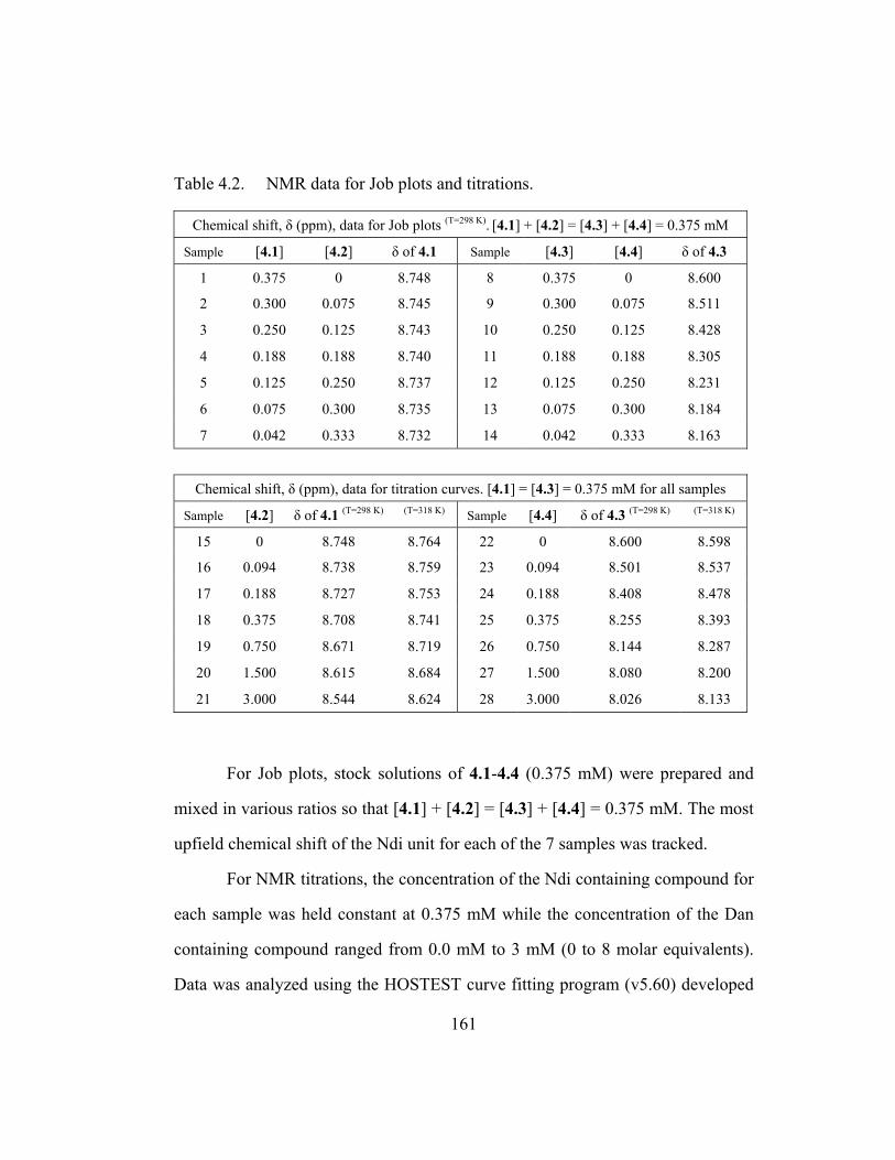
Table 4.2. NMR data for Job plots and titrations.
Chemical shift, δ (ppm), data for Job plots (T=298 K). [4.1] + [4.2] = [4.3] + [4.4] = 0.375 mM
Sample [4.1] [4.2] δ of 4.1 Sample [4.3] [4.4] δ of 4.3
1 0.375 0 8.748 8 0.375 0 8.600
2 0.300 0.075 8.745 9 0.300 0.075 8.511
3 0.250 0.125 8.743 10 0.250 0.125 8.428
4 0.188 0.188 8.740 11 0.188 0.188 8.305
5 0.125 0.250 8.737 12 0.125 0.250 8.231
6 0.075 0.300 8.735 13 0.075 0.300 8.184
7 0.042 0.333 8.732 14 0.042 0.333 8.163
Chemical shift, δ (ppm), data for titration curves. [4.1] = [4.3] = 0.375 mM for all samples
Sample [4.2] δ of 4.1 (T=298 K) (T=318 K) Sample [4.4] δ of 4.3 (T=298 K) (T=318 K)
15 0 8.748 8.764 22 0 8.600 8.598
16 0.094 8.738 8.759 23 0.094 8.501 8.537
17 0.188 8.727 8.753 24 0.188 8.408 8.478
18 0.375 8.708 8.741 25 0.375 8.255 8.393
19 0.750 8.671 8.719 26 0.750 8.144 8.287
20 1.500 8.615 8.684 27 1.500 8.080 8.200
21 3.000 8.544 8.624 28 3.000 8.026 8.133
For Job plots, stock solutions of 4.1-4.4 (0.375 mM) were prepared and
mixed in various ratios so that [4.1] + [4.2] = [4.3] + [4.4] = 0.375 mM. The most
upfield chemical shift of the Ndi unit for each of the 7 samples was tracked.
For NMR titrations, the concentration of the Ndi containing compound for
each sample was held constant at 0.375 mM while the concentration of the Dan
containing compound ranged from 0.0 mM to 3 mM (0 to 8 molar equivalents).
Data was analyzed using the HOSTEST curve fitting program (v5.60) developed
161

by Glagovich and Wilcox at the University of Pittsburgh, Pittsburgh, PA. The
HOSTEST program can apply a number of different models to the chemical shift
data. The reported association constants were obtained using a 1:1 binding model
as in previous studies (Cubberley 2000). Standard deviations of the association
constants, Ka, calculated by the HOSTEST program were reasonable and R2 for
the fitted curves, for all experiments, exceeded a 0.999 value. Examples of the
program outputs are in Figure 4.14 (binding between monomers) and 4.15
(between dimers).
Figure 4.14 Representative HOSTEST output for (4.1:4.2) binding at T = 318 K.
162

Figure 4.16 Representative HOSTEST output for (4.3:4.4) binding at T = 318 K.
ITC. Experiments were performed on a VP-ITC microcalorimeter from
Microcal Inc. (Northhampton, MA 01060) and followed the experimental setups
detailed in literature (Wiseman 1998). Three trials were performed for each
system. Complete binding data is shown in Table 4.3. All solutions were made in
50 mM sodium phosphate buffer and degassed for 15 minutes and stock solution
concentrations were confirmed by extinction coefficients. For a typical
experimental run a solution of the Ndi containing compound was placed in the
instrument chamber at a relatively dilute concentration (4.3, 0.375 mM; 4.5, 0.250
mM; 4.7, 0.188 mM). Relatively concentrated solutions of the Dan containing
compound were taken up in the 250 µL injection syringe (4.4, 3.21 mM; 4.6, 2.11
mM; 4.8, 1.59 mM). After a 10 minute equilibration time injections were started
163

with the first being 2.0 µL and the next 39 being 6.3 µL with 5 minutes between
each injection. After the final injection the molar ratio ([oligo-Dan] / [oligo-Ndi])
reached greater than 1.5 and this was sufficient for analysis by the ORIGIN
software provided with the ITC instrument. Each experiment was performed three
times and the first data point (injection # 1) of each trial was not considered in the
analyses. Also for each experiment, blanks were run where oligo-Dan was
injected into buffer and buffer was injected into oligo-Ndi. Although heats of
dilution were near zero these were still subtracted from raw titration data to
account for any slight heat effects from dilution. Integrated data from titration
runs were fit using a single site model with Ka, ∆H° allowed to vary. The
stoichiometry of binding (N) was not assumed and this value was allowed to vary
as well during the fitting process. In all cases N indicated 1:1 binding with values
within 10 % of 1.00. The c value, as defined in the Wiseman article, fell within
the acceptable range for the determination of accurate binding constants
(Wiseman 1998). As mentioned before the monomeric system (4.1:4.2) could not
be analyzed by ITC due to sensitivity limits. The c value dictated that
concentrations needed to be prohibitively high to achieve an acceptable signal to
noise ratio for heat measured.
164

Table 4.3. Complete hetero-duplex binding data.a
Complex T Conc d Ka N value e ∆G° ∆H° ∆S°
4.1:4.2b 318 0.375 1.3 (0.1) × 102 1 –3.2 (0.01) NA NA
4.3:4.4 b 318 0.750 2.8 (0.3) × 103 1 –5.1 (0.02) NA NA
4.3:4.4 c 318 0.750 2.7 (0.1) × 103 0.91 (0.01) –5.0 (0.02) –12.3 (0.3) –23.0 (0.9)
4.5:4.6 c 318 0.750 4.5 (0.1) × 104 0.97 (0.02) –6.8 (0.02) –17.7 (0.1) –34.2 (0.3)
4.7:4.8 c 318 0.750 3.5 (0.1) × 105 0.91 (0.01) –8.1 (0.03) –19.3 (0.2) –35.3 (0.6)
4.1:4.2 b 298 0.375 3.0 (0.1) × 102 1 –3.2 (0.02) NA NA
4.3:4.4 b 298 0.750 7.5 (0.5) × 103 1 –5.3 (0.03) NA NA
4.3:4.4 c 288 0.750 1.2 (0.1) × 104 0.98 (0.01) –5.4 (0.01) –10.1 (0.2) –16.4 (0.6)
4.3:4.4 c 298 0.750 7.6 (0.1) × 103 1.01 (0.04) –5.3 (0.07) –10.4 (0.2) –17.2 (0.5)
4.3:4.4c 308 0.750 4.7 (0.1) × 103 0.99 (0.01) –5.2 (0.03) –11.1 (0.2) –19.2 (0.1) a Units are T (K), Ka (M–1), ∆G° (kcal·mol–1), ∆H° (kcal·mol–1), ∆S° (cal·mol–1·K–1). For NMR data, errors represent standard deviations calculated by the HOSTEST program (Wilcox 1997). For ITC data, errors represent the standard deviation of three trials. b Analyzed by NMR. c Analyzed by ITC. d This is the initial concentration (mM), pre-titration, of the Ndi unit as oppose to the concentration of the Ndi containing compound. e For titrations analyzed by NMR, a binding stoichiometry of 1:1 was determined by inspection of Job plots. For ITC analysis the N values represents the best fit stoichiometry of binding (oligo-Dan:oligo-Ndi ration) calculated by the ORIGIN software (Microcal, Inc. 1999). NA = Not Applicable since 1:1 fitting model was used and goodness of fit was used to judge accuracy of calculated binding constants. NA = Not applicable. ∆H° and ∆S° derived from the van’t Hoff equation is not applicable since enthalpy for our system was shown to be significantly temperature dependent. (Naghibi 1995).
Figures 4.17-4.22 are representative isotherms showing additional
important ITC data collected during these studies. These are meant to give a
general idea of the curve shapes, trends, chi-square values, and thermodynamic
quantities for these oligomers and may provide a helpful reference as to the
magnitude of heats absorbed or evolved that one could expect when studying
future naphthyl oligomers.
165

Figure 4.16 Typical buffer into buffer titration that results in extremely small heats (0.05-0.30 kcal/mole) released per injection. For comparison the first couple of injections (same volume) in a tetramer run released 20 kcal/mole of heat.
166

Figure 4.17 Typical tetra-Dan injections into buffer. The small heats absorbed were taken into account when processing the tetramer runs where tetra-Dan 4.8 was injected into tetra-Ndi 4.7. For all ITC data reported, heats of dilutions were subtracted from the titration runs to afford the heats associated with binding.
167
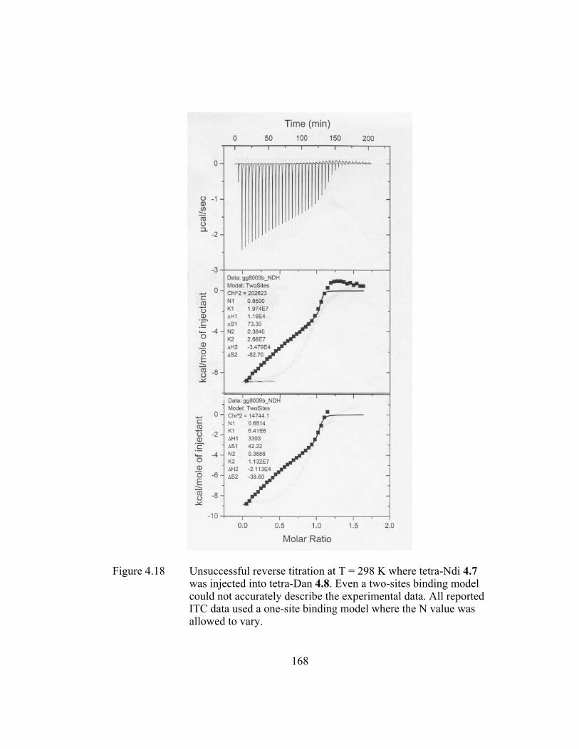
Figure 4.18 Unsuccessful reverse titration at T = 298 K where tetra-Ndi 4.7 was injected into tetra-Dan 4.8. Even a two-sites binding model could not accurately describe the experimental data. All reported ITC data used a one-site binding model where the N value was allowed to vary.
168

Figure 4.19 A representative trimeric system run at T = 298 K. Though the chi-square value is 55,000 the fit appeared to be fairly accurate. On average the Ka(4.5:4.6) was 1.1 × 10⁵ M-1 for the trimeric system at T = 298 K, more than twice as large as that measured at T = 318 K.
169

Figure 4.20 A tetrameric system run at T = 298 K. A chi-square value of 250,000 indicated an inability to describe the binding with a one sites model. Only about three-fourths of the curve fit the model possibly indicating a switch in binding stoichiometry at excess titrant (tetra-Dan 4.8) at room temperature.
170

Figure 4.21 Representative isotherm that afforded excellent results for the tetrameric system (T = 318 K). Notice the chi-square value was below 50,000 and the N value was within a 10% error of 1.0.
171

SEC. All of the SEC runs were performed by Dr. Thomas Mourey from
the Eastman Kodak Company (Rochester, NY 14650). Initially, samples of tetra-
Ndi, 4.7, were tested on glycerol propyl bonded-phase Synchropak columns
(4000, 1000, 500 and 100 Angstrom pore size). Though this type of column is
silica-based and usually works well for small anionic polymers, samples did not
elute after 3 injections. A 60 Angstrom 7.8 x 80 mm Bio-Sil guard column (just
the guard column) was ultimately chosen for SEC runs, which provided minimal
size separation but near complete sample elution. UV (270 nm) and light
scattering (90 degrees) were used for detection. Injections were 0.22 mM in
concentration for each strand and mixtures were heated at 60 °C for 15 minutes
and injected 10 minutes later.
Additional chromatographs (with figure captions by Dr. Mourey) is
included here for future reference should SEC become a standard analytical tool
in the investigations of water-soluble naphthyl oligomers and possibly polymers.
The author thanks his labmate, Joseph Reczek for initiating the correspondence
with Dr. Mourey. In the following chromatographs compound 4a = tetraNdi 4.7
and 4b = tetraDan 4.8.
172

Figure 4.22 90 degree light scattering chromatograms of individual components and 1:1 molar mixture. Note that the intensity of the aggregate peak (1.5 mL) for the 1:1 mixture is less than half that of sample 4A, implying that some of the aggregates are broken during complex formation.
173

Figure 4.23 90 degree light scattering chromatograms of 1:1 molar complex 10 minutes after preparation (15 minute of heating, 60°C) (green), 24 min (cyan), 79 min (red), 93 min (blue). The peak at 1.5 mL is attributed to aggregates; peak at 2.7 mL is from the complex. (Author’s comments – magnitude of 90 degree light scattered is dependent on size of aggregate or complex. See Figure 4.12 for a better quantitative idea of the amount of presumed 1:1 complex to aggregate.)
PAGE. Compounds were dissolved in 50 mM Na phosphate buffer, pH =
7.0, and the duplexes were heated for 5 minutes at 45˚ C and then left at room
temperature before loading into lanes. Non-denaturing polyacrylamide gels were
made (19:1 crosslinker ratio [acrylamide:bis], 20% w/v% acrylamide solution
from Bio-Rad Inc., Hercules, CA 94547). The gel measured 11 cm in vertical
height, with a 1 mm spacer between glass plates. 12 µL of oligomer solution was
174

loaded into each well along with 3 µL of 30% glycerol and gels were ran
vertically at 300V for approximately 2 hr leading to migrations down
approximately two-thirds the length of the gel. Bands were only faintly visible by
UV so the gel was subjected to standard Coomassie staining/destaining followed
by visualization with UV shadowing over a fluorescent TLC plate.
175

CHAPTER 5
Final (Personal) Remarks
You see things; and you say, “Why?”
But I dream things that never were; and I say, “Why not?” -George Bernard Shaw
After the groundbreaking, lab-making work of R. Scott Lokey creating the
first aedamer and after the preliminary study of their gelling properties by John
Nguyen, a good deal of foresight was shown by Mark Cubberley and Andy Zych
who took it upon themselves to study the fundamental reasons of why and how
linked Dan and Ndi units “become” aedamers. Though the paths of combinatorial
library synthesis and supramolecular chemistry must have been tempting them to
synthesize a whole host of super-sized aedamer derivatives, instead they looked
towards more modest sized, but ultimately invaluable molecules.
While Mark found out why aedamers fold, elucidating the driving forces
of Dan:Ndi monomer association, Andy probed the minimal folding unit to
uncover how dimers truly look like when folded. The results of these meticulous
basic science type of projects still impresses. These studies have expanded our
thinking of aromatic interactions and revealed that chemists have tremendous
opportunities to control, more so than previously believed, these noncovalent
interactions for use in molecular assembly. Due to their conclusions, many
176

aedamer projects were being workshopped shortly after their graduation as we
were only limited by our imagination and not the properties of our molecules.
After the studies of Cubberley and Zych there was a promise of
“designablility” based on their rules of association (Chapter 1). In this dissertation
it was shown that the same interaction that caused a string of beads to zig-zag
could also weave the string into a turn (designed folding) and unite two different
strings together (designed binding). Even the gelling properties (folding) and
enzyme refolding inhibition (binding) may be revealed, with better analyses, to be
dependant on (and possibly controllable by) directed aromatic interactions.
Alas, the subtleties of how to fully manipulate these particular properties
have thus far eluded us. Keep in mind though that this was the lab’s first pass at
trying to take advantage of the designability of naphthyl oligomers. Though the
rules of aromatic association have served me well, true mastery of these systems
will likely require constant revision of these rules and several leaps of faith; into
mindsets contrary to previous thinking, into experiments that none of your
labmates believe will work, and into areas where no one in the group has
ventured.
Waiting for the time when I can finally say
That this has all been wonderful but now I'm on my way. But when I think it's time
To leave it all behind I try to find a way
But there's nothing I can say To make it stop.
-Lyrics by Tom Marshall
177

References
Aamer, K. A.; Sardinha, H.; Bhatia, S. R.; Tew, G. N. “Rheological studies of
PLLA-PEO-PLLA triblock copolymer hydrogels” Biomaterials (2003), 35, 1087.
Albrecht, M. “Let's twist again - Double-stranded, triple-stranded, and circular helicates” Chem. Rev. (2001), 101, 3457.
Amabilino, D. B.; Ashton, P. R.; Brown, C. L.; Cordove, E.; Godinez, L. A.; Goodnow, T. T.; Kaifer, A. E.; Newton, S. P.; Pietraszkiewicz, M.; Philp; Raymo, F. M.; Reder, A. S.; Rutland, M. T.; Slawin, A. M. Z.; Spencer, N.; Stoddart, J. F. “Molecular meccano. 2. Self-assembly of [n]catenanes” J. Am. Chem. Soc. (1995), 117, 1271.
Amabilino D. B., Stoddart J. F. “Interlocked and intertwined structures and superstructures” Chem. Rev. (1995), 95, 2725.
Anfinsen, C. B. In “Molecular Organization and Biological Function” Allen, J. M., Ed.; Harper and Row: New York, (1967), 1-19.
Appella, D. H.; Christianson, L. A.; Karle, I. L.; Powell, D. R.; Gellman, S. H. “β-Peptides foldamers: Robust helix formation in a new family of β-amino acid oligomers” J. Am. Chem. Soc. (1996), 118, 13071.
Appella, D. H.; Christianson, L. A.; Klein, D. A.; Powell, D. R.; Huang, X.; Barchi, J. J., Jr.; Gellman, S. H. “Residue-based control of helix shape in β-peptides oligomers” Nature (1997), 387, 381.
Appella, D. H.; LePlae, P. R.; Raguse, T. L. Gellman, S. H. “(R,R,R)-2,5-Diaminocylohexanecarboxylic acid, a building block for water-soluble, helix-forming β-peptides” J. Org. Chem. (2000), 65, 4766.
Archer, E. A.; Gong, H.; Krische, M. J. “Hydrogen bonding in noncovalent synthesis: Selectivity and the directed organization of molecular strands” Tetrahedron (2001), 57, 1139.
Archer, E. A.; Krische, M. J. “Duplex oligomers defined via covalent casting of a one-dimensional hydrogen-bonding motif” J. Am. Chem. Soc. (2002a), 124, 5074.
178

Archer, E. A.; Cauble, D. F.; Lynch, V.; Krische, M. J. “Synthetic duplex oligomers: Optimizing interstrand affinity through the use of a noncovalent constraint” Tetrahedron (2002b), 58, 721.
Benner S. A; Hutter D. “Phosphates, DNA, and the search for nonterrean life: A second generation model for genetic molecules” Bioorg. Chem. (2002), 30, 62.
Berl, V.; Huc, I.; Khoury, R. G.; Krische, M. J.; Lehn, J.-M. “Interconversion of single and double helices formed from synthetic molecular strands” Nature (2000), 407, 720.
Berl, V.; Huc, I.; Khoury, R. G.; Lehn, J.-M. “Helical molecular programming: supramolecular double helices by dimerization of helical oligopyridine-dicarboxamide strands” Chem.-Eur. J. (2001), 7, 2810.
Bisson, A. P.; Carver, F. J.; Hunter, C. A.; Waltho, J. P. “Molecular zippers” J. Am. Chem. Soc. (1994), 116, 10292.
Bisson, A. P.; Carver, F. J.; Eggleston, D. S.; Haltiwanger, R. C.; Hunter, C. A.; Livingstone, D. L.; McCabe, J. F.; Rotger, C.; Rowan, A. E. “Synthesis and recognition properties of aromatic amide oligomers: Molecular zippers” J. Am. Chem. Soc. (2000), 122, 8856.
Brandon, C.; Tooze, J. “Introduction to Protein Structure” 2nd ed; Garland: New York, (1999).
Brenner, M.; Seebach, D. “Synthesis and CD spectra in MeCN, MeOH, and H2O of γ-oligopeptides with hydroxy groups on the backbone” Helv. Chem. Acta (2001), 84, 1181.
Butterfield S. M.; Waters, M. L. “Contribution of aromatic interactions to α-helix stability” J. Am. Chem. Soc. (2002), 124, 9751.
Butterfield S. M.; Waters, M. L. “A designed β-hairpin peptide for molecular recognition of ATP in water” J. Am. Chem. Soc. (2003), 125, 9580.
Butterfield S. M.; Goodman, C. M.; Rotello, V. M.; Waters, M. L. “A peptide flavoprotein mimic: Recognition and redox potential modulation in water by a designed β hairpin” Angew. Chem. Int. Ed. (2004), 43, 724.
Buxbaum, J. N.; Tagoe, C. E. “The genetics of amyloidosis” Annu. Rev. Med. (2000), 51, 543.
179

Cantor, C. R.; Shimmel, P. R. “Biophysical Chemistry, part II: Techniques for the study of biological structure and function” W. H. Freeman: New York, (1980).
Chan, W. C.; White, P.D. eds. “Fmoc solid phase synthesis: A practical approach” Oxford University Press: New York (2000).
Cheng, R. P.; Gellman, S. H.; DeGrado, W. F. “β-Peptides: From structure to function” Chem. Rev. (2001), 101, 3219.
Chichak, K. S.; Cantrill, S. J.; Pease, A. R.; Chiu, S.-H.; Cave, G. W. V.; Atwood, J. L.; Stoddart, J. F. “Molecular Borromean rings” Science (2004), 304, 1308.
Chowdhury, S. “An overlooked dicopper(I) helicate” J. Ind. Chem. Soc. (2004), 81, 153.
Connors, K. A. “Binding constants” Wiley: New York, (1987).
Cozzi, F. Cinquini, M.; Annunziata, R.; Dwyer, T.; Siegel, J. S. “Polar/π interactions between stacked aryls in 1,8-diarylnaphthalenes” J. Am. Chem. Soc. (1992), 114, 5729.
Cubberley, M. S.; Iverson, B. L. “Models of higher-order structure: Foldamers and beyond” Curr. Opin. Chem. Biol. (2001a), 5, 650.
Cubberley, M. S.; Iverson, B. L. “1H NMR investigation of solvent effects in aromatic stacking interactions” J. Am. Chem. Soc. (2001b), 123, 7560.
Cubberley, M. S. Ph.D. thesis, “Investigation of solvent effects in aromatic electron donor-acceptor interactions” The University of Texas at Austin (2000).
Ercolani, G. “Assessment of cooperativity in self-assembly” J. Am. Chem. Soc. (2003), 125, 16097.
Ferguson, S. B.; Sanford, E. M.; Seward, E. M. Diederich, F. “Cyclophane-arene inclusion complexation in protic solvents
Feynman, R. P. “There’s Plenty of Room at the Bottom” Transcript of a talk given on December 29, 1960 at the annual meeting of the American Physical Society. (1960), From http://clsdemo.caltech.edu/archive/ 00000047/.
180

Frackenpohl, J.; Arvidsson, P. I.; Schreiber, J. V.; Seebach, D. “The outstanding biological stability of β- and γ-peptides toward proteolytic enzymes: an in vitro investigation with fifteen peptidases” ChemBioChem (2001), 2, 445.
Gabriel, G. J.; Iverson, B. L. “Aromatic oligomers that form hetero duplexes in aqueous solutions” J. Am. Chem. Soc. (2002), 124, 15174.
Gabriel, G. J.; Iverson, B. L. “Refolding inhibition by an amphiphilic foldamer” (2004a, in preparation).
Gabriel, G. J.; Sorey, S.; Iverson, B. L. “Switching the folding patterns of naphthyl trimers” J. Am. Chem. Soc. (2004b, submitted).
Gellman, S. H. “Foldamers: A manifesto” Acc. Chem. Res. (1998), 31, 173.
Ghosh, S.; Ramakrishnan, S. “Aromatic donor-acceptor charge-transfer and metal-ion-complexation-assisted folding of a synthetic polymer” Angew. Chem., Int. Ed. (2004), 43, 3264.
Gil, V. M. S.; Oliveira, N. C. “On the use of the method of continuous variations” J. Chem. Educ. (1990), 67, 473.
Gill, S. J.; Dec, S. F.; Olofsson, G.; Wadso, I. “Anomalous heat capacity of hydrophobic solvation” J. Phys. Chem. (1985), 89, 3758.
Gin, M. S.; Yokozawa, T.; Prince, R. B.; Moore, J. S. “Helical bias in solvophobically folded oligo(phenylene ethynylene)s” J. Am. Chem. Soc. (1999), 121, 2643.
Glover, J. N.; Harrison, S. C. Crystal structure of the heterodimeric bZIP transcription factor c-Fos-c-Jun bound to DNA. Nature (1995), 373, 257.
Goddard, T. D.; Kneller, D. G. SPARKY 3 (Version 3.85), University of California, San Francisco (2000).
Gong, B. “Specifying non-covalent interactions: Sequence-specific assembly of hydrogen-bonded molecular duplexes” Synlett (2001), 582.
Guelev, V.; Sorey, S.; Hoffman, D.W.; Iverson, B. L. “Changing DNA grooves - A 1,4,5,8-naphthalene tetracarboxylic diimide bis-intercalator with the linker (β-Ala)3-Lys in the minor groove” J. Am. Chem. Soc. (2002), 124, 2864.
181
Guelev, V. M.; Cubberley, M. S.; Murr, M. M.; Lokey, R. S.; Iverson, B. L. “Design, synthesis, and characterization of polyintercalating ligands” Methods in Enzymology (2001a), 340, 556.
Guelev, V.; Lee, J.; Ward, J.; Sorey, S.; Hoffman, D. W.; Iverson, B. L. “Peptide bis-intercalator binds DNA via threading mode with sequence specific contacts in the major groove” Chem. Biol. (2001b), 8, 415.
Guelev, V. M. Ph.D. thesis, “Peptide-based DNA intercalators” The University of Texas at Austin (2001c).
Guelev, V. M.; Harting, M. T.; Lokey, R. S.; Iverson, B. L. “Altered sequence specificity identified from a library of DNA-binding small molecules” Chem. Biol. (2000), 7, 1.
Guinto, E. R.; Di Cera, E. “Large heat capacity change in a protein-monvalent cation interaction” (1996), Biochemistry, 35, 10300.
Hill, D. J.; Mio, M. J.; Prince, R. B.; Hughes, T. S.; Moore, J. S. “A field guide to foldamers” Chem. Rev. (2001), 101, 3893.
Hirao, I.; Kawai, G.; Yoshizawa, S.; Nishimura, Y.; Ishido, Y.; Watanabe, K.; Miura, K.-I. “Most compact hairpin-turn structure exerted by a short DNA fragment, d(GCGAAGC) in solution: an extraordinary stable structure resistant to nucleases and heat” Nuc. Acid Res. (1994), 22, 576.
Hunter, C. A.; Sanders, J. K. M. “The nature of π-π interactions” J. Am. Chem. Soc. (1990), 112, 5525.
Hunter, C. A.; Lawson, K. R.; Perkins, J.; Urch, C. J. “Aromatic interactions” Chem. Soc., Perkin Trans. 2, (2001), 651.
Hunter, C. A.; Jones, P. S.; Tiger, P. M. N.; Tomas, S. “New building blocks for the assembly of sequence selective molecular zippers” Chem. Comm. (2003), 14, 1642.
HyperCube, Inc. HyperChem Computational Chemistry Manual. (Gainesville, FL, 32601) (1996) HyperChem 5.1.
Ilhan, F.; Gray, M.; Blanchette, K.; Rotello, V. M. “Control of polymer solution structure via intra- and intermolecular aromatic stacking” Macromolecules (1999), 32, 6159.
182

Johnston, M. D., Jr.; Martin, G. E. “Establishing long-range connectivities in polynuclear aromatics through the use of two-dimensional MLEV-17-based isotropic mixing” Mag. Reson. Chem. (1989), 27, 529.
Jourdan, M.; Griffiths-Jones, S. R.; Searle, M. S. “Folding of a β-hairpin peptide derived from the N-terminus of ubiquitin” Eur. J. Biochem. (2000), 267, 3539.
Junquera, E.; Nowick, J. S. “Folding of an artificial β-sheet in competetive solvents” J. Org. Chem. (1999), 64, 2527.
Kawano, S.-I.; Fujita, N.; Shinkai, S. “A coordination gelator that shows a reversible chromatic change and sol-gel phase-transition behavior upon oxidative/reductive stimuli” J. Am. Chem. Soc. (2004), 126, 8592.
Kirshenbaum, K.; Barron, A. E.; Goldsmith, R. A.; Armand, P.; Bradley, E. K.; Truong, K. T. V.; Dill, K. A.; Cohen, F. E.; Zuckermann, R. N. “Sequence-specific polypeptoids: a diverse family of heteropolymers with stable secondary structure” Proc. Natl. Acad. Sci. U. S. A. (1998), 95, 4303.
Kubik, S. “High-performance fibers from spider silk” Angew. Chem., Int. Ed. (2002), 41, 2721.
Kuehl, C..J.; Tabellion, F. M.; Arif, A. M.; Stang, P. J. “Single- and double-stranded chains assembled via concomitant metal coordination and hydrogen bonding” Organometallics (2001), 20, 1956.
LaBrenz, S. R.; Kelly, J. W. “Peptidomimetic host that binds a peptide guest affording a β-sheet structure that subsequently self-assembles” A simple receptor mimic. J. Am. Chem. Soc. (1995), 117, 1655.
Langenhan, J. M.; Gellman, S. H. “Effects of alternative side chain pairings and reverse turn sequences on antiparallel sheet structure in β-peptide hairpins” Org. Lett. (2004), 6, 937.
Langer, R.; Peppas, N. A. “Advances in biomaterials, drug delivery, and bionanotechnology” AIChE J. (2003), 49, 2990.
Lehn, J.-M.; Rigault, A.; Siegel, J.; Harrowfield, J.; Chevrier, B.; Moras, D. “Spontaneous assembly of double-stranded helicates from oligobipyridine ligands and copper(I) cations: structure of an inorganic double helix” Proc. Natl. Acad. Sci. U.S.A. (1987), 84, 2565.
183

Lee, J.-Y. Guelev, V.; Sorey, S.; Hoffman, D. W.; Iverson, B. L. “NMR structural analysis of a modular threading tetra-intercalator” J. Am. Chem. Soc. (2004 submitted).
Liao, M.-J.; Prestegard, J. H. “Fusion kinetics of phospatidylcholine-phospatidic acid mixed lipid vesicles” Biochim. Biophys. Acta. (1980), 599, 81.
Lilley, D. M. J. “Folding and catalysis by the hairpin ribozyme” FEBS Lett. (1999), 452, 26.
Liu, D.; DeGrado, W. F. “De novo design, synthesis, and characterization of antimicrobial β-peptides” J. Am. Chem. Soc. (2001), 123, 7553.
Lokey, R. S.; Iverson, B. L. “Synthetic molecules that fold into a pleated secondary structure in solution” Nature (1995), 375, 303.
Lokey, R. S. Ph.D. thesis, “Aedamers: A synthetic approach to higher-order structure” The University of Texas at Austin (1997).
Macomber, R. S. “An introduction to NMR titration for studying rapid reversible complexation” J. Chem. Educ. (1992), 69, 375.
Mao, C.; Sun, W.; Seeman, N. C. “Assembly of Borromean rings from DNA” Nature (1997), 386, 137.
Marshall, K. A.; Robertson, M. P.; Ellington, A. D. “A biopolymer by any other name would bind as well: a comparison of the ligand-binding pockets of nucleic acids and proteins” Structure (London) (1997), 5, 729.
Martin, G. E.; Crouch, R. C. in “Two-Dimensional NMR Spectroscopy: Applications of Chemists and Biochemists” Croasmum, W. R.; Carlson, R. M., Eds; VCH Publishers: New York, (1994).
Menger, F. M.: Peresypkin, A. V. “Strings of vesicles: Flow behavior in an unusual type of aqueous gel” J. Am. Chem. Soc. (2003), 125, 5340.
Meyer, E. A.; Castellano, R. K.; Diederich, F. “Interactions with aromatic rings in chemical and biological recognition” Angew. Chem., Int. Ed. (2003), 42, 1210.
Microcal Inc. VP-ITC MicroCalorimeter user’s manual. ORIGIN software package included. Northhampton, MA 01060, (1999). Personal communications with technicians there were also helpful.
184

Miller, C. T.; Weragoda, R.; Izbicka, E.; Iverson, B. L. “The synthesis and screening of 1,4,5,8-naphthalenetetracarboxylic diimide-peptide conjugates with antibacterial activity” Bio. Med. Chem. (2001), 9, 2015.
Mio, M. J.; Prince, R. B.; Moore, J. S.; Kuebel, C.; Martin, D. C. “Hexagonal packing of oligo(m-phenylene ethynylene)s in the solid state: Helical nanotubules” J. Am. Chem. Soc. (2000), 122, 6134.
Murphy, K. P. “Predicting binding energetics from structure: Looking beyond ∆G” Med. Res. Rev. (1999), 19, 333.
Murphy, J. E.; Uno, T.; Hamer, J. D.; Cohen, F. E.; Dwarki, V.; Zuckermann, R. N. “A combinatorial approach to the discovery of efficient cationic peptoid reagents for gene delivery” Proc. Natl. Acad. Sci. U. S. A. (1998), 95, 1517.
Murr, M. M.; Harting, M. T.; Guelev, V.; Ren, J.; Chaires, J. B.; Iverson, B. L. “An octakis-intercalating molecule” Bio. Med. Chem. (2001), 9, 1141.
Naghibi, H.; Tamura, A.; Sturtevant, J. M. “Significant discrepancies between van't Hoff and calorimetric enthalpies” J. Proc. Natl. Acad. Sci. USA, (1995), 92, 5597.
Nassar, N.; Horn, G.; Herrmann, C.; Scherer, A.; McCormick, F.; Wittinghofer, A. “A crystal structure of the Ras-binding domain of the serine/threonine kinase c-Raf1 in complex with Rap1A and a GTP analogue” Nature (1995), 375, 554.
Nelson, J. C.; Saven, J. G.; Moore, J. S.; Wolynes, P. G. “Solvophobically driven folding of nonbiological oligomers” Science (1997), 277, 1793.
Nguyen, J. Q.; Iverson, B. L. “An amphiphilic folding molecule that undergoes an irreversible conformational change” J. Am. Chem. Soc. (1999), 121, 2639.
Nolan, S. L.; Phillips, R. J.; Cotts, P. M.; Dungan, S. R. “Light scattering study on the effect of polymer composition on the structural properties of PEO-PPO-PEO micelles” J. Coll. Inter. Sci. (1997), 191, 291.
Nowick, J. S.; Powell N. A.; Martinez, E. J.; Smith, E. M.; Noronha, G. “Molecular scaffolds. 1. Intramolecular hydrogen bonding in a family of di- and triureas” J. Org. Chem. (1992), 57, 3763.
185

Nowick, J. S.; Holmes D. L.; Mackin, G.; Noronha, G.; Shaka, A. J.; Smith, E. M. “An artificial β-sheet comprising a molecular scaffold, a β-strand mimic, and a peptide strand” J. Am. Chem. Soc. (1996), 118, 2764.
Nowick, J. S. “Chemical models of protein β-sheets” Acc. Chem. Res. (1999), 32, 287.
Newcomb, L. F.; Gellman, S. H. “Aromatic stacking interactions in aqueous solutions: Evidence that neither classical hydrophobic effects nor dispersion forces are important” J. Am. Chem. Soc. (1994), 116, 4993.
Pecora, R. “Dynamic light scattering: Applications of photon correlation spectroscopy” Plenum Press: New York, (1985).
Perez-Rodriguez, M.; Prieto, G.; Rega, C.; Varela, L. M.; Sarmiento, F.; Mosquera, V. “A comparaqtive study of the determination of the critical micelle concentration by conductivity and dielectric constant measurements” Langmuir (1998), 14, 4422.
Petka, W. A.; Harden, J. L.; McGrath, K. P.; Wirtz, D.; Tirrell, D. A. “Reversible hydrogels from self-assembling artificial proteins” Science (1998), 281, 389.
Philp, D.; Stoddart, J. F. “Self-assembly in natural and unnatural systems” Angew. Chem. Int. Ed. Engl. (1996), 35, 1154.
Pochan, D. J.; Schneider, J. P.; Kretsinger, J.; Ozbas, B.; Rajagopal, K.; Haines, L. “Thermally reversible hydrogels via intramolecular folding and consequent self-assembly of a de novo designed peptide” J. Am. Chem. Soc. (2003), 125, 11802.
Prince, R. B.; Brunsveld, L.; Meijer, E. W.; Moore, J. S. “Twist sense bias induced by chiral side chains in helically folded oligomers” Angew. Chem., Int. Ed. (2000a), 39, 228.
Prince, R. B.; Barnes, S. A.; Moore, J. S. “Foldamer-Based Molecular Recognition” J. Am. Chem. Soc. (2000b), 122, 2758.
Quinn, T. P.; Tweedy, N. B.; Richardson, J. S.; Williams, R. W.; Richardson, D. C. “Betadoublet: de novo design, synthesis, and characterization of a β-sandwich protein” Proc. Natl. Acad. Sci. U. S. A. (1994), 91, 8747.
186

Raguse, T. L.; Lai, J. R.; LePlae, P. R.; Gellman, S. H. “Toward β-peptide tertiary structure: Self-association of an amphphilic 14-helix in aqueous solutions” Org. Lett. (2001), 3, 3963.
Ramachandran, G. N.; Sasisekharan, V. In “Advances in Protein Chemistry” Anfinsen, C. B. J., Anson, M. L., Edsall, J. T., Richards, F. M., Eds.; Academic Press: New York, (1968), Vol. 23.
Reczek, J. J. X-ray quality crystals of the bis(diethylene glycol) Dan in figure 1.7B could only be obtained by sublimation at 180° C at vacuum pump pressures (2002).
Robertson, A. D.; Murphy, K. P. “Protein structure and the energetics of protein stability” Chem. Rev. (1997), 97, 1251.
Rouhi, M; “Intriguing behavior of a folded polymer” Chem. Eng. News (March 29, 1999), 8.
Rowan, S. J.; Cantrill, S. J.; Cousins, G. R. L.; Sanders, J. K. M.; Stoddart, J. F. “Dynamic covalent chemistry” Angew. Chem., Int. Ed. (2002), 41, 899.
Ruso, J. M.; Attwood, D.; Taboada, P.; Mosquera, V.; Sarmiento, F. “Light scattering and NMR studies on the self-aggregation of sodium n-hexyl sulfate in aqueous electrolyte solution” Langmuir (2000), 16, 1620.
Sambrook, J.; Russell, D. W.; “Molecular cloning: A laboratory manual, 3rd edition” Cold Spring Harbor Laboratory Press: New York, (2001).
Searle, M. S. “Peptide models of protein β-sheets: design, folding and insights into stabilizing weak interactions” J. Chem. Soc. Perkin Trans. 2 (2001), 1011.
Seebach, D.; Abele, S.; Gademann, K.; Guichard, G.; Hintermann, T.; Jaun, B.; Matthews, J. L.; Schreiber, J. V.; Oberer, L.; Hommel, U.; Widmer, H. “β2-and β3-peptides with proteinaceous side-chains. Synthesis and solution structures of constitutional isomers, a novel helical secondary structure, and the influence of solvation and hydrophobic interactions on folding” Helv. Chim. Acta (1998), 81, 932.
Seebach, D.; Abele, S.; Gademann, K.; Jaun, B. “Pleated sheets and turns of β-peptides with proteinaceous side-chains” Angew. Chem., Int. Ed. (1999), 38, 1595.
187
Seidler, J.; McGovern, S. L.; Doman, T. N.; Shoichet, B. K. “Identification and prediction of promiscuous aggregating inhibitors among known drugs” J. Med. Chem. (2003), 46, 4477.
Simon, R. J.; Kania, R. S.; Zuckermann, R. N.; Huebner, V. D.; Jewell, D. A.; Banville, S.; Ng, S.; Wang, L.; Rosenberg, S.; Marlowe, C. K.; Spellmeyer, D. C.; Tan, R.; Frankel, A. D.; Santi, D. V.; Cohen, F. E.; Bartlett, P. A. “Peptoids: A modular approach to drug discovery” Proc. Natl. Acad. Sci. U.S.A. (1992), 89, 9367.
Smithrud, D. B.; Diederich, F. “Strength of molecular complexation of apolar solutes in water and in organic solvents is predictable by linear free energy relationships: a general model for solvation effects on apolar binding” J. Am. Chem. Soc. (1990), 112, 339.
Smithrud, D. B.; Wyman, T. B.; Diederich, F. “Enthalpically Driven Cyclophane-Arene Inclusion Complexation: Solvent-Dependent Calorimetric Studies” J. Am. Chem. Soc. (1991), 113, 5420.
Soto, A. M.; Kankia, B. I.; Dande, P.; Gold, B.; Marky, L. A. “Incorporation of a cationic aminopropyl chain in DNA hairpins: thermodynamics and hydration” Nuc. Acid Res. (2001), 29, 3638.
Stoddart, J. F. “Molecular machines” Acc. Chem. Res. (2001), 34, 410.
Stojanovic, M. N.; DePrada, P.; Landry, D. W. “Fluorescent sensors based on aptamer self-assembly” J. Am. Chem. Soc. (2000), 122, 11547.
Stone, M. T.; Moore, J. S. “A water-soluble m-phenylene ethynylene foldamer” Org. Lett. (2004), 6, 469.
Tanatani, A.; Mio, M. J.; Moore, J. S. “Chain length-dependent affinity of helical foldamers for a rodlike guest” J. Am. Chem. Soc. (2001), 123, 1792.
Tanatani, A.; Yamaguchi, K.; Azumaya, I.; Fukutomi, R.; Shudo, K.; Kagechika, H. “N-methylated diphenylguanidines: Conformations, propeller-type molecular chirality, and construction of water soluble oligomers with multilayered aromatic structures” J. Am. Chem. Soc. (1998), 120, 6433.
Tanford, C. “The hydrophobic effect: Formation of micelles and biological membranes” 2nd ed.; Wiley: New York (1980).
188

Tatko, C. D.; Waters, M. L. “Selective aromatic interactions in β-hairpin peptides” J. Am. Chem. Soc. (2002), 124, 9372.
Tatko, C. D.; Waters, M. L. “Comparison of C-H···π and hydrophobic interactions in a β-hairpin peptide: Impact on stability and specificity” J. Am. Chem. Soc. (2004), 126, 2028.
Telfer, S. G.; Sato, T.; Kuroda, R. “Noncovalent ligand strands for transition-metal helicates: The straightforward and stereoselective self-assembly of dinuclear double-stranded helicates using hydrogen bonding” Angew. Che., Int. Ed. (2004), 43, 581.
Tsai, J. H.; Waldman, A. S.; Nowick, J. S. “Two new β-strand mimics” Bioorg. Med. Chem. (1999), 7, 29.
Varela, L. M.; Rega, C.; Suarez-Filloy, M. J.; Ruso, J. M.; Prieto, G.; Attwood, D.; Sarmiento, F.; Mosquera, V. “Self-association of penicillin V in aqueous solution” Langmuir (1999), 15, 6285.
Venkatraman, J.; Shankaramma, S. C.; Balaram, P. “Design of folded peptides” Chem. Rev. (2001), 101, 3131.
Vollrath, F. “Strength and structure of spiders’ silk” Rev. Mol. Biotech. (2000), 74, 67.
von Kiedrowski, G.; Sievers, D. “Self-replication of complementary nucleotide-based oligomers” Nature (1994), 369, 221.
Wadso, I. “Trends in isothermal microcalorimetry” Chem. Soc. Rev. (1997), 26, 79.
Waters, M. L. “Aromatic interactions in model systems” Curr. Opin. Chem. Biol. (2002), 6, 736.
Wang, J. C. “DNA topoisermerases: Why so many?” J. Biol. Chem. (2001), 266, 6659.
Weigard, H.; Wirz, B.; Schweitzer, A.; Camenisch, G. P.; Perez, M. I. R.; Gross, G.; Woessner, R.; Voges, R.; Arvidsson, P. I.; Frackenpohl, J.; Seebach, D. “The outstanding metabolic stability of a 14C-labeled β-nonapeptide in rats - in vitro and in vivo pharmacokinetic studies” Biopharmaceutics & Drug Disposition (2002), 23, 251.
189

Werder, M.; Hauser, H.; Abele, S.; Seebach, D. “β-Peptides as inhibitors of small-intestinal cholesterol and fat absorption” Helv. Chem. Acta (1999), 82, 1774.
Whitford, D.; Concar, D. W.; Williams, R. J. P. “The promotion of self-association of horse-heart cytochrome C by hexametaphosphate anions” Eur. J. Biochem. (1991), 199, 561.
Wilchek, M.; Bayer, E. A. “Methods in enzymology: Avidin-biotin technology” vol 184. Academic Press: San Diego, (1990).
Wilcox, C. S.; Glagovich, N. M. HOSTEST program v.5.60. Department of Chemistry, The University of Pittsburgh; Pittsburgh, PA. (1997).
Wiseman, T.; Williston, S.; Brandts, J. F.; Lin, L. N. “Rapid measurement of binding constants and heats of binding using a new titration calorimeter” Anal. Biochem. (1989), 179, 131.
Yeates, T. O.; Padilla, J. E. “Designing supramolecular assemblies” Curr. Opin. Struct. Biol. (2002), 12, 464.
Zeng, H.; Ickes, H.; Flowers, R. A., II; Gong, B. “Sequence specificity of hydrogen-bonded molecular duplexes” J. Org. Chem. (2001), 66, 3574.
Zeng, H.; Yang, X.; Flowers, R. A., II; Gong, B. “A noncovalent approach to antiparallel β-sheet formation” J. Am. Chem. Soc. (2002), 124, 2903.
Zhang, W.; Horoszewski, D.; Decatur, J.; Nuckolls, C. “A folded, secondary structure in step-growth oligomers from covalently linked, crowded aromatics” J. Am. Chem. Soc. (2003), 125, 4870.
Zhao, X.; Jia, M.-X.; Jiang, X. K.; Wu, L.-Z.; Li, Z.-T.; Chen, G.-J. “Zipper-featured δ-peptide foldamers driven by donor-acceptor interaction. Design, synthesis, and characterization” J. Org. Chem. (2004), 69, 270.
Zhou, Q.-Z.; Jiang, X. K.; Shao, X.-B.; Chen, G.-J.; Jia, M.-X.; Li, Z.-T. “First zipper-featured molecular duplexes driven by cooperative donor-acceptor interaction” Org. Lett. (2003), 5, 1955. [Addition/Correction; (2003), 5, 2763.]
Zutshi, R.; Franciskovich, J.; Shultz, M.; Schweitzer, B.; Bishop, P.; Wilson, M.; Chmielewski, J. “Targeting the dimerization interface of HIV-1 protease:
190

inhibition with cross-linked interfacial peptides” J. Am. Chem. Soc. (1997), 119, 4841.
Zych, A. J.; Iverson, B. L. “Synthesis and conformational characterization of tethered, self-complexing 1,5-dialkoxynaphthalene / 1,4,5,8-naphthalenetetracarboxylic diimide systems” J. Am. Chem. Soc. (2000), 122, 8898.
Zych, A. J. Ph.D. thesis, “Conformational characterization of non-natural secondary structures based on aromatic stacking” The University of Texas at Austin (2001).
Zych, A. J.; Iverson, B. L. “Conformational modularity of an abiotic secondary-structure motif in aqueous solution” Helv. Chim. Acta (2002), 85, 3294.
191

192
Vita
Gregory John Gabriel was born on November 16, 1975 in New York City, the
first of three sons of Dr. Juan B. Gabriel Jr. and Connie Monroy Gabriel. He grew
up mostly in New York (in the neighborhood of Astoria) and Emporia, KS. He
attended Northern Valley Regional High School in Demarest, NJ and graduated in
1994. Crossing the GW Bridge to return to the city, he attended New York
University. In Professor Jim Canary’s group he initiated experiments to measure
association constants of ternary systems modeled after zinc enzymes. After
graduating in 1998 with a BS in Chemistry, he joined Professor Iverson’s group at
the University of Texas at Austin to pursue his PhD degree. At UT he explored
the controlled self-assembly of artificial folding molecules (by making unusual
structures with interlocking building blocks). So, although he has no endearing
stories of childhood scientific curiosities nor has he ever owned a chemistry set,
his days as a kid playing with Legos (making unusual structures with interlocking
building blocks) seemed to have had a lasting effect.
Permanent Address: 46 County Rd., Demarest, NJ 07627
This dissertation was typed by the author.