


Congenital heart disease - Warszawski Uniwersytet...
71
Congenital heart disease Ewa Szczerba M.D. 18.11.2014
Transcript of Congenital heart disease - Warszawski Uniwersytet...
Congenital heart disease
Ewa Szczerba M.D.
18.11.2014

Plan of the seminar
• Embology • Atrial septal defect (ASD) • Ventricular septal defect (VSD) • Atriventricular septal defect (AVSD) • Patent ductus arteriosus (PDA) • Left ventricular outflow tract obstruction (LVOTO) • Coarctation of aorta (CoA) • Tetrafolgy of Fallot (ToF) • Ebstein’s anomaly • Marfan syndrome • Transposition of great arteries (TGA)

Embrology
Source: Congential Malformations of the herat part 1. by R. Rushmer, R Blandau University of Washington http://www.youtube.com/watch?v=5DIUk9IXUaI

Eisenmenger syndrome
• Caused by increased flow through pulmonary vascular system due to left to right shunt
• Pulmonary hipertension develops
• Reversal of the shunt (right to left)
• Signs and symptoms: – Cyanosis
– High red blood cell count
– Nail clubbing of fingers
– Syncope
– Heart failure symptoms
– Arrhythmia

Artial Septal Defect (ASD)
• It may remain undiagnosed until adulthood
• Most common types:
– Ostium primum (15% of ASD)
– Ostium secundum (80% of ASD)

Clinical presentation
• Majority develop sympthoms >40 years old
• Reduced funtional capacity
• Shortness of breath on excercise
• Palpitation
• Pulmonary infections
• Sings of right heart failure
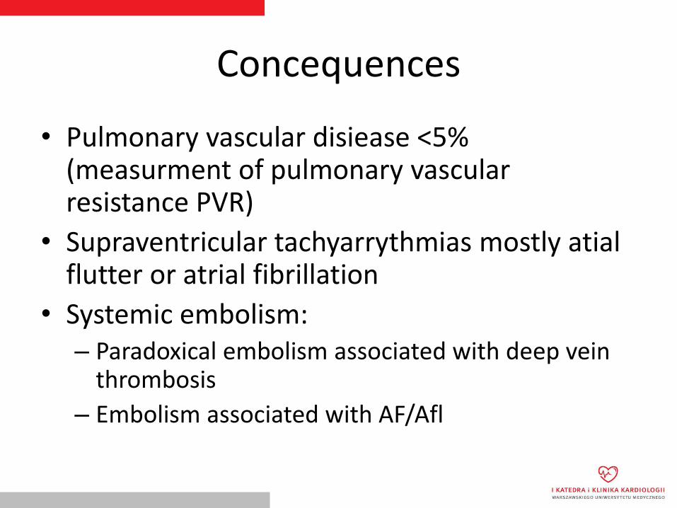
Concequences
• Pulmonary vascular disiease <5% (measurment of pulmonary vascular resistance PVR)
• Supraventricular tachyarrythmias mostly atial flutter or atrial fibrillation
• Systemic embolism: – Paradoxical embolism associated with deep vein
thrombosis
– Embolism associated with AF/Afl
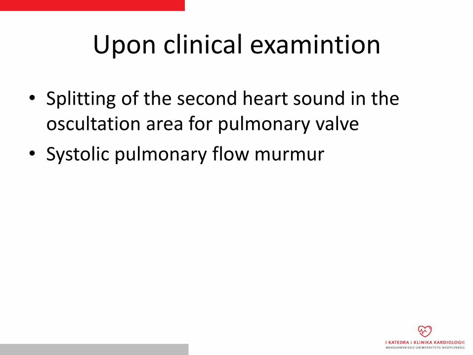
Upon clinical examintion
• Splitting of the second heart sound in the oscultation area for pulmonary valve
• Systolic pulmonary flow murmur
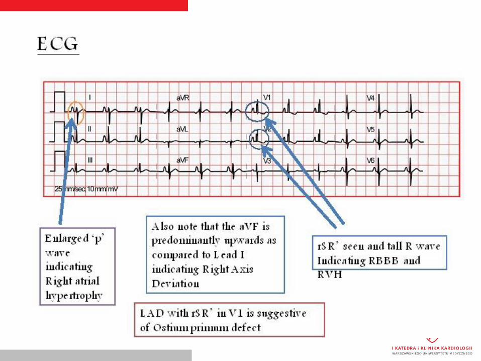
ECG

Echocardiographic view

„Bubble” test
Additional information
• Exercise/sports: No restrictions in asymptomatic patients before or after intervention without pulmonary hypertension, significant arrhythmias, or RV dysfunction.
• Limitation to low-intensity recreational sports in PAH patients • The risk from pregnancy in patients without pulmonary
hypertension is low. • Closure before pregnancy may prevent paradoxical embolism and
worsening of clinical status. • Pregnancy is contraindicated in patients with severe PAH or
Eisenmenger syndrome • The recurrence rate of CHD is 3–10% (excluding familial ASD and
heart –hand syndromes with autosomal dominant inheritance)

Ventricular septal defect

Ventricular septal defect
• Most common, after bicuspid valve, congenital heart malformation at birth as an isolated finding (30-40%)
• Spontanous closure is frequent during childhood (in muscular and perimembranous type)
• Mostly diagnosed before adulthood
• Can be a component of complex anomalies such as tetralogy of Fallot.

Types
• Perimembranous (~80%)
• Muscular/trabecular (15-20%)
• Outlet supracristal/subarterial/subpulmonary/infundibular (~5%)
• Inlet/AV canal/AVSD type (typically in Down syndrome)

Clinical presentation
• Depends on the size of the defect, significancy of the L-R shunt, LV volume overload, pulmonary hipertension
• If small – no clinical manifestation
• If large – dyspnea on excersise
– heart failure symtoms
– Holosystolic murmur over III-IV intercostal space
– pre-cordial thrill

Problems in adulthood
• Eisenmenger syndrome (in large VSD L-R shunt after development of PAH changes into R-L shunt - > cyanosis)
• Endocarditis 6x higher than in the population
• Heart failure
• Arrythmia

Additional information
• Exercise/sports: No restrictions are required in patients after VSD closure, or with small VSD without pulmonary hypertension, significant arrhythmias, or LV dysfunction. Patients with PAH must limit themselves to low-intensity recreational activity/sports
• Pregnancy is contraindicated in Eisenmenger syndrome. The risk is low in asymptomatic patients with normal LV and no PAH
• The recurrence rate of CHD has been reported at 6– 10%.
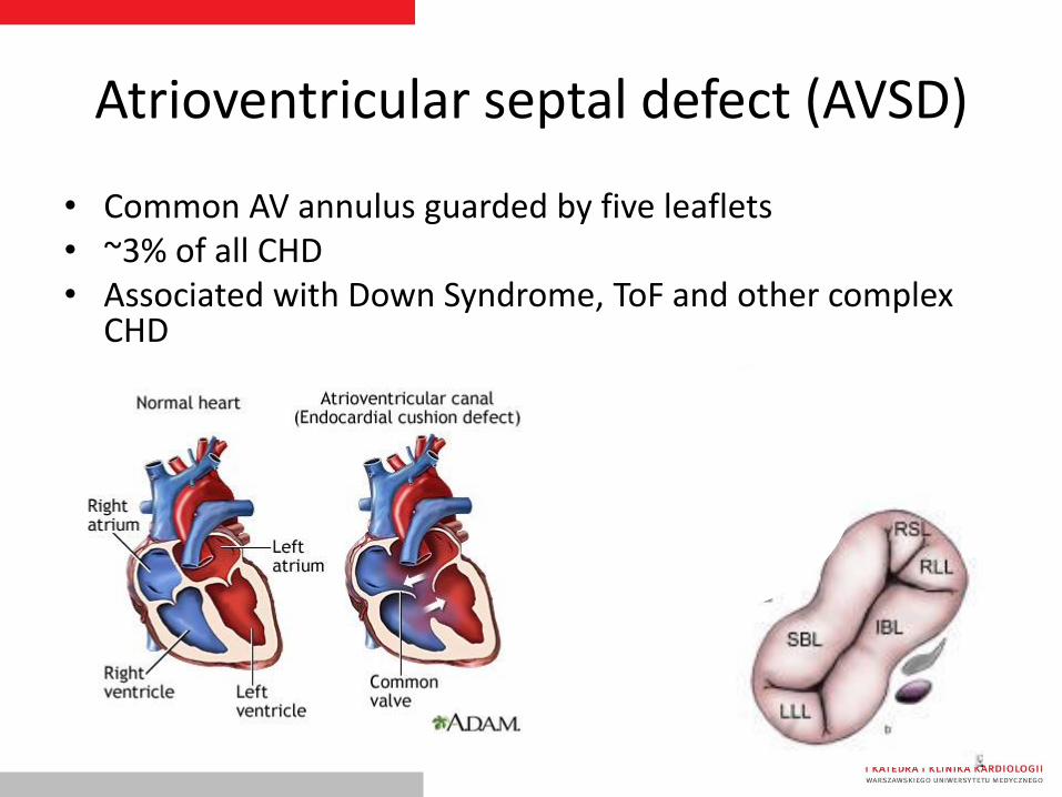
Atrioventricular septal defect (AVSD)
• Common AV annulus guarded by five leaflets • ~3% of all CHD • Associated with Down Syndrome, ToF and other complex
CHD


Clinical presentation
• Depends on the presences and size of ASD and VSD and competences of the left-sided AV valve
• Exercise intolerance, dyspnoea, arrythmia, cyanosis
• If unoperated complete AVSD-> Eisenmenger syndrome

Treatment
• Cases of complete AVSD should be operated in 2-4 month of life,
• If symptoms of heart failure un unmanageble with medication operation even in 1st month

Patent ductus arteriosus (PDA)
• Persistant cominication between the proximal left pulmonary artery and the descending aorta just distal to the left subclavian artery
• Isolated or as a part of complex CHD

Clinical presentation
• Small duct with no LV volume overload (normal LV) and normal PAP (generally asymptomatic)
• Moderate PDA with predominant LV volume overload: large LV with normal or reduced function (may present with left heart failure)
• Moderate PDA with predominant PAH: pressure-overloaded RV (may present with right heart failure)
• Large PDA: Eisenmenger physiology with differential hypoxaemia and differential cyanosis (lower extremities cyanotic, sometimes left arm, too);

Additional information
• Exercise/sports: No restrictions in asymptomatic patients before or after intervention without pulmonary hypertension;
• limitation to low-intensity sports in PAH patients.
• No significantly increased risk for pregnancy in patients without pulmonary hypertension. Pregnancy is contraindicated in patients with severe PAH and Eisenmenger syndrome

Left ventricular outflow tract obstruction (LVOTO)
• Most common site of obstructon is valvular • Other possible levels: subvalvular or supravalvular • Most common cause for congenital valvular aortic stenosis
is bicuspid aortic valve • Patients frequently remain asymptomatic for many years • Prognosis is good and sudden death is rare in asymptomatic • patients with good exercise tolerance, even when stenosis
is severe. • Once symptoms - angina pectoris, dyspnoea, or • Syncope occur the prognosis deteriorates rapidly.
Coarctation of aorta (CoA)
• Its occurs as a discrete stenosis or as a long, hypoplastic aortic segment.
• Typically CoA is located in the area where the ductus arteriosus inserts, and only in rare cases occurs ectopically (ascending, descending, or abdominal aorta).
• CoA accounts for 5–8% of all congenital heart defects.


Clinical presentation
• Signs and symptoms depend on the severity of CoA • Key symptoms may include headache, nosebleeds,
dizziness, tinnitus, shortness of breath, abdominal angina, claudication, leg cramps, exertional leg fatigue, and cold feet.
• The natural course may be complicated by left heart failure, intracranial haemorrhage (from berry aneurysm), IE, aortic rupture/dissection, premature coronary and cerebral artery disease, and associated heart defect
• Screening programme: measurement of blood oxygen saturation on arms and legs

Tetralogy of Fallot (ToF)
• ~10% of all CHD
• four features
– a non-restrictive VSD
– overriding aorta
– Right ventricular outflow tract obstruction infundibular, valvular, or (usually) a combination of both, with or without supravalvular or branch PA stenosis
– Consequent hiperthrophy of the right ventricule


Clinical presentation
• Childhood: – systolic heart murmur, – progressive cyanosis (in exercise or at rest) – anoxemic attacks due to constriction of arteriosus cone – retardation in development
• Unoperated – poor prognosis • Children undergo first surgical repairs between 6-18
months of life: – Total correction – Two-step treatment: Blalock-Taussig operation (junction
between subclavian artery and pulmonary artery)+total correction afterwards
• 85% of children after surgical treatment have proper exercise tolerance and do not require medication

Ebstein’s anomaly
• abnormally formed and apically displaced leaflets of the tricuspid valve
• The apical displacement of the tricuspid valve means that the right heart consists of an RA, an atrialized portion of the RV, and the remaining functional RV.
• The tricuspid valve is often regurgitant


Associated anomalies
• shunt at the atrial level [secundum ASD or patent foramen ovale (PFO)]
• accessory pathways [Wolff–Parkinson– White (WPW) syndrome]
• VSD,
• PS, pulmonary atresia,
• ToF,
• CoA,
• mitral valve abnormalities
Clinical presentation
• Mild forms-> asymptomatic • Severe forms
– high-grade tricuspid regrugitation – Right ventricule disfunction and failure – Cerebral abscesses – Paradoxical embolism – Pulmonary embolism – Tachyarryhythmias – Sudden cardiac death – Infective endocarditits
• Symptoms: – Palpitations, dyspnoea, fatigue, poor exercise tolerance, chest
pain, peripheral and/or central cyanosis


Marfan syndrome
• Autosomal dominant disorder of connective tissue
• Involvment: cardiovascular, skin and skeletal, ocular, pulmonary, and dura mater
• Prevalence ~2–3 per 10 000 births
• Mutations in the FBN1 gene on chromosome 15q21 encoding fibrillin-1, a glycoprotein in the extracellular matrix
Clinical presentation
• Long arms, legs and fingers • Tall and thin body type • Curved spine • Abmormalities of the chest • Flexible joints • Flat feet • Crowded teeth • Stretch marks on the skin that are not related to weight gain or loss
• Prognosis is mainly determined by progressive dilation of the aorta, leading to aortic dissection or rupture, which are the major causes of death
• Men age of untreated patients: 40 years old.


Medical therapy
• Both medical and surgical therapies have improved life expectancy
• substantially up to 60 –70 years.
• Blockers might reduce the rate of aortic dilation and might improve survival, at least in adults.
• Rigorous antihypertensive medical treatment, aiming at a systolic blood pressure ,120 mmHg, and 110 mmHg in patients with aortic dissection, is important.
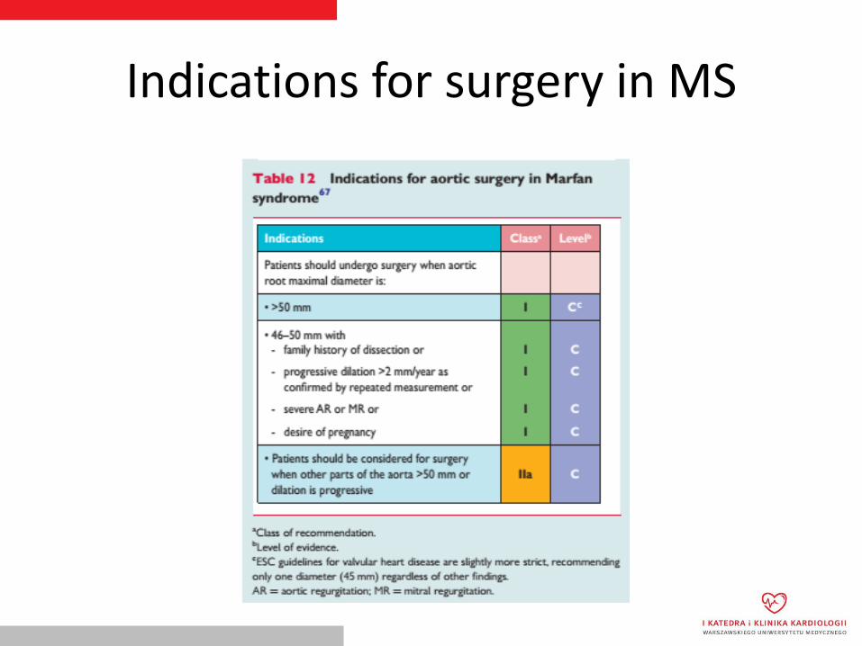
Indications for surgery in MS

Additional information
• Patients should be advised to avoid exertion at maximal capacity, competitive, contact, and isometric sports.
• There is a 50% chance that a child born to a mother with Marfan syndrome would be affected with the condition (genetic counselling).
• Women with an aortic diameter >45 mm are strongly discouraged from becoming pregnant without prior repair because of the high risk of dissection.
• An aortic diameter <40 mm rarely presents a problem, although a completely safe diameter does not exist.
• With an aorta between 40 and 45 mm, previous aortic growth and family history are important for advising pregnancy with or without aortic repair.
• Even after repair of the ascending aorta, Marfan patients remain at risk for dissection of the residual aorta.
Transposition of great arteries (TGA)
• characterized by ventriculo-arterial discordance: the LV gives rise to the PA, and the RV to the aorta
• A complex TGA associates with intracardiac anomalies:
– VSD (in up to 45% of cases),
– LVOTO (~25%),
– CoA (~5%)


After birth
• Requiers either isoprostin admision to keep the ducus arteriosus open
• ENSURE the connection between the two circulations!
• Treatment:
– Mustard or Senning atrial switch procedure
– Arterial switch procedure


Thank you for your attention