Congenital Heart Disease - Healing, Teaching & · PDF fileCongenital Heart Disease A...
82
Congenital Heart Disease Mary Rummell, RN, MN, CPNP, CNS Clinical Nurse Specialist Pediatric Cardiology/Cardiac Surgery
Transcript of Congenital Heart Disease - Healing, Teaching & · PDF fileCongenital Heart Disease A...

Congenital Heart Disease
Mary Rummell, RN, MN, CPNP, CNS
Clinical Nurse Specialist
Pediatric Cardiology/Cardiac Surgery

Objectives The learner will:
Identify 3 areas in the developing heart that result in congenital heart defects.
Discuss 3 important components of a cardiovascular assessment.
List the 3 cardinal signs of congenital heart disease.
Describe the clinical symptoms in each category.
Identify 3 defects that are included in each category.

Congenital Heart Disease
A ―congenital‖anomaly originating in the developing fetus is often considerably modified, at least physiologically, by the dramatic circulatory adjustments at birth. Weeks, months, or even years may then elapse before the anomaly evolves into the ―typical‖ clinical picture. Both physiologic and structural changes subsequently continue, or conversely, the malformation may ―vanish.‖ Joseph Perloff (1978)
Definition:
Congenital (Latin) Con = together, Genital = Born Actually occurs seven months before birth Most defects are compatible with intrauterine life until a term
birth

Incidence of Congenital Heart Disease
8-10 per 1000 live births (approx. 1 %)
(Does not include preterm infants with PDAs or people with slight abnormalities of aortic valve)
Increases to approx 3% with subsequent siblings
Risk increases with left sided lesions – some quote 15%.
Also increases in offspring of parents with CHD
Remains one of the greatest causes on neonatal mortality & morbidity
Leading expense for neonatal health care

Etiology of Congenital Heart Disease: Multifactorial
Maternal: Disease: Diabetes, Collagen disease (Lupus), Seizure disorders Medications: anticonvulsants, diazepam
progesterone/estrogen, alcohol, street drugs (cocaine) , Retin-A, lithium, thalidamide, lithium, warfarin, aspirin, ACE inhibitors
Genetic: Chromosome abnormalities: 13, 18, 21; Turner’s Syndrome, DiGeorge Syndrome (22q11 deletion), Williams Syndrome
Environmental: Viral infections (Rubella, CMV), ???? Toxins

Embryologic Timeline
Week 3: Cardiogenic plate, endocardial tubes Week 4: Fusion of endocardial tubes to single
median tube, first contraction at day 22, cardiogenic looping
Week 5: Beginning of true circulation, septation of primum atrium, AV valves to 3 chamber heart
Week 6: Septum secundum, septation of bulbus and ventricle, divided truncus arteriosus
Week 7: Four chambered heart, absorption of pulmonary veins

Cardiac Looping
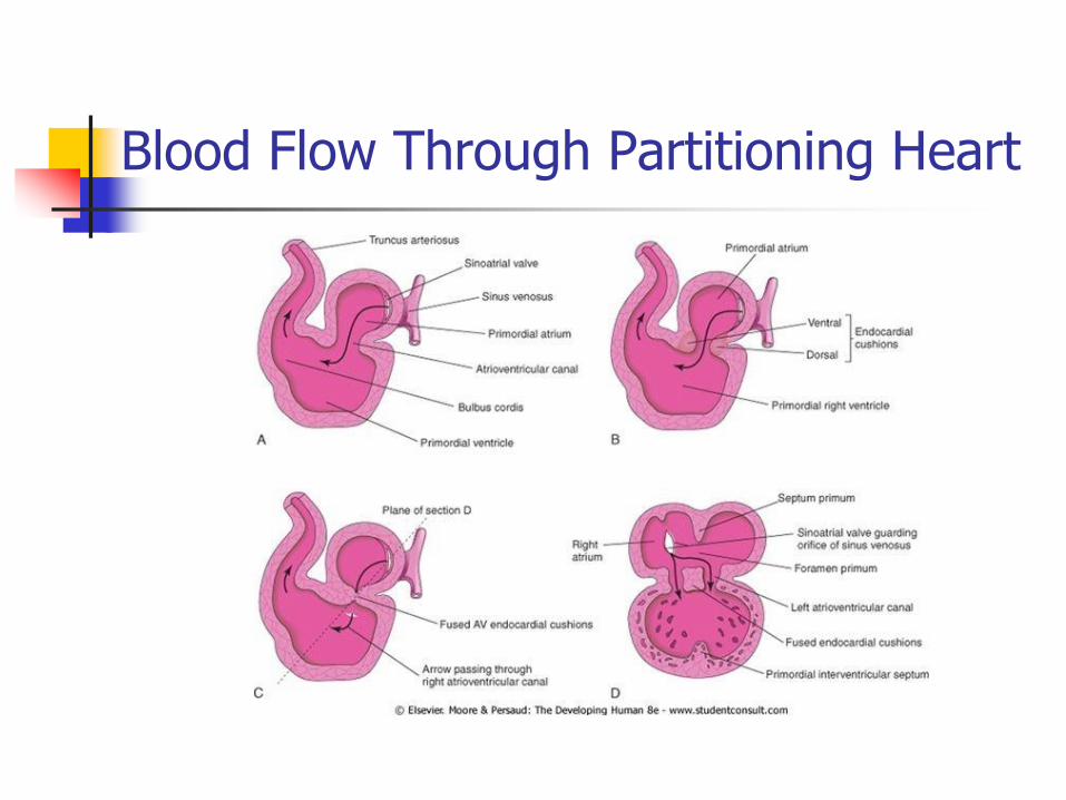
Blood Flow Through Partitioning Heart

Partitioning of Truncus Arteriosus

Development of the Ventricular Septum

Development of Aortic Arch

Fetal Circulation

Newborn Physiology

Closure of fetal shunts
Ductus arteriosus Constricture by 10 to 14 days. Anatomic closure and
formation of ligamentum arteriosus by 12th week
May remain open much longer with prematurity or hypoxia.
Foramen ovalae
Functional closure at birth, anatomic closure has been seen
by 3 months, 50% by 5 years, 25% of adults still have PFO.
Ductus venosus
Constriction of vessel within hours of birth as portal pressure
falls with removal of placenta

Normal Heart Pressures and Saturations

Cardiac Output: Heart Rate X Stroke Volume
Normal Stroke Volume
Newborn = 400 mL/kg/min
Infant = 200mL/kg/min
Child = 100mL/kg/min
Increased oxygen demand (consumption) increase cardiac output: Heart rate and/or stroke volume

Heart Rate: The number of contractions/minute
Principles: Increased heart rate improves Cardiac Output
until diastolic filling time and coronary perfusion are compromised
Decreased heart rate necessitates an increase
in stroke volume to maintain Cardiac Output Neonates are heart rate dependent for Cardiac
Output

Stroke Volume: The volume of blood ejected by the ventricle with each contraction
Factors affecting Stroke Volume:
Preload
Afterload
Contractility

Cardiac Output:
Amount of blood pumped from the ventricle/minute
MUST meet the metabolic demands of the body.
Normalized to BSA (Body Surface Area)
The cardiovascular assessment is your measurement of how well your patient is meeting the metabolic demands.

Assessment Systematic
Consistent
Head to toe
Assess other systems with CV system
Always start with inspection and note general appearance
SICK or WELL

Cardiovascular Assessment
End-organ perfusion
Skin color and temperature
Pulses (peripheral & central)
Capillary refill
Heart rate/rhythm
Heart sounds
Blood pressure

Inspection
Head/neck
Color Cyanosis- central vs
peripheral
Pallor - hemoglobin
Work of breathing Nasal flaring, retractions
Neck veins

Palpation
Peripheral pulses
Rate/rhythm
Quality
Equality
Location
Carotid
Central –Brachial, femoral
Peripheral – Radial,
dorsalis pedis,
posterior tibial
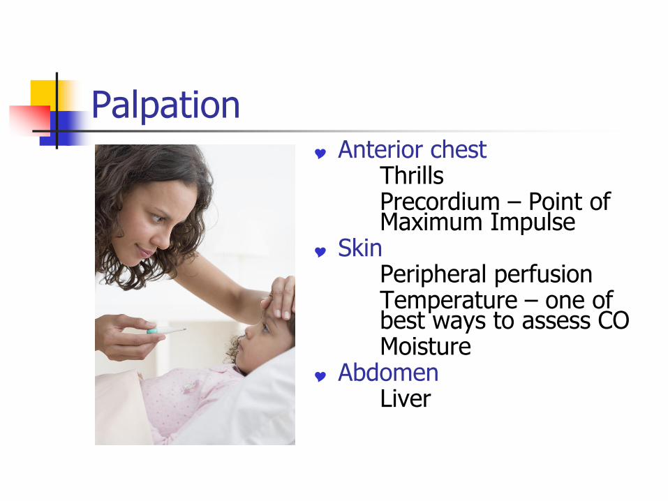
Palpation Anterior chest Thrills Precordium – Point of
Maximum Impulse Skin Peripheral perfusion Temperature – one of
best ways to assess CO Moisture Abdomen Liver

Conduction System

Rhythm Interpretation
Sinus Arrhythmia

Auscultation
Key is to listen to one sound at a time!
Listen in 2 positions.
Listen in systematic pattern.

Heart Sounds
S1– beginning of systole, AV valves close
S2 – end of systole, closure of semilunar valves
S3– normal in children, related to rapid filling of ventricle
S4—abnormal, heard late in diastole or early systole, heard in CHF, decreased ventricular compliance

Murmurs
Definition: ―Whooshing‖ sounds that occur in various stages of diastole and/or systole due to turbulent blood flow.
Causes: Normal heart murmur Increased rate of blood flow Blood flow forced forward through an
incompetent/constricted/irregular valve Blood flows back (regurgitant) through an
incompetent valve, a septal defect, or a patent ductus arteriosus

Characteristics of Murmurs
Timing
Frequency or pitch
Location
Intensity (Graded I – VI)
Radiation
Quality
Effect of respiration
Effect of position

Significance of Murmurs
A systolic murmur may be normal. A diastolic murmur is never normal.
The intensity of the murmur does NOT indicate the severity of heart disease.
You may not hear a murmur in serious CHD.
You may hear a Grade 4-5 murmur in insignificant CHD.

Normal Heart Murmurs
Newborn – Peripheral pulmonary stenosis
Infant/child – Pulmonary ejection murmur
Child – Still’s murmur, vibratory murmur
Teenager – Venous hum
Blood Pressure
Cuff size – EXTREMELY important
Extremity – use right arm
Normal Range
LAST vital sign that will indicate that the child is in trouble

Normal Dinamap Blood Pressure Ranges

Cardiac Reserve
Adaptive mechanisms activated to increase cardiac output.
Mechanical factors
Hypertrophy
Dilation
Biochemical factors
Increase energy production
Activation of the adrenergic nervous system

Cardinal Signs of Heart Disease
Cyanosis
Decreased Systemic Perfusion
Tachypnea

Cyanosis Clinical Presentation
Central rather than peripheral.
May not be immediately present.
Not evident until a significant amount of reduced hemoglobin is present.
Source of systemic arterial blood flow.
Tachypnea without distress
Congenital Defects
•Transposition of the Great Arteries
•Tetralogy of Fallot
•Tricuspid Atresia •Pulmonary Atresia

Consequences of Cyanosis
Polycythemia
Clubbing
Hypoxic spells and squatting
Central nervous system complications
Bleeding disorders

Key Questions
How does blood get to the lungs ? How does blood get to the body? Is the pulmonary/systemic blood flow
dependent on the ductus arteriosus? If the answer to any of these questions is
yes, immediate intervention is mandatory. Prostaglandin E1 (PGE) Atrial septostomy Arterial to pulmonary shunt Palliative/reconstructive surgery

Prostaglandin E1 (PGE)
Indications for use: Right heart outflow obstruction Transposition of the great arteries Left heart outflow obstruction
Contraindications for use: Total anomalous pulmonary vascular return
(TAPVR) Respiratory distress Left-to-right shunts with increase pulmonary
blood flow

Prostaglandin E1
Maintains patency of the ductus arteriosus in ductal dependent congenital heart defects to provide either pulmonary or systemic circulation.
Side effects: Cardiovascular: Vasodilation, arrhythmias, hypotension,
edema Neurological: Seizures, hyperthermia, irritability,
lethargy Respiratory: Apnea, hypoventilation, bronchospasm,
tachypnea Renal: anuria Hematologic: Thrombocytopenia, hemorrhage, DIC

Modified Blalock-Taussig Shunt

Nursing Considerations:
Intracardiac Mixing of Systemic and Pulmonary Circulation
Assessment of Cardiac Output:
Shunt
Murmur present?
Hydration status?
Oxygen saturation?

Transposition of the Great Arteries
5% of all CHD Males > female by 3:1 Must have communication
between left and right sides of the heart
Surgical repair before left ventricular muscle strength decreases
Oxygen saturation may be greater in lower extremities

Arterial Switch Procedure

Coronary Artery Repair for TGA

Tetralogy of Fallot
10 % of all CHD Was most common cyanotic
defect beyond infancy Cyanosis varies by degree of
pulmonary stenosis 4 components:
Large VSD Right ventricular outflow
tract obstruction Overriding aorta Right ventricular
hypertrophy

―Tet‖ Spells Most frequently seen in unrepaired patients with Tetralogy of Fallot. Symptoms:
Rapid and deep respirations (hyperpnea) Increasing cyanosis Irritability and prolonged cry Decreased intensity or absence of heart murmur
Treatment: Pick up and hold infant Knee chest position Morphine sulfate: 0.1-0.2 mg/kg IM 100% oxygen Phenylephrine 0.02 mg/kg IV bolus followed by infusion Sodium bicarbonate: 1 mEq/kg IV

Tetralogy of Fallot with BT Shunt

Repair of Tetralogy of Fallot

Pulmonary Atresia
With Intact Ventricular Septum 1% of all CHD
With VSD 2% of all CHD
Is ductal dependent
Intervention necessary within first few hours after birth.

Tricuspid Atresia
1-2 % of all CHD
Male slightly more than female
An associated defect is necessary for survival.
Systemic saturation is directly related to the amount of pulmonary blood flow.

Tricuspid Atresia with BT Shunt

Tricuspid Atresia with Glenn Shunt

Tricuspid Atresia Lateral Tunnel Fontan

Tricuspid Atresia External Conduit Fontan


Decreased Systemic Perfusion
Congenital Defects
Aortic Valve Stenosis
Coarctation of the Aorta
Hypoplastic Left Heart Syndrome
Clinical Presentation • Poor feeding
• Pallor/grey
• Diaphoresis
• Tachypnea with respiratory distress
• Irritability

Nursing Considerations:
Systemic Blood Flow? Intracardiac Mixing of Systemic
and Pulmonary Circulations? Assessment of cardiac output
Color Temperature Perfusion Blood Pressure Heart Rate/rhythm

Aortic Valve Stenosis
5 % of all CHD Males > female by 4:1 Wide spectrum of disease. Babies with severe stenosis
may be ductal dependent.
Half of patients with Coarctation also have abnormality of aortic valve.
Risk of reoccurance increases by 15 % or greater.
Aortic Valve
Stenosis

Coarctation of the Aorta
8 % of all CHD
Male > female by 2:1
30 % of children with Turners Syndrome
May not be evident until ductus arteriosus closes
Can present with severe cardiogenic shock

Hypoplastic Left Heart Syndrome
2 % of all CHD
10% of all CHD presenting within first month of life
May not be identified until ductus arteriosus closes.
Presents in shock.
Treatment with oxygen will increase severity by decreasing pulmonary vascular resistance resulting in increase pulmonary blood flow and decreased systemic circulation.

Norwood Procedure for HLHS

Norwood Procedure/Sano

Glenn Shunt

HLHS with External Conduit Fontan

Tachypnea
Clinical Manifestations Tachypnea with mild
respiratory distress Hepatomegaly Tachycardia Diaphoresis (secondary
to sympathetic response to increased stress)
Failure to thrive Increased oxygen
consumption Increased work of
breathing
Congenital Defects
Patent Ductus Arteriosus
Atrial Septal Defect
Ventricular Septal Defect
Atrioventricular Septal Defect
Total Anomalous Pulmonary Venous Return (TAPVR)
Truncus Arteriosus

Tachypnea: Excessive Pulmonary Blood Flow
Increased pulmonary blood flow results in pulmonary edema and congestive heart failure.
Increasing inspired oxygen will decrease pulmonary resistance thereby increasing pulmonary blood flow. CHF symptoms will increase and may even have acute decompensation.
Exception is TAPVR below diaphragm. This results in pulmonary venous congestion.

Nursing Considerations:
Intracardiac Mixing of Systemic and Pulmonic Blood
Supplemental Oxygen
Assessment of Cardiac Output
Failure to Thrive Increased work of breathing
Increased heart rate
Decreased caloric intake

Patent Ductus Arteriosus
5-10 % of CHD except for preterm infant
Female > male by 3:1
Elective closure except in preterm infant
Non-surgical closure with coils

Atrial Septal Defect
5-10 % of all CHD 4th most common Females > males by 2:1 Usually asymptomatic Primary types:
Secundum (site of fossa ovalis)
Sinus venosus (often associated with PAPVR)
Ostium primum (AVSD type defect)

Types of Atrial Septal Defects
Park: Pediatric Cardiology for Practitioners, 5th ed. Copyright © 2008 Mosby, An Imprint of Elsevier
> View this image in its location within the book
Figure 12-1
Anatomic types of
atrial septal
defects (ASDs)
viewed with the
right atrial wall
removed. IVC,
inferior vena cava;
SVC, superior
vena cava.

Non surgical closure of ASD

Ventricular Septal Defect
20-25 % of all CHD Most common defect Many will close
spontaneously Intervention
depends on location and symptoms
Increasing use of cath closure devices

Anatomy of Ventricular Septal Defects Figure 12-6 Anatomy of ventricular
septum and ventricular septal defect (VSD). A, Ventricular septum viewed from the right ventricular (RV) side. The membranous septum is small. The large muscular septum has three components: the inlet septum (I), the trabecular septum (T), and the outlet (or infundibular) septum (O). B, Anatomic locations of various VSDs and landmarks, viewed with the RV free wall removed. a, outlet (infundibular) defect; b, papillary muscle of the conus; c, perimembranous defect; d, marginal muscular defect; e, central muscular defect; f, inlet defect; g, apical muscular defect. (From Graham TP Jr, Bender HW, Spach MS: Ventricular septal defect. In Adams FH, Emmanouilides GC, Riemenschneider TA (eds): Moss' Heart Disease in Infants, Children and Adolescents, 4th ed. Baltimore, Williams and Wilkins, 1989.)

Types of Ventricular Septal Defects
Membranous Also called perimembranous Most common Located below aortic valve May require surgical repair
Muscular 70 % close spontaneously
Outlet Located just below pulmonary valve Risk of prolapse of aortic valve leaflet into defect Requires surgical repair, important to protect integrity of aortic
valve
Inlet Located below the leaflet of the tricuspid valve Will need surgical repair

Atrioventricular Septal Defect (AVSD) (AV Canal Defect, Endocardial Cushion Defect)
2 - 5 % of all CHD
30-50 of children with Down’s Syndrome
Usually surgically repaired by 4- 6 months of age. Earlier if increasing CHF.

Total Anomalous Pulmonary Venous Return
Less than 1 % of all CHD No direct connection
between PV and LA Must have an atrial
communication for survival Types:
Supracardiac (50%) Male = female Infracardiac (20%) Male > female by 4:1 Cardiac (20%) Usually to coronary sinus Mixed - Right veins involved
twice as often as left

Truncus Arteriosus
<1 % of all CHD Single trunk from
both ventricles Must have a VSD Truncal valve may
have 3 or 4 cusps Usually presents
shortly after birth. Surgical repair
when diagnosed.

Surgical Repair: Truncus Arteriosus

Ready for Discharge:
Newborn Discharge Considerations Hearing Test
Car Seat Angle Test
Newborn Metabolic Screen
Medications
Wound Healing
Interstage monitoring Oxygen Saturation
Weight Gain
Activity/fussiness

References Allen, HD; Clark, EB; Gutgesell, HP; & Driscoll, D.J. (2001) Moss and
Adams’ Heart Disease in Infants, Children’ and Adolescents, Sixth
Edition. Philadelphia: Lippincott Williams & Wilkins.
Artman, M; Mahony, L; & Teitel, DF (2002) Neonatal Cardiology. New York: The McGraw-Hill Companies.
Barnes, LA (1981) Manual of Pediatric Physical Diagnosis. Fifth Edition. Chicago: Year Book Medical Publishers, Inc.
Curley, MAQ; & Moloney-Harmon, PA. (2001) Critical Care Nursing of
Infants andChildren, 2nd Edition. Philedelphia: W.B. Saunders Company.
Hazinski, MF. (1999) Manual of Pediatric Critical Care. St. Louis: Mosby,
Inc.
Park, MK (2008) Pediatric Cardiology for Practitioners. Chicago: Year Book Medical Publishers, Inc.
Taketomo, CF; Hodding, JH, & Kraus, SM (2007) Pediatric Dosage Handbook. Hudson, Ohio: Lexi-Comp.