Common brain activations for painful and non-painful
18
RESEARCH ARTICLE Open Access Common brain activations for painful and non-painful aversive stimuli Dave J Hayes * and Georg Northoff Abstract Background: Identification of potentially harmful stimuli is necessary for the well-being and self-preservation of all organisms. However, the neural substrates involved in the processing of aversive stimuli are not well understood. For instance, painful and non-painful aversive stimuli are largely thought to activate different neural networks. However, it is presently unclear whether there is a common aversion-related network of brain regions responsible for the basic processing of aversive stimuli. To help clarify this issue, this report used a cross-species translational approach in humans (i.e. meta-analysis) and rodents (i.e. systematic review of functional neuroanatomy). Results: Animal and human data combined to show a core aversion-related network, consisting of similar cortical (i.e. MCC, PCC, AI, DMPFC, RTG, SMA, VLOFC; see results section or abbreviation section for full names) and subcortical (i.e. Amyg, BNST, DS, Hab, Hipp/Parahipp, Hyp, NAc, NTS, PAG, PBN, raphe, septal nuclei, Thal, LC, midbrain) regions. In addition, a number of regions appeared to be more involved in pain-related (e.g. sensory cortex) or non-pain-related (e.g. amygdala) aversive processing. Conclusions: This investigation suggests that aversive processing, at the most basic level, relies on similar neural substrates, and that differential responses may be due, in part, to the recruitment of additional structures as well as the spatio-temporal dynamic activity of the network. This network perspective may provide a clearer understanding of why components of this circuit appear dysfunctional in some psychiatric and pain-related disorders. Keywords: Meta-analysis, Translational, Aversion, Pain, Neuroimaging, Animal models Background Aversion: painful and non-painful stimuli Identification of potentially harmful stimuli is necessary for the well-being and self-preservation of all organisms. Organisms with relatively simple nervous systems (e.g. worms, fruit flies) display motivated approach and avoid- ance behaviours to rewarding and aversive stimuli, respectively, implying the existence of some evolutionar- ily conserved mechanisms [1,2]. Aversive stimuli are those which an organism will generally expend energy to minimize or avoid [3]; in this context, aversion is oper- ationally opposite to reward [4]. However, the strength of aversive stimuli and the context in which they occur can produce a variety of psychophysical (e.g. negative emotion, pain) and behavioural (e.g. reduced behaviour following punishment, avoidance) responses. While recent work has suggested the existence of a common aversion-related network of brain regions responsible for the basic processing of aversive stimuli [5], this work focused only on studies involving non-painful stimuli and studies including painful stimuli were not consid- ered; as such, it is unclear if those results extend to pain-related processing. Pain-associated brain activity Pain, which typically results from activating the nocicep- tive system (but can also involve non-nociceptive mechanisms, such as in neuropathic pain), is experi- enced across mammals and is critical for survival [6]. Studies in humans and non-human animals have gener- ally supported the notion that pain is processed differen- tially in the brain according to affective (e.g. amygdala, anterior insula, hippocampus) and sensory (e.g. somato- sensory cortices, posterior insula) dimensions (e.g. [7-9]; though see also [10] for a review on the influential 3- dimension theory of pain). Nonetheless, the assumption * Correspondence: [email protected] Mind, Brain Imaging and Neuroethics Research Unit, Institute of Mental Health Research, University of Ottawa, 1145 Carling Avenue, Ottawa K1Z 7K4, Canada © 2012 Hayes and Northoff; licensee BioMed Central Ltd. This is an Open Access article distributed under the terms of the Creative Commons Attribution License (http://creativecommons.org/licenses/by/2.0), which permits unrestricted use, distribution, and reproduction in any medium, provided the original work is properly cited. Hayes and Northoff BMC Neuroscience 2012, 13:60 http://www.biomedcentral.com/1471-2202/13/60
Transcript of Common brain activations for painful and non-painful
Hayes and Northoff BMC Neuroscience 2012, 13:60http://www.biomedcentral.com/1471-2202/13/60
RESEARCH ARTICLE Open Access
Common brain activations for painful andnon-painful aversive stimuliDave J Hayes* and Georg Northoff
Abstract
Background: Identification of potentially harmful stimuli is necessary for the well-being and self-preservation of allorganisms. However, the neural substrates involved in the processing of aversive stimuli are not well understood.For instance, painful and non-painful aversive stimuli are largely thought to activate different neural networks.However, it is presently unclear whether there is a common aversion-related network of brain regions responsiblefor the basic processing of aversive stimuli. To help clarify this issue, this report used a cross-species translationalapproach in humans (i.e. meta-analysis) and rodents (i.e. systematic review of functional neuroanatomy).
Results: Animal and human data combined to show a core aversion-related network, consisting of similar cortical(i.e. MCC, PCC, AI, DMPFC, RTG, SMA, VLOFC; see results section or abbreviation section for full names) andsubcortical (i.e. Amyg, BNST, DS, Hab, Hipp/Parahipp, Hyp, NAc, NTS, PAG, PBN, raphe, septal nuclei, Thal, LC,midbrain) regions. In addition, a number of regions appeared to be more involved in pain-related (e.g. sensorycortex) or non-pain-related (e.g. amygdala) aversive processing.
Conclusions: This investigation suggests that aversive processing, at the most basic level, relies on similar neuralsubstrates, and that differential responses may be due, in part, to the recruitment of additional structures as well asthe spatio-temporal dynamic activity of the network. This network perspective may provide a clearer understandingof why components of this circuit appear dysfunctional in some psychiatric and pain-related disorders.
Keywords: Meta-analysis, Translational, Aversion, Pain, Neuroimaging, Animal models
BackgroundAversion: painful and non-painful stimuliIdentification of potentially harmful stimuli is necessaryfor the well-being and self-preservation of all organisms.Organisms with relatively simple nervous systems (e.g.worms, fruit flies) display motivated approach and avoid-ance behaviours to rewarding and aversive stimuli,respectively, implying the existence of some evolutionar-ily conserved mechanisms [1,2]. Aversive stimuli arethose which an organism will generally expend energy tominimize or avoid [3]; in this context, aversion is oper-ationally opposite to reward [4]. However, the strengthof aversive stimuli and the context in which they occurcan produce a variety of psychophysical (e.g. negativeemotion, pain) and behavioural (e.g. reduced behaviourfollowing punishment, avoidance) responses. While
* Correspondence: [email protected], Brain Imaging and Neuroethics Research Unit, Institute of MentalHealth Research, University of Ottawa, 1145 Carling Avenue, Ottawa K1Z 7K4,Canada
© 2012 Hayes and Northoff; licensee BioMedCreative Commons Attribution License (http:/distribution, and reproduction in any medium
recent work has suggested the existence of a commonaversion-related network of brain regions responsiblefor the basic processing of aversive stimuli [5], this workfocused only on studies involving non-painful stimuliand studies including painful stimuli were not consid-ered; as such, it is unclear if those results extend topain-related processing.
Pain-associated brain activityPain, which typically results from activating the nocicep-tive system (but can also involve non-nociceptivemechanisms, such as in neuropathic pain), is experi-enced across mammals and is critical for survival [6].Studies in humans and non-human animals have gener-ally supported the notion that pain is processed differen-tially in the brain according to affective (e.g. amygdala,anterior insula, hippocampus) and sensory (e.g. somato-sensory cortices, posterior insula) dimensions (e.g. [7-9];though see also [10] for a review on the influential 3-dimension theory of pain). Nonetheless, the assumption
Central Ltd. This is an Open Access article distributed under the terms of the/creativecommons.org/licenses/by/2.0), which permits unrestricted use,, provided the original work is properly cited.
Hayes and Northoff BMC Neuroscience 2012, 13:60 Page 2 of 18http://www.biomedcentral.com/1471-2202/13/60
that this network (sometimes referred to as the ‘PainMatrix’) is specifically activated by painful stimuli hasbeen questioned [11-13]. Using fMRI in humans, Mour-aux et al. (2011) uncovered strong support for the no-tion that the typical regions of the Pain Matrix arelargely involved in salience processing [12]. They showedthat multimodal non-painful aversive stimuli and painfulstimuli activate similar regions in the MCC, insula, thal-amus, and sensory cortex and that the BOLD signalsin these regions correlated largely with the perceivedsaliency of the stimulus (regardless of modality or stim-ulus type).
Studies suggest shared regions for pain and non-painaversionWhile much work has identified pain as a uniquelyimportant experience (e.g. [10,14]), numerous studiesusing non-painful aversive stimuli (e.g. unpleasantsounds, sights, etc.) have implicated many of the samecortical and subcortical regions [5], suggesting that theprocessing of various painful and non-painful aversivestimuli require many of the same neurobiological sub-strates. In this regard, human studies have been key tounderstanding the role of cortical regions (e.g. prefrontaland insular cortices; e.g. [15,16]). Alternately, studies inanimals have highlighted the importance of subcorticalareas such as the periaqueductal grey, hypothalamus, bednucleus of the stria terminalis, nucleus accumbens/ventral striatum and ventral tegmental area [17-20].While prior work has identified a network of regionsinvolved in non-painful aversion-related processing [5], itnonetheless remains unclear which, if any, of those identi-fied areas are also involved in processing painful stimuli.
Systematic translational analysis of aversion-relatedcircuitryThe present hypothesis is that there exists a coreaversion-related circuit involved in processing aversivestimuli regardless of whether they are painful or non-painful. In an analogical sense, this network would besimilar to the basic underlying (e.g. mesocorticolimbic)circuitry identified in the field of reward [21-23]. Priormeta-analyses in humans have outlined core regionsassociated with pain processing [6,24], and some animalwork has even suggested the existence of an overlappingpain and non-pain-related aversion network [25]. None-theless, no investigations have used both human andanimal data to directly explore the possibility of a sharednetwork for pain- and non-pain-related processing.To this end, a translational cross-species approach was
used to identify the core components of the potentialaversion-related network. More specifically, our first aimwas to compare brain activations to the passive recep-tion of painful aversive stimuli in healthy adults (using a
meta-analysis of human imaging data; i.e. functionalmagnetic resonance imaging, fMRI, or positron emissiontomography, PET) to those in rodents (using a system-atic review of studies including markers of cellular acti-vation and available imaging studies). Secondly, weaimed to compare the results on pain-related processingto those gathered previously on the processing of passivenon-painful aversive stimuli in humans (meta-analysis)and animals (systematic review) [5].Our main hypothesis is that aversive stimuli, regardless
of origin (e.g. sensory modality) or perception (e.g. pain-ful or non-painful), are processed largely by a commonnetwork of brain regions. However, some areas may bemore (or uniquely) involved in different aspects of pain-and non-pain-related aversive processing. The use of ameta-analytical approach allows for the clear distinctionof areas which have been identified reliably acrossnumerous studies – in comparison to individual studieswhich may have low power and a higher probability ofreporting false positive activations [26]. The incorpor-ation of animal studies allows for a cross-species com-parison and ensures that especially subcortical areas,which may be important for aversion-related processing,are identified. Studying these areas in humans hasproven difficult given limitations in optimal imagingresolution and the correct interpretation of subcorticalactivations (or the lack thereof ) [27]. Importantly, thistranslational approach allowed for the direct comparisonof the overlap between areas identified in pain and non-pain aversion studies.
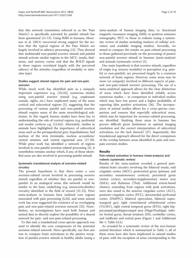
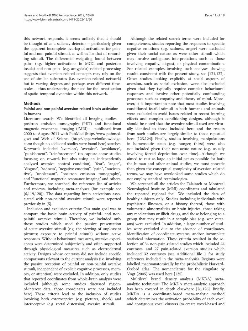
ResultsPain-related activation in humans (meta-analysis) androdents (systematic review)Results of the meta-analysis revealed a general pain-related brain circuitry involving the bilateral insula, midcingulate cortex (MCC), postcentral gyrus (primary andsecondary somatosensory cortices), precentral gyrus(motor cortex), secondary/supplementary motor area(SMA), and thalamus (Thal). Additional extent-basedclusters, extending from regions with peak activations,were also noted in the anterior cingulate cortex (ACC),posterior cingulate cortex (PCC), dorsomedial prefrontalcortex (DMPFC), bilateral operculum, bilateral supra-marginal gyri, right ventrolateral orbitofrontal cortex(VLOFC), right rostral temporal gyrus (RTG), right hip-pocampal/parahippocampal area (Hipp/Parahipp), infer-ior frontal gyrus, dorsal striatum (DS), cerebellar cortex,and midbrain and rostral pons (Figure 1 and Additionalfile 1: Table S1A).As revealed by a systematic review of the non-human
animal literature which is summarized in Table 1, all ofthese areas have also been implicated in animal studiesof pain, with the exception of areas corresponding to the
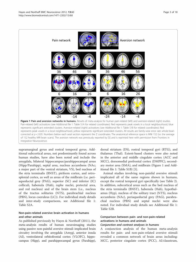
Figure 1 Pain and aversion networks in humans. Results of meta-analysis for human pain-related (left) and aversion-related (right) studies.Pain-related (left) activations (see Additional file 1: Table S1A for related coordinates): Red represents peak voxels in a local neighbourhood, bluerepresents significant extended clusters. Aversion-related (right) activations (see Additional file 1: Table S1B for related coordinates): Redrepresents peak voxels in a local neighbourhood, yellow represents significant extended clusters. All results are family-wise error rate whole-braincorrected at p< 0.05. Numbers below each axial section represent the Z coordinates. The anatomical reference space is MNI 152 (i.e. the averageof 152 healthy MRI brain scans). The aversion network was previously reported by [5] and is reprinted here with permission from Frontiers inIntegrative Neuroscience.
Hayes and Northoff BMC Neuroscience 2012, 13:60 Page 3 of 18http://www.biomedcentral.com/1471-2202/13/60
supramarginal gyrus and rostral temporal gyrus. Add-itional subcortical areas, not predominantly found acrosshuman studies, have also been noted and include theamygdala, bilateral hippocampus/parahippocampal areas(Hipp/Parahipp), septal area, nucleus accumbens (NAc;a major part of the ventral striatum, VS), bed nucleus ofthe stria terminalis (BNST), piriform cortex, and retro-splenial cortex, as well as areas of the midbrain (i.e. peri-aqueductal grey (PAG), superior (SC) and inferior (IC)colliculi, habenula (Hab), raphe nuclei, pretectal area,and red nucleus) and of the brain stem (i.e., nucleusof the tractus solitaries (NTS), parabrachial nucleus(PBN), locus coeruleus (LC)). For individual study detailsand inter-study comparisons, see Additional file 1:Table S2A.
Non-pain-related aversive brain activation in humansand other animalsAs published previously by Hayes & Northoff (2011), themeta-analysis results of human neuroimaging studiesusing passive non-painful aversive stimuli implicated braincircuitry involving the amygdala (Amyg), anterior insula(AI), ventrolateral orbitofrontal cortex (VLOFC), hippo-campus (Hipp), and parahippocampal gyrus (Parahipp),
dorsal striatum (DS), rostral temporal gyri (RTG), andthalamus (Thal). Extent-based clusters were also notedin the anterior and middle cingulate cortex (ACC andMCC), dorsomedial prefrontal cortex (DMPFC), second-ary motor area (SMA), and midbrain (Figure 1 and Add-itional file 1: Table S1B) [5].Animal studies involving non-painful aversive stimuli
implicated all of the same regions shown in humans,except the rostral temporal gyri specifically (see Table 2).In addition, subcortical areas such as the bed nucleus ofthe stria terminalis (BNST), habenula (Hab), hypothal-amus (Hyp), nucleus of the solitary tract (NTS), nucleusaccumbens (NAc), periaqueductal grey (PAG), parabra-chial nucleus (PBN) and septal nuclei were alsonoted. For individual study details see Additional file 1:Table S2B.
Comparison between pain- and non-pain-relatedactivations in humans and animalsConjunction and contrast analyses in humansA conjunction analysis of the human meta-analysisresults for pain- and non-pain-related aversive stimulirevealed a common network of brain areas including:MCC, posterior cingulate cortex (PCC), AI/claustrum,
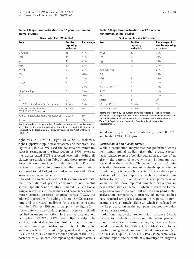
Table 1 Major brain activations in 32 pain non-humananimal studies
Rank order: Pain (32 studies)
Area Studiesreportingactivation
Percentage
Cingulate 15 47%
Thal 14 44%
Sens 13 41%
Hyp 12 38%
Amyg 9 28%
PAG 9 28%
DS 9 28%
Motor 7 22%
Ins 7 22%
Hipp/parahipp 6 19%
NTS 5 16%
IC 5 16%
LC, PBN, Pretectal area, SC, Septal area 4 each 13%
Hab, NAc, Raphe, Piriform,PFC(IL/PL)/OFC, Visual ctx
3 each 9%
Aud ctx, BNST, Cerebellum, Retrosplenial 2 each 6%
Red n 1 3%
Results are ordered by the number of studies reporting specific activations;percent of studies reporting activations is used for comparative illustration. Forindividual study details and inter-study comparisons, see Additional file 1:Table S2A.
Table 2 Major brain activations in 42 aversionnon-human animal studies
Rank order: Aversion (42 studies)
Area Studiesreportingactivation
Percentage ofstudies reportingactivation
Amyg 32 76%
Thal 13 30%
Hyp 12 29%
NTS 10 24%
Parahipp/Hipp 9 21%
PBN 8 19%
PAG 8 19%
Ins 7 17%
PFC (PL, IL)/OFC 7 17%
BNST 5 12%
NAc 5 12%
Septal 3 7%
ACC, DR, DS, LC 2 each 5%
Motor, Hab, VTA 1 each 2%
Results are ordered by the number of studies reporting specific activations;percent of studies reporting activations is used for comparative illustration. Forindividual study details and inter-study comparisons, see Additional file 1:Table S2B. Reprinted with permission from Frontiers in IntegrativeNeuroscience [5].
Hayes and Northoff BMC Neuroscience 2012, 13:60 Page 4 of 18http://www.biomedcentral.com/1471-2202/13/60
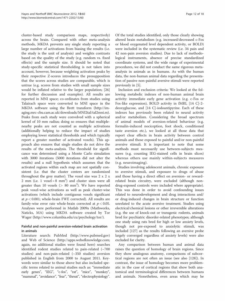
right VLOFC, DMPFC, right RTG, SMA, thalamus,right Hipp/Parahipp, dorsal striatum, and midbrain (seeFigure 2, Table 3). We used the conservative minimumstatistic resulting in the intersection of 5900 voxels atthe cluster-based FWE corrected level [28]. While allclusters are displayed in Table 3, only those greater than10 voxels were considered in the discussion. The per-centage of overlapping voxels in the present studyaccounted for 24% of pain-related activations and 35% ofaversion-related activations.In addition to the activation of this common network,
the presentation of painful compared to non-painfulstimuli (painful> non-painful) resulted in additionalunique activations in the primary and secondary sensori-motor cortices, posterior cingulate cortex (PCC), thebilateral operculum (including bilateral SMG), cerebel-lum, and the rostral midbrain (in a region consistentwith the VTA; see [29]) and rostral pons (see Figure 2).Alternately, non-painful> painful aversive stimuli
resulted in unique activations in the amygdalae and lefthemisphere VLOFC, RTG, and Hipp/Parahipp. Inaddition, extended activation clusters unique to non-painful stimulus presentation were noted for the moreanterior portions of the ACC (pregenual and subgenualACC), the DMPFC, a more anterior portion of the PCC/posterior MCC, an area encompassing the hypothalamus
and dorsal (DS) and ventral striatal (VS) areas, left SMA,and bilateral VLOFC (Figure 2).
Comparison to non-human animalsWhile a conjunction analysis was not performed acrossnon-human animal studies (given that precise coordi-nates related to neural/cellular activation are less oftengiven), the pattern of activation seen in humans wasreflected in these studies. The general pattern of brainactivation between humans and animals appears to bemaintained, as is generally reflected by the relative per-centage of studies reporting such activations (seeTables 3A and 3B). For instance, a large percentage ofanimal studies have reported cingulate activations inpain-related studies (Table 1) which is mirrored by thelarge activation in the pain (but not the non-pain) meta-analysis; in comparison, a majority of animal studieshave reported amygdala activations in response to non-painful aversive stimuli (Table 2), which is reflected bythe large activation in the non-pain (but not the pain)meta-analysis results.Additional subcortical regions of importance (which
may be too difficult to detect or differentiate preciselyusing human brain imaging techniques) have also beennoted in animals (see Tables 1, 2). Most seem to beinvolved in general aversion-related processing (i.e.BNST, Hab, Hyp, LC, NAc, NTS, PAG, PBN, septal area,anterior raphe nuclei) while this investigation suggests
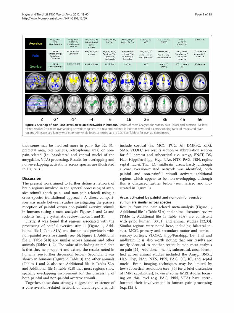
Figure 2 Overlap of pain- and aversion-related networks in humans. Results of meta-analyses for human pain- (blue) and aversion- (yellow)related studies (top row), overlapping activations (green; top row and isolated in bottom row), and a corresponding table of associated brainregions. All results are family-wise error rate whole-brain corrected at p< 0.05. See Table 3 for overlap coordinates.
Hayes and Northoff BMC Neuroscience 2012, 13:60 Page 5 of 18http://www.biomedcentral.com/1471-2202/13/60
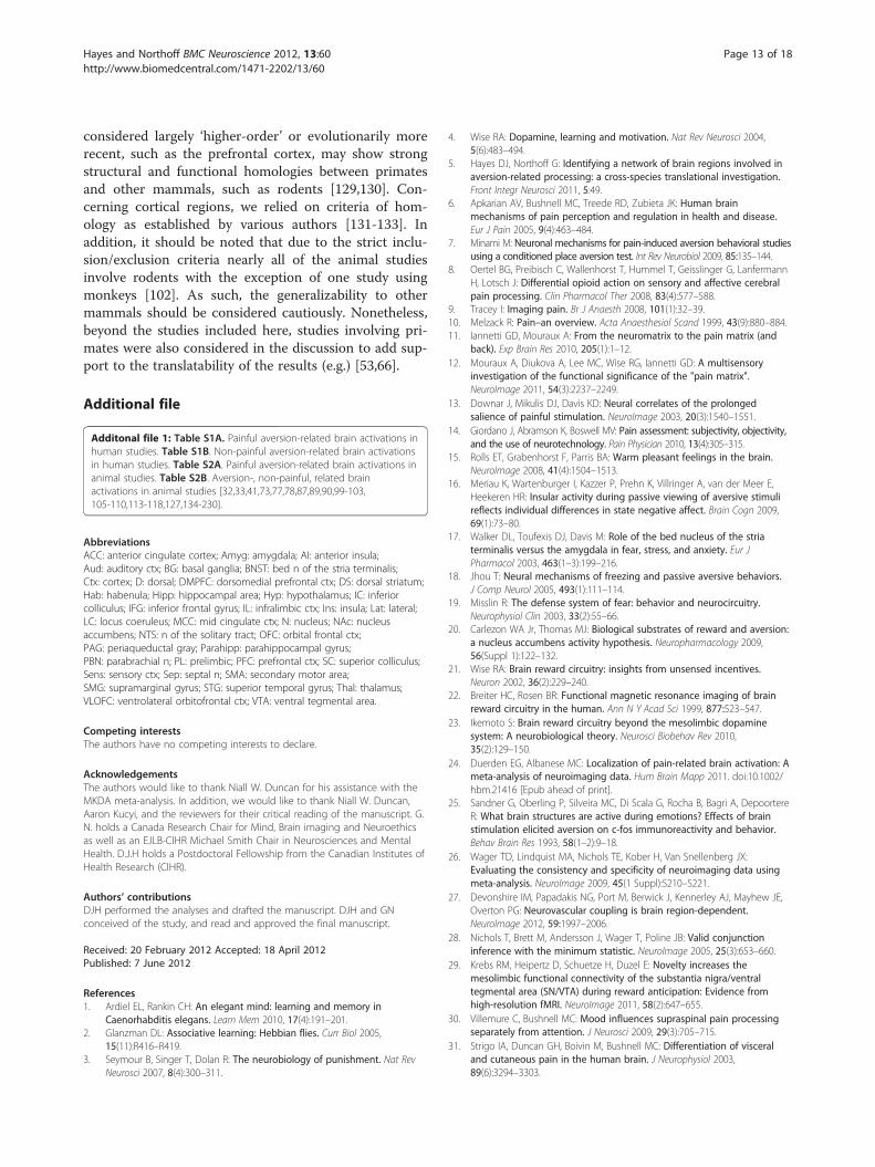
that some may be involved more in pain- (i.e. IC, SC,pretectal area, red nucleus, retrosplenial area) or non-pain-related (i.e. basolateral and central nuclei of theamygdalae, VTA) processing. Results for overlapping andnon-overlapping activations across species are illustratedin Figure 3.
DiscussionThe present work aimed to further define a network ofbrain regions involved in the general processing of aver-sive stimuli (both pain- and non-pain-related) using across-species translational approach. A direct compari-son was made between studies investigating the passivereception of painful versus non-painful aversive stimuliin humans (using a meta-analysis; Figures 1 and 2) androdents (using a systematic review; Tables 1 and 2).Firstly, it was found that regions associated with the
processing of painful aversive stimuli (Figure 1, Add-itional file 1: Table S1A) and those noted previously withnon-painful aversive stimuli (see [5]; Figure 1, Additionalfile 1: Table S1B) are similar across humans and otheranimals (Tables 1, 2). The value of including animal datais that they help support and extend the results noted inhumans (see further discussion below). Secondly, it wasshown in humans (Figure 2, Table 3) and other animals(Tables 1 and 2, also see Additional file 1: Tables S2Aand Additional file 1: Table S2B) that most regions showspatially overlapping involvement for the processing ofboth painful and non-painful aversive stimuli.Together, these data strongly suggest the existence of
a core aversion-related network of brain regions which
include cortical (i.e. MCC, PCC, AI, DMPFC, RTG,SMA, VLOFC; see results section or abbreviation sectionfor full names) and subcortical (i.e. Amyg, BNST, DS,Hab, Hipp/Parahipp, Hyp, NAc, NTS, PAG, PBN, raphe,septal nuclei, Thal, LC, midbrain) areas. Lastly, althougha core aversion-related network was identified, bothpainful and non-painful stimuli activate additionalregions which appear to be non-overlapping, althoughthis is discussed further below (summarized and illu-strated in Figure 3).
Areas activated by painful and non-painful aversivestimuli are similar across speciesResults from the pain-related meta-analysis (Figure 1,Additional file 1: Table S1A) and animal literature review(Table 1, Additional file 1: Table S2A) are consistentwith prior human [30,31] and animal studies [32,33].Similar regions were noted here, including: bilateral in-sula, MCC, primary and secondary motor and somato-sensory cortices, VLOFC, Hipp/Parahipp, DS, Thal andmidbrain. It is also worth noting that our results arenearly identical to another recent human meta-analysison pain [24]. Additional, mainly subcortical, areas identi-fied across animal studies included the Amyg, BNST,Hab, Hyp, NAc, NTS, PBN, PAG, SC, IC, and septalnuclei. Brain imaging techniques may be limited bylow subcortical resolution (see [34] for a brief discussionof fMRI capabilities), however some fMRI studies focus-ing on this level (e.g. PAG, PBN, VTA) have corro-borated their involvement in human pain processing(e.g. [35]).
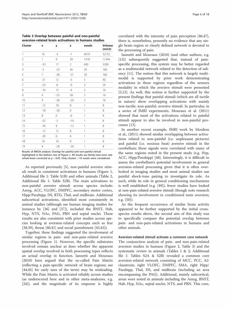
Table 3 Overlap between painful and non-painfulaversion-related brain activations in humans studies
Cluster x y z voxels Volume(mm3)
1 16 -3 -1 4019 32152
2 1 9 39 1418 11344
3 -33 17 2 400 3200
4 -2 -23 36 20 160
5 7 -38 -21 20 160
6 -21 12 -3 5 40
7 -24 -6 -9 3 24
8 -16 15 -4 2 16
9 4 -25 38 2 16
10 9 -30 -20 2 16
11 -12 18 8 1 8
12 -18 -28 0 1 8
13 8 0 -6 1 8
14 -32 -2 -10 1 8
15 -34 4 -12 1 8
16 -12 12 8 1 8
17 -20 6 4 1 8
18 18 16 0 1 8
19 4 -28 -20 1 8
Results of MKDA analysis: Overlap for painful and non-painful stimulihighlighted in the bottom row of Figure 2. All results are family-wise error ratewhole-brain corrected at p< 0.05. Only clusters >10 voxels were considered.
Hayes and Northoff BMC Neuroscience 2012, 13:60 Page 6 of 18http://www.biomedcentral.com/1471-2202/13/60
As reported previously [5], non-painful aversive stim-uli result in consistent activations in humans (Figure 1,Additional file 1: Table S1B) and other animals (Table 2,Additional file 1: Table S2B). The main activations tonon-painful aversive stimuli across species include:Amyg, ACC, VLOFC, DMPFC, secondary motor cortex,Hipp/Parahipp, DS, RTG, Thal, and midbrain. Additionalsubcortical activations, identified most consistently inanimal studies (although see human imaging studies forinstance by [36] and [37]), included the BNST, Hab,Hyp, NTS, NAc, PAG, PBN and septal nuclei. Theseresults are also consistent with prior studies across spe-cies looking at aversion-related concepts such as fear[38,39], threat [40,41] and social punishment [42,43]).Together, these findings suggested the involvement of
similar regions in pain- and non-pain-related aversiveprocessing (Figure 1). However, the specific substratesinvolved remain unclear as does whether the apparentspatial overlap involved in both processing types reflectsan actual overlap in function. Iannetti and Mouraux(2010) have argued that the so-called Pain Matrix(reflecting a pain-specific network of brain regions; see[44,45] for early uses of the term) may be misleading.While the Pain Matrix is activated reliably across studies(as underscored here and in other meta-analsyses, e.g.[24]), and the magnitude of its response is highly
correlated with the intensity of pain perception [46,47],there is, nonetheless, presently no evidence that any sin-gle brain region or clearly defined network is devoted tothe processing of pain.Iannetti and Mouraux (2010) (and other authors, e.g.
[13]) subsequently suggested that, instead of pain-specific processing, this system may be better regardedas a multimodal network related to the detection of sali-ency [11]. The notion that this network is largely multi-modal is supported by prior work demonstratingactivations in these regions regardless of the sensorymodality in which the aversive stimuli were presented[5,12]. As well, this notion is further supported by thepresent findings that painful stimuli (which are all tactilein nature) show overlapping activations with mainlynon-tactile, non-painful, aversive stimuli. In particular, ina series of fMRI experiments, Mouraux et al. (2011)showed that most of the activations related to painfulstimuli appear to also be involved in non-painful pro-cesses [12].In another recent example, fMRI work by Moulton
et al., (2011) showed similar overlapping between activa-tions related to non-painful (i.e. unpleasant pictures)and painful (i.e. noxious heat) aversive stimuli in thecerebellum; these signals were correlated with many ofthe same regions noted in the present study (e.g. Hyp,ACC, Hipp/Parahipp) [48]. Interestingly, it is difficult toassess the cerebellum’s potential involvement in generalaversion-related processing given that it is often over-looked in imaging studies and most animal studies usepainful shock-tone pairing to investigate its role. Assuch, while its role in general conditioning mechanismsis well established (e.g. [49]), fewer studies have lookedat non-pain-related aversive stimuli (though note researchshowing its involvement in conditioned taste aversion;e.g. [50]).As the frequent occurrence of similar brain activity
appeared to be further supported by the initial cross-species results above, the second aim of this study wasto specifically compare the potential overlap betweenpain- and non-pain-related activations in humans andother animals.
Aversion-related stimuli activate a common core networkThe conjunction analysis of pain- and non-pain-relatedaversion studies in humans (Figure 2, Table 3) and thesystematic review in animals (Tables 1 & 2; Additionalfile 1: Tables S2A & S2B) revealed a common coreaversion-related network consisting of MCC, PCC, AI/claustrum, right VLOFC, DMPFC, SMA, right Hipp/Parahipp, Thal, DS, and midbrain (including an areaencompassing the PAG). Additional, mainly subcortical,areas were noted in animals including the Amyg, BNST,Hab, Hyp, NAc, septal nuclei, NTS, and PBN. This core,
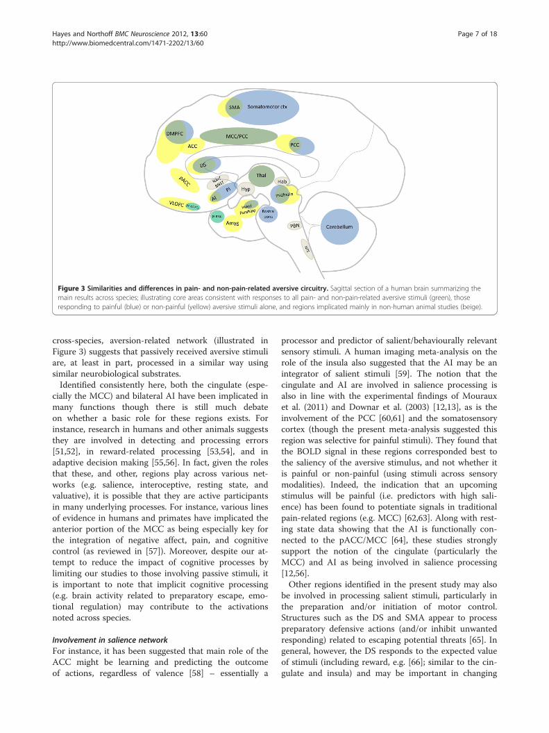
Figure 3 Similarities and differences in pain- and non-pain-related aversive circuitry. Sagittal section of a human brain summarizing themain results across species; illustrating core areas consistent with responses to all pain- and non-pain-related aversive stimuli (green), thoseresponding to painful (blue) or non-painful (yellow) aversive stimuli alone, and regions implicated mainly in non-human animal studies (beige).
Hayes and Northoff BMC Neuroscience 2012, 13:60 Page 7 of 18http://www.biomedcentral.com/1471-2202/13/60
cross-species, aversion-related network (illustrated inFigure 3) suggests that passively received aversive stimuliare, at least in part, processed in a similar way usingsimilar neurobiological substrates.Identified consistently here, both the cingulate (espe-
cially the MCC) and bilateral AI have been implicated inmany functions though there is still much debateon whether a basic role for these regions exists. Forinstance, research in humans and other animals suggeststhey are involved in detecting and processing errors[51,52], in reward-related processing [53,54], and inadaptive decision making [55,56]. In fact, given the rolesthat these, and other, regions play across various net-works (e.g. salience, interoceptive, resting state, andvaluative), it is possible that they are active participantsin many underlying processes. For instance, various linesof evidence in humans and primates have implicated theanterior portion of the MCC as being especially key forthe integration of negative affect, pain, and cognitivecontrol (as reviewed in [57]). Moreover, despite our at-tempt to reduce the impact of cognitive processes bylimiting our studies to those involving passive stimuli, itis important to note that implicit cognitive processing(e.g. brain activity related to preparatory escape, emo-tional regulation) may contribute to the activationsnoted across species.
Involvement in salience networkFor instance, it has been suggested that main role of theACC might be learning and predicting the outcomeof actions, regardless of valence [58] – essentially a
processor and predictor of salient/behaviourally relevantsensory stimuli. A human imaging meta-analysis on therole of the insula also suggested that the AI may be anintegrator of salient stimuli [59]. The notion that thecingulate and AI are involved in salience processing isalso in line with the experimental findings of Mourauxet al. (2011) and Downar et al. (2003) [12,13], as is theinvolvement of the PCC [60,61] and the somatosensorycortex (though the present meta-analysis suggested thisregion was selective for painful stimuli). They found thatthe BOLD signal in these regions corresponded best tothe saliency of the aversive stimulus, and not whether itis painful or non-painful (using stimuli across sensorymodalities). Indeed, the indication that an upcomingstimulus will be painful (i.e. predictors with high sali-ence) has been found to potentiate signals in traditionalpain-related regions (e.g. MCC) [62,63]. Along with rest-ing state data showing that the AI is functionally con-nected to the pACC/MCC [64], these studies stronglysupport the notion of the cingulate (particularly theMCC) and AI as being involved in salience processing[12,56].Other regions identified in the present study may also
be involved in processing salient stimuli, particularly inthe preparation and/or initiation of motor control.Structures such as the DS and SMA appear to processpreparatory defensive actions (and/or inhibit unwantedresponding) related to escaping potential threats [65]. Ingeneral, however, the DS responds to the expected valueof stimuli (including reward, e.g. [66]; similar to the cin-gulate and insula) and may be important in changing
Hayes and Northoff BMC Neuroscience 2012, 13:60 Page 8 of 18http://www.biomedcentral.com/1471-2202/13/60
expectations based on past and present contexts [67].The PAG also responds to highly salient (particularlyaversive) stimuli, is a well-known source for descendingcontrol over spinal pain pathways [68], and appears tobe involved in autonomic-somatomotor integrationrelated to orchestrating defensive behaviours [69]. Thisintegration is achieved via its close connections to Thal,Hyp, AMYG, PFC, and other brain stem nuclei involvedin autonomic processing (e.g. NTS, PBN, raphe; as notedacross the present animal data) [70]. (For a comprehen-sive review on the PAG in neuroimaging studies see[71].) Considered together, the cross-species data notedhere suggest that the core regions in general aversiveprocessing may be involved in processing salience infor-mation. This is in line with the findings from Mourauxet al. (2011), who suggested that these regions form asalience (as opposed to a pain) network [12].
Involvement in interoceptive networkThe AI and ACC/MCC are also considered key compo-nents of a circuit mediating interoceptive awareness.Studies have shown that both the insula and ACC/MCCare involved in processing the interoceptive awareness ofstimuli (e.g. heartbeat, respiration; e.g. [72]), while ani-mal studies have demonstrated similar roles (e.g. [73]).Menon and Uddin (2010) take a network perspective de-scribing these regions as core components of a saliencenetwork which act as integrators of overlapping net-works (carrying information related to interoception,homeostasis, working memory, higher-order controlprocesses) [74]. Additionally, the authors posit that theactivity of these hub regions and the interaction betweenthem may be involved, partly, in choosing/switchingbetween relevant task-related and resting state networks(e.g. engaging task-relevant memory and attentional pro-cesses while disengaging from non-task-related activity).The notion of the ACC/MCC and insula as a key integra-tor network is also supported by others (e.g. [64]) whohave noted functional connectivity between AI-pACC/MCC (which they suggest may integrate interoceptiveand emotional information) and between insula-MCC(which may be more involved in exteroceptive processingand response selection).
Involvement in resting state networksThe baseline activity of the brain is essential in deter-mining the relative stimulus-induced activation in bothanimal and human studies. As such, how the baseline isdefined conceptually and experimentally may ultimatelyaffect the results (see [75,76] for further discussion).As noted in the Methods section, most of the animalstudies included here necessarily use between-subjectsmeasures, whereas the human neuroimaging studies typ-ically use within-subjects measures – suggesting that the
baselines may be quite different. As such, caution shouldbe taken when interpreting the results across species.Nonetheless, the animal neuroimaging studies includedhere (e.g. [77,78]) are able to help bridge the gap some-what and their findings are congruent with the otherstudies. Moreover, human imaging studies are moreoften including a baseline (or so-called resting state)period in their designs (in which the subject is instructedto stare at a fixation cross or close their eyes and notfocus on any particular thoughts), which results in theidentification of baseline or resting state brain activity.Given the apparent overlap and interactivity between
these resting state regions (e.g. VMPFC/pACC and PCC)and exteroceptive/salience regions, some authors haverecently explored their potential relationship. For example,one recent study investigated the potential relationshipbetween resting state activity (within the default modenetwork) and emotion- and intero-/exteroceptive-relatedactivity [79]. They demonstrated that increased activity inthe default mode regions (e.g. VMPFC/pACC, PCC) dur-ing rest was associated with decreased emotional percep-tion ability, without any noted relationships to theperception of intero-/exteroceptive stimuli. This raises thequestion of whether the activity, particularly in the MCC,is selective for salience or rather for value-related proces-sing (related to determining the positive or negative valueof a stimulus). Moreover, it questions whether differentregions of the cingulate (or similar regions activated differ-entially across time) are involved separately in processingsalience-, emotion-, and interoceptive-related information(e.g. [64]).
Involvement in valuative networkFinally, recent reviews of the reward literature for bothhumans [80] and animals [23] describe many of thesame regions noted here for the aversion-related net-work. This raises the issue of whether, and to whatdegree, the core aversion-related regions noted inthe present study are involved in aversion-specific (notreward-related) processing. If both aversion- and reward-related activity are found equally in these regions, thiswould provide further support of a salience network.However, if different regions are involved in each, and/orto different degrees, this would suggest the existence ofinteracting, or even separate, neural networks for proces-sing value-related information. While there are, to ourknowledge, no meta-analyses or systematic reviewsoutlining the similarities and differences for reward- andaversion-related brain activity, a number of studies(particularly at the neuronal level) have suggested thatperhaps both salience-selective and value-selective pro-cessing occur in both overlapping and separate networks.For instance, many animal (e.g. [81]; and see [20] for
review) and some human studies (e.g. [36,37] and see
Hayes and Northoff BMC Neuroscience 2012, 13:60 Page 9 of 18http://www.biomedcentral.com/1471-2202/13/60
[82] for review) have implicated the NAc in coding bothaversive and rewarding states. Lammel et al. (2011)showed that among dopamine cells of the VTA thereappear to be many distinct (reward-, aversion-, or saliency-related) populations defined by receptor/channel type,activity, location and density, and axonal projections[83]. Another study demonstrated that while the dopa-minergic modulation of the basal ganglia’s direct striato-nigral and indirect striatopallidal pathways are involvedin both processing types, primary activation is shifted tothe indirect pathway during aversion-related processing[84]. Furthermore, single-unit ACC recordings in fourhumans viewing emotional pictures showed that of cellsresponding, most were selective for general aversion-related stimuli – although some cells responded to bothaversive and rewarding stimuli [85]. These findings helpexplain how a regional population of cells can contributeto differential processing (e.g. aversion and reward).Taken together, these results support the existence of a
core group of brain regions involved in basic aversion-related processing (Figure 3); the inclusion of non-human animal data has helped confirm and furtherextend these findings (mainly regarding subcorticalregions). The evidence suggests that components ofthis network are involved in processing various typesof information (e.g. saliency, interoception, valuative).Future neuroimaging studies should consider parsing,for instance, value and salience (e.g. using multi-levelledstimuli, to investigate ‘dose–response-like’ curves) as hasbeen attempted in some behavioural experiments [86].Though the focus has been on the overlap betweenpain- and non-pain-related aversive processing, it shouldbe noted that some activations (particularly in the meta-analysis) appeared to be selective for painful over non-painful stimuli (and vice versa). These activations arediscussed briefly in the following section.
Differential activations related to painful and non-painfulaversive stimuliAlthough a core network of brain regions for the proces-sing of painful and non-painful aversive stimuli wasidentified, the areas of activation noted in the humanmeta-analysis do not overlap completely. In fact, thereare a number of regions which appear to be unique topainful or non-painful stimuli. Specifically, painful stim-uli resulted in unique activations in the primary and sec-ondary sensory cortices, the PCC, bilateral operculum/mid and posterior insula, and cerebellum. Non-painfulaversive stimuli resulted in unique activations in theAMYG, L VLOFC, L RTG, and L Hipp/Parahipp as wellas additional extended activations in the pACC, DMPFC,HYP, posterior MCC, DS/VS, and bilateral VLOFC.Similar subcortical regions were identified in animalstudies of both pain- and non-pain-related processing
(e.g. NTS, PBN, BNST). Nonetheless, from the animalstudies included here, a few areas are noted in onlypain- (i.e. red nucleus, pretectal area, IC/SC) or non-pain- (i.e. VTA) related studies.The differences may in fact be unique, thus providing
unique neural signatures for painful and non-painfulaversive stimuli. However, there are alternative explana-tions for these apparently unique activations which seemmore tenable. Firstly, the differential weighting of activa-tions (i.e. that some regions are more involved) may helpexplain the absence of activations in human studies onpain (e.g. amygdala) compared to non-pain aversion.Secondly, as the analysis of imaging studies on pain andnon-pain aversion were limited to the passive receptionperiod, this is equivalent to taking a temporal snapshotof brain activity. While intended to be similar acrosspain and non-pain studies, this snapshot may in factreflect unique brain processing for two types of stimulidifferentiated by their timing properties (i.e. painfulstimuli result in fast aversive responding; non-painfulstimuli are slower and more variable).
Differential weightingThat nearly all regions were noted in both animal stud-ies of pain and non-pain aversion (Tables 1 and 2; e.g.involvement of the amygdala, [32,87]) supports thenotion that entirely unique activations for either are rareor unlikely. However, a differential weighting of activa-tions may be reflected in the ranking of regions in ani-mal studies (Tables 1 and 2), which generally reflectthe core regions noted in the human meta-analysis(Figure 2). For instance, the amygdala is activated moreconsistently in non-pain imaging studies, whereas thecingulate and sensory cortex appear more involved inpain studies (Figure 1). However, these rankings (i.e. thepercentage of animal studies noting specific brain activa-tions) should only be considered illustrative, as animalstudies typically choose regions a priori (compared to awhole-brain approach). Findings for a set of regions maylead to a disproportionately higher investigation rate byother researchers, which can inflate or mask the relativeimportance of some regions (though brain imaging isalso not immune to such biases; see [88] for a brief dis-cussion of this in relation to meta-analyses). Nonetheless,most animal studies included here investigated ≥5 brainregions, and the results are similar to those in humansand other animals (including studies investigating otheraversion-related concepts such as fear [39], threat [41]and social punishment [43].
Differential temporal dynamicsAnother potential explanation for differences betweenpain and non-pain aversion includes the timing of activ-ity, as noted above. While the temporal dynamics of this
Hayes and Northoff BMC Neuroscience 2012, 13:60 Page 10 of 18http://www.biomedcentral.com/1471-2202/13/60
circuitry have not been worked out, conditioning studiesin animals [89,90] and humans [91,92] have suggestedtemporal and/or subregional differences between theprocessing of conditioned stimuli predicting an aversivestimulus and the reception itself (underscoring theimportance of spatio-temporal dynamics). For instance,the amygdala is known to be more involved in assessingthe expectation (especially involving the timing) of aver-sive stimuli [93,94] – though see [95] for an fMRI studyin which long stimulation periods of pain perceptionresulted in amygdala activation. Another study by Gui-marais et al. (2011) showed that increasing the timeinterval between a predictive tone and a shock changedthe involvement of some structures in rats [96]. For in-stance, at ~5 s intervals, the mPFC became more active,whereas at longer (~40s) intervals, dorsal hippocampalactivity became necessary for learning about the aversivestimulus. Nonetheless, it is difficult to compare theresults from animal studies directly to those in humanimaging (especially without direct translational map-ping), and further study on the temporal aspects of aver-sive processing should be undertaken.
Strengths and limitationsThe greatest strength of the current work is its transla-tional nature. The inclusion of animal studies has twomain advantages. Firstly, they help to support the find-ings from human imaging and add insight regardingsubregional differences and underlying mechanisms.Secondly, they underscore the involvement of many sub-cortical regions which are generally underreported inimaging studies (as noted previously). We believe usingthis approach somewhat offsets the potential selectionbiases which may be found across human imaging stud-ies (e.g. lowering statistical thresholds for a prioriregions) and animal studies (e.g. looking only for activityin a priori regions; also see below).The strict and narrow criteria used in both human and
animal studies allowed for a clearer interpretation ofresults (e.g. the use of passive and acute aversive stimulionly; the exclusion of studies/subjects using explicit cog-nitive tasks; see Methods section for all criteria). Thesecriteria were used to isolate, as clearly as the presentmethods allow, the period of brain activation duringwhich acute aversive stimuli are present (e.g. most neu-roimaging studies look at periods around 5–10 seconds;the animal studies included here extract the brains assoon as possible following stimulus presentation) – thus,attempting to separate this period from others (e.g.anticipation, termination). It is in this sense that wehave attempted to identify a network associated withaversion-related processing (see also [26] for further dis-cussion on using meta-analyses to identify functionallyrelated brain regions). Nonetheless, it is worth pointing
out again (as discussed briefly above in the Differentialweighting and Differential temporal dynamics sections)that the inference of a temporal relationship betweenregional activations relies heavily on the inclusion, andexclusion, of appropriate studies. Ultimately, the identifi-cation of such networks through meta-analyses andsystematic review should be used as the basis for testingfuture hypotheses regarding co-activation.This approach also shed some light on one inherent
and important limitation of meta-analyses – particularlythose using human imaging studies. While some neuroi-maging studies do report subcortical activations inaversion-related processing (e.g. [37,97,98]), their relativescarcity means some subcortical regions may not benoted in the final meta-analysis results. This absence ofactivation likely also extends to highly variable corticalregions. The corollary is that meta-analysis resultsunderscore the most consistent nodes of activationacross studies (with the coordinates being more inform-ative than the size or shape of the clusters per se; seealso Differential weighting discussion above), whileregions not identified may still be active (and evenessential) components – findings that are made clearerthrough animal studies looking directly at brain tissue.Although the results of the animal studies outlined in
Tables 1 and 2 (i.e. listing the percentage of reportedbrain activations) should be considered illustrative dueto reporting and researcher interest biases and the lackof a whole-brain approach (as insisted upon for the im-aging data), most studies investigated at least 5 brainregions. In fact, only 9 of the 42 non-pain-related studies[89,90,99-106] and 13 of the 32 pain-related studies[87,107-118] focused on 4 or less regions. In addition,none of the pain-related studies focused solely on thecingulate (a key node identified in both human and ani-mal data), and only 3 studies from the non-pain-relatedaversion studies [100,105,106] focused solely on theamygdala (perhaps the single best described aversion-related region). Although the issue of selection bias andthe reporting of positive data (the so-called file-drawerproblem) cannot be fully accounted for, taken together,the animal and human data allow for a more confidentinterpretation regarding the inclusion of brain areasinvolved in aversion-related processing.
ConclusionsThe results from this translational approach stronglysuggest that humans and animals have a common coreaversion-related network, consisting of similar corticaland subcortical regions. This work extends from previ-ous work [5] by demonstrating that most of the regionstypically associated with a pain network are involved inthe generalized processing of all aversive stimuli to someextent. While saliency may be an integral factor to which
Hayes and Northoff BMC Neuroscience 2012, 13:60 Page 11 of 18http://www.biomedcentral.com/1471-2202/13/60
this network responds, it seems unlikely that it shouldbe thought of as a saliency detector – particularly giventhe apparent incomplete overlap of activations for pain-ful and non-painful stimuli, as well as for that of reward-ing stimuli. The differential weighting found betweenpain- (e.g. higher activations in MCC and posteriorinsula) and non-pain- (e.g. amygdala) related processingsuggests that aversion-related concepts may rely on theuse of similar substrates (i.e. aversion-related network)but to varying degrees and perhaps over different time-scales – thus underscoring the need for the investigationof spatio-temporal dynamics within this network.
MethodsPainful and non-painful aversion-related brain activationin humansLiterature search: We identified all imaging studies –positron emission tomography (PET) and functionalmagnetic resonance imaging (fMRI) – published from2000 to August 2011 with PubMed (http://www.pubmed.gov) and Web of Science (http://apps.webofknowledge.com; though no additional studies were found here) searches.Keywords included "aversion", "aversive", “avoidance”,“punishment”, “reinforcement” (to capture some studiesfocusing on reward, but also using an independentlyanalysed aversive control condition), “fear”, “anger”,“disgust”, “sadness”, “negative emotion”, “pain”, “nocicep-tive”, “unpleasant”, "positron emission tomography",and "functional magnetic resonance imaging" and others.Furthermore, we searched the reference list of articlesand reviews, including meta-analyses (for example see[6,119,120]). The data regarding brain activations asso-ciated with non-painful aversive stimuli were reportedpreviously in [5].Inclusion and exclusion criteria: Our main goal was to
compare the basic brain activity of painful- and non-painful aversive stimuli. Therefore, we included onlythose studies which used the passive presentationof acute aversive stimuli (e.g. the viewing of unpleasantpictures; exposure to painful stimuli) without activeresponses. Without behavioural measures, aversive experi-ences were determined subjectively and often supportedthrough physiological measures such as electrodermalactivity. Designs whose contrasts did not include specificcomparisons relevant to the current analysis (i.e. involvingthe passive reception of painful or non-painful aversivestimuli, independent of explicit cognitive processes, mem-ory, or attention) were excluded. In addition, only studiesthat reported coordinates from whole-brain analysis wereincluded (although some studies discussed region-of-interest data, those coordinates were not includedhere). These criteria lead to the inclusion of studiesinvolving both exteroceptive (e.g. pictures, shock) andinteroceptive (e.g. rectal distension) aversive stimuli.
Although the related search terms were included forcompleteness, studies reporting the responses to specificnegative emotions (e.g. sadness, anger) were excludedgiven their social nature as were other stimuli whichmay involve ambiguous interpretations such as thoseinvolving empathy, disgust, or physical contamination.For related examples involving such analyses showingresults consistent with the present study, see [121,122].Other studies looking explicitly at social aspects ofaversion, such as social exclusion, were also excludedgiven that they typically require complex behaviouralresponses and involve other potentially confoundingprocesses such as empathy and theory of mind. More-over, it is important to note that most studies involvingconditioned fearful stimuli in both humans and animalswere excluded to avoid issues related to recent learningeffects and complex conditioning designs, although itshould be noted that the aversive stimuli used are virtu-ally identical to those included here and the resultsfrom such studies are largely similar to those reportedhere [123,124]. Finally, studies involving manipulationsin homeostatic states (e.g. hunger, thirst) were alsonot included given their non-acute nature (e.g. usuallyinvolving forced deprivation over time). Although weaimed to cast as large an initial net as possible for boththe human and other animal studies, we must concedethat, given the conceptual complexity of aversion-relatedterms, we may have overlooked some studies which donot employ standard terminologies.We screened all the articles for Talairach or Montreal
Neurological Institute (MNI) coordinates and tabulatedthe reported regional foci. We included the data ofhealthy subjects only. Studies including individuals withpsychiatric illnesses, or a history thereof, those withvolumetric abnormalities or brain injuries, those takingany medications or illicit drugs, and those belonging to agroup that may result in a sample bias (e.g. war veter-ans) were excluded. In addition, a large number of stud-ies were excluded due to the absence of coordinates,identification of coordinate systems, and/or incompletestatistical information. These criteria resulted in the se-lection of 34 non-pain-related studies which included 44contrasts, and 27 pain-related aversion studies whichincluded 32 contrasts (see Additional file 1 for studyreferences included in the meta-analysis). Regions werelabelled macroanatomically by the probabilistic Harvard-Oxford atlas. The nomenclature for the cingulate byVogt (2005) was used here [125].Multilevel kernel density analysis (MKDA) meta-
analytic technique: The MKDA meta-analytic approachhas been covered in depth elsewhere [26,126]. Briefly,MKDA is a coordinate-based meta-analytic methodwhich determines the activation probability of each voxeland contiguous voxel clusters (to create voxel-based and
Hayes and Northoff BMC Neuroscience 2012, 13:60 Page 12 of 18http://www.biomedcentral.com/1471-2202/13/60
cluster-based study comparison maps, respectively)across the brain. Compared with other meta-analysismethods, MKDA prevents any single study reporting alarge number of activations from biasing the results (i.e.the study is the unit of analysis) and weights contrastsbased on the quality of the study (e.g. random vs. fixedeffects) and the sample size. It should be noted thatstudy-specific statistical thresholding is not taken intoaccount, however, because weighting activation peaks bytheir respective Z-scores introduces the presuppositionthat the scores across studies are comparable, which isuntrue (e.g. scores from studies with small sample sizeswould be inflated relative to the larger population; [26]for further discussion and examples). All results arereported in MNI space; co-ordinates from studies usingTalairach space were converted to MNI space in theMKDA software using the Brett transform (http://im-aging.mrc-cbu.cam.ac.uk/downloads/MNI2tal/tal2mni.m).Peaks from each study were convolved with a sphericalkernel of 10 mm radius; doing so ensures that multiple-nearby peaks are not counted as multiple activations(additionally helping to reduce the impact of studiesemploying lower statistical thresholds and which typicallyreport a greater number of activated voxels). This ap-proach also ensures that single studies do not drive theresults of the meta-analysis. The threshold for signifi-cance was determined using a Monte Carlo simulationwith 3000 iterations (5000 iterations did not alter theresults) and a null hypothesis which assumes that theactivated regions within each map are not spatially con-sistent (i.e. that the cluster centers are randomizedthroughout the grey matter). The voxel size was 2 x 2 x2 mm (i.e. 1 voxel = 8 mm3) and cluster sizes were allgreater than 10 voxels (> 80 mm3). We have reportedpeak voxel-wise activations as well as peak cluster-wiseactivations (which include contiguous voxels significantat p< 0.001; whole-brain FWE corrected). All results arefamily-wise error rate whole-brain corrected at p< 0.05.Analyses were performed in Matlab 2009a (Mathworks,Naticks, MA) using MKDA software created by TorWager (http://www.columbia.edu/cu/psychology/tor/).
Painful and non-painful aversion-related brain activationin animalsLiterature search: PubMed (http://www.pubmed.gov)and Web of Science (http://apps.webofknowledge.com;again, no additional studies were found here) searchesidentified rodent studies related to pain-related (~700studies) and non-pain-related (~350 studies) aversionpublished in English from 2000 to August 2011. Key-words were similar to those above but also included spe-cific terms related to animal studies such as: “immediateearly genes”, “IEG”, “c-fos”, “rat”, “mice”, “monkey”,“mammal”, “avoidance”, “fear”, “threat”, “electrophysiology”.
Of the total studies identified, only those clearly showingaltered brain metabolism (e.g. increased/decreased c-Fosor blood oxygenated level dependent activity, or BOLD)were included in the systematic review (i.e. 34 pain and42 non-pain aversion studies). Due to lack of methodo-logical instruments, absence of precise standardizedcoordinate systems, and the wide range of experimentalprocedures, we did not conduct the same rigorous meta-analysis in animals as in humans. As with the humandata, the non-human animal data regarding the presenta-tion of passive non-painful aversive stimuli were reportedpreviously in [5].Inclusion and exclusion criteria: We looked at the fol-
lowing metabolic indexes of non-human animal brainactivity: immediate early gene activation (e.g. c-Fos orFos-like expression), BOLD activity in fMRI, [14 C]-2-deoxyglucose, and [14 C]-iodoantipyrine. Each of theseindexes has previously been related to neural activityand/or metabolism. Considering the broad spectrumof animal models of aversion-related behaviour (e.g.formalin-induced nociception, foot shock, conditionedtaste aversion etc.), we looked at all those data thatreport clear effects in brain activity between controlanimals and those exposed to painful and/or non-painfulaversive stimuli. It is important to note that somemethods must necessarily use between-subjects mea-sures (e.g. counting IEG-stained cells in brain slices)whereas others use mainly within-subjects measures(e.g. neuroimaging).Studies involving adolescent animals, chronic exposure
to aversive stimuli, and exposure to drugs of abuseand those having a direct effect on aversion- or reward-related brain circuitry, were excluded (although non-drug-exposed controls were included where appropriate).This was done in order to avoid confounding issuesrelated to neurodevelopment and drug interactions and/or drug-induced changes in brain structure or functionunrelated to the acute aversive treatment. Studies usingelectrical/chemical lesions or other irreversible alterations(e.g. the use of knock-out or transgenic rodents, animalsbred for psychiatric disorder-related phenotypes; althoughone study using rats bred for high or low anxiety levels,though not pre-exposed to anxiolytic stimuli, wasincluded [127] as the results following an aversive probelargely converged regardless of anxiety levels) were alsoexcluded for clarity.Any comparison between human and animal data
raises the question of homology of brain regions. Sincethey show analogous anatomy, comparisons of subcor-tical regions are not often an issue (see also [128]). Incontrast, the issue of homology becomes more problem-atic in the case of cortical regions that show both ana-tomical and terminological differences between humansand animals. Nonetheless, even areas which may be
Hayes and Northoff BMC Neuroscience 2012, 13:60 Page 13 of 18http://www.biomedcentral.com/1471-2202/13/60
considered largely ‘higher-order’ or evolutionarily morerecent, such as the prefrontal cortex, may show strongstructural and functional homologies between primatesand other mammals, such as rodents [129,130]. Con-cerning cortical regions, we relied on criteria of hom-ology as established by various authors [131-133]. Inaddition, it should be noted that due to the strict inclu-sion/exclusion criteria nearly all of the animal studiesinvolve rodents with the exception of one study usingmonkeys [102]. As such, the generalizability to othermammals should be considered cautiously. Nonetheless,beyond the studies included here, studies involving pri-mates were also considered in the discussion to add sup-port to the translatability of the results (e.g.) [53,66].
Additional file
Additonal file 1: Table S1A. Painful aversion-related brain activations inhuman studies. Table S1B. Non-painful aversion-related brain activationsin human studies. Table S2A. Painful aversion-related brain activations inanimal studies. Table S2B. Aversion-, non-painful, related brainactivations in animal studies [32,33,41,73,77,78,87,89,90,99-103,105-110,113-118,127,134-230].
AbbreviationsACC: anterior cingulate cortex; Amyg: amygdala; AI: anterior insula;Aud: auditory ctx; BG: basal ganglia; BNST: bed n of the stria terminalis;Ctx: cortex; D: dorsal; DMPFC: dorsomedial prefrontal ctx; DS: dorsal striatum;Hab: habenula; Hipp: hippocampal area; Hyp: hypothalamus; IC: inferiorcolliculus; IFG: inferior frontal gyrus; IL: infralimbic ctx; Ins: insula; Lat: lateral;LC: locus coeruleus; MCC: mid cingulate ctx; N: nucleus; NAc: nucleusaccumbens; NTS: n of the solitary tract; OFC: orbital frontal ctx;PAG: periaqueductal gray; Parahipp: parahippocampal gyrus;PBN: parabrachial n; PL: prelimbic; PFC: prefrontal ctx; SC: superior colliculus;Sens: sensory ctx; Sep: septal n; SMA: secondary motor area;SMG: supramarginal gyrus; STG: superior temporal gyrus; Thal: thalamus;VLOFC: ventrolateral orbitofrontal ctx; VTA: ventral tegmental area.
Competing interestsThe authors have no competing interests to declare.
AcknowledgementsThe authors would like to thank Niall W. Duncan for his assistance with theMKDA meta-analysis. In addition, we would like to thank Niall W. Duncan,Aaron Kucyi, and the reviewers for their critical reading of the manuscript. G.N. holds a Canada Research Chair for Mind, Brain imaging and Neuroethicsas well as an EJLB-CIHR Michael Smith Chair in Neurosciences and MentalHealth. D.J.H holds a Postdoctoral Fellowship from the Canadian Institutes ofHealth Research (CIHR).
Authors’ contributionsDJH performed the analyses and drafted the manuscript. DJH and GNconceived of the study, and read and approved the final manuscript.
Received: 20 February 2012 Accepted: 18 April 2012Published: 7 June 2012
References1. Ardiel EL, Rankin CH: An elegant mind: learning and memory in
Caenorhabditis elegans. Learn Mem 2010, 17(4):191–201.2. Glanzman DL: Associative learning: Hebbian flies. Curr Biol 2005,
15(11):R416–R419.3. Seymour B, Singer T, Dolan R: The neurobiology of punishment. Nat Rev
Neurosci 2007, 8(4):300–311.
4. Wise RA: Dopamine, learning and motivation. Nat Rev Neurosci 2004,5(6):483–494.
5. Hayes DJ, Northoff G: Identifying a network of brain regions involved inaversion-related processing: a cross-species translational investigation.Front Integr Neurosci 2011, 5:49.
6. Apkarian AV, Bushnell MC, Treede RD, Zubieta JK: Human brainmechanisms of pain perception and regulation in health and disease.Eur J Pain 2005, 9(4):463–484.
7. Minami M: Neuronal mechanisms for pain-induced aversion behavioral studiesusing a conditioned place aversion test. Int Rev Neurobiol 2009, 85:135–144.
8. Oertel BG, Preibisch C, Wallenhorst T, Hummel T, Geisslinger G, LanfermannH, Lotsch J: Differential opioid action on sensory and affective cerebralpain processing. Clin Pharmacol Ther 2008, 83(4):577–588.
9. Tracey I: Imaging pain. Br J Anaesth 2008, 101(1):32–39.10. Melzack R: Pain–an overview. Acta Anaesthesiol Scand 1999, 43(9):880–884.11. Iannetti GD, Mouraux A: From the neuromatrix to the pain matrix (and
back). Exp Brain Res 2010, 205(1):1–12.12. Mouraux A, Diukova A, Lee MC, Wise RG, Iannetti GD: A multisensory
investigation of the functional significance of the "pain matrix".NeuroImage 2011, 54(3):2237–2249.
13. Downar J, Mikulis DJ, Davis KD: Neural correlates of the prolongedsalience of painful stimulation. NeuroImage 2003, 20(3):1540–1551.
14. Giordano J, Abramson K, Boswell MV: Pain assessment: subjectivity, objectivity,and the use of neurotechnology. Pain Physician 2010, 13(4):305–315.
15. Rolls ET, Grabenhorst F, Parris BA: Warm pleasant feelings in the brain.NeuroImage 2008, 41(4):1504–1513.
16. Meriau K, Wartenburger I, Kazzer P, Prehn K, Villringer A, van der Meer E,Heekeren HR: Insular activity during passive viewing of aversive stimulireflects individual differences in state negative affect. Brain Cogn 2009,69(1):73–80.
17. Walker DL, Toufexis DJ, Davis M: Role of the bed nucleus of the striaterminalis versus the amygdala in fear, stress, and anxiety. Eur JPharmacol 2003, 463(1–3):199–216.
18. Jhou T: Neural mechanisms of freezing and passive aversive behaviors.J Comp Neurol 2005, 493(1):111–114.
19. Misslin R: The defense system of fear: behavior and neurocircuitry.Neurophysiol Clin 2003, 33(2):55–66.
20. Carlezon WA Jr, Thomas MJ: Biological substrates of reward and aversion:a nucleus accumbens activity hypothesis. Neuropharmacology 2009,56(Suppl 1):122–132.
21. Wise RA: Brain reward circuitry: insights from unsensed incentives.Neuron 2002, 36(2):229–240.
22. Breiter HC, Rosen BR: Functional magnetic resonance imaging of brainreward circuitry in the human. Ann N Y Acad Sci 1999, 877:523–547.
23. Ikemoto S: Brain reward circuitry beyond the mesolimbic dopaminesystem: A neurobiological theory. Neurosci Biobehav Rev 2010,35(2):129–150.
24. Duerden EG, Albanese MC: Localization of pain-related brain activation: Ameta-analysis of neuroimaging data. Hum Brain Mapp 2011. doi:10.1002/hbm.21416 [Epub ahead of print].
25. Sandner G, Oberling P, Silveira MC, Di Scala G, Rocha B, Bagri A, DepoortereR: What brain structures are active during emotions? Effects of brainstimulation elicited aversion on c-fos immunoreactivity and behavior.Behav Brain Res 1993, 58(1–2):9–18.
26. Wager TD, Lindquist MA, Nichols TE, Kober H, Van Snellenberg JX:Evaluating the consistency and specificity of neuroimaging data usingmeta-analysis. NeuroImage 2009, 45(1 Suppl):S210–S221.
27. Devonshire IM, Papadakis NG, Port M, Berwick J, Kennerley AJ, Mayhew JE,Overton PG: Neurovascular coupling is brain region-dependent.NeuroImage 2012, 59:1997–2006.
28. Nichols T, Brett M, Andersson J, Wager T, Poline JB: Valid conjunctioninference with the minimum statistic. NeuroImage 2005, 25(3):653–660.
29. Krebs RM, Heipertz D, Schuetze H, Duzel E: Novelty increases themesolimbic functional connectivity of the substantia nigra/ventraltegmental area (SN/VTA) during reward anticipation: Evidence fromhigh-resolution fMRI. NeuroImage 2011, 58(2):647–655.
30. Villemure C, Bushnell MC: Mood influences supraspinal pain processingseparately from attention. J Neurosci 2009, 29(3):705–715.
31. Strigo IA, Duncan GH, Boivin M, Bushnell MC: Differentiation of visceraland cutaneous pain in the human brain. J Neurophysiol 2003,89(6):3294–3303.
Hayes and Northoff BMC Neuroscience 2012, 13:60 Page 14 of 18http://www.biomedcentral.com/1471-2202/13/60
32. Shih YY, Chiang YC, Chen JC, Huang CH, Chen YY, Liu RS, Chang C, Jaw FS:Brain nociceptive imaging in rats using (18)f-fluorodeoxyglucosesmall-animal positron emission tomography. Neuroscience 2008,155(4):1221–1226.
33. Hess A, Sergejeva M, Budinsky L, Zeilhofer HU, Brune K: Imaging ofhyperalgesia in rats by functional MRI. Eur J Pain 2007, 11(1):109–119.
34. Logothetis NK: What we can do and what we cannot do with fMRI.Nature 2008, 453(7197):869–878.
35. Fairhurst M, Wiech K, Dunckley P, Tracey I: Anticipatory brainstemactivity predicts neural processing of pain in humans. Pain 2007,128(1–2):101–110.
36. Becerra L, Breiter HC, Wise R, Gonzalez RG, Borsook D: Reward circuitryactivation by noxious thermal stimuli. Neuron 2001, 32(5):927–946.
37. Levita L, Hare TA, Voss HU, Glover G, Ballon DJ, Casey BJ: The bivalent sideof the nucleus accumbens. NeuroImage 2009, 44(3):1178–1187.
38. Klucken T, Kagerer S, Schweckendiek J, Tabbert K, Vaitl D, Stark R: Neural,electrodermal and behavioral response patterns in contingency awareand unaware subjects during a picture-picture conditioning paradigm.Neuroscience 2009, 158(2):721–731.
39. Lim LW, Temel Y, Visser-Vandewalle V, Blokland A, Steinbusch H: Fosimmunoreactivity in the rat forebrain induced by electrical stimulationof the dorsolateral periaqueductal gray matter. J Chem Neuroanat 2009,38(2):83–96.
40. Mobbs D, Petrovic P, Marchant JL, Hassabis D, Weiskopf N, Seymour B,Dolan RJ, Frith CD: When fear is near: threat imminence elicits prefrontal-periaqueductal gray shifts in humans. Science 2007, 317(5841):1079–1083.
41. Day HE, Masini CV, Campeau S: The pattern of brain c-fos mRNA inducedby a component of fox odor, 2,5-dihydro-2,4,5-trimethylthiazoline (TMT),in rats, suggests both systemic and processive stress characteristics.Brain Res 2004, 1025(1–2):139–151.
42. Eisenberger NI, Gable SL, Lieberman MD: Functional magnetic resonanceimaging responses relate to differences in real-world social experience.Emotion 2007, 7(4):745–754.
43. Nikulina EM, Arrillaga-Romany I, Miczek KA, Hammer RP Jr: Long-lastingalteration in mesocorticolimbic structures after repeated social defeatstress in rats: time course of mu-opioid receptor mRNA and FosB/DeltaFosB immunoreactivity. Eur J Neurosci 2008, 27(9):2272–2284.
44. Talbot JD, Marrett S, Evans AC, Meyer E, Bushnell MC, Duncan GH: Multiplerepresentations of pain in human cerebral cortex. Science 1991,251(4999):1355–1358.
45. Jones A: The pain matrix and neuropathic pain. Brain 1998,121(Pt 5):783–784.
46. Derbyshire SW, Jones AK, Gyulai F, Clark S, Townsend D, Firestone LL: Painprocessing during three levels of noxious stimulation producesdifferential patterns of central activity. Pain 1997, 73(3):431–445.
47. Rainville P: Brain mechanisms of pain affect and pain modulation. CurrOpin Neurobiol 2002, 12(2):195–204.
48. Moulton EA, Elman I, Pendse G, Schmahmann J, Becerra L, Borsook D:Aversion-related circuitry in the cerebellum: responses to noxious heatand unpleasant images. J Neurosci 2011, 31(10):3795–3804.
49. Bellebaum C, Daum I: Mechanisms of cerebellar involvement inassociative learning. Cortex 2011, 47(1):128–136.
50. Mediavilla C, Molina F, Puerto A: Retention of concurrent taste aversionlearning after electrolytic lesioning of the interpositus-dentate region ofthe cerebellum. Brain Res 2000, 868(2):329–337.
51. Simons RF: The way of our errors: theme and variations. Psychophysiology2010, 47(1):1–14.
52. Bossaerts P: Risk and risk prediction error signals in anterior insula. BrainStruct Funct 2010, 214(5–6):645–653.
53. Haber SN, Knutson B: The reward circuit: linking primate anatomy andhuman imaging. Neuropsychopharmacology 2010, 35(1):4–26.
54. Wittmann M, Lovero KL, Lane SD, Paulus MP: Now or later? Striatum andinsula activation to immediate versus delayed rewards. J Neurosci PsycholEcon 2010, 3(1):15–26.
55. Walton ME, Croxson PL, Behrens TE, Kennerley SW, Rushworth MF: Adaptivedecision making and value in the anterior cingulate cortex. NeuroImage2007, 36(Suppl 2):T142–T154.
56. Wiech K, Lin CS, Brodersen KH, Bingel U, Ploner M, Tracey I: Anterior insulaintegrates information about salience into perceptual decisions aboutpain. J Neurosci 2010, 30(48):16324–16331.
57. Shackman AJ, Salomons TV, Slagter HA, Fox AS, Winter JJ, Davidson RJ: Theintegration of negative affect, pain and cognitive control in thecingulate cortex. Nat Rev Neurosci 2011, 12(3):154–167.
58. Alexander WH, Brown JW: Medial prefrontal cortex as an action-outcomepredictor. Nat Neurosci 2011, 14(10):1338–1344.
59. Kurth F, Zilles K, Fox PT, Laird AR, Eickhoff SB: A link between the systems:functional differentiation and integration within the human insularevealed by meta-analysis. Brain Struct Funct 2010, 214(5–6):519–534.
60. Maddock RJ: The retrosplenial cortex and emotion: new insights fromfunctional neuroimaging of the human brain. Trends Neurosci 1999,22(7):310–316.
61. Small DM, Zatorre RJ, Dagher A, Evans AC, Jones-Gotman M: Changes inbrain activity related to eating chocolate: from pleasure to aversion.Brain 2001, 124(Pt 9):1720–1733.
62. Sawamoto N, Honda M, Okada T, Hanakawa T, Kanda M, Fukuyama H,Konishi J, Shibasaki H: Expectation of pain enhances responses tononpainful somatosensory stimulation in the anterior cingulate cortexand parietal operculum/posterior insula: an event-related functionalmagnetic resonance imaging study. J Neurosci 2000, 20(19):7438–7445.
63. Atlas LY, Bolger N, Lindquist MA, Wager TD: Brain mediators of predictivecue effects on perceived pain. J Neurosci 2010, 30(39):12964–12977.
64. Taylor KS, Seminowicz DA, Davis KD: Two systems of resting stateconnectivity between the insula and cingulate cortex. Hum Brain Mapp2009, 30(9):2731–2745.
65. Sagaspe P, Schwartz S, Vuilleumier P: Fear and stop: a role for theamygdala in motor inhibition by emotional signals. NeuroImage 2011,55(4):1825–1835.
66. Schultz W, Tremblay L, Hollerman JR: Reward processing in primateorbitofrontal cortex and basal ganglia. Cereb Cortex 2000, 10(3):272–284.
67. Sharot T, Shiner T, Dolan RJ: Experience and choice shape expectedaversive outcomes. J Neurosci 2010, 30(27):9209–9215.
68. Sanders KH, Klein CE, Mayor TE, Heym C, Handwerker HO: Differentialeffects of noxious and non-noxious input on neurones according tolocation in ventral periaqueductal grey or dorsal raphe nucleus. Brain Res1980, 186(1):83–97.
69. Schenberg LC, Povoa RM, Costa AL, Caldellas AV, Tufik S, Bittencourt AS:Functional specializations within the tectum defense systems of the rat.Neurosci Biobehav Rev 2005, 29(8):1279–1298.
70. Hadjipavlou G, Dunckley P, Behrens TE, Tracey I: Determining anatomicalconnectivities between cortical and brainstem pain processing regionsin humans: a diffusion tensor imaging study in healthy controls. Pain2006, 123(1–2):169–178.
71. Linnman C, Moulton EA, Barmettler G, Becerra L, Borsook D: Neuroimagingof the periaqueductal gray: state of the field. NeuroImage 2012,60(1):505–522.
72. Critchley HD, Wiens S, Rotshtein P, Ohman A, Dolan RJ: Neural systemssupporting interoceptive awareness. Nat Neurosci 2004, 7(2):189–195.
73. Wang Z, Guo Y, Bradesi S, Labus JS, Maarek JM, Lee K, Winchester WJ, MayerEA, Holschneider DP: Sex differences in functional brain activation duringnoxious visceral stimulation in rats. Pain 2009, 145(1–2):120–128.
74. Menon V, Uddin LQ: Saliency, switching, attention and control: a networkmodel of insula function. Brain Struct Funct 2010, 214(5–6):655–667.
75. Hayes DJ, Huxtable AG: Interpreting deactivations in neuroimaging. FrontPsychol 2012, 3:27.
76. Northoff G, Duncan NW, Hayes DJ: The brain and its resting state activity–experimental and methodological implications. Prog Neurobiol 2010,92(4):593–600.
77. Inui-Yamamoto C, Yoshioka Y, Inui T, Sasaki KS, Ooi Y, Ueda K, Seiyama A,Ohzawa I: The brain mapping of the retrieval of conditioned tasteaversion memory using manganese-enhanced magnetic resonanceimaging in rats. Neuroscience 2010, 167(2):199–204.
78. Shih YY, Chen YY, Chen CC, Chen JC, Chang C, Jaw FS: Whole-brainfunctional magnetic resonance imaging mapping of acute nociceptiveresponses induced by formalin in rats using atlas registration-basedevent-related analysis. J Neurosci Res 2008, 86(8):1801–1811.
79. Wiebking C, de Greck M, Duncan NW, Heinzel A, Tempelmann C, NorthoffG: Are emotions associated with activity during rest or interoception? Anexploratory fMRI study in healthy subjects. Neurosci Lett 2011, 491(1):87–92.
80. Liu X, Hairston J, Schrier M, Fan J: Common and distinct networksunderlying reward valence and processing stages: a meta-analysis of
Hayes and Northoff BMC Neuroscience 2012, 13:60 Page 15 of 18http://www.biomedcentral.com/1471-2202/13/60
functional neuroimaging studies. Neurosci Biobehav Rev 2011,35(5):1219–1236.
81. Hayes DJ, Hoang J, Greenshaw AJ: The role of nucleus accumbens shellGABA receptors on ventral tegmental area intracranial self-stimulationand a potential role for the 5-HT2C receptor. J Psychopharmacol 2011,25(12):1661–1675.
82. Leknes S, Tracey I: A common neurobiology for pain and pleasure. NatRev Neurosci 2008, 9(4):314–320.
83. Lammel S, Ion DI, Roeper J, Malenka RC: Projection-specific modulation ofdopamine neuron synapses by aversive and rewarding stimuli. Neuron2011, 70(5):855–862.
84. Hikida T, Kimura K, Wada N, Funabiki K, Nakanishi S: Distinct roles ofsynaptic transmission in direct and indirect striatal pathways to rewardand aversive behavior. Neuron 2010, 66(6):896–907.
85. Kawasaki H, Adolphs R, Oya H, Kovach C, Damasio H, Kaufman O,Howard M 3rd: Analysis of single-unit responses to emotional scenes inhuman ventromedial prefrontal cortex. J Cogn Neurosci 2005,17(10):1509–1518.
86. Madan CR, Spetch ML: Is the enhancement of memory due to rewarddriven by value or salience? Acta Psychol (Amst) 2012, 139(2):343–349.
87. Nakagawa T, Katsuya A, Tanimoto S, Yamamoto J, Yamauchi Y, Minami M,Satoh M: Differential patterns of c-fos mRNA expression in theamygdaloid nuclei induced by chemical somatic and visceral noxiousstimuli in rats. Neurosci Lett 2003, 344(3):197–200.
88. Rottschy C, Langner R, Dogan I, Reetz K, Laird AR, Schulz JB, Fox PT, EickhoffSB: Modelling neural correlates of working memory: A coordinate-basedmeta-analysis. NeuroImage 2011, 60(1):830–846.
89. Calandreau L, Jaffard R, Desmedt A: Dissociated roles for the lateral andmedial septum in elemental and contextual fear conditioning. LearnMem 2007, 14(6):422–429.
90. Yasoshima Y, Scott TR, Yamamoto T: Differential activation of anterior andmidline thalamic nuclei following retrieval of aversively motivatedlearning tasks. Neuroscience 2007, 146(3):922–930.
91. Straube T, Weiss T, Mentzel HJ, Miltner WH: Time course of amygdalaactivation during aversive conditioning depends on attention.NeuroImage 2007, 34(1):462–469.
92. Marschner A, Kalisch R, Vervliet B, Vansteenwegen D, Buchel C: Dissociableroles for the hippocampus and the amygdala in human cued versuscontext fear conditioning. J Neurosci 2008, 28(36):9030–9036.
93. Strigo IA, Simmons AN, Matthews SC, Craig AD: The relationship betweenamygdala activation and passive exposure time to an aversive cueduring a continuous performance task. PLoS One 2010, 5(11):e15093.
94. Ziv M, Tomer R, Defrin R, Hendler T: Individual sensitivity to painexpectancy is related to differential activation of the hippocampus andamygdala. Hum Brain Mapp 2010, 31(2):326–338.
95. Dube AA, Duquette M, Roy M, Lepore F, Duncan G, Rainville P: Brainactivity associated with the electrodermal reactivity to acute heat pain.NeuroImage 2009, 45(1):169–180.
96. Guimarais M, Gregorio A, Cruz A, Guyon N, Moita MA: Time determines theneural circuit underlying associative fear learning. Front Behav Neurosci2011, 5:89.
97. Herwig U, Abler B, Walter H, Erk S: Expecting unpleasant stimuli–an fMRIstudy. Psychiatry Res 2007, 154(1):1–12.
98. Jensen J, McIntosh AR, Crawley AP, Mikulis DJ, Remington G, Kapur S: Directactivation of the ventral striatum in anticipation of aversive stimuli.Neuron 2003, 40(6):1251–1257.
99. Radwanska K, Nikolaev E, Knapska E, Kaczmarek L: Differential responseof two subdivisions of lateral amygdala to aversive conditioningas revealed by c-Fos and P-ERK mapping. NeuroReport 2002,13(17):2241–2246.
100. Badowska-Szalewska E, Ludkiewicz B, Domaradzka-Pytel B, Dziewiatkowski J,Spodnik JH, Morys J: The immunoreactivity of c-Fos, NGF and its receptorTrkA after open-field exposure in the central and medial nuclei of therat amygdala. Folia Morphol (Warsz) 2006, 65(2):145–151.
101. Calfa G, Bussolino D, Molina VA: Involvement of the lateral septum andthe ventral Hippocampus in the emotional sequelae induced bysocial defeat: role of glucocorticoid receptors. Behav Brain Res 2007,181(1):23–34.
102. Hoffman KL, Gothard KM, Schmid MC, Logothetis NK: Facial-expressionand gaze-selective responses in the monkey amygdala. Curr Biol 2007,17(9):766–772.
103. Mediavilla C, Bernal A, Puerto A: Taste aversion learning induced c-fosexpression in the nucleus of the solitary tract after spontaneous flavorintake: role of the inter-stimulus interval. Neurobiol Learn Mem 2007,88(2):264–268.
104. Baumgartel K, Genoux D, Welzl H, Tweedie-Cullen RY, Koshibu K,Livingstone-Zatchej M, Mamie C, Mansuy IM: Control of the establishmentof aversive memory by calcineurin and Zif268. Nat Neurosci 2008,11(5):572–578.
105. Kwon B, Goltz M, Houpt TA: Expression of AP-1 family transcriptionfactors in the amygdala during conditioned taste aversion learning: rolefor Fra-2. Brain Res 2008, 1207:128–141.
106. Butler RK, Sharko AC, Oliver EM, Brito-Vargas P, Kaigler KF, Fadel JR, WilsonMA: Activation of phenotypically-distinct neuronal subpopulations of therat amygdala following exposure to predator odor. Neuroscience 2011,175:133–144.
107. Culman J, Das G, Ohlendorf C, Haass M, Maser-Gluth C, Zuhayra M, Zhao Y,Itoi K: Blockade of tachykinin NK1/NK2 receptors in the brain attenuatesthe activation of corticotrophin-releasing hormone neurones in thehypothalamic paraventricular nucleus and the sympathoadrenaland pituitary-adrenal responses to formalin-induced pain in the rat.J Neuroendocrinol 2010, 22(5):467–476.
108. Hagiwara H, Kimura F, Mitsushima D, Funabashi T: Formalin-inducednociceptive behavior and c-Fos expression in middle-aged female rats.Physiol Behav 2010, 100(2):101–104.
109. Vrang N, Phifer CB, Corkern MM, Berthoud HR: Gastric distension inducesc-Fos in medullary GLP-1/2-containing neurons. Am J Physiol Regul IntegrComp Physiol 2003, 285(2):R470–R478.
110. Shih YY, Chen CC, Shyu BC, Lin ZJ, Chiang YC, Jaw FS, Chen YY, Chang C: Anew scenario for negative functional magnetic resonance imagingsignals: endogenous neurotransmission. J Neurosci 2009, 29(10):3036–3044.
111. Edelsbrunner ME, Nakano M, Holzer P: Afferent signalling from the acid-challenged rat stomach is inhibited and gastric acid elimination isenhanced by lafutidine. BMC Gastroenterol 2009, 9:40.
112. Ren Y, Zhang L, Lu Y, Yang H, Westlund KN: Central lateral thalamicneurons receive noxious visceral mechanical and chemical input in rats.J Neurophysiol 2009, 102(1):244–258.
113. Hayashi T, Miyata M, Nagata T, Izawa Y, Kawakami Y:Intracerebroventricular fluvoxamine administration inhibited painbehavior but increased Fos expression in affective pain pathways.Pharmacol Biochem Behav 2009, 91(3):441–446.
114. Novikova NS, Kazakova TB, Rogers V, Korneva EA: Expression of the c-Fosgene in the rat hypothalamus in electrical pain stimulation and UHFstimulation of the skin. Neurosci Behav Physiol 2008, 38(4):415–420.
115. Morano TJ, Bailey NJ, Cahill CM, Dumont EC: Nuclei-and condition-specificresponses to pain in the bed nucleus of the stria terminalis. ProgNeuropsychopharmacol Biol Psychiatry 2008, 32(3):643–650.
116. Gavrilov YV, Perekrest SV, Novikova NS: Intracellular expression of c-Fosprotein in various structures of the hypothalamus in electrical painstimulation and administration of antigens. Neurosci Behav Physiol 2008,38(1):87–92.
117. Li L, Ding J, Ren Z, Han Q, Hu G, Xiao M: Expression and colocalization ofNADPH-diaphorase and Fos in the subnuclei of the parabrachial nucleusin rats following visceral noxious stimulation. Brain Res 2006, 1114(1):41–52.
118. Coizet V, Dommett EJ, Redgrave P, Overton PG: Nociceptive responses ofmidbrain dopaminergic neurones are modulated by the superiorcolliculus in the rat. Neuroscience 2006, 139(4):1479–1493.
119. Costafreda SG, Brammer MJ, David AS, Fu CH: Predictors of amygdalaactivation during the processing of emotional stimuli: a meta-analysis of385 PET and fMRI studies. Brain Res Rev 2008, 58(1):57–70.
120. Mechias ML, Etkin A, Kalisch R: A meta-analysis of instructed fear studies:implications for conscious appraisal of threat. NeuroImage 2010,49(2):1760–1768.
121. Fan Y, Duncan NW, de Greck M, Northoff G: Is there a core neural networkin empathy? An fMRI based quantitative meta-analysis. Neurosci BiobehavRev 2011, 35(3):903–911.
122. Kober H, Barrett LF, Joseph J, Bliss-Moreau E, Lindquist K, Wager TD:Functional grouping and cortical-subcortical interactions in emotion: ameta-analysis of neuroimaging studies. NeuroImage 2008, 42(2):998–1031.
123. Delgado MR, Jou RL, Phelps EA: Neural systems underlying aversiveconditioning in humans with primary and secondary reinforcers. FrontNeurosci 2011, 5:71.
Hayes and Northoff BMC Neuroscience 2012, 13:60 Page 16 of 18http://www.biomedcentral.com/1471-2202/13/60
124. Johnson LR, Hou M, Prager EM, Ledoux JE: Regulation of the Fear Networkby Mediators of Stress: Norepinephrine Alters the Balance betweenCortical and Subcortical Afferent Excitation of the Lateral Amygdala.Front Behav Neurosci 2011, 5:23.
125. Vogt BA: Pain and emotion interactions in subregions of the cingulategyrus. Nat Rev Neurosci 2005, 6(7):533–544.
126. Salimi-Khorshidi G, Smith SM, Keltner JR, Wager TD, Nichols TE: Meta-analysis of neuroimaging data: a comparison of image-based andcoordinate-based pooling of studies. NeuroImage 2009, 45(3):810–823.
127. Salchner P, Sartori SB, Sinner C, Wigger A, Frank E, Landgraf R, Singewald N:Airjet and FG-7142-induced Fos expression differs in rats selectively bredfor high and low anxiety-related behavior. Neuropharmacology 2006,50(8):1048–1058.
128. Panksepp J: Affectvie neuroscience: the foundations of human and animalemotions. New York: Oxford University Press; 1998.
129. Dalley JW, Cardinal RN, Robbins TW: Prefrontal executive and cognitivefunctions in rodents: neural and neurochemical substrates. NeurosciBiobehav Rev 2004, 28(7):771–784.
130. Heidbreder CA, Groenewegen HJ: The medial prefrontal cortex in the rat:evidence for a dorso-ventral distinction based upon functional andanatomical characteristics. Neurosci Biobehav Rev 2003, 27(6):555–579.
131. Ongur D, Price JL: The organization of networks within the orbital andmedial prefrontal cortex of rats, monkeys and humans. Cereb Cortex 2000,10(3):206–219.
132. Shumake J, Gonzalez-Lima F: Brain systems underlying susceptibility tohelplessness and depression. Behav Cogn Neurosci Rev 2003, 2(3):198–221.
133. Vertes RP: Interactions among the medial prefrontal cortex, hippocampusand midline thalamus in emotional and cognitive processing in the rat.Neuroscience 2006, 142(1):1–20.
134. Albrechet-Souza L, Borelli KG, Carvalho MC, Brandao ML: The anteriorcingulate cortex is a target structure for the anxiolytic-like effects ofbenzodiazepines assessed by repeated exposure to the elevated plusmaze and Fos immunoreactivity. Neuroscience 2009, 164:387–397.
135. Baffi JS, Palkovits M: Fine topography of brain areas activated by coldstress. A fos immunohistochemical study in rats. Neuroendocrinology 2000,72:102–113.
136. Bernstein IL, Koh MT: Molecular signaling during taste aversion learning.Chem Senses 2007, 32:99–103.
137. Chang C, Shyu BC: A fMRI study of brain activations during non-noxiousand noxious electrical stimulation of the sciatic nerve of rats. Brain Res2001, 897:71–81.
138. Chen W, Tenney J, Kulkarni P, King JA: Imaging unconditioned fearresponse with manganese-enhanced MRI (MEMRI). Neuroimage 2007,37:221–229.
139. Dardou D, Datiche F, Cattarelli M: Fos and Egr1 expression in the rat brainin response to olfactory cue after taste-potentiated odor aversionretrieval. Learn Mem 2006, 13:150–160.
140. Dardou D, Datiche F, Cattarelli M: Does taste or odor activate the samebrain networks after retrieval of taste potentiated odor aversion?Neurobiol Learn Mem 2007, 88:186–197.
141. Dielenberg RA, Hunt GE, McGregor IS: "When a rat smells a cat": thedistribution of Fos immunoreactivity in rat brain following exposure to apredatory odor. Neuroscience 2001, 104:1085–1097.
142. Fekete EM, Zhao Y, Li C, Sabino V, Vale WW, Zorrilla EP: Social defeat stressactivates medial amygdala cells that express type 2 corticotropin-releasing factor receptor mRNA. Neuroscience 2009, 162:5–13.
143. Ferreira G, Ferry B, Meurisse M, Levy F: Forebrain structures specificallyactivated by conditioned taste aversion. Behav Neurosci 2006,120:952–962.
144. Funk D, Amir S: Enhanced fos expression within the primary olfactoryand limbic pathways induced by an aversive conditioned odor stimulus.Neuroscience 2000, 98:403–406.
145. Furlong TM, Cole S, Hamlin AS, McNally GP: The role of prefrontal cortex inpredictive fear learning. Behav Neurosci 2010, 124:574–586.
146. Garcia-Medina NE, Jimenez-Capdeville ME, Ciucci M, Martinez LM, DelgadoJM, Horn CC: Conditioned flavor aversion and brain Fos expressionfollowing exposure to arsenic. Toxicology 2007, 235:73–82.
147. Hao S, Dulake M, Espero E, Sternini C, Raybould HE, Rinaman L: Central Fosexpression and conditioned flavor avoidance in rats followingintragastric administration of bitter taste receptor ligands. Am J PhysiolRegul Integr Comp Physiol 2009, 296:R528–R536.
148. Jhou TC, Fields HL, Baxter MG, Saper CB, Holland PC: The rostromedialtegmental nucleus (RMTg), a GABAergic afferent to midbrain dopamineneurons, encodes aversive stimuli and inhibits motor responses. Neuron2009, 61:786–800.
149. Johnson PL, Fitz SD, Hollis JH, Moratalla R, Lightman SL, Shekhar A, LowryCA: Induction of c-Fos in 'panic/defence'-related brain circuits followingbrief hypercarbic gas exposure. J Psychopharmacol 2010a.
150. Johnson ZV, Revis AA, Burdick MA, Rhodes JS: A similar pattern ofneuronal Fos activation in 10 brain regions following exposure toreward- or aversion-associated contextual cues in mice. Physiol Behav2010, 99:412–418.
151. Koh MT, Bernstein IL: Mapping conditioned taste aversion associationsusing c-Fos reveals a dynamic role for insular cortex. Behav Neurosci 2005,119:388–398.
152. Lamprea MR, Cardenas FP, Vianna DM, Castilho VM, Cruz-Morales SE,Brandao ML: The distribution of fos immunoreactivity in rat brainfollowing freezing and escape responses elicited by electricalstimulation of the inferior colliculus. Brain Res 2002, 950:186–194.
153. Lehner M, Taracha E, Skorzewska A, Turzynska D, Sobolewska A, Maciejak P,Szyndler J, Hamed A, Bidzinski A, Wislowska-Stanek A, Plaznik A: Expressionof c-Fos and CRF in the brains of rats differing in the strength of a fearresponse. Behav Brain Res 2008, 188:154–167.
154. Lehner M, Taracha E, Skorzewska A, Wislowska A, Zienowicz M, Maciejak P,Szyndler J, Bidzinski A, Plaznik A: Sensitivity to pain and c-Fos expressionin brain structures in rats. Neurosci Lett 2004, 370:74–79.
155. Lei LG, Zhang YQ, Zhao ZQ: Pain-related aversion and Fos expression inthe central nervous system in rats. Neuroreport 2004, 15:67–71.
156. Lemos JI, Resstel LB, Guimaraes FS: Involvement of the prelimbicprefrontal cortex on cannabidiol-induced attenuation of contextualconditioned fear in rats. Behav Brain Res 2010, 207:105–111.
157. Lowe AS, Beech JS, Williams SC: Small animal, whole brain fMRI:innocuous and nociceptive forepaw stimulation. Neuroimage 2007,35:719–728.
158. Malisza KL, Docherty JC: Capsaicin as a source for painful stimulation infunctional MRI. J Magn Reson Imaging 2001, 14:341–347.
159. Malisza KL, Gregorash L, Turner A, Foniok T, Stroman PW, Allman AA,Summers R, Wright A: Functional MRI involving painful stimulation of theankle and the effect of physiotherapy joint mobilization. Magn ResonImaging 2003, 21:489–496.
160. Malkani S, Rosen JB: Induction of NGFI-B mRNA following contextual fearconditioning and its blockade by diazepam. Brain Res Mol Brain Res 2000,80:153–165.
161. Martinez RC, Carvalho-Netto EF, Ribeiro-Barbosa ER, Baldo MV, Canteras NS:Amygdalar roles during exposure to a live predator and to a predator-associated context. Neuroscience 2011, 172:314–328.
162. Martinez V, Wang L, Tache Y: Proximal colon distension induces Fosexpression in the brain and inhibits gastric emptying through capsaicin-sensitive pathways in conscious rats. Brain Res 2006, 1086:168–180.
163. Mickley GA, Kenmuir CL, McMullen CA, Yocom AM, Valentine EL, Dengler-Crish CM, Weber B, Wellman JA, Remmers-Roeber DR: Dynamic processingof taste aversion extinction in the brain. Brain Res 2004, 1016:79–89.
164. Moylan Governo RJ, Morris PG, Prior MJ, Marsden CA, Chapman V:Capsaicin-evoked brain activation and central sensitization inanaesthetised rats: a functional magnetic resonance imaging study. Pain2006, 126:35–45.
165. Navarro M, Spray KJ, Cubero I, Thiele TE, Bernstein IL: cFos induction duringconditioned taste aversion expression varies with aversion strength.Brain Res 2000, 887:450–453.
166. Nikolaev E, Kaczmarek L, Zhu SW, Winblad B, Mohammed AH:Environmental manipulation differentially alters c-Fos expression inamygdaloid nuclei following aversive conditioning. Brain Res 2002,957:91–98.
167. Roche M, Johnston P, Mhuircheartaigh ON, Olango WM, Mackie K, Finn DP:Eur J Pain 2009.
168. Roseboom PH, Nanda SA, Bakshi VP, Trentani A, Newman SM, Kalin NH:Predator threat induces behavioral inhibition, pituitary-adrenal activationand changes in amygdala CRF-binding protein gene expression.Psychoneuroendocrinology 2007, 32:44–55.
169. Sadananda M, Wohr M, Schwarting RK: Playback of 22-kHz and 50-kHzultrasonic vocalizations induces differential c-fos expression in rat brain.Neurosci Lett 2008, 435:17–23.
Hayes and Northoff BMC Neuroscience 2012, 13:60 Page 17 of 18http://www.biomedcentral.com/1471-2202/13/60
170. Shah YB, Haynes L, Prior MJ, Marsden CA, Morris PG, Chapman V:Functional magnetic resonance imaging studies of opioid receptor-mediated modulation of noxious-evoked BOLD contrast in rats.Psychopharmacology (Berl) 2005, 180:761–773.
171. Shih YY, Chang C, Chen JC, Jaw FS: BOLD fMRI mapping of brainresponses to nociceptive stimuli in rats under ketamine anesthesia. MedEng Phys 2008, 30:953–958.
172. Shih YY, Chen YY, Chen JM, Lung Y, Chen JY, Chang C, Chen JC, Jaw FS:Exploring nociceptive response by BOLD fMRI in alpha-chloraloseanesthetized rats. Conf Proc IEEE Eng Med Biol Soc 2006, 1:33–36.
173. Shih YY, Wey HY, De La Garza BH, Duong TQ: Striatal and cortical BOLD,blood flow, blood volume, oxygen consumption, and glucoseconsumption changes in noxious forepaw electrical stimulation. J CerebBlood Flow Metab 2011, 31:832–841.
174. Sinniger V, Porcher C, Mouchet P, Juhem A, Bonaz B: c-fos and CRFreceptor gene transcription in the brain of acetic acid-induced somato-visceral pain in rats. Pain 2004, 110:738–750.
175. St Andre J, Albanos K, Reilly S: C-fos expression in the rat brain followinglithium chloride-induced illness. Brain Res 2007, 1135:122–128.
176. Tuor UI, Malisza K, Foniok T, Papadimitropoulos R, Jarmasz M, Somorjai R,Kozlowski P: Functional magnetic resonance imaging in rats subjected tointense electrical and noxious chemical stimulation of the forepaw.Pain 2000, 87:315–324.
177. Vianna DM, Borelli KG, Ferreira-Netto C, Macedo CE, Brandao ML: Fos-likeimmunoreactive neurons following electrical stimulation of the dorsalperiaqueductal gray at freezing and escape thresholds. Brain Res Bull2003, 62:179–189.
178. Wang L, Martinez V, Larauche M, Tache Y: Proximal colon distensioninduces Fos expression in oxytocin-, vasopressin-, CRF- andcatecholamines-containing neurons in rat brain. Brain Res 2009,1247:79–91.
179. Wilkins EE, Bernstein IL: Conditioning method determines patterns of c-fos expression following novel taste-illness pairing. Behav Brain Res 2006,169:93–97.
180. Yasoshima Y, Scott TR, Yamamoto T: Memory-dependent c-Fos expressionin the nucleus accumbens and extended amygdala following theexpression of a conditioned taste aversive in the rat. Neuroscience 2006,141:35–45.
181. Yu O, Parizel N, Pain L, Guignard B, Eclancher B, Mauss Y, Grucker D: Textureanalysis of brain MRI evidences the amygdala activation by nociceptivestimuli under deep anesthesia in the propofol-formalin rat model. MagnReson Imaging 2007, 25:144–146.
182. Zanoveli JM, Ferreira-Netto C, Brandao ML: Conditioned place aversionorganized in the dorsal periaqueductal gray recruits the laterodorsalnucleus of the thalamus and the basolateral amygdala. Exp Neurol 2007,208:127–136.
183. Apkarian AV, Gelnar PA, Krauss BR, Szeverenyi NM: Cortical responses tothermal pain depend on stimulus size: a functional MRI study.J Neurophysiol 2000, 83:3113–3122.
184. Becerra L, Iadarola M, Borsook D: CNS activation by noxious heat to thehand or foot: site-dependent delay in sensory but not emotion circuitry.J Neurophysiol 2004, 91:533–541.
185. Casey KL, Morrow TJ, Lorenz J, Minoshima S: Temporal and spatialdynamics of human forebrain activity during heat pain: analysis bypositron emission tomography. J Neurophysiol 2001, 85:951–959.
186. Coghill RC, Sang CN, Maisog JM, Iadarola MJ: Pain intensity processingwithin the human brain: a bilateral, distributed mechanism.J Neurophysiol 1999, 82:1934–1943.
187. Derbyshire SW, Jones AK, Creed F, Starz T, Meltzer CC, Townsend DW,Peterson AM, Firestone L: Cerebral responses to noxious thermalstimulation in chronic low back pain patients and normal controls.Neuroimage 2002, 16:158–168.
188. Derbyshire SW, Whalley MG, Stenger VA, Oakley DA: Cerebral activationduring hypnotically induced and imagined pain. Neuroimage 2004,23:392–401.
189. Dimitrova A, Kolb FP, Elles HG, Maschke M, Gerwig M, Gizewski E, TimmannD: Cerebellar activation during leg withdrawal reflex conditioning: anfMRI study. Clin Neurophysiol 2004, 115:849–857.
190. Gelnar PA, Krauss BR, Sheehe PR, Szeverenyi NM, Apkarian AV: Acomparative fMRI study of cortical representations for thermal painful,vibrotactile, and motor performance tasks. Neuroimage 1999, 10:460–482.
191. Helmchen C, Mohr C, Erdmann C, Petersen D, Nitschke MF: Differentialcerebellar activation related to perceived pain intensity during noxiousthermal stimulation in humans: a functional magnetic resonanceimaging study. Neurosci Lett 2003, 335:202–206.
192. Hofbauer RK, Rainville P, Duncan GH, Bushnell MC: Corticalrepresentation of the sensory dimension of pain. J Neurophysiol 2001,86:402–411.
193. Kurata J, Thulborn KR, Gyulai FE, Firestone LL: Early decay of pain-relatedcerebral activation in functional magnetic resonance imaging:comparison with visual and motor tasks. Anesthesiology 2002,96:35–44.
194. Mohr C, Leyendecker S, Mangels I, Machner B, Sander T, Helmchen C:Central representation of cold-evoked pain relief in capsaicin inducedpain: an event-related fMRI study. Pain 2009, 139:416–430.
195. Morrison I, Lloyd D, di Pellegrino G, Roberts N: Vicarious responses to painin anterior cingulate cortex: is empathy a multisensory issue? Cogn AffectBehav Neurosci 2004, 4:270–278.
196. Niddam DM, Yeh TC, Wu YT, Lee PL, Ho LT, Arendt-Nielsen L, Chen AC,Hsieh JC: Event-related functional MRI study on central representation ofacute muscle pain induced by electrical stimulation. Neuroimage 2002,17:1437–1450.
197. Ochsner KN, Ludlow DH, Knierim K, Hanelin J, Ramachandran T, Glover GC,Mackey SC: Neural correlates of individual differences in pain-related fearand anxiety. Pain 2006, 120:69–77.
198. Petrovic P, Carlsson K, Petersson KM, Hansson P, Ingvar M: Context-dependent deactivation of the amygdala during pain. J Cogn Neurosci2004, 16:1289–1301.
199. Peyron R, Garcia-Larrea L, Gregoire MC, Costes N, Convers P, Lavenne F,Mauguiere F, Michel D, Laurent B: Haemodynamic brain responses toacute pain in humans: sensory and attentional networks. Brain 1999,122(Pt 9):1765–1780.
200. Rolls ET, Kringelbach ML, de Araujo IE: Different representations ofpleasant and unpleasant odours in the human brain. Eur J Neurosci 2003,18:695–703.
201. Scott DJ, Heitzeg MM, Koeppe RA, Stohler CS, Zubieta JK: Variations in thehuman pain stress experience mediated by ventral and dorsal basalganglia dopamine activity. J Neurosci 2006, 26:10789–10795.
202. Smith KA, Ploghaus A, Cowen PJ, McCleery JM, Goodwin GM, Smith S,Tracey I, Matthews PM: Cerebellar responses during anticipation ofnoxious stimuli in subjects recovered from depression. Functionalmagnetic resonance imaging study. Br J Psychiatry 2002, 181:411–415.
203. Tolle TR, Kaufmann T, Siessmeier T, Lautenbacher S, Berthele A, Munz F,Zieglgansberger W, Willoch F, Schwaiger M, Conrad B, Bartenstein P:Region-specific encoding of sensory and affective components of painin the human brain: a positron emission tomography correlationanalysis. Ann Neurol 1999, 45:40–47.
204. Abler B, Erk S, Herwig U, Walter H: Anticipation of aversive stimuliactivates extended amygdala in unipolar depression. J Psychiatr Res 2007,41:511–522.
205. Blood AJ, Zatorre RJ, Bermudez P, Evans AC: Emotional responses topleasant and unpleasant music correlate with activity in paralimbic brainregions. Nat Neurosci 1999, 2:382–387.
206. Botvinick M, Jha AP, Bylsma LM, Fabian SA, Solomon PE, Prkachin KM:Viewing facial expressions of pain engages cortical areas involved in thedirect experience of pain. Neuroimage 2005, 25:312–319.
207. Buchel C, Bornhovd K, Quante M, Glauche V, Bromm B, Weiller C:Dissociable neural responses related to pain intensity, stimulus intensity,and stimulus awareness within the anterior cingulate cortex: aparametric single-trial laser functional magnetic resonance imagingstudy. J Neurosci 2002, 22:970–976.
208. Buchel C, Dolan RJ, Armony JL, Friston KJ: Amygdala-hippocampalinvolvement in human aversive trace conditioning revealed throughevent-related functional magnetic resonance imaging. J Neurosci 1999,19:10869–10876.
209. Frings M, Maschke M, Erichsen M, Jentzen W, Muller SP, Kolb FP, Diener HC,Timmann D: Involvement of the human cerebellum in fear-conditionedpotentiation of the acoustic startle response: a PET study. Neuroreport2002, 13:1275–1278.
210. Garrett AS, Maddock RJ: Separating subjective emotion from theperception of emotion-inducing stimuli: an fMRI study. Neuroimage 2006,33:263–274.
Hayes and Northoff BMC Neuroscience 2012, 13:60 Page 18 of 18http://www.biomedcentral.com/1471-2202/13/60
211. Gottfried JA, O'doherty J, Dolan RJ: Appetitive and aversive olfactorylearning in humans studied using event-related functional magneticresonance imaging. J Neurosci 2002, 22:10829–10837.
212. Grabenhorst F, Rolls ET, Margot C, Da Silva MA, Velazco MI: How pleasantand unpleasant stimuli combine in different brain regions: odormixtures. J Neurosci 2007, 27:13532–13540.
213. Hamann S, Mao H: Positive and negative emotional verbal stimuli elicitactivity in the left amygdala. Neuroreport 2002, 13:15–19.
214. Hamann SB, Ely TD, Hoffman JM, Kilts CD: Ecstasy and agony: activation ofthe human amygdala in positive and negative emotion. Psychol Sci 2002,13:135–141.
215. Herpertz SC, Dietrich TM, Wenning B, Krings T, Erberich SG, Willmes K, ThronA, Sass H: Evidence of abnormal amygdala functioning in borderlinepersonality disorder: a functional MRI study. Biol Psychiatry 2001,50:292–298.
216. Liberzon I, Phan KL, Decker LR, Taylor SF: Extended amygdala andemotional salience: a PET activation study of positive and negativeaffect. Neuropsychopharmacology 2003, 28:726–733.
217. Liberzon I, Taylor SF, Fig LM, Decker LR, Koeppe RA, Minoshima S: Limbicactivation and psychophysiologic responses to aversive visual stimuli.Interaction with cognitive task. Neuropsychopharmacology 2000,23:508–516.
218. Liberzon I, Zubieta JK, Fig LM, Phan KL, Koeppe RA, Taylor SF: mu-Opioidreceptors and limbic responses to aversive emotional stimuli. Proc NatlAcad Sci U S A 2002, 99:7084–7089.
219. Maddock RJ, Garrett AS, Buonocore MH: Posterior cingulate cortexactivation by emotional words: fMRI evidence from a valence decisiontask. Hum Brain Mapp 2003, 18:30–41.
220. Meseguer V, Romero MJ, Barros-Loscertales A, Belloch V, Bosch-Morell F,Romero J, Avila C: Mapping the apetitive and aversive systems withemotional pictures using a block-design fMRI procedure. Psicothema2007, 19:483–488.
221. Mirz F, Gjedde A, Sodkilde-Jrgensen H, Pedersen CB: Functional brainimaging of tinnitus-like perception induced by aversive auditory stimuli.Neuroreport 2000, 11:633–637.
222. Nitschke JB, Sarinopoulos I, Mackiewicz KL, Schaefer HS, Davidson RJ:Functional neuroanatomy of aversion and its anticipation. Neuroimage2006, 29:106–116.
223. O'doherty JP, Deichmann R, Critchley HD, Dolan RJ: Neural responsesduring anticipation of a primary taste reward. Neuron 2002, 33:815–826.
224. Phan KL, Liberzon I, Welsh RC, Britton JC, Taylor SF: Habituation of rostralanterior cingulate cortex to repeated emotionally salient pictures.Neuropsychopharmacology 2003, 28:1344–1350.
225. Rolls ET: Functions of the orbitofrontal and pregenual cingulate cortexin taste, olfaction, appetite and emotion. Acta Physiol Hung 2008,95:131–164.
226. Shirao N, Okamoto Y, Mantani T, Yamawaki S: Gender differences in brainactivity generated by unpleasant word stimuli concerning body image:an fMRI study. Br J Psychiatry 2005, 186:48–53.
227. Tabbert K, Stark R, Kirsch P, Vaitl D: Hemodynamic responses of theamygdala, the orbitofrontal cortex and the visual cortex during a fearconditioning paradigm. Int J Psychophysiol 2005, 57:15–23.
228. Taylor SF, Liberzon I, Koeppe RA: The effect of graded aversive stimuli onlimbic and visual activation. Neuropsychologia 2000, 38:1415–1425.
229. Taylor SF, Phan KL, Decker LR, Liberzon I: Subjective rating of emotionallysalient stimuli modulates neural activity. Neuroimage 2003, 18:650–659.
230. Zald DH, Pardo JV: The neural correlates of aversive auditory stimulation.Neuroimage 2002, 16:746–753.
doi:10.1186/1471-2202-13-60Cite this article as: Hayes and Northoff: Common brain activations forpainful and non-painful aversive stimuli. BMC Neuroscience 2012 13:60.
Submit your next manuscript to BioMed Centraland take full advantage of:
• Convenient online submission
• Thorough peer review
• No space constraints or color figure charges
• Immediate publication on acceptance
• Inclusion in PubMed, CAS, Scopus and Google Scholar
• Research which is freely available for redistribution
Submit your manuscript at www.biomedcentral.com/submit