
Common and unusual pathologies - BMUS - … · Common and unusual pathologies Dr Anne Marie Coady...
50
The uterus and the endometrium Common and unusual pathologies Dr Anne Marie Coady Consultant Radiologist Head of Obstetric and Gynaecological Ultrasound HEY WACH
Transcript of Common and unusual pathologies - BMUS - … · Common and unusual pathologies Dr Anne Marie Coady...

The uterus and the endometrium Common and unusual pathologies
Dr Anne Marie Coady Consultant Radiologist
Head of Obstetric and Gynaecological Ultrasound HEY WACH
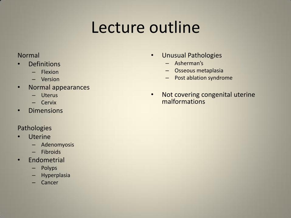
Lecture outline
Normal • Definitions
– Flexion – Version
• Normal appearances – Uterus – Cervix
• Dimensions
Pathologies • Uterine
– Adenomyosis – Fibroids
• Endometrial – Polyps – Hyperplasia – Cancer
• Unusual Pathologies – Asherman’s – Osseous metaplasia – Post ablation syndrome
• Not covering congenital uterine
malformations

To be avoided at all costs
• Do not describe every uterus with two endometrial cavities as a bicornuate uterus
• Do not use “malignancy cannot be excluded” as a blanket term to
describe a mass that you cannot categorize
• Do not use “ectopic cannot be excluded” just because you cannot determine the site of the pregnancy
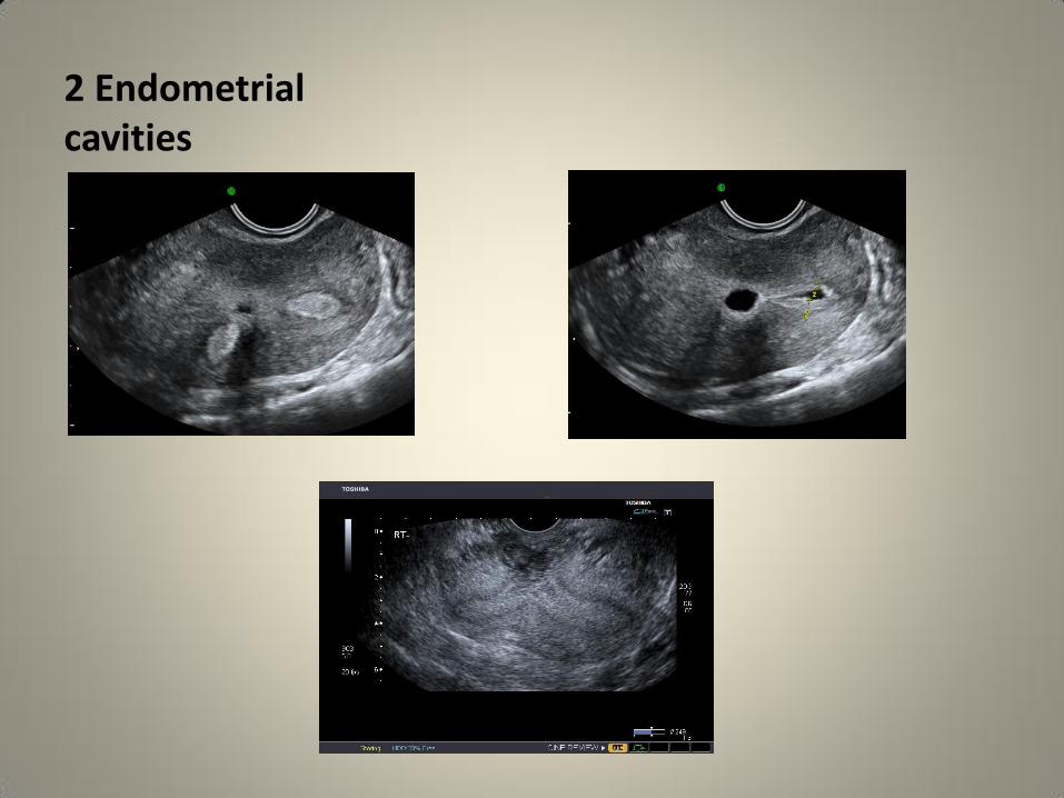
2 Endometrial cavities
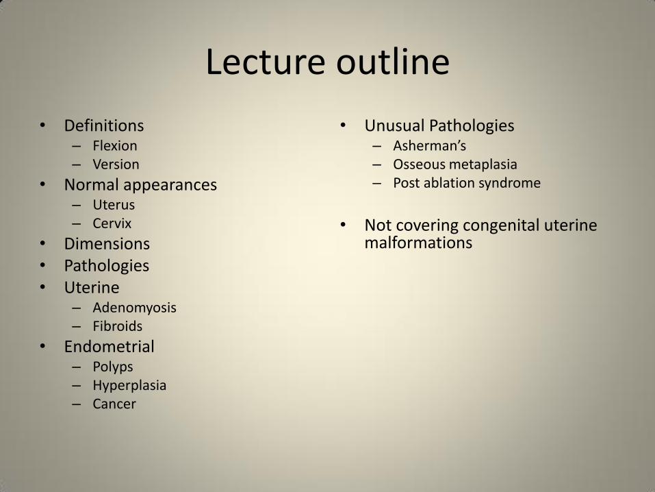
Lecture outline
• Definitions – Flexion – Version
• Normal appearances – Uterus – Cervix
• Dimensions • Pathologies • Uterine
– Adenomyosis – Fibroids
• Endometrial – Polyps – Hyperplasia – Cancer
• Unusual Pathologies – Asherman’s – Osseous metaplasia – Post ablation syndrome
• Not covering congenital uterine
malformations
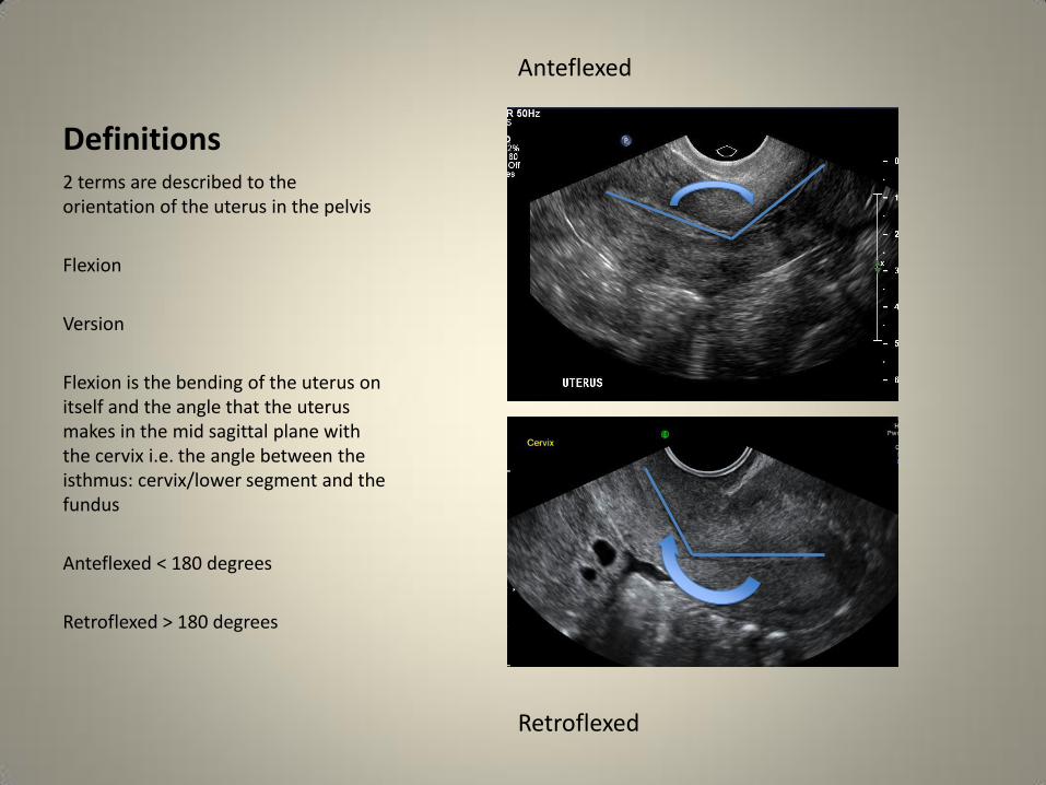
Definitions 2 terms are described to the orientation of the uterus in the pelvis
Flexion
Version
Flexion is the bending of the uterus on itself and the angle that the uterus makes in the mid sagittal plane with the cervix i.e. the angle between the isthmus: cervix/lower segment and the fundus
Anteflexed < 180 degrees
Retroflexed > 180 degrees
Retroflexed
Anteflexed

Definitions 2 terms are described to the orientation of the uterus in the pelvis
Version
When there is NO angle between the uterus and the cervix the uterus is described in terms of version, which describes the displacement of the entire uterus either backwards or forwards
Anteverted
The fundus is close to the bladder
Retroverted
The fundus is close to the recto sigmoid
Retroflexed

Normal sonographic appearances The uterus is a homogenous layer of smooth muscle and blood vessels and it is less echogenic than the adjacent endometrium
It is composed of three layers
Inner/junctional thin and hypo echoic
Middle thick and homogenous
Outer thin and hypo echoic
“arcuate vessels”

Normal sonographic appearances
The cervix has 3 layers also
Ectocervix
Endocervix
Endocervical canal
The cervical stroma is the same echogenicity as the myometrium
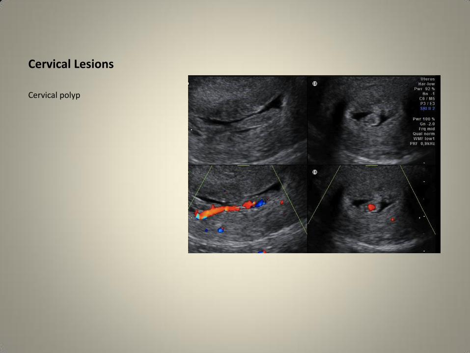
Cervical Lesions
Cervical polyp

Cervical lesions
Cervical cancer
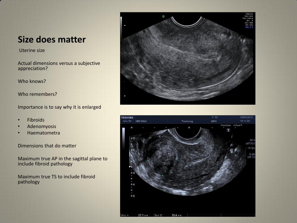
Size does matter Uterine size Actual dimensions versus a subjective appreciation? Who knows? Who remembers? Importance is to say why it is enlarged • Fibroids • Adenomyosis • Haematometra Dimensions that do matter Maximum true AP in the sagittal plane to include fibroid pathology Maximum true TS to include fibroid pathology
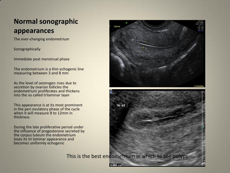
Normal sonographic appearances The ever-changing endometrium Sonographically Immediate post menstrual phase The endometrium is a thin echogenic line measuring between 3 and 8 mm As the level of oestrogen rises due to secretion by ovarian follicles the endometrium proliferates and thickens into the so called trilaminar layer This appearance is at its most prominent in the peri ovulatory phase of the cycle when it will measure 8 to 12mm in thickness During the late proliferative period under the influence of progesterone secreted by the corpus luteum the endometrium loses its tri laminar appearance and becomes uniformly echogenic
This is the best endometrium in which to see polyps
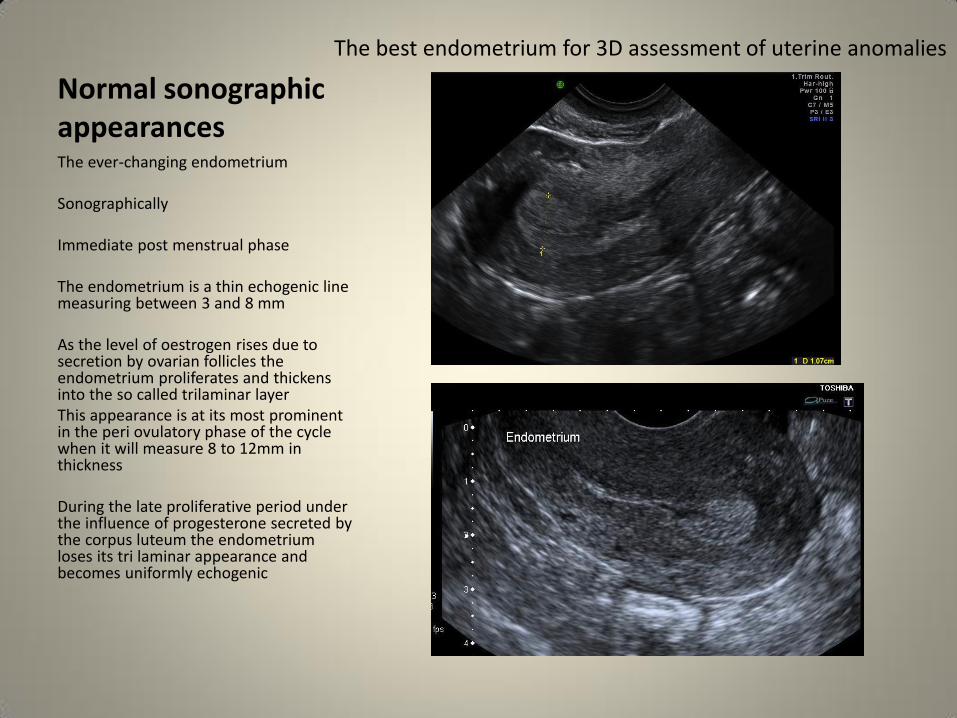
Normal sonographic appearances The ever-changing endometrium Sonographically Immediate post menstrual phase The endometrium is a thin echogenic line measuring between 3 and 8 mm As the level of oestrogen rises due to secretion by ovarian follicles the endometrium proliferates and thickens into the so called trilaminar layer This appearance is at its most prominent in the peri ovulatory phase of the cycle when it will measure 8 to 12mm in thickness During the late proliferative period under the influence of progesterone secreted by the corpus luteum the endometrium loses its tri laminar appearance and becomes uniformly echogenic
The best endometrium for 3D assessment of uterine anomalies
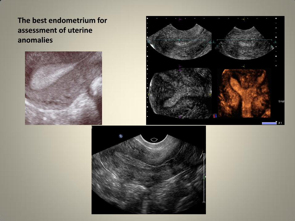
The best endometrium for assessment of uterine anomalies
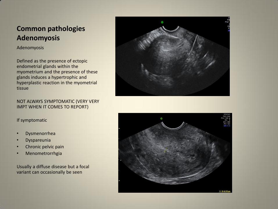
Common pathologies Adenomyosis Adenomyosis
Defined as the presence of ectopic endometrial glands within the myometrium and the presence of these glands induces a hypertrophic and hyperplastic reaction in the myometrial tissue
NOT ALWAYS SYMPTOMATIC (VERY VERY IMPT WHEN IT COMES TO REPORT)
If symptomatic
• Dysmenorrhea
• Dyspareunia
• Chronic pelvic pain
• Menometrorrhgia
Usually a diffuse disease but a focal variant can occasionally be seen

Common pathologies Adenomyosis
Adenomyosis
Sonographic features
• Globular enlargement of the uterus
• Asymmetric anterior and posterior uterine wall thickening
• Anechoic spaces in the myometrium
• Heterogenous myometrial echo texture
• Sub endometrial linear striations (venetian blind appearances)
• Obscuration of the endometrial myometrial border DIFFICULTY MEASURING THE ENDOMETRIUM
• Thickening of the transition zone MRI


Common pathologies Fibroids Fibroids
Commonest benign tumour
30 % of women > 35 years
40% of women over the age of 40 years
They are composed of smooth muscle with various amounts of connective tissue and their growth is oestrogen dependent
Leiomyomas have pseudo capsules, which are formed of compressed surrounding myometrium
Leiomyosarcomatous change is rare < 0.2%
Only 50% of leiomyomas actually grow in pregnancy

Fibroids: the fibroid map
Location is key to symptoms and management
• Intra mural
• Sub serosal
• Pedunculated
• Sub mucosal
• Intracavitary
Intra mural: no bulging into the endometrium or the serosa Sub serosal: a significant portion of the leiomyoma is bulging into the serosal surface Pedunculated: the leiomyoma is exophytic and attached to the uterus by a pedicle Sub mucosal: a significant portion is bulging into the endometrial cavity > < 50 % is important Intracavitary: the leiomyoma is within the endometrial cavity and it is attached to the myometrium by a pedicle

Common pathologies Fibroids
Fibroids
Location is key to symptoms and management
INTRA MURAL
Intra mural: no bulging into the endometrium or the serosa

Fibroids
Fibroids
Location is key to symptoms and management
SUB SEROSAL
Sub serosal: a significant portion of the leiomyoma is bulging into the serosal surface

Fibroids
Location is key to symptoms and management
PEDUNCULATED
Pedunculated: the leiomyoma is exophytic and attached to the uterus by a pedicle
BEWARE THE “SOLID OVARIAN TUMOUR” OCCAMS RAZOR

Occam’s razor When you hear the sound of hooves
Think horses Not zebras
Simple arithmetic says that a solid adnexal mass is far more likely to be an abnormal location of a common pathology i.e. a fibroid than a rare pathology i.e. a solid ovarian tumour FIND THE OVARY!!!!!!

Fibroids
Location is key to symptoms and management
SUB MUCOSAL
Sub mucosal: a significant portion is bulging into the endometrial cavity > < 50 % is important

Fibroids
Location is key to symptoms and management
INTRA CAVITARY
Intracavitary: the leiomyoma is within the endometrial cavity and it is attached to the myometrium by a pedicle
It may even prolapse into the cervix!!!
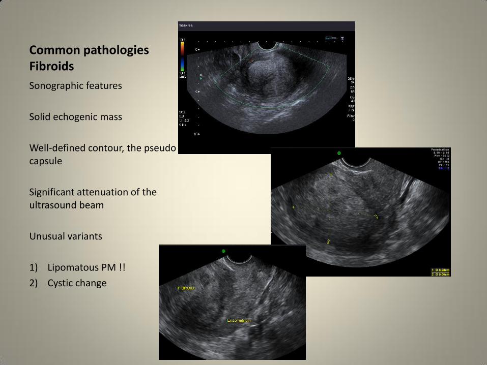
Common pathologies Fibroids
Sonographic features
Solid echogenic mass
Well-defined contour, the pseudo capsule
Significant attenuation of the ultrasound beam
Unusual variants
1) Lipomatous PM !!
2) Cystic change

Common pathologies Fibroids
Cystic change is NOT always a feature of malignancy and it is due to hyaline degeneration
Fibroid calcification
Usually central
Peripheral post embolisation


Abnormal uterine bleeding
AUB abnormal menstrual flow in women of reproductive age Classification • Polyps • Adenomyosis • Leiomyomas • Malignancy • Coagulopathy • Ovarian dysfunction • Endometrial iatrogenic • Not yet classified

Common pathologies Endometrial
Polyps
30% of cases of post menopausal bleeding
Echogenic
Completely contained in the endometrial cavity with NO extension into the myometrium
Homogenous usually but occasional cystic changes within
Narrow base of attachment, which may have a vascular pedicle within
Common pathologies Endometrial
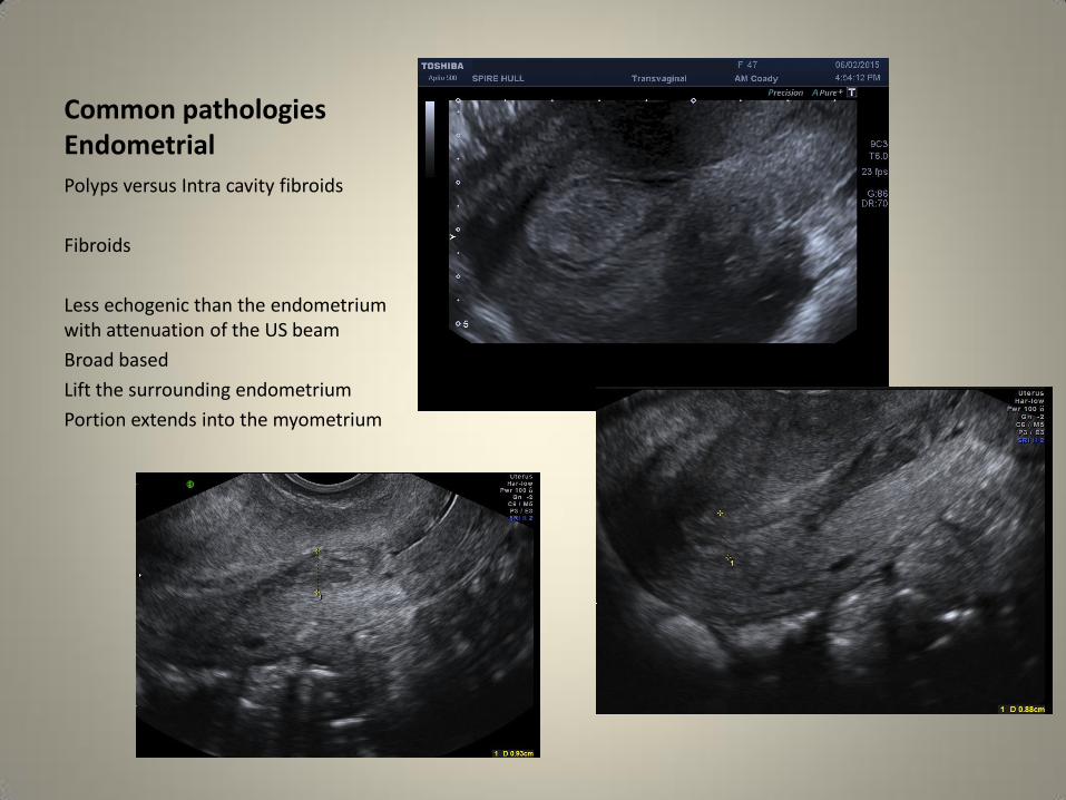
Polyps versus Intra cavity fibroids
Fibroids
Less echogenic than the endometrium with attenuation of the US beam
Broad based
Lift the surrounding endometrium
Portion extends into the myometrium

Endometrial hyperplasia
Endometrial hyperplasia is an abnormal proliferation of endometrial stroma and glands and represents a spectrum of endometrial changes
A definitive diagnosis can be made only with biopsy, imaging cannot reliably allow differentiation between hyperplasia and carcinoma.
Up to one-third of endometrial carcinoma is believed to be preceded by hyperplasia.
All types of endometrial hyperplasia can cause diffusely smooth endometrial thickening
Far less commonly, hyperplasia produces focal hypo echoic endometrial thickening.

Common pathologies Endometrial
Endometrial hyperplasia and cancer
Endometrial cancer most common gynaecological cancer
Endometrial hyperplasia can be diffuse or focal and differentiating this from a polyp can at times be difficult
Endometrial cancer has similar sonographic characteristics to hyperplasia or an enlarged polyp with of course the exception of myometrial invasion
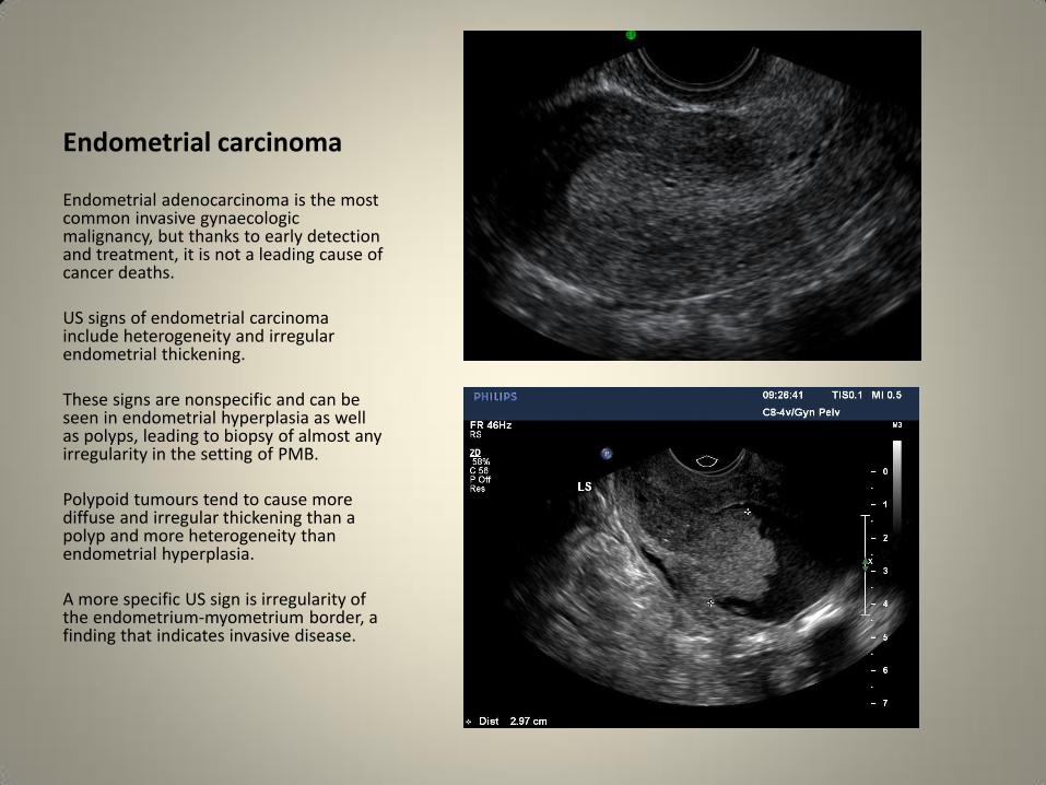
Endometrial carcinoma Endometrial adenocarcinoma is the most common invasive gynaecologic malignancy, but thanks to early detection and treatment, it is not a leading cause of cancer deaths. US signs of endometrial carcinoma include heterogeneity and irregular endometrial thickening. These signs are nonspecific and can be seen in endometrial hyperplasia as well as polyps, leading to biopsy of almost any irregularity in the setting of PMB. Polypoid tumours tend to cause more diffuse and irregular thickening than a polyp and more heterogeneity than endometrial hyperplasia. A more specific US sign is irregularity of the endometrium-myometrium border, a finding that indicates invasive disease.

Endometrial carcinoma
Endometrial Adenocarcinoma
A more specific US sign is irregularity of the endometrium-myometrium border, a finding that indicates invasive disease.

Intra uterine fluid collections
Importance depends on the age of the patient
In premenopausal patients, fluid collections are most commonly associated with
• menstruation,
• early IUP,
• the pseudogestational sac in an ectopic pregnancy
Other benign causes of obstruction leading to intrauterine fluid production include polyps, infection and submucosal fibroids.
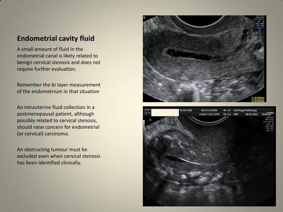
Endometrial cavity fluid
A small amount of fluid in the endometrial canal is likely related to benign cervical stenosis and does not require further evaluation.
Remember the bi layer measurement of the endometrium in that situation
An intrauterine fluid collection in a postmenopausal patient, although possibly related to cervical stenosis, should raise concern for endometrial (or cervical) carcinoma.
An obstructing tumour must be excluded even when cervical stenosis has been identified clinically.

Intra uterine fluid collections
Importance depends on the age of the patient
In prepubertal patients, fluid in the endometrial canal may be related to haematometrocolpos.
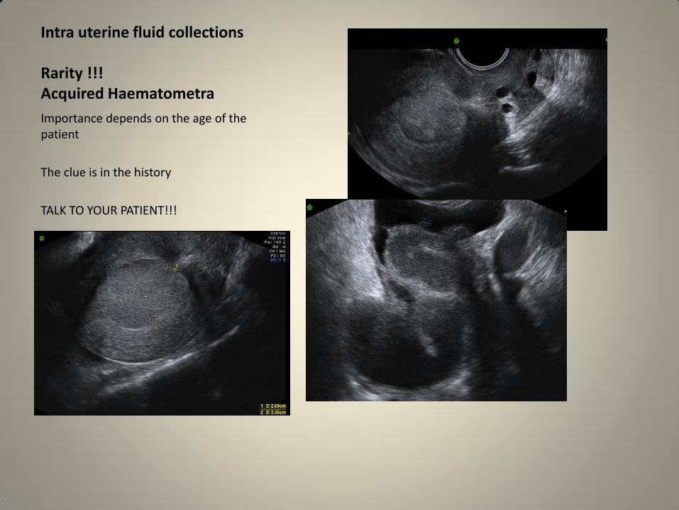
Intra uterine fluid collections Rarity !!! Acquired Haematometra
Importance depends on the age of the patient
The clue is in the history
TALK TO YOUR PATIENT!!!

Intra uterine fluid collections Rarity !!! Acquired Haematometra
Importance depends on the age of the patient
The clue is in the history
TALK TO YOUR PATIENT!!!

Uncommon pathologies Endometrial
• Endometrial adhesions
• AVM
• Osseous metaplasia

Endometrial adhesions Endometrial adhesions are posttraumatic or postsurgical in nature and can cause Asherman syndrome, which includes infertility, recurrent pregnancy loss, and amenorrhea.
Adequate distention of the endometrial cavity seen at sonohysterography or HSG is necessary for radiologic diagnosis.
Sonohysterography may demonstrate synaechiae as echogenic bands bridging the uterine cavity.
If the bands are thick and fibrotic, they may prevent complete uterine distention.
.

Uncommon pathologies Endometrial
AVM These are actually very rare and typically arise following instrumentation of the endometrial cavity commonly in association with pregnancy loss or delivery They can be associated with Malignancies Infections RPOCS Molar gestations They can be congenital and these are even less common and less symptomatic than acquired variety Symptoms Heavy bleeding Pelvic pain and dyspareunia

Uncommon pathologies AV Malformation
The diagnosis is made by TV ultrasound with colour and pulsed doppler
Grey scale ultrasound
Anechoic spaces with an irregular contour
Turbulent flow with aliasing and pulsed doppler demonstrates high velocity flow with low impedance

VERY Uncommon pathologies Endometrial Osseous metaplasia
Endometrial ossification is a rare disease, and its aetiology and pathogenesis are controversial.
It is often misdiagnosed.
Most reported cases are related to pregnancy ? RPOCS
It has also been related to transformation of uterine tissue to bone in response to inflammation and the reparative process induced by abortion
Osseus metaplasia has been associated with secondary infertility and dysmenorrhea and has mimicked
a retained intrauterine device
It can be successfully treated
with hysteroscopic resection.

Prolonged haematometra

Key points
• Meticulous scan technique: always strive to – Visualise the cervix – Define the endometrium – Find the ovaries – Find the answer
• Fibroid diagnosis is not enough, it is also about location location location
• Size matters but it is the cause that is crucial • Talk to your patients, the clue is often in the
history especially in the weird and wonderful

The End
Thank you for your attention
Questions please?