Coiled-coil protein Scy is a key component of growth in ...
10
Coiled-coil protein Scy is a key component of a multiprotein assembly controlling polarized growth in Streptomyces Neil A. Holmes a , John Walshaw b,c , Richard M. Leggett a,c , Annabelle Thibessard a,1 , Kate A. Dalton a,2 , Michael D. Gillespie a , Andrew M. Hemmings a,d , Bertolt Gust b,3 , and Gabriella H. Kelemen a,4 a School of Biological Sciences, c School of Computing Sciences, and d School of Chemistry, University of East Anglia, Norwich Research Park, Norwich NR4 7TJ, United Kingdom; and b John Innes Centre, Norwich Research Park, Norwich NR4 7UH, United Kingdom Edited by Richard Losick, Harvard University, Cambridge, MA, and approved December 7, 2012 (received for review July 5, 2012) Polarized growth in eukaryotes requires polar multiprotein com- plexes. Here, we establish that selection and maintenance of cell polarity for growth also requires a dedicated multiprotein assem- bly in the filamentous bacterium, Streptomyces coelicolor. We present evidence for a tip organizing center and confirm two of its main components: Scy (Streptomyces cytoskeletal element), a unique bacterial coiled-coil protein with an unusual repeat period- icity, and the known polarity determinant DivIVA. We also estab- lish a link between the tip organizing center and the filament- forming protein FilP. Interestingly, both deletion and overproduction of Scy generated multiple polarity centers, suggesting a mechanism wherein Scy can both promote and limit the number of emerging polarity centers via the organization of the Scy-DivIVA assemblies. We propose that Scy is a molecular “assembler,” which, by seques- tering DivIVA, promotes the establishment of new polarity centers for de novo tip formation during branching, as well as supporting polarized growth at existing hyphal tips. bacterial polarized growth | polar multiprotein assembly | cell division | modified hendecad coiled coil H ow organisms, cells, or tissues establish polarity is one of the fundamental questions in developmental biology. In eukar- yotes, from unicellular organisms to multicellular plants and animals, some of the core mechanisms for generating polarity are conserved (1). Sites for polarization must be selected using po- sitional markers, followed by the recruitment of a complex assem- bly, which, in turn, orients cytoskeletal filaments that deliver vesicles to the particular sites. A specific example of polarity is polarized growth, during which cells select a single polarization site and generate elongated, cylindrical shapes, such as neuronal dendrites in animals, root hairs and pollen tubes in plants, hyphal growth of filamentous fungi, elongation of Schizosaccharomyces pombe, or the short period of polarized growth during budding in Saccharo- myces cerevisiae. However, the presumed earliest examples of po- larized growth can be found in bacteria. These include the actin- omycete Streptomyces coelicolor, which is used as a model organism for studying morphological differentiation and filamentous growth. The complex life cycle of S. coelicolor begins with an ovoid spore that contains a single chromosome. During germination, long, multigenomic filaments (germ tubes) are formed, which branch regularly to generate a network of hyphal filaments. Branching is a necessity for exponential growth because the rate of tip elongation cannot exceed a certain maximum; hence, there is an exponential increase in the number of new tips. New tips develop on the lateral wall well behind the existing tip, a phe- nomenon also observed and described as apical dominance in eukaryotic filamentous fungi. When grown on semisolid agar medium, these hyphal filaments first grow across and into the solid medium, generating the vegetative mycelium, followed by the formation of an aerial mycelium by hyphal extension into the air. In addition to branching, the multichromosomal hyphae are segmented by occasional septation, but this is not followed by separation of the neighboring segments. Regular septation is exclusive to the sporogenic aerial hyphae, where a highly co- ordinated but only partly understood mechanism generates syn- chronous septation and chromosome segregation, leading to chains of unigenomic spores (2). The shape of bacterial cells is defined by their cell wall, which largely comprises peptidoglycan. During bacterial growth, peptido- glycan synthesis and deposition does not occur in a random fashion but is highly organized and localized to specific sites identified by cytoskeletal scaffolding proteins. In a typical rod-shaped bacterium, such as Bacillus subtilis, there are two phases of nascent peptido- glycan synthesis: during cell division at a midcell division site, where the primary cytoskeletal scaffold is provided by the tubulin-like FtsZ, and during elongation at the lateral wall, where members of the actin-like MreB family, including MreB, Mbl, and MreBH, are im- plicated in forming the helical pattern of nascent peptidoglycan synthesis (3). In spherical cells such as those of Staphylococcus au- reus, only the FtsZ-driven cell wall synthesis takes place and occurs at the division site (4), whereas the curved rod-shaped bacterium Caulobacter crescentus also employs a third, intermediate filament- like cytoskeletal protein, CreS, to establish its crescent shape (5, 6). In the filamentous bacterium S. coelicolor, hyphal extension takes place exclusively at hyphal tips, as was shown by monitoring fluorescent vancomycin (Van-FL) labeling of newly externalized and/or incorporated peptidoglycan precursors (7, 8). However, the mechanism of polarized growth in Streptomyces does not rely on the organization of either MreB helices or FtsZ rings, as is known for common rod-shaped or spherical bacteria. The MreB homologs of S. coelicolor are implicated in the development of the rod-shaped compartments that become the spores and not in hyphal tip extension (9–11). Similarly, although S. coelicolor FtsZ is essential for septum formation and sporulation, it is not in- volved in filamentous growth (12). Polarized hyphal extension in S. coelicolor depends on the coiled-coil protein DivIVA (8, 13). The divIVA gene is essential in S. coelicolor, where either partial depletion or overproduction of DivIVA resulted in aberrant Author contributions: N.A.H. and G.H.K. designed research; N.A.H., J.W., R.M.L., A.T., K.A.D., M.D.G., B.G., and G.H.K. performed research; J.W., R.M.L., and A.M.H. analyzed data; and J.W., A.M.H., and G.H.K. wrote the paper. The authors declare no conflict of interest. This article is a PNAS Direct Submission. 1 Present address: Université de Lorraine, Genetique et Microbiologie, Unité Mixte de Recherche, Institut National de la Recherche Agronomique, BP239-54506 Nancy, France. 2 Present address: ImmBio, Babraham Research Campus, Cambridge CB22 3AT, United Kingdom. 3 Present address: Eberhard-Karls-Universität Tübingen, 72074 Tubingen, Germany. 4 To whom correspondence should be addressed. E-mail: [email protected]. See Author Summary on page 1580 (volume 110, number 5). This article contains supporting information online at www.pnas.org/lookup/suppl/doi:10. 1073/pnas.1210657110/-/DCSupplemental. www.pnas.org/cgi/doi/10.1073/pnas.1210657110 PNAS | Published online January 7, 2013 | E397–E406 GENETICS PNAS PLUS Downloaded by guest on December 1, 2021
Transcript of Coiled-coil protein Scy is a key component of growth in ...
Coiled-coil protein Scy is a key component ofa multiprotein assembly controlling polarizedgrowth in StreptomycesNeil A. Holmesa, John Walshawb,c, Richard M. Leggetta,c, Annabelle Thibessarda,1, Kate A. Daltona,2,Michael D. Gillespiea, Andrew M. Hemmingsa,d, Bertolt Gustb,3, and Gabriella H. Kelemena,4
aSchool of Biological Sciences, cSchool of Computing Sciences, and dSchool of Chemistry, University of East Anglia, Norwich Research Park, Norwich NR4 7TJ,United Kingdom; and bJohn Innes Centre, Norwich Research Park, Norwich NR4 7UH, United Kingdom
Edited by Richard Losick, Harvard University, Cambridge, MA, and approved December 7, 2012 (received for review July 5, 2012)
Polarized growth in eukaryotes requires polar multiprotein com-plexes. Here, we establish that selection and maintenance of cellpolarity for growth also requires a dedicated multiprotein assem-bly in the filamentous bacterium, Streptomyces coelicolor. Wepresent evidence for a tip organizing center and confirm two ofits main components: Scy (Streptomyces cytoskeletal element), aunique bacterial coiled-coil protein with an unusual repeat period-icity, and the known polarity determinant DivIVA. We also estab-lish a link between the tip organizing center and the filament-formingprotein FilP. Interestingly, both deletion andoverproductionof Scy generatedmultiple polarity centers, suggesting amechanismwherein Scy can both promote and limit the number of emergingpolarity centers via the organization of the Scy-DivIVA assemblies.We propose that Scy is a molecular “assembler,” which, by seques-tering DivIVA, promotes the establishment of new polarity centersfor de novo tip formation during branching, as well as supportingpolarized growth at existing hyphal tips.
bacterial polarized growth | polar multiprotein assembly | cell division |modified hendecad coiled coil
How organisms, cells, or tissues establish polarity is one of thefundamental questions in developmental biology. In eukar-
yotes, from unicellular organisms to multicellular plants andanimals, some of the core mechanisms for generating polarity areconserved (1). Sites for polarization must be selected using po-sitional markers, followed by the recruitment of a complex assem-bly, which, in turn, orients cytoskeletal filaments that deliver vesiclesto the particular sites. A specific example of polarity is polarizedgrowth, during which cells select a single polarization site andgenerate elongated, cylindrical shapes, such as neuronal dendritesin animals, root hairs and pollen tubes in plants, hyphal growth offilamentous fungi, elongation of Schizosaccharomyces pombe, orthe short period of polarized growth during budding in Saccharo-myces cerevisiae. However, the presumed earliest examples of po-larized growth can be found in bacteria. These include the actin-omycete Streptomyces coelicolor, which is used as a model organismfor studying morphological differentiation and filamentous growth.The complex life cycle of S. coelicolor begins with an ovoid
spore that contains a single chromosome. During germination,long, multigenomic filaments (germ tubes) are formed, whichbranch regularly to generate a network of hyphal filaments.Branching is a necessity for exponential growth because the rateof tip elongation cannot exceed a certain maximum; hence, thereis an exponential increase in the number of new tips. New tipsdevelop on the lateral wall well behind the existing tip, a phe-nomenon also observed and described as apical dominance ineukaryotic filamentous fungi. When grown on semisolid agarmedium, these hyphal filaments first grow across and into thesolid medium, generating the vegetative mycelium, followed bythe formation of an aerial mycelium by hyphal extension into theair. In addition to branching, the multichromosomal hyphae aresegmented by occasional septation, but this is not followed by
separation of the neighboring segments. Regular septation isexclusive to the sporogenic aerial hyphae, where a highly co-ordinated but only partly understood mechanism generates syn-chronous septation and chromosome segregation, leading tochains of unigenomic spores (2).The shape of bacterial cells is defined by their cell wall, which
largely comprises peptidoglycan. During bacterial growth, peptido-glycan synthesis and deposition does not occur in a random fashionbut is highly organized and localized to specific sites identified bycytoskeletal scaffolding proteins. In a typical rod-shaped bacterium,such as Bacillus subtilis, there are two phases of nascent peptido-glycan synthesis: during cell division at a midcell division site, wherethe primary cytoskeletal scaffold is provided by the tubulin-like FtsZ,and during elongation at the lateral wall, where members of theactin-like MreB family, including MreB, Mbl, and MreBH, are im-plicated in forming the helical pattern of nascent peptidoglycansynthesis (3). In spherical cells such as those of Staphylococcus au-reus, only the FtsZ-driven cell wall synthesis takes place and occursat the division site (4), whereas the curved rod-shaped bacteriumCaulobacter crescentus also employs a third, intermediate filament-like cytoskeletal protein, CreS, to establish its crescent shape (5, 6).In the filamentous bacterium S. coelicolor, hyphal extension
takes place exclusively at hyphal tips, as was shown by monitoringfluorescent vancomycin (Van-FL) labeling of newly externalizedand/or incorporated peptidoglycan precursors (7, 8). However,the mechanism of polarized growth in Streptomyces does not relyon the organization of either MreB helices or FtsZ rings, as isknown for common rod-shaped or spherical bacteria. The MreBhomologs of S. coelicolor are implicated in the development ofthe rod-shaped compartments that become the spores and not inhyphal tip extension (9–11). Similarly, although S. coelicolor FtsZis essential for septum formation and sporulation, it is not in-volved in filamentous growth (12). Polarized hyphal extension inS. coelicolor depends on the coiled-coil protein DivIVA (8, 13).The divIVA gene is essential in S. coelicolor, where either partialdepletion or overproduction of DivIVA resulted in aberrant
Author contributions: N.A.H. and G.H.K. designed research; N.A.H., J.W., R.M.L., A.T.,K.A.D., M.D.G., B.G., and G.H.K. performed research; J.W., R.M.L., and A.M.H. analyzeddata; and J.W., A.M.H., and G.H.K. wrote the paper.
The authors declare no conflict of interest.
This article is a PNAS Direct Submission.1Present address: Université de Lorraine, Genetique et Microbiologie, Unité Mixte deRecherche, Institut National de la Recherche Agronomique, BP239-54506 Nancy, France.
2Present address: ImmBio, Babraham Research Campus, Cambridge CB22 3AT,United Kingdom.
3Present address: Eberhard-Karls-Universität Tübingen, 72074 Tubingen, Germany.4To whom correspondence should be addressed. E-mail: [email protected].
See Author Summary on page 1580 (volume 110, number 5).
This article contains supporting information online at www.pnas.org/lookup/suppl/doi:10.1073/pnas.1210657110/-/DCSupplemental.
www.pnas.org/cgi/doi/10.1073/pnas.1210657110 PNAS | Published online January 7, 2013 | E397–E406
GEN
ETICS
PNASPL
US
Dow
nloa
ded
by g
uest
on
Dec
embe
r 1,
202
1
branching and altered hyphal characteristics (8, 14). DivIVA-Egfplocalized to growing hyphal tips and also to sites in the lateral wall,marking future branch points before the appearance of the newlateral tip (14). This suggested thatDivIVAwas a positionalmarkerfor site selection of de novo lateral branch formation. Interestingly,in the rod-shaped actinomycetes Corynebacterium glutamicum,Mycobacterium smegmatis, andMycobacterium tuberculosis, nascentpeptidoglycan synthesis also appears to be directed at the poles ina DivIVA-dependent manner (15), a pattern apparently specific toactinomycetes. The direct interaction between DivIVA and PBP3,a penicillin-binding protein inM. tuberculosis (16), supports the roleof DivIVA as a polarity marker that recruits the cell wall syntheticmachinery during polarized growth in actinomycetes.DivIVA itself is widespread in Gram-positive bacteria but has
different roles outside of the actinomycetes. In B. subtilis, for ex-ample, DivIVA controls cell division via positioning of the MinCandMinDdivision inhibitor proteins to the cell poles, allowing FtsZring formation at midcell (17). InB. subtilis, DivIVA localizes to themidcell during cell division and to mature cell poles by targeting tonegatively curved, concave membranes (18, 19).Apart from the site selection for cell wall extension by DivIVA
in the actinomycetes, such as S. coelicolor, C. glutamicum, M.smegmatis, and M. tuberculosis, very little is known of the mech-anism and the molecular interactions that drive polarized growthin bacteria. Here, we report that although DivIVA is necessary,it is not sufficient for normal polarized growth in Streptomyces,and we present Scy (Streptomyces cytoskeletal element), a uniquecoiled-coil protein, which, together with DivIVA, is critical forestablishing and maintaining polarized growth in S. coelicolor.We provide evidence of a complex, multiprotein, polar assemblyfor polarized growth in bacteria, and we demonstrate that Scycontrols the number of new polarity centers established.
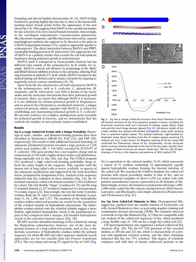
ResultsScy Is a Large Coiled-Coil Protein with a Unique Periodicity. Homo-logs of actin-, tubulin-, and filament-forming proteins have beenidentified in Streptomyces [MreB (9); FtsZ (12), and FilP (20),respectively]. Our search to find additional bacterial homologs ofeukaryote cytoskeletal proteins revealed a large protein of 1,326amino acid residues (Mr = 146 kDa) encoded by SCO5397 ofS. coelicolor. This gene product has a net negative charge and anestimated pI of 4.94 with a highly skewed amino acid distribution,being especially rich in Ala, Glu, and Arg. The COILS program(21) predicted a high coiled-coil–forming probability along al-most the entire length of the sequence. This, together with theknown role of long coiled coils as levers, scaffolds, or spacers ofthe eukaryote cytoskeleton and supported by the work describedbelow, prompted the designation of Scy. Analysis of the sequenceindicated that Scy consisted of three domains (Fig. 1A): the N-terminal canonical, coiled-coil domain (residues 1–64) is followedby a short, Gly-rich flexible “hinge” (residues 64–72) and the longC-terminal domain (1,227 residues) composed of a noncanonical,51-residue repeat (22). Interestingly, the very few (only 4) prolineresidues flank the N- and C-terminal coiled-coil domains at posi-tions 11, 64, 1,300, and 1,317. The positions of the hydrophobicresidues within coiled-coil proteins are crucial for the associationof the α-helical strands via hydrophobic interactions. The hydro-phobic residues plotted onto the α-helical cylinder suggest a moreshallow, right-handed hydrophobic streak in the penindaenad re-peat of Scy compared with a steeper, left-handed hydrophobicstreak in the canonical heptad repeat (Fig. 1B).BLAST searches identified homologs of Scy exclusively among
the filamentous actinomycetes; however, one of the most im-portant features of a long coiled-coil protein, such as Scy, is theperiodic occurrence of hydrophobic residues within the primarysequence, for which BLAST and other sequence similarity searchapproaches are far less suitable than fast Fourier transforms(FTs). The very sharp and strong FT signal for Scy at 3.643 (Fig.
S1) is equivalent to the rational number 51/14, which representsa repeat of 51 residues containing 14 approximately equallyspaced hydrophobic residues forming the internal interface ofthe coiled coil. We searched the UniProt database for coiled-coilproteins with repeat periodicity similar to that of Scy, and wefound numerous examples of short (<250 aa) coiled coils withputative penindaenad repeats represented in all domains of life.Interestingly, we have also found several proteinswith long (∼600–1,000 amino acids) Scy-like repeats among bacteria (Helicobacter,Leptospira, and Brachyspira) and simple eukaryotes (Parameciumand Trichomonas) (Fig. S1).
Scy Can Form Coiled-Coil Filaments in Vitro. Overexpressed His-tagged Scy, purified from the soluble fraction of Escherichia colicells, formed filaments in vitro, as seen using negative staining andtransmission electron microscopy (TEM). We routinely observeda network of rope-like filaments (Fig. 1C) that are compatible withour analysis of the coiled-coil sequences of Scy, which predicteda long, flexible rope of ∼190 nm for a single Scy coiled coil (22).CD spectropolarimetry also supported a helical coiled-coil Scy
structure (Fig. 1D). The far-UV CD spectrum of Scy revealedminima at 208 nm and 222 nm, which is characteristic of a pro-tein with significant α-helical content, and the latter minimumindicated that Scy was 73% α-helical. This degree of α-helicitycompares well with that of known coiled-coil proteins at the
Fig. 1. Scy has a unique coiled-coil structure that forms filaments in vitro.(A) Domain structure of Scy from sequence analysis is shown, including theN-terminal canonical (red) and C-terminal 51-residue repeat (blue) coiledcoils and the short hinge domain (green) (Fig. S1). (B) Atomic model of theα-helix exhibits the stylized left-handed hydrophobic stripe (red) resultingfrom a canonical heptad repeat. The stylized shallower, right-handed hy-drophobic stripe (blue) resulting from the 51-residue repeat, assuming 3.6residues per turn, is also shown. (C) Negative staining of His-Scy using TEMconfirmed the filamentous nature of Scy. Occasionally, circular structures(white arrows) could be discerned at the end of the Scy ropes. (Insets) Close-up images of these structures are shown. (Scale bars: 100 nm.) (D) CD spec-troscopy of His-Scy in the absence and presence of 50% (vol/vol) TFE.
E398 | www.pnas.org/cgi/doi/10.1073/pnas.1210657110 Holmes et al.
Dow
nloa
ded
by g
uest
on
Dec
embe
r 1,
202
1
same temperature. Another significant feature of Scy’s spec-trum is a ratio of [θ]222/[θ]208 > 1, indicating a stable coiled coil.Exposure to 50% (vol/vol) trifluoroethanol (TFE), which blockshydrophobic interactions and so decreases the oligomerizationof amphipathic α-helices into coiled coils, changed this ratio inScy from 1.07 to 0.94. Consistent with its coiled-coil structure,spectral deconvolution of Scy also indicated that the TFE-in-duced disruption of oligomerization resulted in the decrease ofthe helical content to 55%, implying cooperativity between helixformation and the coiled-coil assembly.
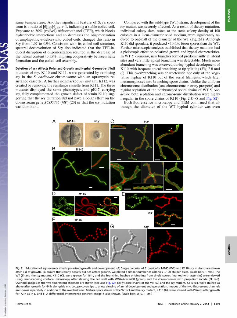
Deletion of scy Affects Polarized Growth and Hyphal Geometry. Nullmutants of scy, K110 and K111, were generated by replacingscy in the S. coelicolor chromosome with an apramycin re-sistance cassette. A further nonmarked scy mutant, K112, wascreated by removing the resistance cassette from K111. The threemutants displayed the same phenotypes, and pK47, carryingscy, fully complemented the growth defect of strain K110, sug-gesting that the scy mutation did not have a polar effect on thedownstream gene SCO5396 (filP) (20) or that the scy mutationwas dominant.
Compared with the wild-type (WT) strain, development of thescy mutant was severely affected. As a result of the scy mutation,individual colony sizes, tested at the same colony density of 100colonies in a 9-cm–diameter solid medium, were significantly re-duced to one-half of the diameter of the WT (Fig. 2A). AlthoughK110 did sporulate, it produced ∼10-fold fewer spores than theWT.Further microscopic analyses established that the scy mutation hada pleiotropic effect on polarized growth and hyphal characteristics.In WT S. coelicolor, new branches formed predominantly at lateralsites and very little apical branching was detectable. Much moreabundant branching was observed during hyphal development ofK110, with frequent apical branching or tip splitting (Fig. 2 B andC). This overbranching was characteristic not only of the vege-tative hyphae of K110 but of the aerial filaments, which latermetamorphosed into branching spore chains. Unlike the uniformchromosome distribution (one chromosome in every prespore) andregular septation of the nonbranched spore chains of WT S. coe-licolor, both septation and chromosome distribution were highlyirregular in the spore chains of K110 (Fig. 2 D–G and Fig. S2).Both fluorescence microscopy and TEM confirmed that al-
though the diameter of the WT hyphal cylinder was even
Fig. 2. Mutation of scy severely affects polarized growth and development. (A) Single colonies of S. coelicolor M145 (WT) and K110 (scy mutant) are shownafter 6 d of growth. To ensure that colony density did not affect growth, we plated a similar number of colonies, ∼100 cfu per plate. (Scale bars: 1 mm.) TheWT (B) and the scy mutant, K110 (C), were grown for 16 h, and the branching hyphae originating from single spores (marked with asterisks) were viewedusing laser-scanning confocal microscopy after staining the cell wall with WGA-Alexa488 (green) and the chromosomes with propidium iodide (PI; red).Overlaid images of the two fluorescent channels are shown (see also Fig. S2). Early spore chains of the WT (D) and the scy mutant, K110 (E), were stained asabove after growth for 44 h alongside microscope coverslips to allow viewing of aerial development and sporulation. Images of the two fluorescent channelsare shown separately in addition to the overlaid view. Mature spore chains of the WT (F) and the scymutant, K110 (G), were stained with PI (red) after growthfor 72 h as in D and E. A differential interference contrast image is also shown. (Scale bars: B–G, 1 μm.)
Holmes et al. PNAS | Published online January 7, 2013 | E399
GEN
ETICS
PNASPL
US
Dow
nloa
ded
by g
uest
on
Dec
embe
r 1,
202
1
throughout its length, varying between 0.4 and 0.7 μm, the scymutant had a very irregular hyphal width, varying between 0.25and 1.1 μm. Thin cross-sections of the WT hyphal cylindergenerated circular or ellipsoidal shapes when the TEM sec-tioning was nearly perpendicular to the hyphal axis, whereas inthe scy mutant, irregular “amoeboid” cross-sections were typ-ical (Fig. S2). Because hyphal extension occurs at the hyphal tipin Streptomyces, to explain the irregularities in the hyphal di-ameter of scy, we looked for cues in the tip architecture. In theWT, the tip dome was symmetrical with at least one plane ofsymmetry, whereas the hyphal tip of the scy mutant was oftenbulging and asymmetrical.
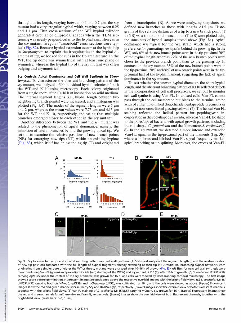
Scy Controls Apical Dominance and Cell Wall Synthesis in Strep-tomyces. To characterize the aberrant branching pattern of thescy mutant, we analyzed ∼300 individual hyphal colonies of boththe WT and K110 using microscopy. Each colony originatedfrom a single spore after 10–16 h of incubation on solid medium.The internal segment lengths (i.e., hyphal length between twoneighboring branch points) were measured, and a histogram wasplotted (Fig. 3A). The modes of the segment lengths were 5 μmand 2 μm, whereas the mean values were 7.73 μm and 4.15 μmfor the WT and K110, respectively, indicating that multiplebranches emerged closer to each other in the scy mutant.Another difference between the WT and the scy mutant was
related to the phenomenon of apical dominance, namely, theinhibition of lateral branches behind the growing apical tip. Weset out to examine the relative positions of new branch points(NB) for emerging new tips (NT) within an existing hyphae(Fig. S3), which itself has an extending tip (T) and originated
from a branchpoint (B). As we were analyzing snapshots, wedefined new branches as those with lengths <1.5 μm. Histo-grams of the relative distances of a tip to a new branch point (Tto NB) vs. a tip to an old branch point (T to B) were plotted usingthe same sets of hyphal samples tested above (Fig. 3A). Apicaldominance was typical for the WT strain, which had a strongpreference for generating new tips far behind the growing tip. In theWT, only 6%of the new branch points were in the tip-proximal 20%of the hyphal length, whereas 77% of the new branch points werecloser to the previous branch point than to the growing tip. Incontrast, in the scy mutant, 33% of the new branch points were inthe tip-proximal 20% and 66%of new branch points were in the tip-proximal half of the hyphal filament, suggesting the lack of apicaldominance in the scy mutant.To test whether the uneven hyphal diameter, the short hyphal
length, and the aberrant branching pattern of K110 reflected defectsin the incorporation of cell wall precursors, we set out to monitorcell wall synthesis using Van-FL. In unfixed cells, Van-FL cannotpass through the cell membrane but binds to the terminal aminoacids of either lipid-linked disaccharide pentapeptide precursors orthe as yet non–cross-linked growing cell wall (7). The helical Van-FLstaining reflected the helical pattern for peptidoglycan in-corporation in the rod-shapedB. subtilis,whereasVan-FL localizedto the poles/tips of bacteria with apical growth patterns, includingthe rod-shaped C. glutamicum and the filamentous S. coelicolor (7,8). In the scy mutant, we detected a more intense and extendedVan-FL signal in the tip-proximal part of the filaments (Fig. 3B),where a bulbous and bilobed Van-FL signal frequently markedapical branching or tip splitting. Moreover, the excess of Van-FL
Fig. 3. Scy localizes to the tips and affects branching patterns and cell wall synthesis. (A) Statistical analysis of the segment length (i) and the relative locationof new tip positions compared with the full-length of hyphal fragments already extending at their tip (ii). Around 300 branching hyphal networks, eachoriginating from a single spore of either the WT or the scy mutant, were analyzed after 10–16 h of growth (Fig. S3). (B) Sites for new cell wall synthesis weremonitored using Van-FL (green) and propidium iodide (red) staining of the WT (i) and scy mutant, K110 (ii), after 16 h of growth. (C) S. coelicolor M145/pK56,carrying egfp-scy under the control of the scy promoter, was grown for 16 h, and cells were viewed by laser-scanning confocal microscopy. The first imageshows a spore before germination. Fluorescent images are positioned above the respective overlaid images with the bright-field views. (D) S. coelicolorM145/pKF59/pK57, carrying both divIVA-egfp (pKF59) and mCherry-scy (pK57), was cultivated for 16 h, and the cells were viewed as above. (Upper) Fluorescentimages show the red and green channels for mCherry-Scy and DivIVA-Egfp, respectively. (Lower) Images show the overlaid view of both fluorescent channels,together with the bright-field views. (E) Van-FL staining of S. coelicolor M145/pK57 carrying mCherry-Scy grown for 16 h. (Upper) Fluorescent images showthe red and green channels for mCherry-Scy and Van-FL, respectively. (Lower) Images show the overlaid view of both fluorescent channels, together with thebright-field view. (Scale bars: B–E, 1 μm.)
E400 | www.pnas.org/cgi/doi/10.1073/pnas.1210657110 Holmes et al.
Dow
nloa
ded
by g
uest
on
Dec
embe
r 1,
202
1
staining at lateral positions resulted in multiple branches origi-nating from the same lateral sites.
Scy Localizes to Growing Tips and Marks the Sites for de Novo TipEmergence. To establish the localization of Scy, we generatedpK56, carrying an Egfp-Scy translational fusion under the controlof the native scy promoter, allowing Egfp-Scy localization in thepresence of native Scy. Apart from some hyphalmeandering, pK56fully complemented the branching defects and altered sporulationproperties of K112, the unmarked null mutant of scy, confirmingthat Egfp-Scy was mostly functional. Egfp-Scy localized to growingestablished tips and young tips that had been formed recently.Bright Egfp-Scy foci were also identified in spores before germi-nation at potential sites for germ tube outgrowth and at sites in thelateral hyphal wall, possibly marking the positions of future branchpoints (Fig. 3C). DivIVA, the known polarity determinant of S.coelicolor, was shown to localize to future tip positions ∼30 minbefore the time when the dome of the tip became visible (14).
Therefore, to confirm that Scy not only localized to existing tips butmarked the positions of future tips, we localized Scy and DivIVAsimultaneously using two different fluorescent fusions, mCherry-Scy and DivIVA-Egfp (Fig. 3D). The colocalization of mCherry-Scy and DivIVA-Egfp not only at the hyphal apex but at sites onthe lateral wall confirmed that Scy, together with DivIVA, is im-plicated in the selection of sites for de novo tip formation.Furthermore, to establish a link between Scy and cell wall
synthesis, we monitored Van-FL incorporation, together withlocalization of mCherry-Scy (Fig. 3E). The colocalization ofVan-FL and mCherry-Scy confirmed that, similar to DivIVA, Scyalso marks the sites of new cell wall synthesis, which was notexclusive to growing tip ends but also to several distinct, lateralfoci that presumably marked future branchpoints.
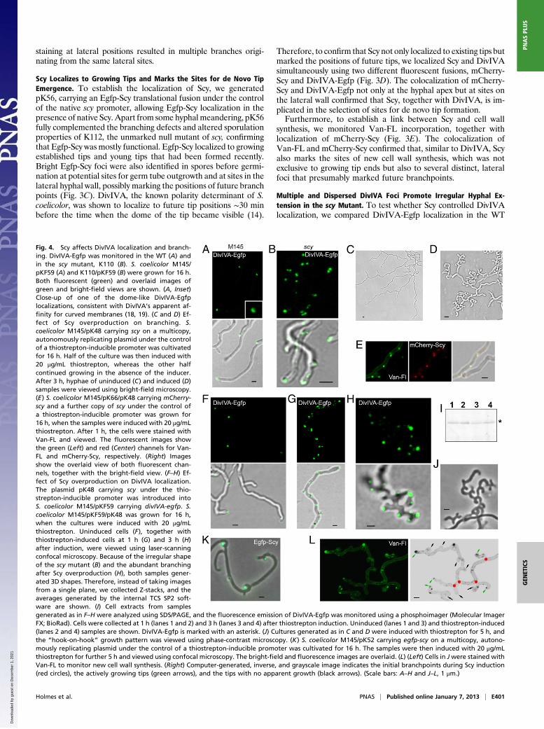
Multiple and Dispersed DivIVA Foci Promote Irregular Hyphal Ex-tension in the scy Mutant. To test whether Scy controlled DivIVAlocalization, we compared DivIVA-Egfp localization in the WT
Fig. 4. Scy affects DivIVA localization and branch-ing. DivIVA-Egfp was monitored in the WT (A) andin the scy mutant, K110 (B). S. coelicolor M145/pKF59 (A) and K110/pKF59 (B) were grown for 16 h.Both fluorescent (green) and overlaid images ofgreen and bright-field views are shown. (A, Inset)Close-up of one of the dome-like DivIVA-Egfplocalizations, consistent with DivIVA’s apparent af-finity for curved membranes (18, 19). (C and D) Ef-fect of Scy overproduction on branching. S.coelicolor M145/pK48 carrying scy on a multicopy,autonomously replicating plasmid under the controlof a thiostrepton-inducible promoter was cultivatedfor 16 h. Half of the culture was then induced with20 μg/mL thiostrepton, whereas the other halfcontinued growing in the absence of the inducer.After 3 h, hyphae of uninduced (C) and induced (D)samples were viewed using bright-field microscopy.(E) S. coelicolor M145/pK66/pK48 carrying mCherry-scy and a further copy of scy under the control ofa thiostrepton-inducible promoter was grown for16 h, when the samples were induced with 20 μg/mLthiostrepton. After 1 h, the cells were stained withVan-FL and viewed. The fluorescent images showthe green (Left) and red (Center) channels for Van-FL and mCherry-Scy, respectively. (Right) Imagesshow the overlaid view of both fluorescent chan-nels, together with the bright-field view. (F–H) Ef-fect of Scy overproduction on DivIVA localization.The plasmid pK48 carrying scy under the thio-strepton-inducible promoter was introduced intoS. coelicolor M145/pKF59 carrying divIVA-egfp. S.coelicolor M145/pKF59/pK48 was grown for 16 h,when the cultures were induced with 20 μg/mLthiostrepton. Uninduced cells (F), together withthiostrepton-induced cells at 1 h (G) and 3 h (H)after induction, were viewed using laser-scanningconfocal microscopy. Because of the irregular shapeof the scy mutant (B) and the abundant branchingafter Scy overproduction (H), both samples gener-ated 3D shapes. Therefore, instead of taking imagesfrom a single plane, we collected Z-stacks, and theaverages generated by the internal TCS SP2 soft-ware are shown. (I) Cell extracts from samplesgenerated as in F–H were analyzed using SDS/PAGE, and the fluorescence emission of DivIVA-Egfp was monitored using a phosphoimager (Molecular ImagerFX; BioRad). Cells were collected at 1 h (lanes 1 and 2) and 3 h (lanes 3 and 4) after thiostrepton induction. Uninduced (lanes 1 and 3) and thiostrepton-induced(lanes 2 and 4) samples are shown. DivIVA-Egfp is marked with an asterisk. (J) Cultures generated as in C and D were induced with thiostrepton for 5 h, andthe “hook-on-hook” growth pattern was viewed using phase-contrast microscopy. (K) S. coelicolor M145/pK52 carrying egfp-scy on a multicopy, autono-mously replicating plasmid under the control of a thiostrepton-inducible promoter was cultivated for 16 h. The samples were then induced with 20 μg/mLthiostrepton for further 5 h and viewed using confocal microscopy. The bright-field and fluorescence images are overlaid. (L) (Left) Cells in Jwere stained withVan-FL to monitor new cell wall synthesis. (Right) Computer-generated, inverse, and grayscale image indicates the initial branchpoints during Scy induction(red circles), the actively growing tips (green arrows), and the tips with no apparent growth (black arrows). (Scale bars: A–H and J–L, 1 μm.)
Holmes et al. PNAS | Published online January 7, 2013 | E401
GEN
ETICS
PNASPL
US
Dow
nloa
ded
by g
uest
on
Dec
embe
r 1,
202
1
and scy mutant. In WT hyphae, a single, point-like DivIVA-Egfpfocusmarked the new tip sites, followed by amore extended, dome-shaped DivIVA-Egfp signal that kept to the curvature of the tip(Fig. 4A). This is consistent with the recent demonstration that B.subtilis DivIVA binds to membranes with concave curvatures (18,19). In contrast, in the scymutant, the DivIVA foci appeared to bepatchier and dispersed. At least ∼76% of the DivIVA foci werewithin less than 1 μm from a neighboring DivIVA foci, and two tothree DivIVA foci were found at ∼40% of existing tips (Fig. 4B),consistent with apical branching in K110 (Figs. 2C and 3B). Themultiple DivIVA patches, which promote cell wall extension atthese irregular sites, can explain the asymmetrical tip curvature anduneven hyphal diameter of the scy mutant. This suggests that Scycontrols the number and organization of the DivIVA complexes inthe Streptomyces hyphae.
Overexpression of Scy Initiates Copious de Novo Tip Formation viaRecruitment of DivIVA to Ectopic Locations. To understand how Scycontrolled hyphal branching, we monitored the effect of Scyoverproduction in S. coelicolor using pK48, a multicopy plasmidcarrying scy under the control of a thiostrepton-inducible pro-moter. In the absence of thiostrepton, hyphal growth and bran-ching of S. coelicolorM145/pK48 were indistinguishable from thatof the WT, M145 (Fig. 4C). However, when Scy overproductionwas induced in the presence of thiostrepton, there were dramaticeffects on hyphal morphology, with the prolific emergence ofnew tips generating short and curved hyphae (Fig. 4D). Todissect the effect of Scy overproduction, we first monitored Scyitself by introducing the single-copy, integrative plasmid pK66carryingmCherry-scy, together with the multicopy plasmid pK48carrying the thiostrepton-inducible scy. After induction of Scyoverproduction for 1 h, we observed the appearance of multiplemCherry-Scy foci, which localized to new cell wall synthesiswhen monitored using Van-FL staining (Fig. 4E). This suggestedthat Scy controlled the selection of sites for de novo tip pro-duction and the establishment of new cell wall synthesis duringlateral branching.The abundant emergence of new tips after the ectopic pro-
duction of Scy was reminiscent of the effect of DivIVA over-production (8, 14). To establish whether de novo tip formation inthe Scy overproducing strain was via recruitment of DivIVA, wemonitored DivIVA-Egfp after thiostrepton induction of Scy ex-pression (Fig. 4 F–H). One hour after Scy induction, multipleDivIVA-Egfp foci were detected along the lateral hyphal wall,and these DivIVA-Egfp foci marked the location of new tipsdetected about 3 h after thiostrepton induction of Scy over-production. This suggested that Scy can recruit DivIVA andmark the sites for de novo tip formation, generating lateralbranching. Monitoring the level of DivIVA-Egfp using activitygels (Fig. 4I) confirmed that Scy overproduction had no effecton DivIVA levels, only on its localization. Interestingly, over-production of Scy for a further 5 h produced a fractal-like,“hook-on-hook” growth pattern (Fig. 4J). Monitoring Egfp-Scyunder these conditions confirmed the presence of Scy at the endof all of the “hooks” (Fig. 4K); however, Van-FL staining sug-gested abortive branching, because most of the hook ends lackedactive cell wall synthesis apart from the last, leading hook (Fig.4L). One possible explanation for this unusual growth pattern isthat although Scy levels were high, levels of DivIVA, and pre-sumably also levels of cell wall synthetic enzymes, were un-changed. Therefore, the repeated generation of a large numberof actively growing tips could not be maintained, which led toabortive branching generating the hook-on-hook pattern.
Roles of Scy and DivIVA Are Closely Linked.Clearly, both the absenceand overproduction of Scy had significant effects on DivIVA lo-calization. Conversely, to test the effect of DivIVA on Scy local-ization, we monitored mCherry-Scy in the DivIVA-depleted strain,
K115, and in the DivIVA overproducer, K114. The former strainhas a chromosomal divIVA deletion but carries a single copy ofdivIVA under the thiostrepton-inducible promoter (8). In thepresence of low levels of thiostrepton, K115 exhibits normalgrowth and branching and mCherry-Scy localizes at the growingtips (Fig. S4A). When K115 cells carrying mCherry-Scy weremoved onto media lacking thiostrepton, although we observed thesame hyphal deformations due to DivIVA depletion as describedbefore (8), mCherry-Scy clearly localized to the hyphal tips (Fig.S4B). This might suggest that Scy does not require DivIVA for itspolar localization. On the other hand, thiostrepton depletion doesnot completely abolish DivIVA synthesis in K115, and the lowlevels of DivIVA [1/10th of the WT levels (8)] might be sufficientfor the recruitment of Scy. The DivIVA depletion studies suggesteither that Scy can localize independent of DivIVA or that verylittle DivIVA is sufficient to recruit Scy.When DivIVA was overproduced by exposing K114 carrying
pK66 to high levels of thiostrepton, mCherry-Scy was detected atthe multiple, ballooned tips (Fig. S4D), confirming that DivIVAcan recruit Scy to newly established tip locations. To test whetheroverproduction of DivIVA affected Scy levels, we monitoredmCherry-Scy in K114/pK66 after thiostrepton induction of DivIVAproduction (Fig. S4E); however, just as Scy overproduction didnot change the DivIVA levels, DivIVA overproduction did notlead to elevated Scy levels.
Protein–Protein Interactions Confirm a Polar Assembly of Coiled-CoilProteins. We tested whether colocalization of Scy and DivIVAwas due to a direct interaction between these two proteins usinga bacterial two-hybrid (BTH) assay, wherein two domains, T18and T25, of a bacterial adenylate cyclase were fused to the Nterminus of Scy or DivIVA. Protein–protein interactions wereestablished when the appropriate T18 and T25 fusions restoreda functional adenylate cyclase screened using a cAMP-dependentβ-galactosidase assay. We have demonstrated interactions be-tween T18-Scy and T25-Scy previously (22), suggesting eitherparallel homodimerization of Scy and/or interactions withina higher order assembly. All the homo- and hetero-pairs of T18-Scy, T25-Scy, T18-DivIVA, and T25-DivIVA were tested (Fig.5A), and we could clearly demonstrate an in vivo interaction be-tween Scy and DivIVA in the heterologous host E. coli.To demonstrate direct interaction between Scy and DivIVA in
vitro, both Scy and DivIVA were overexpressed, purified, andanalyzed using a pelleting assay (Fig. 5B). After ultracentrifu-gation, the Scy, DivIVA, and Scy-DivIVA mixtures were ana-lyzed using SDS/PAGE. The fact that Scy pulled DivIVA to thepellet fraction confirmed interactions between these two pro-teins. Moreover, Scy could form higher order assemblies withDivIVA even when the latter was present in excess (Fig. 5C).Although we do not know the stoichiometry of this interaction,the molar DivIVA/Scy ratio was higher than 3 in our in vitroassay. To assess the Scy-DivIVA interaction, we tested DivIVA,together with three different C-terminal Scy fragments, using theBTH assay (Fig. 5D). The positive interaction of DivIVA with allthese Scy fragments suggests that Scy could function as a mo-lecular scaffold and could sequester DivIVA, generating someform of higher order assembly.The unusually long and regular C-terminal coiled-coil repeat
could make Scy particularly suitable for the role of a molecularscaffold that can interact and organize numerous proteins at thehyphal tip. This raises the possibility of the existence of a multi-protein, polar assembly in Streptomyces similar to the polarisomefound in yeast (23). To establish the presence of a multiproteincomplex, we searched for other proteins that have already beenlocalized to the tip in S. coelicolor. One of these, FilP, anothercoiled-coil protein that has noncanonical repeats (22), had beenshown to form filaments within the Streptomyces hyphae withadditional occasional tip localization (20). Interestingly, the gene
E402 | www.pnas.org/cgi/doi/10.1073/pnas.1210657110 Holmes et al.
Dow
nloa
ded
by g
uest
on
Dec
embe
r 1,
202
1
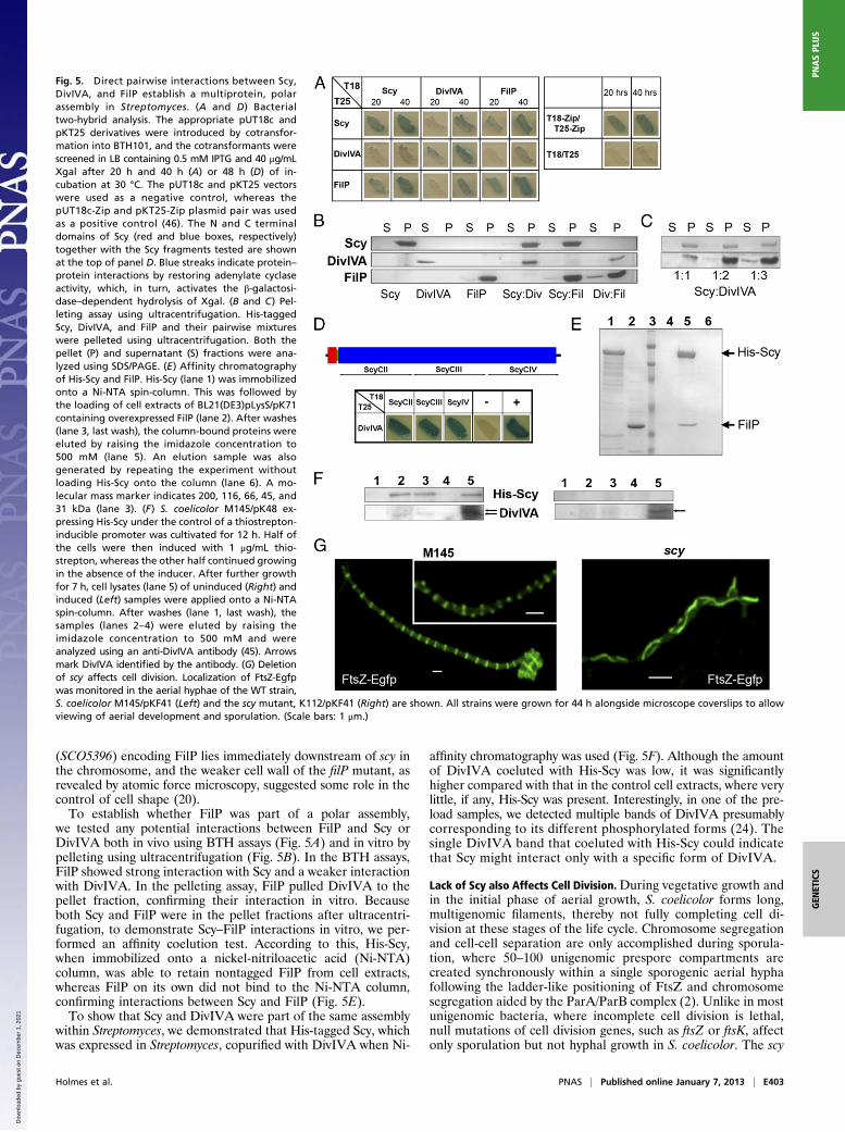
(SCO5396) encoding FilP lies immediately downstream of scy inthe chromosome, and the weaker cell wall of the filP mutant, asrevealed by atomic force microscopy, suggested some role in thecontrol of cell shape (20).To establish whether FilP was part of a polar assembly,
we tested any potential interactions between FilP and Scy orDivIVA both in vivo using BTH assays (Fig. 5A) and in vitro bypelleting using ultracentrifugation (Fig. 5B). In the BTH assays,FilP showed strong interaction with Scy and a weaker interactionwith DivIVA. In the pelleting assay, FilP pulled DivIVA to thepellet fraction, confirming their interaction in vitro. Becauseboth Scy and FilP were in the pellet fractions after ultracentri-fugation, to demonstrate Scy–FilP interactions in vitro, we per-formed an affinity coelution test. According to this, His-Scy,when immobilized onto a nickel-nitriloacetic acid (Ni-NTA)column, was able to retain nontagged FilP from cell extracts,whereas FilP on its own did not bind to the Ni-NTA column,confirming interactions between Scy and FilP (Fig. 5E).To show that Scy and DivIVA were part of the same assembly
within Streptomyces, we demonstrated that His-tagged Scy, whichwas expressed in Streptomyces, copurified with DivIVA when Ni-
affinity chromatography was used (Fig. 5F). Although the amountof DivIVA coeluted with His-Scy was low, it was significantlyhigher compared with that in the control cell extracts, where verylittle, if any, His-Scy was present. Interestingly, in one of the pre-load samples, we detected multiple bands of DivIVA presumablycorresponding to its different phosphorylated forms (24). Thesingle DivIVA band that coeluted with His-Scy could indicatethat Scy might interact only with a specific form of DivIVA.
Lack of Scy also Affects Cell Division.During vegetative growth andin the initial phase of aerial growth, S. coelicolor forms long,multigenomic filaments, thereby not fully completing cell di-vision at these stages of the life cycle. Chromosome segregationand cell-cell separation are only accomplished during sporula-tion, where 50–100 unigenomic prespore compartments arecreated synchronously within a single sporogenic aerial hyphafollowing the ladder-like positioning of FtsZ and chromosomesegregation aided by the ParA/ParB complex (2). Unlike in mostunigenomic bacteria, where incomplete cell division is lethal,null mutations of cell division genes, such as ftsZ or ftsK, affectonly sporulation but not hyphal growth in S. coelicolor. The scy
Fig. 5. Direct pairwise interactions between Scy,DivIVA, and FilP establish a multiprotein, polarassembly in Streptomyces. (A and D) Bacterialtwo-hybrid analysis. The appropriate pUT18c andpKT25 derivatives were introduced by cotransfor-mation into BTH101, and the cotransformants werescreened in LB containing 0.5 mM IPTG and 40 μg/mLXgal after 20 h and 40 h (A) or 48 h (D) of in-cubation at 30 °C. The pUT18c and pKT25 vectorswere used as a negative control, whereas thepUT18c-Zip and pKT25-Zip plasmid pair was usedas a positive control (46). The N and C terminaldomains of Scy (red and blue boxes, respectively)together with the Scy fragments tested are shownat the top of panel D. Blue streaks indicate protein–protein interactions by restoring adenylate cyclaseactivity, which, in turn, activates the β-galactosi-dase–dependent hydrolysis of Xgal. (B and C) Pel-leting assay using ultracentrifugation. His-taggedScy, DivIVA, and FilP and their pairwise mixtureswere pelleted using ultracentrifugation. Both thepellet (P) and supernatant (S) fractions were ana-lyzed using SDS/PAGE. (E) Affinity chromatographyof His-Scy and FilP. His-Scy (lane 1) was immobilizedonto a Ni-NTA spin-column. This was followed bythe loading of cell extracts of BL21(DE3)pLysS/pK71containing overexpressed FilP (lane 2). After washes(lane 3, last wash), the column-bound proteins wereeluted by raising the imidazole concentration to500 mM (lane 5). An elution sample was alsogenerated by repeating the experiment withoutloading His-Scy onto the column (lane 6). A mo-lecular mass marker indicates 200, 116, 66, 45, and31 kDa (lane 3). (F) S. coelicolor M145/pK48 ex-pressing His-Scy under the control of a thiostrepton-inducible promoter was cultivated for 12 h. Half ofthe cells were then induced with 1 μg/mL thio-strepton, whereas the other half continued growingin the absence of the inducer. After further growthfor 7 h, cell lysates (lane 5) of uninduced (Right) andinduced (Left) samples were applied onto a Ni-NTAspin-column. After washes (lane 1, last wash), thesamples (lanes 2–4) were eluted by raising theimidazole concentration to 500 mM and wereanalyzed using an anti-DivIVA antibody (45). Arrowsmark DivIVA identified by the antibody. (G) Deletionof scy affects cell division. Localization of FtsZ-Egfpwas monitored in the aerial hyphae of the WT strain,S. coelicolor M145/pKF41 (Left) and the scy mutant, K112/pKF41 (Right) are shown. All strains were grown for 44 h alongside microscope coverslips to allowviewing of aerial development and sporulation. (Scale bars: 1 μm.)
Holmes et al. PNAS | Published online January 7, 2013 | E403
GEN
ETICS
PNASPL
US
Dow
nloa
ded
by g
uest
on
Dec
embe
r 1,
202
1
mutant produced significantly fewer spores (10%) than the WT,and the irregularly spaced and often tilted sporulation septationand the uneven chromosome distribution in the spore chainssuggested that cell shape and cell division were both affected incells lacking Scy (Fig. 2 and Fig. S2). To test the effect of Scy oncell division, we monitored FtsZ localization in both WT and scymutant strains by introducing pKF41 (25) carrying ftsZ-egfp. Inthe WT strain, FtsZ-Egfp rings were easily detectable and werepositioned as in a ladder, marking the sites for future sporula-tion septation (Fig. 5G). However, in K112/pKF41, there werevery few aerial hyphae, with short, unsynchronized stretches ofnoticeable but very faint FtsZ-Egfp rings. Instead, strong FtsZ-Egfp cables were detected in the majority (∼80%) of the aerialhyphae of K112/pKF41, which did not show sporulation septa-tion and where, instead of discrete and orderly FtsZ rings, long,loosely curved FtsZ cables run alongside and often parallel tothe lateral walls of the hyphae (Fig. 5G).
DiscussionScy Is a Long Coiled-Coil Protein with Multiple Partners. Scy is un-usually long, and it is dominated by a novel repeat that appearsto form a continuous coiled coil, constituting >90% of the wholesequence. If indeed this is an uninterrupted single domain, itslength exceeds those of any previously characterized bacterialor archaeal coiled-coil domain (26), although it is slightly shorterthan two eukaryotic proteins, the spindle pole component NuMA(1,500 amino acids) and the Golgi-localized Giantin (3,150 aminoacids). The most notable feature of Scy is the nature of the coiled-coil repeat in this long domain, which is compatible with a 51-merrepeat that can be interpreted as a hendecad (11-mer) repeatmodified by an extra seven residues after every four repeats. Al-though there are cases of highly periodically modified heptadrepeats (27), we are unaware of such a regular modification ofhendecad repeats in any other protein.Although no structural studies are available, Scy was suggested
to form parallel homodimer coiled coils (22). These homodimersare likely to form higher order assemblies, because soluble Scypreparations pelleted when exposed to ultracentrifugation (Fig.5B). We have shown that Scy directly interacted with the fila-ment-forming protein FilP and the polarity marker DivIVA invitro, and we have shown that Scy and DivIVA colocalized invivo at newly established and growing tips. Both the BTH assay(Fig. 5D) and the pelleting assay (Fig. 5C) confirmed that Scy canbind multiple DivIVAs throughout its long C-terminal domain.This suggests that Scy can accommodate, sequester, and organizeseveral of its partner proteins.
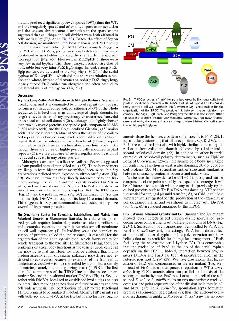
Tip Organizing Center for Selecting, Establishing, and MaintainingPolarized Growth in Filamentous Bacteria. In eukaryotes, polar-ized growth requires landmark proteins to select specific sitesand a complex assembly that recruits vesicles for cell membraneor cell wall expansion (1). In budding yeast, the complex as-sembly of proteins, called the “polarisome,” is essential for theorganization of the actin cytoskeleton, which forms cables forvesicle transport to the bud site. In filamentous fungi, the Spit-zenkörper or apical body functions as the vesicle supply center atthe growing hyphal tip. Here, we provide evidence that multi-protein assemblies for organizing polarized growth are not re-stricted to eukaryotes, because tip extension of the filamentousbacterium S. coelicolor is also controlled by a complex assemblyof proteins, namely, the tip organizing center (TIPOC). The firstidentifed components of the TIPOC include the molecular or-ganizer Scy and the positional marker DivIVA (Fig. 6). Scy, to-gether with DivIVA, localized to established hyphal tips and alsoto lateral sites marking the positions of future branches and newcell wall synthesis. The contribution of FilP to the functionalTIPOC remains to be analyzed in detail. Clearly, FilP can interactwith both Scy and DivIVA at the tip, but it also forms strong fil-
aments along the hyphae, a pattern so far specific to FilP (20). Itis particularly interesting that all three proteins, Scy, DivIVA, andFilP, are coiled-coil proteins with highly similar domain organi-zation: a short coiled-coil domain, followed by a linker and asecond coiled-coil domain (22). In addition to other bacterialexamples of coiled-coil polarity determinants, such as TipN orPopZ of C. crescentus (28–32), the spindle pole body, specializedfor centromere function in yeast, is also dominated by long coiled-coil proteins (33, 34), suggesting further structural similaritiesbetween organizing centers in bacteria and eukaryotes.We believe that the evidence for a TIPOC is strong, and further
components of the polar assembly are expected to emerge. It willbe of interest to establish whether any of the previously tip-lo-calized proteins, such as TraB, a DNA-translocating ATPase thatis essential for conjugal plasmid transfer (35), or CslA, a cellulosesynthase that is suggested for the production of the extracellularpolysaccharide matrix and was shown to interact with DivIVA(36) (Fig. 6), are indeed organized by the TIPOC.
Link Between Polarized Growth and Cell Division? The scy mutantshowed severe defects in cell division during sporulation, pro-ducing spore compartments with different amounts of DNA (Fig.2 D–G). Segregation of chromosomes is controlled by ParA andParB in S. coelicolor and, interestingly, ParA forms distinct fociat the tips of the aerial hyphae before polymerization into ParAhelices that act as scaffolds for the regular arrangement of ParBfoci along the sporogenic aerial hyphae (37). It is conceivablethat the nucleation of ParA at the tip of the aerial hyphaedepends on the TIPOC. Indeed, interaction between Strepto-myces DivIVA and ParB has been demonstrated, albeit in theheterologous host E. coli (38). We have also shown that locali-zation of FtsZ was compromised in the scy mutant (Fig. 5G).Instead of FtsZ ladders that precede septation in WT S. coeli-color, long FtsZ filaments often run parallel to the axis of thesporogenic aerial hyphae. FtsZ positioning at midcell of the rod-shaped E. coli or B. subtilis relies on two mechanisms: nucleoidocclusion and polar sequestration of the division inhibitors, MinDand MinC (17). In S. coelicolor, sporulation septa formationprecedes chromosome segregation (2); thus, the nucleoid occlu-sion mechanism is unlikely. Moreover, S. coelicolor has no obvi-
Fig. 6. TIPOC serves as a “hub” for polarized growth. The long, coiled-coilprotein Scy directly interacts with DivIVA and FilP at hyphal tips. DivIVA di-rectly controls cell wall synthesis (PBP), whereas Scy is responsible for theorganization of the TIPOC. The possible link between the cell division ma-chinery (FtsZ, SsgA, SsgB, ParA, and ParB) and the TIPOC is also shown. Othertip-localized proteins include CslA (cellulose synthase), TraB (DNA translo-case) and AfsK, the kinase that can phosphorylate DivIVA. CM, cell mem-brane; PG, peptidoglycan.
E404 | www.pnas.org/cgi/doi/10.1073/pnas.1210657110 Holmes et al.
Dow
nloa
ded
by g
uest
on
Dec
embe
r 1,
202
1
ous MinC or MinE homologs. Recently, FtsZ positioning in S.coelicolor was shown to be under positive control by an FtsZpartner protein, SsgB, which, in turn, depends on SsgA (39). In-terestingly, SsgA localizes at the hyphal tips in a dynamic fashion(40); therefore, it could, in principle, be part of the TIPOC (Fig.6). Although it is very attractive to propose a direct link betweenthe TIPOC and cytokinesis, we cannot rule out an indirect linkbecause the severe defect of the hyphal geometry of the scy mu-tant might affect spatial cues for the formation of the FtsZ ladder.
Scy Is Important for the Assembly and Integrity of the TIPOC. Al-though the roles of DivIVA and Scy are very closely linked, wecan distinguish their individual contribution to establishing po-larity in Streptomyces. DivIVA is essential for branching andpolarized growth, and it directly recruits the cell wall syntheticmachinery. For the latter, we have only indirect, although strong,evidence. First, direct interaction between DivIVA and PBP3was shown in another actinomycete, M. tuberculosis (16). Fur-thermore, during DivIVA overproduction, isotropic tip expan-sion was observed (8) (Fig. S4D), which was inhibited bybacitracin, a cell wall synthesis inhibitor, demonstrating that theballooning tip expansion was dependent on active cell wall syn-thesis (14). Scy overproduction did not lead to similar ballooningtips but, instead, generated copious branching followed by ahook-on-hook hyphal growth pattern in which only selected tipswere actively growing (Fig. 4). This suggested that although Scycontrolled the number of tips established, it did not directly re-cruit the cell wall synthetic enzymes. Also, deletion of scy did notabolish tip formation. On the contrary, it promoted over-branching; thus, Scy is not essential for tip formation, per se.Identifying the mechanism for DivIVA positioning and orga-
nization is the key to establishing a new polarity center in Strep-tomyces. Special compositional cues of the membrane might actas site-specific markers. Anionic phospholipids localized to tipsand branchpoints in S. coelicolor, and changing the levels ofthese phospholipids altered branching and hyphal geometry (41).In B. subtilis, membrane composition was suggested to affectthe localization of transmembrane proteins that stabilize theMreB cables (15). More importantly, a geometrical cue, negativemembrane curvature, was shown to recruit DivIVA to the polesand division sites in the rod-shaped B. subtilis (18, 19). However,unlike the limited sites of convex membrane curvatures in B.subtilis, the long and often meandering Streptomyces hyphae havenumerous convex curvatures that could, in principle, target DivIVAto these sites. Although most of the new branch points appear tobe from convex hyphal curvatures (14), there are many morenegative hyphal curvatures than emerging tips; thus, althoughmembrane curvature might facilitate DivIVA recruitment,Streptomyces must have a mechanism to restrict DivIVA to se-lected sites only. We propose that it is the Scy assembly, which, bysequestering multiple DivIVA tetramers (42), regulates thenumber of DivIVA foci, and therefore controls the number ofpolarity centers in Streptomyces.Lack of Scy produces multiple polarity centers, suggesting that
Scy assemblies can limit the number of TIPOCs established,perhaps by organizing the otherwise more dispersed DivIVAassemblies into a limited number of polarity centers. Over-production of both Scy and DivIVA also results in an increasingnumber of new polarity centers, confirming that the cellularlevels of the TIPOC components can control the assembly of theTIPOC and de novo tip emergence. On the other hand, thedispersal of the DivIVA patches at existing tips of the scy mutantsuggests that Scy is important not only for the assembly of theTIPOC but for the integrity of existing, actively growing tips.Hence, lack of Scy will destabilize the TIPOC, leading to thedispersal of DivIVA patches followed by the emergence of newtips. Recently, it was shown that new branches can be initiatedby DivIVA complexes breaking off from existing tips and initi-
ating new tip emergence. Mathematical modeling supported themechanism whereby increasing DivIVA levels at already estab-lished and growing tips result in tip focus splitting (43). Similarly,when Scy is overproduced in the presence of unchanging DivIVAlevels, the hook-on-hook growth pattern is compatible with thedispersal of excess TIPOC components, in this case Scy, gener-ating Scy-containing but not actively growing hook tips, togetherwith Scy-DivIVA assemblies at the growing tip (Fig. 4 J–L). In-terestingly, DivIVA phosphorylation at the hyphal tip by AfsK,a Ser/Thr kinase, was shown to affect branching in Streptomyces(24). Therefore, it will be of special interest to establish in thefuture how AfsK is recruited to the tips and what the effect ofDivIVA phosphorylation is on Scy-DivIVA interaction and theintegrity of the TIPOC.The suggested mechanism for controlling the assembly and
integrity of a bacterial polarity center by the sequestration andorganization of a multiprotein polarity complex, the TIPOC, isconsistent with our experimental findings, and it reconciles theapparent paradox concerning the nucleation of multiple polaritycenters both in the absence and excess of a polarity determinant.We propose that the TIPOC, including Scy, serves not only asa “hub” for polarized growth but, indirectly or directly, for co-ordinating cell division during sporulation in Streptomyces.
Materials and MethodsBacterial Strains and Growth Conditions. All bacterial strains, plasmids, andoligonucleotides used in this work are listed in Tables S1–S3, respectively. Gen-eration of KO mutants and all plasmid constructions is detailed in SI MaterialsandMethods. Plasmids were introduced into S. coelicolor using conjugation viaE. coli ET12567/pUZ8002, with the exception of pKF59, which was introducedinto S. coelicolor using protoplast transformation (44). Streptomyces strainswere grown at 30 °C on soya flour medium (SFM) containing 1%mannitol (44).
Overexpression and Purification of Scy, DivIVA, and FilP. Plasmid pGS2 wasintroduced to E. coli BL21 (DE3)pLysS (Novagen). Cultures of BL21(DE3)pLysS/pGS2 were grown at 37 °C for 3 h and, after induction with 1 mM isopropyl-β-D-thiogalactopyranoside (IPTG), for a further 4 h. After harvesting themycelium, His-Scy was purified from the supernatant of cell lysates in bufferA: 50 mM NaH2PO4, 500 mM NaCl, and 10 mM imidazole under nativeconditions using a HisTrap HP 1-mL affinity column (GE Healthcare). Afterwashing with buffer A containing 30 mM imidazole, His-Scy was eluted witha linear gradient of 30–300 mM imidazole in buffer A. His-Scy fractions weredialyzed against 20 mM Tris·HCl (pH 8) and further purified using a MonoQ5/50 anion exchange column (GE Healthcare). Elution was carried out usinga linear gradient of 0.01–1 M NaCl in 20 mM Tris·HCl (pH 8.0). Fractionscontaining His-Scy were dialyzed against 10 mM NaH2PO4 (pH 8.0) and wereused for CD spectroscopy.
For the ultracentrifugation and affinity coelution studies, His-Scy, His-DivIVA, and His-FilP were purified with a simplified version of the above.Accordingly, cultures of BL21(DE3)pLysS carrying pGS2, pK68, and pK70 weregrown at 37 °C for 3 h and, after induction with 1 mM IPTG, for a further 4 h.The His-tagged proteins were eluted with a step elution of 300 mM imidazolein buffer A, and the fractions were dialyzed either against 20 mM Tris·HCl and200 mM NaCl (pH 8) for the ultracentrifugation or against 50 mM NaH2PO4,300 mM NaCl, and 10 mM imidazole for the affinity coelution studies.
For the ultracentrifugation assays, the purified proteins were measuredusing a Bradford assay (Bio-Rad), and ∼1-μMproteins were incubated at 30 °Cfor 20 min and spun at 100,000 × g for 30 min at 4 °C. The supernatant andthe pellet were analyzed on 10% (wt/vol) PAGE.
Affinity Coelution of FilP with His-Scy. Purified His-Scy was immobilized ontoa Ni-NTA spin-column (Qiagen) and was exposed to cell extracts from BL21(DE3)pLysS/pK71 containing overexpressed FilP (nontagged) in a buffer (50mM NaH2PO4, 300 mM NaCl, 20 mM MgCl2, and 50 mM imidazole) opti-mized to avoid binding of nontagged proteins to the column. After severalwashes, His-Scy was eluted using 500 mM imidazole and the eluted sampleswere analyzed on 10% (wt/vol) PAGE. As a control, the experiment wasrepeated without loading His-Scy onto the Ni-NTA column to confirm thatFilP on its own did not bind to the column.
Affinity Coelution of DivIVA with His-Scy from Streptomyces. S. coelicolorM145/pK48 expressing His-Scy under the control of a thiostrepton-inducible
Holmes et al. PNAS | Published online January 7, 2013 | E405
GEN
ETICS
PNASPL
US
Dow
nloa
ded
by g
uest
on
Dec
embe
r 1,
202
1
promoter was cultivated for 12 h. Half of the samples were then inducedusing a low level of 1 μg/mL thiostrepton, whereas the other half continuedgrowing in the absence of the inducer. After further growth for 7 h, celllysates of uninduced and induced samples were applied onto a Ni-NTA spin-column in a buffer of 50 mM NaH2PO4 and 300 mM NaCl. After severalwashes, His-Scy was eluted by raising the imidazole concentration to 500mM and the samples were analyzed on 8% (wt/vol) PAGE. Western blotanalysis utilizing anti-DivIVA primary antibody (45) raised in rabbit diluted at1:10,000 and a secondary antibody of anti-rabbit IgG-HRP conjugate (DAKO)diluted at 1:5,000 was used to confirm that DivIVA copurified with His-Scyfrom Streptomyces cell extracts.
Microscopy. Light microscopy. For laser-scanning confocal microscopy and forstatistical analysis, spores were inoculated on the surface of cellophanemembranes positioned on solid SFMmedium to restrict hyphal growth in twodimensions when hyphal characteristics were monitored. When the purposeof the study was aerial development and sporulation, spores were inoculatedalongside microscope coverslips inserted at angles of 45° into SFM medium.Cells on both cellophane membrane and coverslips were fixed with ice-coldmethanol for 1 min before further staining with WGA-Alexa488 (50 μg/mL;Molecular Probes) and/or propidium iodide (25 μg/mL; Sigma) for 20 min.After several washes with water, samples were mounted using 20% (vol/vol)glycerol and viewed using a Leica TCS SP2 laser-scanning confocal microscopewith a 63×, 1.4-N.A. oil immersion objective. Staining of nascent peptidoglycan
was performed by staining the growing cells with 1 μg/mL BODIPY Van FL(Molecular Probes) and 1 μg/mL unlabeled vancomycin (Sigma) for 5 min.TEM. Colonies were fixed with 2% (vol/vol) glutaraldehyde in 0.05 M sodiumcacodylate (pH 7.0), stained with osmic acid, and embedded in LRWhite resin(The London Resin Co.) according to the manufacturer’s instructions. Aftersectioning, the samples were examined in a Jeol 1200 EX transmissionelectron microscope at 80 KV and tagged image file format (TIFF) files weregenerated using a Deben AMT 1.3k camera.
Protein samples of His-Scy in 20 mM Tris and 150 mM NaCl (pH 8.0) werenegatively stained using freshly prepared 2% ammoniummolybdate (pH 8.0)on a 400-mesh copper grid with a carbon-coated pyroxylin support film. Thegrids were viewed at 200 kV in a Tecnai 20 transmission electron microscope(FEI) fitted with an AMT XR60B digital camera (Deben), and digital TIFFimages were saved.
ACKNOWLEDGMENTS. We thank Kim C. Findlay and Richard Ewans-Gowingfor assistance with the TEM and SEM; George Smith, Laura Kerry, and JamesHunter for generating some of the constructs; and Ruchita Desai fortechnical assistance. We also thank Bartek Ditkowski and Dagmara Jakimo-wicz for sharing some of their constructs prior to publication and Prof.Richard Morris for stimulating discussions. We are very grateful to Prof. SirDavid Hopwood, Prof. Andy Johnston, and Dr. Margaret Wexler for theirvaluable comments on the manuscript. This work was supported by RoyalSociety Research Grant 23646 (to G.H.K.), the John and Pamela SaltersFoundation (G.H.K.), and Biotechnology and Biological Sciences ResearchCouncil Studentship BB/D526196/1 (to N.A.H.).
1. Nelson WJ (2003) Adaptation of core mechanisms to generate cell polarity. Nature422(6933):766–774.
2. Flärdh K, Buttner MJ (2009) Streptomyces morphogenetics: Dissecting differentiationin a filamentous bacterium. Nat Rev Microbiol 7(1):36–49.
3. Cabeen MT, Jacobs-Wagner C (2010) The bacterial cytoskeleton. Annu Rev Genet 44:365–392.
4. Pinho MG, Errington J (2003) Dispersed mode of Staphylococcus aureus cell wallsynthesis in the absence of the division machinery. Mol Microbiol 50(3):871–881.
5. Ausmees N, Kuhn JR, Jacobs-Wagner C (2003) The bacterial cytoskeleton: Anintermediate filament-like function in cell shape. Cell 115(6):705–713.
6. Charbon G, Cabeen MT, Jacobs-Wagner C (2009) Bacterial intermediate filaments: Invivo assembly, organization, and dynamics of crescentin. Genes Dev 23(9):1131–1144.
7. Daniel RA, Errington J (2003) Control of cell morphogenesis in bacteria: Two distinctways to make a rod-shaped cell. Cell 113(6):767–776.
8. Flärdh K (2003) Essential role of DivIVA in polar growth and morphogenesis inStreptomyces coelicolor A3(2). Mol Microbiol 49(6):1523–1536.
9. Mazza P, et al. (2006) MreB of Streptomyces coelicolor is not essential for vegetativegrowth but is required for the integrity of aerial hyphae and spores. Mol Microbiol60(4):838–852.
10. Heichlinger A, et al. (2011) The MreB-like protein Mbl of Streptomyces coelicolor A3(2) depends on MreB for proper localization and contributes to spore wall synthesis.J Bacteriol 193(7):1533–1542.
11. Kleinschnitz EM, et al. (2011) Proteins encoded by the mre gene cluster inStreptomyces coelicolor A3(2) cooperate in spore wall synthesis. Mol Microbiol 79(5):1367–1379.
12. McCormick JR, Su EP, Driks A, Losick R (1994) Growth and viability of Streptomycescoelicolor mutant for the cell division gene ftsZ. Mol Microbiol 14(2):243–254.
13. Flärdh K (2010) Cell polarity and the control of apical growth in Streptomyces. CurrOpin Microbiol 13(6):758–765.
14. Hempel AM, Wang SB, Letek M, Gil JA, Flärdh K (2008) Assemblies of DivIVA marksites for hyphal branching and can establish new zones of cell wall growth inStreptomyces coelicolor. J Bacteriol 190(22):7579–7583.
15. Margolin W (2009) Sculpting the bacterial cell. Curr Biol 19(17):812–822.16. Mukherjee P, et al. (2009) Novel role of Wag31 in protection of mycobacteria under
oxidative stress. Mol Microbiol 73(1):103–119.17. Wu LJ, Errington J (2012) Nucleoid occlusion and bacterial cell division. Nat Rev
Microbiol 10(1):8–12.18. Ramamurthi KS, Losick R (2009) Negative membrane curvature as a cue for subcellular
localization of a bacterial protein. Proc Natl Acad Sci USA 106(32):13541–13545.19. Lenarcic R, et al. (2009) Localisation of DivIVA by targeting to negatively curved
membranes. EMBO J 28(15):2272–2282.20. Bagchi S, Tomenius H, Belova LM, Ausmees N (2008) Intermediate filament-like proteins
in bacteria and a cytoskeletal function in Streptomyces. Mol Microbiol 70(4):1037–1050.21. Lupas A, Van Dyke M, Stock J (1991) Predicting coiled coils from protein sequences.
Science 252(5009):1162–1164.22. Walshaw J, Gillespie MD, Kelemen GH (2010) A novel coiled-coil repeat variant in
a class of bacterial cytoskeletal proteins. J Struct Biol 170(2):202–215.23. Moseley JB, Goode BL (2006) The yeast actin cytoskeleton: From cellular function to
biochemical mechanism. Microbiol Mol Biol Rev 70(3):605–645.24. Hempel AM, et al. (2012) The Ser/Thr protein kinase AfsK regulates polar growth and
hyphal branching in the filamentous bacteria Streptomyces. Proc Natl Acad Sci USA109(35):2371–2379.
25. Grantcharova N, Lustig U, Flärdh K (2005) Dynamics of FtsZ assembly during sporulationin Streptomyces coelicolor A3(2). J Bacteriol 187(9):3227–3237.
26. Rose A, Schraegle SJ, Stahlberg EA, Meier I (2005) Coiled-coil protein composition of22 proteomes—Differences and common themes in subcellular infrastructure andtraffic control. BMC Evol Biol 5:66.
27. Lupas AN, Gruber M (2005) The structure of alpha-helical coiled coils. Adv ProteinChem 70:37–78.
28. Huitema E, Pritchard S, Matteson D, Radhakrishnan SK, Viollier PH (2006) Bacterialbirth scar proteins mark future flagellum assembly site. Cell 124(5):1025–1037.
29. Lam H, Schofield WB, Jacobs-Wagner C (2006) A landmark protein essential forestablishing and perpetuating the polarity of a bacterial cell. Cell 124(5):1011–1023.
30. Bowman GR, et al. (2008) A polymeric protein anchors the chromosomal origin/ParBcomplex at a bacterial cell pole. Cell 134(6):945–955.
31. Ebersbach G, Briegel A, Jensen GJ, Jacobs-Wagner C (2008) A self-associating proteincritical for chromosome attachment, division, and polar organization in Caulobacter.Cell 134(6):956–968.
32. Bowman GR, et al. (2010) Caulobacter PopZ forms a polar subdomain dictatingsequential changes in pole composition and function. Mol Microbiol 76(1):173–189.
33. Zizlsperger N, Malashkevich VN, Pillay S, Keating AE (2008) Analysis of coiled-coilinteractions between core proteins of the spindle pole body. Biochemistry 47(45):11858–11868.
34. Zizlsperger N, Keating AE (2010) Specific coiled-coil interactions contribute to a globalmodel of the structure of the spindle pole body. J Struct Biol 170(2):246–256.
35. Reuther J, Gekeler C, Tiffert Y, Wohlleben W, Muth G (2006) Unique conjugationmechanism inmycelial streptomycetes:ADNA-bindingATPase translocates unprocessedplasmid DNA at the hyphal tip. Mol Microbiol 61(2):436–446.
36. Xu H, Chater KF, Deng Z, Tao M (2008) A cellulose synthase-like protein involved inhyphal tip growth and morphological differentiation in Streptomyces. J Bacteriol190(14):4971–4978.
37. Jakimowicz D, Zydek P, Kois A, Zakrzewska-Czerwi�nska J, Chater KF (2007) Alignmentof multiple chromosomes along helical ParA scaffolding in sporulating Streptomyceshyphae. Mol Microbiol 65(3):625–641.
38. Donovan C, Sieger B, Krämer R, Bramkamp M (2012) A synthetic Escherichia colisystem identifies a conserved origin tethering factor in Actinobacteria. Mol Microbiol84(1):105–116.
39. Willemse J, Borst JW, de Waal E, Bisseling T, van Wezel GP (2011) Positive control ofcell division: FtsZ is recruited by SsgB during sporulation of Streptomyces. Genes Dev25(1):89–99.
40. Noens EE, et al. (2007) Loss of the controlled localization of growth stage-specific cell-wall synthesis pleiotropically affects developmental gene expression in an ssgAmutant of Streptomyces coelicolor. Mol Microbiol 64(5):1244–1259.
41. Jyothikumar V, et al. (2012) Cardiolipin synthase is required for Streptomyces coelicolormorphogenesis. Mol Microbiol 84(1):181–197.
42. Oliva MA, et al. (2010) Features critical for membrane binding revealed by DivIVAcrystal structure. EMBO J 29(12):1988–2001.
43. Richards DM, Hempel AM, Flärdh K, Buttner MJ, Howard M (2012) Mechanistic basisof branch-site selection in filamentous bacteria. PLOS Comput Biol 8(3):e1002423.
44. Kieser T, Bibb MJ, Buttner MJ, Chater KF, Hopwood DA (2000) Practical StreptomycesGenetics (The John Innes Foundation, Norwich, UK).
45. Wang SB, et al. (2009) Domains involved in the in vivo function and oligomerizationof apical growth determinant DivIVA in Streptomyces coelicolor. FEMS Microbiol Lett297(1):101–109.
46. Karimova G, Pidoux J, Ullmann A, Ladant D (1998) A bacterial two-hybrid systembased on a reconstituted signal transduction pathway. Proc Natl Acad Sci USA 95(10):5752–5756.
E406 | www.pnas.org/cgi/doi/10.1073/pnas.1210657110 Holmes et al.
Dow
nloa
ded
by g
uest
on
Dec
embe
r 1,
202
1