Coiled-coil 1-mediated fastening of the neck and motor ...coiled-coil segment associates with both...
10
Coiled-coil 1-mediated fastening of the neck and motor domains for kinesin-3 autoinhibition Jinqi Ren a,1 , Shuang Wang a,b,1 , Han Chen a,b , Wenjuan Wang a , Lin Huo a , and Wei Feng a,b,2 a National Laboratory of Biomacromolecules, CAS Center for Excellence in Biomacromolecules, Institute of Biophysics, Chinese Academy of Sciences, 100101 Beijing, China; and b College of Life Sciences, University of Chinese Academy of Sciences, 100049 Beijing, China Edited by Ronald D. Vale, Howard Hughes Medical Institute and Department of Cellular and Molecular Pharmacology, University of California, San Francisco, CA, and approved October 29, 2018 (received for review June 29, 2018) In kinesin-3, the coiled-coil 1 (CC1) can sequester the preceding neck coil (NC) for autoinhibition, but the underlying mechanism is poorly understood. Here, we determined the structures of the uninhibited motor domain (MD)-NC dimer and inhibited MD-NC- CC1 monomer of kinesin-3 KIF13B. In the MD-NC-CC1 monomer, CC1 is broken into two short helices that unexpectedly interact with both the NC and the MD. Compared with the MD-NC dimer, the CC1- mediated integration of NC and MD not only blocks the NC dimer formation, but also prevents the neck linker (NL) undocking and the ADP release from the MD. Mutations of the essential residues in the interdomain interaction interface in the MD-NC-CC1 monomer re- stored the MD activity. Thus, CC1 fastens the neck domain and MD and inhibits both NC and NL. This CC1-mediated lockdown of the entire neck domain may represent a paradigm for kinesin autoinhibi- tion that could be applicable to other kinesin-3 motors. intracellular transport | molecular motor | kinesin-3 | autoinhibition | coiled-coil K inesins are a superfamily of microtubule (MT)-based mo- lecular motors that can convert the chemical energy of ATP hydrolysis into the mechanical power to drive long-range in- tracellular transport or dictate MT dynamics (1–5). To transport cellular cargoes, in addition to the core motor domain (MD; referred to as the “head”) that can bind to MTs and undergoes nucleotide-dependent conformational changes, processive kine- sin motors (kinesin-1 to -3) require a characteristic neck domain [containing a neck linker (NL) and a neck coil (NC)] and a globular tail domain (2, 6). The neck domain forms a dimeric “neck” for assembling a functional “two-headed” motor and controlling the directionality and processivity, while the tail do- main is responsible for binding to cargoes or cargo adaptors (7– 9). However, in the absence of cargoes, most of these processive kinesin motors exist in an autoinhibited state to avoid futile ATP hydrolysis and the potential congestion of MT tracks (10). In kinesin-1 (the conventional kinesin), the compact autoinhibited conformation is predominantly mediated by its tail domain that folds back to directly interact with the MD (11, 12). This “tail-to-head” intramolecular interaction interferes with either the MT-binding capacity or the ATPase activity of the MD (or both) and keeps the motor in a folded inactive state (13). Similar tail-to-head intra- molecular interaction happens in kinesin-2 to maintain the motor in an autoinhibited conformation as well (14, 15). The tail domain- mediated autoinhibition of kinesin-1 and -2 would prevent their improper cargo loading and erroneous movement and thus ensure the precise spatial and temporal regulation of motor motility (10). Kinesin-3 (including KIF1A/B/C, KIF13A/B, and KIF16A/B in mammals) is a unique family of kinesin motors for in- tracellular transport with superprocessivity (2, 3, 16). In com- parison with other kinesins, kinesin-3 is characterized by a family-specific forkhead-associated (FHA) domain that is sand- wiched between two coiled-coils [coiled-coil 1 (CC1) and CC2] and, together, follows the neck domain (Fig. 1A). When not transporting cargoes, most of kinesin-3 motors also adopt an autoinhibited conformation (10). The founding member KIF1A was found to exist as a globular compact monomer that is largely inactive (only with a capacity of slow diffusive motion) and needs the cargo-mediated dimerization for fast processive movement (17–19). The monomeric autoinhibited conformation of KIF1A is partly contributed by the intramolecular interaction between CC2 and the FHA domain that led to the decreased propensity of dimerization and the inhibition of MT binding (20). More- over, in KIF13B and KIF16B, a central stalk region immediately following CC2 was found to directly interact with the MD (21, 22), and this “stalk-to-head” intramolecular interaction prevents the binding of MTs and keeps the motor in a folded inactive conformation. In addition to the inactivation of the MD, the neck domain of kinesin-3 is also inhibited by following CC1. The earlier studies of UNC-104 (KIF1A homolog in Caenorhabditis elegans) demonstrated that CC1 can fold back to associate with the NC to form a compact self-folded helix bundle that seques- ters the NC and prevents its dimerization (23). The intra- molecular interaction between the NC and CC1 was suggested to be able to control the monomer–dimer transition of KIF1A/ UNC-104 by switching of the NC between the intramolecular monomer and intermolecular dimer (16, 24). More intriguingly, the CC1-mediated inhibition of the NC was also found in Significance Kinesins are microtubule-based molecular motors that can drive long-range intracellular transport. When not transporting cargoes, processive kinesin motors often adopt an auto- inhibited state. The autoinhibited conformation of kinesin-3 is predominantly mediated by a coiled-coil segment that follows the neck and motor domains. In this study, we found that this coiled-coil segment associates with both the neck and motor domains and fastens them together. With the aid of the motor domain (or, namely, head), the autoinhibitory coiled-coil seg- ment tightly locks down the entire neck domain and inhibits both the neck domain-mediated dimerization and the ADP re- lease from the motor head. This “head-aided lockdown of neck” mechanism mediated by the internal coiled-coil segment may represent a paradigm for kinesin autoinhibition. Author contributions: J.R., S.W., and W.F. designed research; J.R. and S.W. performed research; J.R., S.W., H.C., W.W., L.H., and W.F. analyzed data; and J.R. and W.F. wrote the paper. The authors declare no conflict of interest. This article is a PNAS Direct Submission. Published under the PNAS license. Data deposition: The atomic coordinates of the MD-NC dimer and the MD-NC-CC1-Y73C monomer have been deposited in the Protein Data Bank [PDB ID codes 6A1Z (MD-NC dimer) and 6A20 (MD-NC-CC1-Y73C monomer)]. See Commentary on page 12845. 1 J.R. and S.W. contributed equally to this work. 2 To whom correspondence should be addressed. Email: [email protected]. This article contains supporting information online at www.pnas.org/lookup/suppl/doi:10. 1073/pnas.1811209115/-/DCSupplemental. Published online November 21, 2018. www.pnas.org/cgi/doi/10.1073/pnas.1811209115 PNAS | vol. 115 | no. 51 | E11933–E11942 BIOPHYSICS AND COMPUTATIONAL BIOLOGY SEE COMMENTARY Downloaded by guest on March 12, 2020
Transcript of Coiled-coil 1-mediated fastening of the neck and motor ...coiled-coil segment associates with both...

Coiled-coil 1-mediated fastening of the neck and motordomains for kinesin-3 autoinhibitionJinqi Rena,1, Shuang Wanga,b,1, Han Chena,b, Wenjuan Wanga, Lin Huoa, and Wei Fenga,b,2
aNational Laboratory of Biomacromolecules, CAS Center for Excellence in Biomacromolecules, Institute of Biophysics, Chinese Academy of Sciences, 100101Beijing, China; and bCollege of Life Sciences, University of Chinese Academy of Sciences, 100049 Beijing, China
Edited by Ronald D. Vale, Howard Hughes Medical Institute and Department of Cellular and Molecular Pharmacology, University of California, San Francisco,CA, and approved October 29, 2018 (received for review June 29, 2018)
In kinesin-3, the coiled-coil 1 (CC1) can sequester the precedingneck coil (NC) for autoinhibition, but the underlying mechanism ispoorly understood. Here, we determined the structures of theuninhibited motor domain (MD)-NC dimer and inhibited MD-NC-CC1 monomer of kinesin-3 KIF13B. In the MD-NC-CC1 monomer,CC1 is broken into two short helices that unexpectedly interact withboth the NC and the MD. Compared with the MD-NC dimer, the CC1-mediated integration of NC and MD not only blocks the NC dimerformation, but also prevents the neck linker (NL) undocking and theADP release from the MD. Mutations of the essential residues in theinterdomain interaction interface in the MD-NC-CC1 monomer re-stored the MD activity. Thus, CC1 fastens the neck domain and MDand inhibits both NC and NL. This CC1-mediated lockdown of theentire neck domain may represent a paradigm for kinesin autoinhibi-tion that could be applicable to other kinesin-3 motors.
intracellular transport | molecular motor | kinesin-3 | autoinhibition |coiled-coil
Kinesins are a superfamily of microtubule (MT)-based mo-lecular motors that can convert the chemical energy of ATP
hydrolysis into the mechanical power to drive long-range in-tracellular transport or dictate MT dynamics (1–5). To transportcellular cargoes, in addition to the core motor domain (MD;referred to as the “head”) that can bind to MTs and undergoesnucleotide-dependent conformational changes, processive kine-sin motors (kinesin-1 to -3) require a characteristic neck domain[containing a neck linker (NL) and a neck coil (NC)] and aglobular tail domain (2, 6). The neck domain forms a dimeric“neck” for assembling a functional “two-headed” motor andcontrolling the directionality and processivity, while the tail do-main is responsible for binding to cargoes or cargo adaptors (7–9). However, in the absence of cargoes, most of these processivekinesin motors exist in an autoinhibited state to avoid futile ATPhydrolysis and the potential congestion of MT tracks (10). Inkinesin-1 (the conventional kinesin), the compact autoinhibitedconformation is predominantly mediated by its tail domain that foldsback to directly interact with the MD (11, 12). This “tail-to-head”intramolecular interaction interferes with either the MT-bindingcapacity or the ATPase activity of the MD (or both) and keepsthe motor in a folded inactive state (13). Similar tail-to-head intra-molecular interaction happens in kinesin-2 to maintain the motor inan autoinhibited conformation as well (14, 15). The tail domain-mediated autoinhibition of kinesin-1 and -2 would prevent theirimproper cargo loading and erroneous movement and thus ensurethe precise spatial and temporal regulation of motor motility (10).Kinesin-3 (including KIF1A/B/C, KIF13A/B, and KIF16A/B
in mammals) is a unique family of kinesin motors for in-tracellular transport with superprocessivity (2, 3, 16). In com-parison with other kinesins, kinesin-3 is characterized by afamily-specific forkhead-associated (FHA) domain that is sand-wiched between two coiled-coils [coiled-coil 1 (CC1) and CC2]and, together, follows the neck domain (Fig. 1A). When nottransporting cargoes, most of kinesin-3 motors also adopt anautoinhibited conformation (10). The founding member KIF1A
was found to exist as a globular compact monomer that is largelyinactive (only with a capacity of slow diffusive motion) and needsthe cargo-mediated dimerization for fast processive movement(17–19). The monomeric autoinhibited conformation of KIF1Ais partly contributed by the intramolecular interaction betweenCC2 and the FHA domain that led to the decreased propensityof dimerization and the inhibition of MT binding (20). More-over, in KIF13B and KIF16B, a central stalk region immediatelyfollowing CC2 was found to directly interact with the MD (21,22), and this “stalk-to-head” intramolecular interaction preventsthe binding of MTs and keeps the motor in a folded inactiveconformation. In addition to the inactivation of the MD, theneck domain of kinesin-3 is also inhibited by following CC1. Theearlier studies of UNC-104 (KIF1A homolog in Caenorhabditiselegans) demonstrated that CC1 can fold back to associate withthe NC to form a compact self-folded helix bundle that seques-ters the NC and prevents its dimerization (23). The intra-molecular interaction between the NC and CC1 was suggested tobe able to control the monomer–dimer transition of KIF1A/UNC-104 by switching of the NC between the intramolecularmonomer and intermolecular dimer (16, 24). More intriguingly,the CC1-mediated inhibition of the NC was also found in
Significance
Kinesins are microtubule-based molecular motors that candrive long-range intracellular transport. When not transportingcargoes, processive kinesin motors often adopt an auto-inhibited state. The autoinhibited conformation of kinesin-3 ispredominantly mediated by a coiled-coil segment that followsthe neck and motor domains. In this study, we found that thiscoiled-coil segment associates with both the neck and motordomains and fastens them together. With the aid of the motordomain (or, namely, head), the autoinhibitory coiled-coil seg-ment tightly locks down the entire neck domain and inhibitsboth the neck domain-mediated dimerization and the ADP re-lease from the motor head. This “head-aided lockdown ofneck” mechanism mediated by the internal coiled-coil segmentmay represent a paradigm for kinesin autoinhibition.
Author contributions: J.R., S.W., and W.F. designed research; J.R. and S.W. performedresearch; J.R., S.W., H.C., W.W., L.H., and W.F. analyzed data; and J.R. and W.F. wrotethe paper.
The authors declare no conflict of interest.
This article is a PNAS Direct Submission.
Published under the PNAS license.
Data deposition: The atomic coordinates of the MD-NC dimer and the MD-NC-CC1-Y73Cmonomer have been deposited in the Protein Data Bank [PDB ID codes 6A1Z (MD-NCdimer) and 6A20 (MD-NC-CC1-Y73C monomer)].
See Commentary on page 12845.1J.R. and S.W. contributed equally to this work.2To whom correspondence should be addressed. Email: [email protected].
This article contains supporting information online at www.pnas.org/lookup/suppl/doi:10.1073/pnas.1811209115/-/DCSupplemental.
Published online November 21, 2018.
www.pnas.org/cgi/doi/10.1073/pnas.1811209115 PNAS | vol. 115 | no. 51 | E11933–E11942
BIOPH
YSICSAND
COMPU
TATIONALBIOLO
GY
SEECO
MMEN
TARY
Dow
nloa
ded
by g
uest
on
Mar
ch 1
2, 2
020

KIF13A, KIF13B, and KIF16B, and the cargo-mediated di-merization of these motors resulted in their superprocessivemotions (16), supporting that the CC1-mediated autoinhibitionis highly conserved in kinesin-3 motors and plays a prominentrole in regulating the motor motility, although the mechanism ofinhibition remains poorly understood.In this study, we determined the structures of the uninhibited
MD-NC dimer and the inhibited MD-NC-CC1 monomer of thekinesin-3 motor KIF13B [that plays a prominent role in trans-porting neuronal cargoes and controlling endocytosis (25, 26)].In the MD-NC dimer, the NC forms a coiled-coil dimer for as-sembling an uninhibited two-headed motor. In the MD-NC-CC1 monomer, CC1 is broken into two short helices (CC1a andCC1b) that unexpectedly interact with both the NC and the MDto form a compact self-folded monomer. CC1a folds back topack with the NC in an antiparallel manner, and this NC-CC1atwo-helix bundle further leans against the MD to integrate thethree domains together and fix both the NC and NL, while CC1bdoes not contact with the NC, but anchors at a distinct site of theMD to stabilize the integrated monomer. The CC1-mediatedintegration of the NC and the MD not only inhibits the NC di-mer formation but also prevents the NL undocking and the ADPrelease from the MD. Mutations of the essential residues in theinterdomain interaction interface in the MD-NC-CC1 monomerrestored the MD activity. Thus, CC1 fastens the neck domainand MD and sequesters both the NC and NL for kinesin-3 autoinhibition.
ResultsBiochemical Characterization of the MD-Containing Fragments ofKIF13B. To investigate the mechanism underlying the CC1-mediated autoinhibition of kinesin-3, we initiated this workwith biochemically characterizing the various fragments ofKIF13B. We focused on the N-terminal MD-containing frag-ments with or without CC1 [i.e., MD-NL, MD-NC, and MD-NC-CC1 (Fig. 1A)] and characterized the oligomeric states of thesefragments by using the analytical gel-filtration and ultracentri-fugation analysis (Fig. 1 B and C). As expected, the MD (with theNL) predominantly exists in a monomeric state in solution (Fig.1 B and C). Extension of the MD to the NC induced di-merization of the MD (Fig. 1 B and C). However, the NC-mediated dimerization was blocked by further inclusion ofCC1, and the resulting MD-NC-CC1 tandem adopted a stablemonomeric state in solution (Fig. 1 B and C). Thus, consistentwith the previous studies of KIF13B (16), CC1 is an inhibitorysegment capable of interfering with the NC-mediated di-merization.In addition to the oligomeric states, we also performed the
MT-simulated ATPase assay to characterize the ATPase activi-ties of these KIF13B fragments. Without MTs, the basal ATPaseactivities of MD-NL, MD-NC, and MD-NC-CC1 are extremelylow (Fig. 1D), consistent with the established critical role of MTsin the ATP hydrolysis cycle of kinesin motors (27). Upon addingMTs, both the MD (with the NL) and the MD-NC tandem showsthe similar high ATPase activities (Fig. 1D), indicating that ex-tension of the MD to the NC has little impact on its ATPaseactivity. In contrast, the MD-NC-CC1 tandem shows the signif-icantly lower ATPase activity compared with the MD-NL and -NCtandems (Fig. 1D), suggesting that further inclusion of CC1 inhibitsthe MT-simulated ATPase activity of the MD. Taken together, allof the biochemical data demonstrated that CC1 prevents the NCdimer formation as well as the MT-simulated ATPase activity ofthe MD.
Structure of the Uninhibited MD-NC Dimer. The NC can mediatedimerization of the MD, and the MD-NC dimer is in an un-inhibited state (Fig. 1 B–D). To dissect the NC-mediated di-merization, we performed the crystal screening of the MD-NCtandem. After extensive trials, the diffraction-quality crystalswere obtained. The structure of the MD-NC tandem was solvedby the molecular-replacement method and refined to 2.6 Å (Fig.2 and SI Appendix, Table S1). Although one molecule was foundin the asymmetric unit of the crystal, there exists a twofoldsymmetry-related MD-NC dimer (Fig. 2 A and B). In the MD-NC dimer structure, the MD adopts a canonical kinesin-motorfold with eight β-strands (β1–β8) to form a central β-sheet that issurrounded by six α-helices (α1–α6), The NL adopts a linearconformation and docks onto the MD, and the NC forms acoiled-coil dimer that brings the two MDs together to assemblean uninhibited two-headed motor (Fig. 2 B and C). Although noextra nucleotides were added during crystallization, ADP andMg2+ were still found to reside in the nucleotide-binding pocketof the MD based on their well-traced electron densities (Fig. 2 Aand B).In the MD-NC dimer structure, the NC initiates from P363
that produces a characteristic turn-like structure capping at theN terminus of the coiled-coil dimer (Fig. 2D). Consistent withthe previous structural studies of the NC (28), the interhelicalpacking between the NC coiled-coil dimer (buried with a surfacearea of ∼1,390 Å2) is largely contributed by the hydrophobicresidues from the a and d sites of the NC [based on the heptadrepeat pattern (a–g) analysis]—that is, I367, I368, L371, V375,L378, and L382 form the hydrophobic packing core of the coiled-coil dimer (Fig. 2D). Moreover, the electrostatic interactions
Fig. 1. Biochemical characterization of the various MD-containing frag-ments of KIF13B. (A) Domain organization of kinesin-3 KIF13B. KIF13B con-tains an N-terminal MD, a neck domain (containing the NL and NC), a CC1-FHA-CC2 tandem followed by a MBS domain, and a C-terminal CAP-Glydomain. The residue numbers for each domain are indicated below thedomain organization. (B) Analytical gel-filtration analysis of the variousKIF13B fragments, including MD-NL (solid black lines), MD-NC (solid graylines), and MD-ND-CC1 (dotted lines). The elution volumes of molecular massmarkers are indicated at the top of the elution profile. mAU, milliabsorbanceunit. (C) Analytical ultracentrifugation characterization of the variousKIF13B fragments. The calculated molecular masses for MD-NL, MD-NC, andMD-NC-CC1 are indicated. (D) ATPase activities of the various KIF13B frag-ments in the presence (+) or the absence (−) of MTs. Data were normalized bythe MT-stimulated ATPase activity of KHC as 100%. Each bar represents themean value ± SD. NS, no significant differences. ***P < 0.001 (Student’s t test).
E11934 | www.pnas.org/cgi/doi/10.1073/pnas.1811209115 Ren et al.
Dow
nloa
ded
by g
uest
on
Mar
ch 1
2, 2
020

between E374 and R379 from the two NC helices further stabilizethe coiled-coil formation (Fig. 2D).We compared the structure of the uninhibited MD-NC dimer
of kinesin-3 with that of kinesin-1 [Protein Data Bank (PDB) IDcode 3KIN; in which the NL also docks onto the MD] (SI Ap-pendix, Fig. S1). From the overall structure, the MD-NC dimer ofkinesin-3 largely resembles that of kinesin-1 (SI Appendix, Fig.S1), indicating that the NC-mediated dimerization is a similarmechanism in processive kinesin motors. However, the distancebetween the two domains (both upon NL docking) is somewhatdifferent between the two dimer structures—that is, ∼70 Å inkinesin-3 vs. ∼90 Å in kinesin-1 (based on the distance between thenucleotide-binding pockets of the two MDs) (Fig. 2E and SI Ap-pendix, Fig. S1). In the MD-NC dimer of kinesin-3, the two MDsare related by a rotation of ∼180° rather than ∼120° observed inthat of kinesin-1 (SI Appendix, Fig. S1). Upon superimposition ofone subunit in the dimer, the other one of kinesin-3 undergoes arotation of ∼60° and moves closer to the superimposed one (Fig.2E). These structural differences between the two MD-NC di-mers of kinesin-1/-3 are likely caused by the different lengths ofNL that link the two MDs through the NC coiled-coil dimer.Taken together, the structure of the MD-NC dimer of KIF13Bdemonstrated the NC-mediated dimerization of the MD andrevealed the potential active conformation of kinesin-3 motors.
Crystallization of the Inhibited MD-NC-CC1 Monomer. In contrast tothe uninhibited MD-NC dimer, the MD-NC-CC1 tandem formsan inhibited monomer in which both the NC-mediated di-merization and the MT-simulated ATPase activity of the MD areblocked (Fig. 1). To investigate the mechanism of inhibition, weperformed the crystal screening of the MD-NC-CC1 tandem.However, all of the crystals of this fragment were diffractedpoorly, despite extensive efforts. We then had to resort to theMD-NC-CC1 tandem with a Y73C mutation in the MD forcrystallization. In our previous studies, the Y73C mutation wasdemonstrated to be capable of enhancing the crystal packingwith little impact on the MD (29), suggesting that this pointmutation would not interfere with the inhibited monomeric stateof the MD-NC-CC1 tandem. Supporting this hypothesis, theMD-NC-CC1-(Y73C) mutant adopted a monomeric conforma-tion in solution with the low MT-simulated ATPase activitysimilar to that of the MD-NC-CC1 tandem (SI Appendix, Fig.S2A). With the Y73C mutation in the MD-NC-CC1 tandem, wesuccessfully obtained the diffraction-quality crystals that aresuitable for structural determination.
Structure of the Compact MD-NC-CC1 Monomer. The structure ofthe MD-NC-CC1 tandem with the Y73C mutation was solved bythe molecular-replacement method and refined to 2.4 Å (Fig. 3and SI Appendix, Table S1). Consistent with the monomeric statein solution (Fig. 1 and SI Appendix, Fig. S2A), only one molecule
Fig. 2. Structure of the MD-NC dimer of KIF13B. (A) A ribbon diagram of the structure of the MD-NC subunit. The MD, NL, and NC are colored in pale cyan,pink, and yellow, respectively. Missing loops in the structure are marked with dashed lines. A, Inset shows the omit electron-density maps of ADP and Mg2+
(contoured at 1.5σ level). (B and C) A ribbon diagram (B) and surface representation (C) of the structure of the MD-NC dimer. The MD-NC dimer is assembledby the NC coiled-coil dimer (highlighted by a dashed box in B). (D) A combined ribbon and stick diagram showing the interhelical packing between the NCcoiled-coil dimer. The side chains of the interhelical packing residues are shown as sticks. (E) Structural comparison of the dimeric MD-NC structures of kinesin-1/-3. Here, kinesin-1 KIF5C (PDB ID code 3KIN) is colored in pink, and kinesin-3 KIF13B is colored in pale cyan. The distance between the nucleotide-bindingpockets of the two MDs for both kinesin-1 and -3 is labeled.
Ren et al. PNAS | vol. 115 | no. 51 | E11935
BIOPH
YSICSAND
COMPU
TATIONALBIOLO
GY
SEECO
MMEN
TARY
Dow
nloa
ded
by g
uest
on
Mar
ch 1
2, 2
020

was found in the asymmetric unit of the crystal. In the structureof the MD-NC-CC1-(Y73C) mutant, the Y73C mutation is lo-cated in loop 3 of the MD, and this position is far away from thebinding sites for contacting with NC and CC1 (SI Appendix, Fig.S2B), supporting the fact that the Y73C mutation has limitedeffect on the overall structure of the MD-NC-CC1 tandem anddoes not interfere with the interdomain interactions between theMD, the NC, and CC1. Based on the crystal-packing analysis andstructural comparison, the Y73C mutation stabilizes the flexibleloop 3 and extends the following helix α1a that happens to sit atthe intermolecular packing interface of the crystal (SI Appendix,Fig. S2 C and D). The electrostatic interactions between theextended helix α1a and the neighboring molecule significantlyenhance the crystal packing (SI Appendix, Fig. S2D). Thus, boththe biochemical data and the structural analysis demonstrate thatthe Y73C mutation facilitates the crystal packing with minimalimpact on the overall structure and motor activity of the MD-NC-CC1 tandem, and the structure of the MD-NC-CC1-(Y73C)mutant can be reasonably used to represent that of the wild-typeinhibited MD-NC-CC1 monomer.In the structure of the MD-NC-CC1 monomer, the MD
adopts a canonical kinesin-motor fold, the NL linearly docksonto the MD, and the NC forms a single α-helical structurerather than a coiled-coil dimer (Fig. 3A). Additionally, ADP andMg2+ were also well traced in the nucleotide-binding pocket ofthe MD, even without extra nucleotides being added duringcrystallization (Fig. 3A). Instead of forming an extended α-helix,CC1 is broken into two short α-helices (CC1a and CC1b) aroundthe middle that unexpectedly interact with both the NC and theMD to assemble a compact, self-folded monomer (Fig. 3). TheN-terminal helix CC1a folds back to pack with the NC in anantiparallel manner, and together they form a two-helix bundle
that directly sequesters the NC and prevents its coiled-coil dimerformation (Fig. 3A). Moreover, this two-helix bundle furtherleans against the MD and interacts with a short β-hairpin(formed by β5a and β5b) from the MT-binding subdomain tointegrate the three domains together (Fig. 3). On the other hand,the C-terminal helix CC1b (perpendicular to CC1a) does notcontact with the NC, but anchors onto the MD at a distinct site(Fig. 3A), which would further stabilize the self-folded confor-mation of the MD-NC-CC1 tandem.To avoid the potential artifacts in the MD-NC-CC1 monomer
caused by the crystal packing, we further performed the molec-ular dynamics simulations of this monomer structure in solution(SI Appendix, Fig. S3). The overall structure of the MD-NC-CC1 monomer underwent little conformational change duringthe simulations, supporting that the compact MD-NC-CC1monomer is relatively stable (SI Appendix, Fig. S3). However, incomparison with the core structure of the MD, several loops(loops 2 and 10–12) of the MD and the hinge between the NCand CC1 exhibit more dynamic conformations (SI Appendix, Fig.S3), suggesting that these regions are intrinsically flexible. Con-sistent with this feature, in the MD-NC-CC1 monomer structure,loops 2 and 10–12 of the MD and the NC/CC1 hinge could notbe clearly defined due to the poor electron-density maps (asindicated by the dashed lines in Fig. 3A).
Interdomain Interaction Interface in the MD-NC-CC1 Monomer. In theself-folded MD-NC-CC1 monomer, the interdomain interactioninterface (buried with a surface area of ∼2,327 Å2) between thethree domains is highly conserved in KIF13B and can be furtherdivided into two sites (sites I and II) (Fig. 4A and SI Appendix,Fig. S4). Site I is contributed by the MD, NC, and CC1a thatform the primary tripartite interaction interface (Fig. 4A). In this
Fig. 3. Structure of the MD-NC-CC1-Y73C monomer of KIF13B. (A) A ribbon diagram of the structure of the MD-NC-CC1-Y73C monomer. MD, NL, NC, andCC1 are colored in pale cyan, pink, yellow, and light orange, respectively. Missing loops in the structure are marked with dashed lines. The omit electron-density maps of ADP, Mg2+, and the coordinated water molecules are shown in A, Inset and contoured at the 1.5σ level. (B) A surface representation of thestructure of the MD-NC-CC1-Y73C monomer. The color scheme follows that in A. This shows that the MD-NC-CC1 tandem forms a compact self-foldedmonomer.
E11936 | www.pnas.org/cgi/doi/10.1073/pnas.1811209115 Ren et al.
Dow
nloa
ded
by g
uest
on
Mar
ch 1
2, 2
020
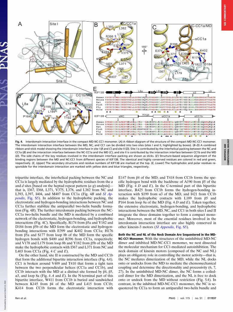
tripartite interface, the interhelical packing between the NC andCC1a is largely mediated by the hydrophobic residues from the aand d sites [based on the heptad repeat pattern (a–g) analysis]—that is, I367, I368, L371, V375, L378, and L382 from NC andL393, L397, I404, and M407 from CC1a (Fig. 4B and SI Ap-pendix, Fig. S5). In addition to the hydrophobic packing, theelectrostatic and hydrogen-bonding interactions between NC andCC1a further stabilize the antiparallel two-helix bundle forma-tion (Fig. 4B). The further interdomain packing between the NC-CC1a two-helix bundle and the MD is mediated by a combinednetwork of the electrostatic, hydrogen-bonding, and hydrophobicinteractions (Fig. 4C). Specifically, R174 from β5a and Y182 andD184 from β5b of the MD form the electrostatic and hydrogen-bonding interactions with E399 and K402 from CC1a; H176from β5a and S177 from loop 8b of the MD form the specifichydrogen bonds with S400 and R396 from CC1a, respectively;and V178 and L179 from loop 8b and Y182 from β5b of the MDmake the hydrophobic contacts with I367 and L371 from NC andL403 from CC1a (Fig. 4 C and E).On the other hand, site II is constructed by the MD and CC1b
that form the additional bipartite interaction interface (Fig. 4A).CC1 is broken around V409 and T410 that forms a tight turnbetween the two perpendicular helices (CC1a and CC1b), andCC1b interacts with the MD at a distinct site formed by β4, β5,α3, and loop 8a (Fig. 4 A and E). In the N-terminal part of thisbipartite interface, W411 from CC1b is buried and sandwichedbetween K145 from β4 of the MD and L415 from CC1b;K414 from CC1b forms the electrostatic interaction with
E147 from β4 of the MD; and T418 from CC1b forms the spe-cific hydrogen bond with the backbone of A190 from β5 of theMD (Fig. 4 D and E). In the C-terminal part of this bipartiteinterface, R425 from CC1b forms the hydrogen-bonding in-teraction with S199 from α3 of the MD, and I421 from CC1bmakes the hydrophobic contacts with L189 from β5 andP164 from loop 8a of the MD (Fig. 4 D and E). Taken together,the extensive electrostatic, hydrogen-bonding, and hydrophobicinteractions between the MD, NC, and CC1 in both sites I and IIintegrate the three domains together to form a compact mono-mer. Moreover, most of the essential residues involved in theinterdomain interaction interface are also highly conserved inother kinesin-3 motors (SI Appendix, Fig. S5).
Both the NC and NL of the Neck Domain Are Sequestered in the MD-NC-CC1 Monomer. With the structures of the uninhibited MD-NCdimer and inhibited MD-NC-CC1 monomer, we next dissectedthe molecular mechanism for CC1-mediated autoinhibition. Theneck domain of kinesin motors (composed of the NC and NL)plays an obligatory role in controlling the motor activity—that is,the NC mediates dimerization of the MD, while the NL docksonto or undocks from the MD to mediate the chemomechanicalcoupling and determine the directionality and processivity (6, 7,27). In the uninhibited MD-NC dimer, the NC forms a coiled-coil dimer for the MD dimerization, and the NL is free to dockonto or undock from the MD without restriction (Fig. 5A). Incontrast, in the inhibited MD-NC-CC1 monomer, the NC is se-questered by CC1a to form an antiparallel two-helix bundle and
Fig. 4. Interdomain interaction interface in the compact MD-NC-CC1 monomer. (A) A ribbon diagram of the structure of the compact MD-NC-CC1 monomer.The interdomain interaction interface between the MD, NC, and CC1 can be divided into two sites (sites I and II, highlighted by boxes). (B–D) A combinedribbon-and-stick model showing the interdomain interface in site I (B and C) and site II (D). Site I is contributed by the interhelical packing between the NC andCC1a (B) and the interaction interface between the NC-CC1a and the MD (C), and site II is contributed by the interaction interface between CC1b and the MD(D). The side chains of the key residues involved in the interdomain interface packing are shown as sticks. (E) Structure-based sequence alignment of thebinding regions between the MD and NC-CC1 from different species of KIF13B. The identical and highly conserved residues are colored in red and green,respectively. (E, Upper) The secondary structures and residue numbers of KIF13B are marked at the top. (E, Lower) The hydrophobic and polar residues re-sponsible for the interdomain interaction are marked with yellow dots and blue triangles, respectively.
Ren et al. PNAS | vol. 115 | no. 51 | E11937
BIOPH
YSICSAND
COMPU
TATIONALBIOLO
GY
SEECO
MMEN
TARY
Dow
nloa
ded
by g
uest
on
Mar
ch 1
2, 2
020

is unable to form a coiled-coil dimer for dimerization of the MD(Fig. 5B). Thus, the NC-mediated dimerization is blocked due to thedirect intramolecular interaction between the NC and CC1, con-sistent with the previous studies and biochemical characterization.More intriguingly, the NC-CC1a bundle and CC1b further
interact with the MD, which would tether and fix the NC ontothe MD firmly (Fig. 3). With superimposition of the MD, thebinding of the NC-CC1a bundle and CC1b to the MD drives theNC to undergo a rotation of ∼90° toward the MD (Fig. 5B).Thus, in addition to preventing the NC coiled-coil dimer for-mation, CC1 also restrains its freedom of motion throughbinding to the MD. Given that the NC immediately follows the
NL, restriction of the NC motion subsequently sequesters the NLand locks it in a docked conformation (Fig. 5C), suggesting thatthe NL would hardly undock from the MD in the inhibited MD-NC-CC1 monomer. Taken together, CC1 fastens the neck do-main and MD by interacting with the two domains and seques-ters both the NC and NL of the neck domain for autoinhibition.
Release of ADP from the MD Is Inhibited in the MD-NC-CC1 Monomer.The initial biochemical characterization of the MD-NC-CC1 tandem demonstrated that its MT-stimulated ATPase ac-tivity is also inhibited (Fig. 1). The decreased activity may becaused by the direct interference of binding to MTs. However, in
Fig. 5. Both NC and NL of the neck domain are sequestered in the MD-NC-CC1 monomer. (A) A combined ribbon-and-surface representation of the MD-NCsubunit from the dimer. The MD is shown in surface and can be divided into the P-loop, SwitchI/II, and MT-binding subdomains colored in salmon, slate, andgreen, respectively, and the NC is in a cylinder representation and colored in gray. (B) Structural comparison of the MD-NC and MD-NC-CC1 tandems. The MDand NC of the MD-NC tandem are colored as in A, and NC and CC1 of the MD-NC-CC1 tandem are colored in yellow and orange, respectively. After su-perimposition of the two structures, the NC undergoes dramatic conformational changes. (C) A close-up view of the NL regions of the MD-NC and MD-ND-CC1 structures. The NL can dock/undock freely in the MD-NC tandem, but is locked in the docked state in the MD-NC-CC1 tandem. (D) A schematic workingmodel for explaining the conformational changes between the three subdomains coupled with the NL undocking and ADP release. The color scheme for thethree subdomains follows that in A. The NL is shown as a pink line, and the NC and CC1 are colored in yellow and orange, respectively. (E) Kinetics of themant-ADP release from the MD-NL (red), the MD-NC-CC1 (black), and the various MD-NC-CC1 mutants (V375E, blue; ΔP391, pink; and L415A, green). Nor-malized fluorescence variations on release of ADP from the different fragments in the absence of MTs (solid lines) or the presence of MTs (dashed lines) areshown. (F) MT-stimulated ATPase activity of the wild type and various mutants of the MD-NC-CC1 tandem of KIF13B. Each bar represents the mean value ±SD. ***P < 0.001 (Student’s t test).
E11938 | www.pnas.org/cgi/doi/10.1073/pnas.1811209115 Ren et al.
Dow
nloa
ded
by g
uest
on
Mar
ch 1
2, 2
020

the MD-NC-CC1 monomer structure, the NC-CC1a bundle onlyleans against the periphery of the MT-binding subdomain of theMD and the major binding site of this subdomain is completelyexposed (Fig. 3), indicating that the MD-NC-CC1 tandem is stillcapable of interacting with MTs. To test this hypothesis, weperformed the MT-binding assay for the MD (with the NL) andthe MD-NC-CC1 tandem. As expected, the MD-NC-CC1 tandemcan bind to MTs similarly to the MD-NL tandem (SI Appendix,Fig. S6), demonstrating that the MT-binding capacity of the MDin the MD-NC-CC1 tandem is not severely impaired. Thus, thedecreased MT-stimulated ATPase activity of the MD-NC-CC1tandem is unlikely to be caused by interfering with the MT-binding property of the MD.Based on previous studies, the MD can be divided into three
subdomains—that is, the P-loop, SwitchI/II, and MT-bindingsubdomains (30) (Fig. 5A). The intramolecular structuralchanges between the three subdomains are intimately coupledwith the ATP hydrolysis cycle of the MD and its ATPase activity(27, 30). Upon docking onto the MD, the NL anchors betweenthe P-loop and MT-binding subdomains, and its undocking al-lows the intersubdomain rearrangement required for the MT-stimulated ADP release from the MD (Fig. 5D). However, inthe inhibited MD-NC-CC1 monomer, the NL is sequestered in adocked state, which would block the movement between the P-loop and MT-binding subdomains and prevent the subsequentMT-stimulated ADP release (Fig. 5D). To test this hypothesis,we performed the MT-stimulated mant-ADP release assay forthe MD (with the NL) and the MD-NC-CC1 tandem. Consistentwith the essential role of MTs for the ADP release, the ADPrelease from the MD-NL tandem is slow without MTs but issignificantly accelerated upon adding them (Fig. 5E and SI Ap-pendix, Table S2). In the absence of MTs, the ADP release fromthe MD-NC-CC1 tandem is also slow and remains slow afteradding MTs compared with the MD-NL tandem (Fig. 5E and SIAppendix, Table S2), indicating that the MT-stimulated ADPrelease is severely impaired in the MD-NC-CC1 monomer.Taken together, consistent with the structural analysis, the MT-stimulated ADP release from the MD in the MD-NC-CC1 tan-dem is inhibited by sequestering the NL, which would cause thedecreased ATPase activity.
Disruptions of the MD-NC-CC1 Monomer Restore the MD Activity.Based on the structural analysis of the inhibited MD-NC-CC1 monomer, we introduced a series of the point mutations inthe MD, NC, and CC1 to disrupt the interdomain interactioninterface—that is, V178Q in the MD, V375E in the NC, andE399A, K414A, and L415A in CC1—and checked the MT-stimulated ATPase activity of these mutants. We also removeda proline from the NC/CC1 hinge (ΔP391), which was previouslydemonstrated to disrupt the folded-back conformation of CC1(16, 28). All of the mutations did not have major effects on thesecondary structures of the MD-NC-CC1 tandem, as indicatedby the similar profile in the CD spectra (SI Appendix, Fig. S7A).However, most of the mutations in the interdomain interactioninterface destabilized the MD-NC-CC1 tandem in the limitedproteolysis assay (SI Appendix, Fig. S7B), suggesting that thecompact self-folded structure is disrupted by these mutations. Asexpected, in comparison with the wild-type MD-NC-CC1 tan-dem, the mutations in the interdomain interaction interface or inthe NC/CC1 hinge all significantly enhanced the MT-simulatedATPase activity (Fig. 5F), indicating that disruptions of thecontacts between the three domains or the folded-back confor-mation of CC1 restore the MD activity. Consistent with the in-creased ATPase activity, the MT-stimulated ADP release of theMD-NC-CC1 tandem was also markedly accelerated by themutations (V375E, ΔP391, and L415A that keep the MD intact)(Fig. 5E and SI Appendix, Table S2), further implicating theloosening of the NL for the ADP release. On the other hand,
disruptions of the interactions between the three domains or thefolded-back conformation of CC1 would also release the NC fordimerization of the MD. Consistent with this hypothesis, theΔP391-mutant adopts a dimeric conformation, and most ofthe other mutants (V178Q, E399A, K414A, and L415A with theintact NC) have a tendency to form a dimer and exist in amonomer/dimer equilibrium in solution (SI Appendix, Fig. S8).In contrast, the V375E mutant [with the mutation in the NC thatdisrupts the MD-NC-CC1 monomer as well as the NC coiled-coildimer (Fig. 2D)] remains as a monomer (SI Appendix, Fig. S8),further supporting the essential role of the NC for dimerizationof the MD.Finally, we assessed the effects of these mutations on the full-
length KIF13B in vivo by using a cell-based assay (Fig. 6A).Consistent with previous results (16), the wild-type KIF13Badopts an inactive state and was largely localized in the cell body(Fig. 6 A and B). Similar to the wild-type KIF13B, the Y73C–KIF13B mutant was also largely localized in the cell body (SIAppendix, Fig. S2 E and F), supporting the minimal impact ofthis mutation on the full-length protein. As expected, most of theKIF13B mutants (V178Q, ΔP391, E399A, K414A, and L415A)showed the enhanced accumulations at the cell tips in compar-ison with the wild-type KIF13B (Fig. 6 A and B), indicating therestored motor motility by these mutations. In contrast, theV375E–KIF13B mutant did not show the significantly increasedtip accumulations (Fig. 6 A and B), also consistent with the es-sential role of the NC-mediated dimerization for the motor ac-tivity. Taken together, all of the biochemical and cellular datademonstrated that disruptions of the MD-NC-CC1 monomer re-lease the sequestered neck domain and restore the MD activity.
DiscussionIntrinsic Structural Versatility of CC1 for Autoinhibition. In kinesin-3 motors, the coiled-coil domain CC1 is a well-known auto-inhibitory segment that folds back to sequester the preceding NCfor inhibiting the motor activity (2, 16, 23), but the underlyingmechanism is not well understood. In this study, we determinedthe structure of the inhibited MD-NC-CC1 monomer of KIF13Bin which CC1 is broken into two short α-helices (CC1a andCC1b) that interact with both the NC and the MD to sequesterthe entire neck domain (Figs. 3 and 4). Thus, instead of formingan extended coiled-coil, CC1 adopts a distinct two-helix con-formation for autoinhibition. Consistent with this unexpectedstructural feature, our previous studies of the NC-CC1-FHAtandem of KIF13A demonstrated that CC1 is not a perfectcoiled-coil for dimerization (28). Moreover, CC1 contains sev-eral intrinsic unusual features (i.e., the potential defects in theinterhelical packing and a “stutter” break in the middle) that candistort the coiled-coil formation and break the extended helix(28) (SI Appendix, Fig. S5), supporting the propensity of CC1 toform a two-helix conformation in the autoinhibited state. Takentogether, CC1 of kinesin-3 motors is not a canonical coiled-coilbut possesses the intrinsic structural versatility to adopt a distinctconformation for autoinhibition.In the MD-NC-CC1 monomer, CC1a folds back to pack with
the NC in an antiparallel manner to form a two-helix bundle witha short hinge (Figs. 3 and 4), consistent with previous structuralprediction (16). Removal of the signature proline (P391) fromthe short hinge disrupted the folded-back conformation ofCC1 and promoted the motor dimerization (SI Appendix, Fig.S8), also consistent with the previous studies (16, 28). It has beenproposed that, for different kinesin-3 motors, the length of thehinge between the NC and CC1 may determine their packingorientation (16)—i.e., for the long hinge, CC1 forms an extendedhelix to pack with the NC in a parallel manner based on theelectron-microscopy studies of UNC-104 (23), while for the shorthinge, CC1 interacts with the NC in an antiparallel manner, asdemonstrated in this study (Fig. 3). However, given the high
Ren et al. PNAS | vol. 115 | no. 51 | E11939
BIOPH
YSICSAND
COMPU
TATIONALBIOLO
GY
SEECO
MMEN
TARY
Dow
nloa
ded
by g
uest
on
Mar
ch 1
2, 2
020

sequence similarity of CC1 among different kinesin-3 motors (SIAppendix, Fig. S5), CC1 in the long-hinged kinesin-3 motors mayalso be capable of adopting the two-helix conformation for se-questering the NC. Moreover, due to the short length of the NC,the half of CC1 (CC1a) is sufficient to cover the whole NC, asshown in the MD-NC-CC1 monomer structure (Fig. 4 and SIAppendix, Fig. S5). Thus, we may not exclude the possibility thatthe antiparallel packing between CC1a and the NC for auto-inhibition could also exist in other long-hinged kinesin-3 motors.The essential role of CC1 for regulating the NC-mediated
dimerization was described in the work of Al-Bassam et al. (23).They worked on the different constructs of UNC-104 with MTsby the cryo-EM method at low resolution and found that, uponbinding to MTs, AMPPNP (the nonhydrolyzable ATP analog)appears to trap the MD-NC-CC1 tandem in a monomeric state(23). In this work, the similar MD-NC-CC1 tandem of KIF13Badopts a monomeric conformation in solution and is also capableof binding to MTs (Fig. 1 and SI Appendix, Fig. S6), consistentwith the work of Al-Bassam et al. work. However, the MD of theMD-NC-CC1 monomer of KIF13B was tapped in an ADP-bound state (since we did not add extra nucleotides during theprotein purification and crystallization) (Fig. 3). The discrepancy
of the MD-NC-CC1 monomer with different nucleotide-boundstates between the two studies might be caused by different ex-perimental conditions—i.e., in the study of Al-Bassam et al., thework was carried out with MTs, whereas in this study, the workwas largely performed without MTs.
The MD Is Unexpectedly Involved in the CC1-Mediated Autoinhibition.In addition to the broken CC1, the other unexpected structuralfeature in the MD-NC-CC1 monomer is that the MD is involvedin the CC1-mediated autoinhibition (Fig. 3). The MD containstwo distinct sites (in the β4-α3 region, referred to as the NC-CC1-binding region) to capture the NC-CC1a bundle and CC1b,respectively, to form a compact self-folded structure (Fig. 4). Incomparison with the MD-NC dimer, the binding of the NC-CC1abundle and CC1b induced some local conformational changes inthe β5a/β5b region of the MD, while no significant structuralchanges were found in the Switch I and II elements (since theMDs from the two structures were both trapped in the similarADP-bound state) (SI Appendix, Fig. S9). The two site-mediatedinteractions between the MD and NC-CC1a/CC1b not onlytether the NC to theMD, but also sequester the NL, which ultimatelyleads to the complete lockdown of the entire neck domain from the
Fig. 6. Disruptions of the MD-NC-CC1 monomer restore the motor activity of KIF13B. (A) Cellular localizations of the full-length KIF13B and its variousmutants in the N2A cells. The wild-type KIF13B was largely localized in the cell body (A1). Most of the mutants showed the significantly increased accu-mulations at the cell tips, except for the V375E mutant (with the mutation in NC) (A2–A7). (Scale bar: 20 μm.) (B) Quantification of the cellular distributiondata shown in A. The ratio of the tip to cell body average FI was quantified for each construct for >15 cells (n > 15). Each bar represents the mean value ± SEM.***P < 0.001 (Student’s t test). (C) A working model for the CC1-mediated autoinhibition of kinesin-3. In the uninhibited state (C, Upper), the NC mediatesdimerization of the MD to assemble a dimeric motor, and the NL freely docks onto or undocks from the MD for processive movement. The MD is in the surfacerepresentation (colored in gray), and the NL and NC are in the ribbon representation (colored in pink and yellow, respectively). The gray oval represents theMD with the NL undocking. In the inhibited state (C, Lower), CC1 (colored in orange) folds back to interact with both the NC and the MD, which not onlyinhibits the NC-mediated dimerization, but also prevents the NL undocking and the ADP release from the MD.
E11940 | www.pnas.org/cgi/doi/10.1073/pnas.1811209115 Ren et al.
Dow
nloa
ded
by g
uest
on
Mar
ch 1
2, 2
020
NL to the NC (Fig. 5). Without binding to the MD, both the NC-CC1a bundle and CCb1 would be flexible and unstable in solution [asindicated in the limited proteolysis assay (SI Appendix, Fig. S7B)], andthe neck domain would be only partially inhibited due to the fact thatthe NL could freely undock from the MD without restraints (Fig. 5).Thus, with two distinct binding sites, the MD tightly packs with NC-CC1a/CC1b and facilitates CC1 to inhibit the entire neck domain.Consistent with this hypothesis, mutations of the residues of the MD(that are in the interdomain interaction interface) disrupted the MD-NC-CC1 monomer and restored the MD activity (Figs. 5 and 6).Intriguingly, most of the essential residues in the NC-CC1-bindingregion of the MD are highly conserved in other kinesin-3 motors (SIAppendix, Fig. S5), suggesting that the MD in these motors may alsoparticipate in the CC1-mediated autoinhibition. Moreover, mutationsof the MD of kinesin-3 KIF1A are coupled with a number of humanneuronal disorders, and some of the disease-related mutations arelocated in the NC-CC1-binding region (31–33) (SI Appendix, Fig. S5),indicating that the identified NC-CC1-binding region of the MD ofkinesin-3 motors may also be of functional importance.
CC1 Fastens the Neck Domain and MD for Kinesin-3 Autoinhibition.Based on the structures of the uninhibited MD-NC dimer andinhibited MD-NC-CC1 monomer, the molecular mechanism forthe CC1-mediated autoinhibition could be proposed (Fig. 6C).In the uninhibited state, the NC mediates dimerization of theMD to assemble a two-headed motor, and the NL freely docksonto or undocks from the MD without restraints (Fig. 6C). In theCC1-mediated autoinhibited state, CC1 folds back to interactwith both the NC and the MD to fasten the neck domain and theMD, which not only inhibits the NC-mediated dimerization butalso prevents the NL undocking and the ADP release from theMD (Fig. 6C). In contrast, the MT-binding capacity of the MD isnot severely impaired by CC1 due to the exposure of the MT-binding subdomain (Fig. 3 and SI Appendix, Fig. S6). Thus, withthe aid of the MD, CC1 sequesters both the NC and NL of theneck domain without interfering with the MD (Fig. 6C). Thisdual inhibition of the neck domain by CC1 would secure theinactive state of the motor without any futile ATP hydrolysis.The CC1-mediated lockdown of the entire neck domain mayrepresent a paradigm for kinesin autoinhibition and could beapplicable to other kinesin-3 motors.The CC1-mediated prevention of the NL undocking and the
ADP release found in kinesin-3 is also reminiscent of themechanism of kinesin-1 autoinhibition. In kinesin-1, a shortpeptide from the tail domain was found to directly bind to theMD (11–13). The structure of the motor-tail complex of kinesin-1 demonstrated that a single tail peptide can cross-link the twoMDs and, together with the NC dimer, prevents the movementof the two MDs that is required for the NL undocking and theADP release (34, 35). In kinesin-3, the CC1-mediated fasteningof the neck domain and MD locks down the entire neck domain,and the NL is prevented from undocking from the MD for theADP release (Fig. 6C). Thus, different approaches used by kinesin-1 and -3 (tail peptide-mediated vs. CC1-mediated) come to thesame result, and prevention of the NL undocking and the ADPrelease may be a general mechanism for kinesin autoinhibition.In the full-length motor, in addition to the key autoinhibitory
segment CC1, other regions have been shown to further facilitatethe autoinhibited conformation of kinesin-3. In KIF1A, CC2 wasshown to be able to fold back to interact with the FHA domain(20), and in KIF13B, the membrane-associated guanylate kinasehomolog binding stalk (MBS) segment immediately followingCC2 was found to interact with the MD (21). On the other hand,in our previous work, we found that the CC1-FHA tandem ofkinesin-3 can form an extended dimer that would release theCC1-mediated inhibition and couple with the NC dimer to as-semble a dimeric motor for processive movement (28, 36).Moreover, the activation and dimerization of kinesin-3 were
demonstrated to be chiefly mediated by cargo binding (16, 19,21). Based on all of the previous studies and this work, a workingmodel for the cargo-mediated transition of the full-lengthKIF13B from an autoinhibited monomer to an uninhibited di-mer could also be proposed (SI Appendix, Fig. S10). In themonomeric autoinhibited state, CC1 interacts with both NC andthe MD, CC2 interacts with the FHA domain, and the MBSsegment binds to the MD (SI Appendix, Fig. S10). Upon bindingto cargoes or cargo adaptors likely through the MBS segment inKIF13B (21), the FHA domain could be somehow released topromote the formation of the CC1-FHA dimer that would fur-ther release the CC1-mediated inhibition of the neck domain (SIAppendix, Fig. S10). In the dimeric uninhibited state, the FHAdomain, CC1, and the NC would work together to form a stabledimer that brings the two MDs together for walking along MTs(SI Appendix, Fig. S10).
Materials and MethodsProtein Expression and Purification. DNA sequences encoding the rat andhuman KIF13B MD-NL (1–371), MD-NC (1–389), MD-NC-CC1 (1–432), andvarious mutants were each cloned into a modified pET32a vector with a C-terminal His6-tag. Point mutations were introduced by using the standardPCR-based mutagenesis method and confirmed by DNA sequencing.Recombinant proteins were expressed in Escherichia coli BL21 (DE3) hostcells at 16 °C. The His6-tagged fusion proteins were purified by Ni2+-Sepharose 6 Fast Flow (GE Healthcare) affinity chromatography followed bysize-exclusion chromatography (Superdex-200 26/60; GE Healthcare) in thebuffer containing 50 mM Tris·HCl (pH 7.5), 150 mM NaCl, 2 mM MgCl2, 1 mMEGTA, and 1 mM DTT. For the analytical gel-filtration analysis, proteinsamples were concentrated to ∼5.0 mg/mL and loaded onto the Superdex-200 10/300 GL column (GE Healthcare).
Analytical Ultracentrifugation. Sedimentation velocity experiments wereperformed on a Beckman XL-I analytical ultracentrifuge equipped with a four-cell An-60 Ti rotor (Beckman Coulter) under 220,000 × g at 25 °C. The partialspecific volume of protein samples and the buffer density were calculated byusing the program SEDNTERP (bitcwiki.sr.unh.edu/index.php/Main_Page). Thefinal sedimentation velocity data were analyzed and fitted to a continuoussedimentation coefficient distribution model by using the program SEDFIT.
Crystallization. Crystals of the MD-NC tandem (∼25 mg/mL) were obtained in1.4 M Na/KPO4 (pH 6.9). Crystals of the MD-NC-CC1-Y73C mutant (∼20 mg/mL)were obtained in 0.2 M proline, 0.1 M Hepes (pH 7.5), and 14% (wt/vol) PEG3350. All of the crystals were obtained by using the sitting-drop vapor-diffusionmethod at 16 °C. Before being flash-frozen in liquid nitrogen, crystals weresoaked in the mother liquor supplemented with 15% (vol/vol) ethylene glycolfor cryoprotection.
Data Collection, Structural Determination, and Refinement. All of the dif-fraction datasets were collected at the beamline BL17U1 and BL19U of theShanghai Synchrotron Radiation Facility with a wavelength of 0.989 Å at 100K (37). Datasets were integrated and scaled with HKL2000 (38). The struc-tures of the MD-NC tandem and the MD-NC-CC1-Y73C mutant were de-termined by the molecular-replacement methods using the MD of KIF13B(PDB ID code 5ZBR) as the searching model with Phaser (39). Additionalmissing residues were manually modeled into the structures according to the2Fo − Fc and Fo − Fc electron-density maps using COOT (40). The structureswere further refined and validated with PHENIX (41). The statistics for thedata collection and structural refinement are summarized in SI Appendix,Table S1. The protein structure figures were prepared by using the programPyMOL (https://pymol.org/2/).
MT-Stimulated ATPase Assay. Measurements of the MT-stimulated ATPaseactivities of various KIF13B fragments were performed by using the HTSKinesin ATPase Endpoint Assay Biochem Kit (Cytoskeleton, Inc.). Briefly, all ofthe measurements were based on the malachite green phosphate assay toprobe inorganic phosphate generated during the reaction. A standard curveof phosphatewasmade to estimate the amount of phosphate generated. Thebasal ATPase activities (without MTs) of these KIF13B fragments were alsomeasured by using the same method. Each protein sample had three repli-cates, and eachmeasurement was repeated at least three times independently.The kinesin-1 heavy chain (KHC) supplied in the kit was used as the control. Allof the data were analyzed by using the GraphPad Prism5 program.
Ren et al. PNAS | vol. 115 | no. 51 | E11941
BIOPH
YSICSAND
COMPU
TATIONALBIOLO
GY
SEECO
MMEN
TARY
Dow
nloa
ded
by g
uest
on
Mar
ch 1
2, 2
020

Kinetics of MT-Stimulated Mant-ADP Dissociation. The bound ADPs in the MD(with the NL) and the MD-NC-CC1 tandem of KIF13B were first exchanged formant-ADPs by equilibration with a 10-fold excess of mant-ADP at 25 °C for30 min. The excess mant-ADPs were removed by exchanging into the re-action buffer containing 80 mM Pipes (pH 6.9), 1 mM MgCl2, 1 mM EGTA,and 0.1% Tween 20 using a Sephadex G-25 column. Release of mant-ADPwas started by mixing the mant-ADP–loaded protein with a 50-fold excess ofunlabeled ATP in the same buffer. The reaction was monitored by using aSX20 Stopped-Flow spectrometer at 25 °C. Excitation was at 365 nm, andfluorescence was recorded by using a 400-nm long-pass filter. For the MT-stimulated mant-ADP dissociation experiments, additional preprepared1 μMMTs and 100 μM taxol were supplemented in the buffer. All of the datawere analyzed and fitted by using the GraphPad Prism5 program.
Cell Culture, Imaging, and Data Analysis. The full-length human KIF13B andvarious mutants were each cloned into a pEGFP-N3 vector. N2A cells werecultured in DMEM containing 10% (vol/vol) FBS and were grown at 37 °C. Thecells were transfected with the wild-type KIF13B and various mutants byLipofectamine 3000 (Invitrogen), according to the manufacturer’s instruc-tions. Fluorescence images were obtained by using Olympus FV1000 Laser
Scanning Confocal Microscopy with a 60× (NA = 1.42) oil objective. Confocalsettings used for image capture were held constant in comparison experi-ments. For the cellular distribution data analysis, the specific regions of the cellbody (excluding the nucleus) and the tip of each cell were chosen, and theaverage fluorescence intensities (FIs) were calculated, respectively. All of thefluorescence images were processed and analyzed by ImageJ (NIH). The finalquantification graphs were generated by the GraphPad Prism5 program.
Accession Numbers. The atomic coordinates of the MD-NC dimer and the MD-NC-CC1-Y73C monomer have been deposited in the Protein Data Bank, withPDB ID codes 6A1Z and 6A20, respectively.
ACKNOWLEDGMENTS. We thank Dr. Yong Zhang for the help of moleculardynamics simulations. We thank the beamline BL17U of the ShanghaiSynchrotron Radiation Facility for the beam time. This work was supportedby National Key R&D Program of China Grant 2017YFA0503501; NationalMajor Basic Research Program of China Grant 2014CB910202; and NationalNatural Science Foundation of China Grants 31470746, 31770786, 31600608,and 31600622.
1. Vale RD (2003) The molecular motor toolbox for intracellular transport. Cell 112:467–480.
2. Verhey KJ, Kaul N, Soppina V (2011) Kinesin assembly and movement in cells. AnnuRev Biophys 40:267–288.
3. Hirokawa N, Noda Y, Tanaka Y, Niwa S (2009) Kinesin superfamily motor proteins andintracellular transport. Nat Rev Mol Cell Biol 10:682–696.
4. Cross RA, McAinsh A (2014) Prime movers: The mechanochemistry of mitotic kinesins.Nat Rev Mol Cell Biol 15:257–271.
5. Walczak CE, Gayek S, Ohi R (2013) Microtubule-depolymerizing kinesins. Annu RevCell Dev Biol 29:417–441.
6. Vale RD, Fletterick RJ (1997) The design plan of kinesin motors. Annu Rev Cell Dev Biol13:745–777.
7. Woehlke G, Schliwa M (2000) Walking on two heads: The many talents of kinesin. NatRev Mol Cell Biol 1:50–58.
8. Endow SA (1999) Determinants of molecular motor directionality. Nat Cell Biol 1:E163–E167.
9. Gennerich A, Vale RD (2009) Walking the walk: How kinesin and dynein coordinatetheir steps. Curr Opin Cell Biol 21:59–67.
10. Verhey KJ, Hammond JW (2009) Traffic control: Regulation of kinesin motors. Nat RevMol Cell Biol 10:765–777.
11. Friedman DS, Vale RD (1999) Single-molecule analysis of kinesin motility revealsregulation by the cargo-binding tail domain. Nat Cell Biol 1:293–297.
12. Coy DL, Hancock WO, Wagenbach M, Howard J (1999) Kinesin’s tail domain is aninhibitory regulator of the motor domain. Nat Cell Biol 1:288–292.
13. Hackney DD, Stock MF (2000) Kinesin’s IAK tail domain inhibits initial microtubule-stimulated ADP release. Nat Cell Biol 2:257–260.
14. Imanishi M, Endres NF, Gennerich A, Vale RD (2006) Autoinhibition regulates themotility of the C. elegans intraflagellar transport motor OSM-3. J Cell Biol 174:931–937.
15. Hammond JW, Blasius TL, Soppina V, Cai D, Verhey KJ (2010) Autoinhibition of thekinesin-2 motor KIF17 via dual intramolecular mechanisms. J Cell Biol 189:1013–1025.
16. Soppina V, et al. (2014) Dimerization of mammalian kinesin-3 motors results in su-perprocessive motion. Proc Natl Acad Sci USA 111:5562–5567.
17. Okada Y, Yamazaki H, Sekine-Aizawa Y, Hirokawa N (1995) The neuron-specific ki-nesin superfamily protein KIF1A is a unique monomeric motor for anterograde ax-onal transport of synaptic vesicle precursors. Cell 81:769–780.
18. Tomishige M, Klopfenstein DR, Vale RD (2002) Conversion of Unc104/KIF1A kinesininto a processive motor after dimerization. Science 297:2263–2267.
19. Klopfenstein DR, Tomishige M, Stuurman N, Vale RD (2002) Role of phosphatidyli-nositol(4,5)bisphosphate organization in membrane transport by the Unc104 kinesinmotor. Cell 109:347–358.
20. Lee JR, et al. (2004) An intramolecular interaction between the FHA domain and acoiled coil negatively regulates the kinesin motor KIF1A. EMBO J 23:1506–1515.
21. Yamada KH, Hanada T, Chishti AH (2007) The effector domain of human Dlg tumorsuppressor acts as a switch that relieves autoinhibition of kinesin-3 motor GAKIN/KIF13B. Biochemistry 46:10039–10045.
22. Farkhondeh A, Niwa S, Takei Y, Hirokawa N (2015) Characterizing KIF16B in neuronsreveals a novel intramolecular “stalk inhibition” mechanism that regulates its ca-pacity to potentiate the selective somatodendritic localization of early endosomes.J Neurosci 35:5067–5086.
23. Al-Bassam J, et al. (2003) Distinct conformations of the kinesin Unc104 neck regulate amonomer to dimer motor transition. J Cell Biol 163:743–753.
24. Hammond JW, et al. (2009) Mammalian kinesin-3 motors are dimeric in vivo andmove by processive motility upon release of autoinhibition. PLoS Biol 7:e72.
25. Horiguchi K, Hanada T, Fukui Y, Chishti AH (2006) Transport of PIP3 by GAKIN, akinesin-3 family protein, regulates neuronal cell polarity. J Cell Biol 174:425–436.
26. Kanai Y, Wang D, Hirokawa N (2014) KIF13B enhances the endocytosis of LRP1 byrecruiting LRP1 to caveolae. J Cell Biol 204:395–408.
27. Cross RA (2016) Review: Mechanochemistry of the kinesin-1 ATPase. Biopolymers 105:476–482.
28. Ren J, et al. (2016) Structural correlation of the neck coil with the coiled-coil (CC1)-forkhead-associated (FHA) tandem for active kinesin-3 KIF13A. J Biol Chem 291:3581–3594.
29. Ren J, et al. (2018) Structural delineation of the neck linker of kinesin-3 for processivemovement. J Mol Biol 430:2030–2041.
30. Cao L, et al. (2014) The structure of apo-kinesin bound to tubulin links the nucleotidecycle to movement. Nat Commun 5:5364.
31. Citterio A, et al. (2015) Variants in KIF1A gene in dominant and sporadic forms ofhereditary spastic paraparesis. J Neurol 262:2684–2690.
32. Hotchkiss L, et al. (2016) Novel de novo mutations in KIF1A as a cause of hereditaryspastic paraplegia with progressive central nervous system involvement. J ChildNeurol 31:1114–1119.
33. Lee JR, et al. (2015) De novo mutations in the motor domain of KIF1A cause cognitiveimpairment, spastic paraparesis, axonal neuropathy, and cerebellar atrophy. HumMutat 36:69–78.
34. Hackney DD, Baek N, Snyder AC (2009) Half-site inhibition of dimeric kinesin headdomains by monomeric tail domains. Biochemistry 48:3448–3456.
35. Kaan HY, Hackney DD, Kozielski F (2011) The structure of the kinesin-1 motor-tailcomplex reveals the mechanism of autoinhibition. Science 333:883–885.
36. Huo L, et al. (2012) The CC1-FHA tandem as a central hub for controlling the di-merization and activation of kinesin-3 KIF1A. Structure 20:1550–1561.
37. Wang Q-S, et al. (2018) Upgrade of macromolecular crystallography beamlineBL17U1 at SSRF. Nucl Sci Tech 29:68.
38. Otwinowski Z, Minor W (1997) Processing of X-ray diffraction data collected in os-cillation mode. Methods Enzymol 276:307–326.
39. McCoy AJ (2007) Solving structures of protein complexes by molecular replacementwith Phaser. Acta Crystallogr D Biol Crystallogr 63:32–41.
40. Emsley P, Cowtan K (2004) Coot: Model-building tools for molecular graphics. ActaCrystallogr D Biol Crystallogr 60:2126–2132.
41. Adams PD, et al. (2010) PHENIX: A comprehensive Python-based system for macro-molecular structure solution. Acta Crystallogr D Biol Crystallogr 66:213–221.
E11942 | www.pnas.org/cgi/doi/10.1073/pnas.1811209115 Ren et al.
Dow
nloa
ded
by g
uest
on
Mar
ch 1
2, 2
020