CO-releasing molecule (CORM) conjugate systems
19
Dalton Transactions PERSPECTIVE Cite this: Dalton Trans., 2016, 45, 18045 Received 9th September 2016, Accepted 17th October 2016 DOI: 10.1039/c6dt03515a www.rsc.org/dalton CO-releasing molecule (CORM) conjugate systems Anna Christin Kautz, Peter C. Kunz and Christoph Janiak* The development of CORMs (CO-releasing molecules) as a prodrug for CO administration in living organ- isms has attracted significant attention. CORMs offer the promising possibility of a safe and controllable release of CO in low amounts triggered by light, ligands, enzymes, etc. For the targeting of specific tissues or diseases and to prevent possible side effects from metals and other residues after CO release, these CORMs are attached to biocompatible systems, like peptides, polymers, nanoparticles, dendrimers, protein cages, non-wovens, tablets, and metal–organic frameworks. We discuss in this review the known CORM carrier conjugates, in short CORM conjugates, with covalently-bound or incorporated CORMs for medicinal and therapeutic applications. Most conjugates are nontoxic, show increasing half-lives of CO release, and make use of the EPR-effect, but still show problems because of a continuous background of CO release and the absence of an on/off-switch for the CO release. Introduction In recent years, carbon monoxide releasing molecules (CORMs) have attracted much attention due to their possibility to deliver low amounts of carbon monoxide in a controllable therapeutic way. 1 For decades, carbon monoxide (CO) has been known as “the silent killer”, as first described by Haldane, because of its strong affinity to bind to hemoglobin and therefore it was only seen as an obstacle to oxygen transport in the blood system. 2,3 However, it has been proven that CO is not only toxic but also occurs in small amounts within the human body (produced by the oxidation of heme to biliverdin catalyzed by heme oxygen- ase) and plays important physiological roles as a small mole- cule messenger (such as stimulating guanylyl cyclase to form cyclic guanosine monophosphate (cGMP), activating cGMP, inhibiting the DNA binding activity of holo-NPAS2 (neuronal Anna Christin Kautz Anna Christin Kautz studied business chemistry at Heinrich- Heine-University in Düsseldorf and received her Diploma in 2012. Her current research (dis- sertation) focuses on metal– organic frameworks and encap- sulation of drugs, for example, CO releasing molecules (CORMs). Peter C. Kunz Peter C. Kunz studied Chemistry at Heinrich-Heine-University in Düsseldorf where he received his Diploma in 1999 on studying the nickel-catalysed copolymerisa- tion of ethene and CO. Under the guidance of Wolfgang Kläui he completed his Dissertation in 2003 on biomimetic tripodal ligands. In 2004 he stayed with Roger Alberto as Postdoc in Zürich, where he investigated organometallic Re and Tc com- plexes and focused on medicinal bioinorganic topics. From 2004 to 2010 he worked on his Habilitation at Heinrich-Heine-University in Düsseldorf and as an extraordinary lecturer in the Pharmaceutical Chemistry Department. Since 2012, he has been a chemistry and physics teacher at Annette-von-Droste-Hülshoff-Gymnasium in Düsseldorf. Institut für Anorganische Chemie und Strukturchemie, Heinrich-Heine-Universität, Universitätsstr. 1, D-40225 Düsseldorf, Germany. E-mail: [email protected] This journal is © The Royal Society of Chemistry 2016 Dalton Trans. , 2016, 45, 18045–18063 | 18045 Open Access Article. Published on 18 October 2016. Downloaded on 1/28/2022 12:56:58 AM. This article is licensed under a Creative Commons Attribution-NonCommercial 3.0 Unported Licence. View Article Online View Journal | View Issue
Transcript of CO-releasing molecule (CORM) conjugate systems

DaltonTransactions
PERSPECTIVE
Cite this: Dalton Trans., 2016, 45,18045
Received 9th September 2016,Accepted 17th October 2016
DOI: 10.1039/c6dt03515a
www.rsc.org/dalton
CO-releasing molecule (CORM) conjugate systems
Anna Christin Kautz, Peter C. Kunz and Christoph Janiak*
The development of CORMs (CO-releasing molecules) as a prodrug for CO administration in living organ-
isms has attracted significant attention. CORMs offer the promising possibility of a safe and controllable
release of CO in low amounts triggered by light, ligands, enzymes, etc. For the targeting of specific tissues
or diseases and to prevent possible side effects from metals and other residues after CO release, these
CORMs are attached to biocompatible systems, like peptides, polymers, nanoparticles, dendrimers,
protein cages, non-wovens, tablets, and metal–organic frameworks. We discuss in this review the known
CORM carrier conjugates, in short CORM conjugates, with covalently-bound or incorporated CORMs for
medicinal and therapeutic applications. Most conjugates are nontoxic, show increasing half-lives of CO
release, and make use of the EPR-effect, but still show problems because of a continuous background of
CO release and the absence of an on/off-switch for the CO release.
Introduction
In recent years, carbon monoxide releasing molecules(CORMs) have attracted much attention due to their possibilityto deliver low amounts of carbon monoxide in a controllabletherapeutic way.1
For decades, carbon monoxide (CO) has been known as“the silent killer”, as first described by Haldane, because of itsstrong affinity to bind to hemoglobin and therefore it was onlyseen as an obstacle to oxygen transport in the blood system.2,3
However, it has been proven that CO is not only toxic but alsooccurs in small amounts within the human body (produced bythe oxidation of heme to biliverdin catalyzed by heme oxygen-ase) and plays important physiological roles as a small mole-cule messenger (such as stimulating guanylyl cyclase to formcyclic guanosine monophosphate (cGMP), activating cGMP,inhibiting the DNA binding activity of holo-NPAS2 (neuronal
Anna Christin Kautz
Anna Christin Kautz studiedbusiness chemistry at Heinrich-Heine-University in Düsseldorfand received her Diploma in2012. Her current research (dis-sertation) focuses on metal–organic frameworks and encap-sulation of drugs, for example,CO releasing molecules(CORMs).
Peter C. Kunz
Peter C. Kunz studied Chemistryat Heinrich-Heine-University inDüsseldorf where he received hisDiploma in 1999 on studying thenickel-catalysed copolymerisa-tion of ethene and CO. Under theguidance of Wolfgang Kläui hecompleted his Dissertation in2003 on biomimetic tripodalligands. In 2004 he stayed withRoger Alberto as Postdoc inZürich, where he investigatedorganometallic Re and Tc com-plexes and focused on medicinal
bioinorganic topics. From 2004 to 2010 he worked on hisHabilitation at Heinrich-Heine-University in Düsseldorf and as anextraordinary lecturer in the Pharmaceutical ChemistryDepartment. Since 2012, he has been a chemistry and physicsteacher at Annette-von-Droste-Hülshoff-Gymnasium in Düsseldorf.
Institut für Anorganische Chemie und Strukturchemie, Heinrich-Heine-Universität,
Universitätsstr. 1, D-40225 Düsseldorf, Germany. E-mail: [email protected]
This journal is © The Royal Society of Chemistry 2016 Dalton Trans., 2016, 45, 18045–18063 | 18045
Ope
n A
cces
s A
rtic
le. P
ublis
hed
on 1
8 O
ctob
er 2
016.
Dow
nloa
ded
on 1
/28/
2022
12:
56:5
8 A
M.
Thi
s ar
ticle
is li
cens
ed u
nder
a C
reat
ive
Com
mon
s A
ttrib
utio
n-N
onC
omm
erci
al 3
.0 U
npor
ted
Lic
ence
.
View Article OnlineView Journal | View Issue

Per-Arnt-Sim-domain protein 2)) like nitric oxide or hydrogensulfide.4–7 In addition, in low concentrations, carbon monox-ide has a beneficial effect on cardiovascular diseases, inflam-matory disorders, tumor growth, bacterial infections, andorgan transplantations.8,9
In order to achieve the release of CO at specific sites intissue or organs with a controllable and tunable quantity,Motterlini et al. proposed the pharmaceutical use of carbonmonoxide releasing molecules (CORMs), a chemically boundform of CO as a prodrug.10 This initial work described the bio-chemical properties of transition metal carbonyl complexes,like the commercially available [Mn2(CO)10] (CORM-1) and[RuCl2(CO)3]2 (CORM-2) (Fig. 1).10 Over a period of time,more CORMs, like the water-soluble [Ru(CO)3Cl(glycinate)](CORM-3) and [Mn(CO)3(tpm)]PF6 (CORM-L1, tpm: tris(pyrazo-lyl) methane), were synthesized and analyzed in terms of theirCO-releasing properties (Fig. 1).11–13
The CO release of CORMs can be initiated through diversespecific triggers.1 One possible mechanism leading to the
Fig. 1 Examples of some CO-releasing molecules (CORMs) with their accepted acronym (CORM-A1, CORM-2 etc.), principal investigator, andspecific release trigger, where known (in green: hν = photolysis, ΔT = temperature increase, pH change, oxidation or ligand, L substitution). ForCORMs with a spontaneous or unclear release trigger, no indication is given. The released or replaceable CO ligands are depicted in blue.17
Christoph Janiak
Christoph Janiak studiedChemistry at TU Berlin and theUniv. of Oklahoma, followed bya postdoc at Cornell Univ. and atBASF AG and an associateprofessorship at the Univ. ofFreiburg. Since 2010, he hasbeen a full professor ofBioinorganic Chemistry and cat-alysis at the University ofDüsseldorf with further researchinterests in metal and porousorganic frameworks (MOFs,COFs) and metal nanoparticlesand catalysis.
Perspective Dalton Transactions
18046 | Dalton Trans., 2016, 45, 18045–18063 This journal is © The Royal Society of Chemistry 2016
Ope
n A
cces
s A
rtic
le. P
ublis
hed
on 1
8 O
ctob
er 2
016.
Dow
nloa
ded
on 1
/28/
2022
12:
56:5
8 A
M.
Thi
s ar
ticle
is li
cens
ed u
nder
a C
reat
ive
Com
mon
s A
ttrib
utio
n-N
onC
omm
erci
al 3
.0 U
npor
ted
Lic
ence
.View Article Online

CO release of CORMs is photochemical external activation bydifferent wavelengths of light. These CORMs are often calledPhotoCORMs and examples include CORM-1 and CORM-L1.13
CORM-2 releases CO spontaneously through ligand substi-tution/exchange by forming a new bond with, for example,sulfur.1 In particular for application under physiological con-ditions, the trigger temperature may be important. SomeCORMs, like CORM-3, release CO also by a trigger combi-nation, e.g., by thermal degradation and ligand substitution.Other triggers are degradation by enzymes (ET-CORMs, ET:enzyme triggered), pH change, and oxidation.1,14 The proto-typical oxidatively triggered CORM is ALF186.15,16
Unfortunately, it is also possible that this oxidation trigger isresponsible for the background or steady CO release of otherCORMs in solution, which are thought to be triggered byligand exchange. Only one electron is necessary to start therelease and this oxidation trigger could hide behind severaltriggers. Fig. 1 provides a summarizing overview of CORMsand their specific release triggers.
Vibrational spectroscopy (infrared, IR, and Raman) is thesimplest and quickest method to analyze CORMs and to proveif the CO groups are bound to the transition metal complex.The aforementioned CORM-L1, for example, shows the typicaltwo CvO bands at 1941 and 2047 cm−1 for a facial coordi-nation of the tpm ligand to the manganese tricarbonyl unit.12
In contrast to the inert CORM-L1, the IR spectra of CORM-3show pH-dependency. After the addition of aqueous HCl, theIR spectrum shows three bands at 2134, 2070, and 2052 cm−1,because of the reversible conversion of [Ru(CO)3Cl(NH2CH2CO2)] to [Ru(CO)3Cl2(NH2CH2CO2H)]. According tothe added amount of NaOH, the two bands of a cis-dicarbonylruthenium fragment in a basic solution are 2058 and1985 cm−1 (1 equivalent of NaOH) or 2044 and 1968 cm−1
(2 equivalents of NaOH), respectively. In basic solution,the reversible reactions of all the CORM-3 derivates, like[Ru(CO)3Cl2L], with OH− leads to [Ru(CO)2(COOH)Cl2L] com-pounds showing the two mentioned CO stretching bands.18,19
There are a plethora of methods known in order to detectthe CO upon release from the compounds, with the so-calledmyoglobin assay being the most widely used technique andtherefore something like a “gold standard”. In this protocol,first reported by Motterlini et al., the CORM analyte isadded to a buffered aqueous solution containing sodiumdithionite (Na2S2O4) and deoxy-myoglobin (Mb) under anatmosphere of nitrogen gas.10 The buffers used (pH = 7.4) canbe 3-(N-morpholino)-propanesulfonic acid (MOPS), phosphate-buffered saline (PBS), potassium phosphate (KPI), KrebsHenseleit buffer (KHB), or others.10,20,21 Sodium dithionite isused to reduce any Fe(III) in oxymyoglobin to desoxymyoglobin.During CO release from the CORM, the formation of carboxy-myoglobin (Mb-CO) is observed by a decreasing intensity ofthe absorption band of myoglobin (556 nm) and two increas-ing absorption bands of the carboxy-myoglobin (541 nm,578 nm) in the visible spectra (eqn (1)). By this method, COrelease over time can be followed easily and the resultant half-life for the decay of the CO complex as well as the amount of
CO released can be calculated. In general, the myoglobin assayis performed using conditions found in living organisms(pH 7.4, 37 °C).10,22 It should be noted, however, that thereducing agent, sodium dithionite, also induces CO releasefrom several CORMs, namely CORM-2 and CORM-3.20 Ligand-labile CORMs, such as CORM-3 and its analogs, are also sus-ceptible to CO abstraction by heme proteins.23
Mbþ CO ! Mb� CO ð1Þλmax ¼ 556 nm 541; 578 nm
Fluorescent probes, electrochemical sensing, gas chromato-graphy, and gas-phase IR spectroscopy are other used methodsto measure the CO release of CORMs.24–28 Fluorescent probesfor the measurement of the CO release of CORMs can detectCO in lower concentrations compared to the myoglobin assay,but they are not suitable for short-time kinetic measurementsas they have relatively long response times (about onehour).24,25 Electrochemical sensors can determine the CO con-centration directly in solution and do not require anaerobicconditions. A disadvantage of this method is the restrictedview on the release kinetics of CO due to the indirect measure-ment of the electrochemical oxidation of CO. Furthermore,additional studies on the influence of electrochemical reac-tions on the CO release are not available.26 Using gas chrom-atography as a direct CO sensing method, various detectors,like GC-RGD (reduction gas detector),27 GC-MS,28 and GC-TCD(thermal conductivity detector), are available. However, forGC-TDC, the samples have to be taken out of the reactionmixture or from the headspace, thereby making this methodnot suitable for continuous, long-term measurements.26 Anoutstanding, high-resolution method for the measurement ofthe CO release is gas-phase IR spectroscopy. It is suited for thedetermination of CO released in the gas phase as well as insolution (ATR technique). A further advantage of this methodis that there is no need for additives, like a buffer or sodiumdithionite reductant.26
In addition to the cytotoxicity and stability issues of CORMsin solution, their delivery to specific targets and the suitablehalf-life of CO release are also essential for therapeutic appli-cations. Indeed, half-lives constitute a fundamental problemof CORMs. Compared to the perhaps less practicable and con-trollable CO gas, which is administered by inhalation (t1/2 =3–7 h under normoxic conditions in humans),8 the half-livesof some of the above-mentioned unprotected CORMs (t1/2 =1 min in PBS buffer at 37 °C and pH 7.4 for CORM-1, -2, and-3; t1/2 = 3.6 min in human plasma for CORM-3)29,30 are oftenlow. Such half-lives are relatively short considering the CORMshave to circulate in body fluids to reach the target tissue ororgans before the CO is fully released.31
Furthermore, there is a wide range of different specifictherapeutic applications and so their transport, interactionswith tissue compounds, and a suitable degradation of theCORMs needs to be addressed for the controlled and effectiveuse of CORMs within the human body.22 Furthermore, thedegradation products of the CORM after CO release could have
Dalton Transactions Perspective
This journal is © The Royal Society of Chemistry 2016 Dalton Trans., 2016, 45, 18045–18063 | 18047
Ope
n A
cces
s A
rtic
le. P
ublis
hed
on 1
8 O
ctob
er 2
016.
Dow
nloa
ded
on 1
/28/
2022
12:
56:5
8 A
M.
Thi
s ar
ticle
is li
cens
ed u
nder
a C
reat
ive
Com
mon
s A
ttrib
utio
n-N
onC
omm
erci
al 3
.0 U
npor
ted
Lic
ence
.View Article Online

side effects on the organism, which one needs to be aware ofand which has to be controlled.31
These problems might be addressed using a conjugatedCORM carrier or guest–host systems, which could open doorsfor the future applications of CORMs and offer new aspects inmedicinal chemistry.32 Covalent binding or encapsulatingCORMs in a protective host structure should increase theirhalf-lives, especially for CORMs where the CO release isinduced by ligand exchange reactions. Also, the carrier or hoststructure should be easier and better adapted to the relevantapplication and through the corresponding trigger of thespecific CORM, CO release should still be controllable. A pre-condition for the carrier system is still its non-toxicity as wellas the non-toxicity of the remaining CORM-fragments after COrelease. Furthermore, due to covalent binding or incorporationof the CORM, specific delivery to a biological target couldbecome possible through the carrier and the CORM stabilityunder physiological conditions would also be increased.31
Target control could be exerted through the tissue specificaccumulation of conjugated CORM carrier systems, such asCORM-functionalized nanoparticles or polymers. This can ingeneral be achieved by two different strategies: (a) by “active”decoration of the material surface, e.g., with site-directing pro-teins, or (b) by “passive” accumulation based on the enhancedpermeability and retention (EPR) effect.33–35 The growth oftumors (>2 mm) and especially their supporting blood vesselsis very fast and leads to destabilization and higher per-meability of the newly formed blood vessel tissue compared tonormal tissue.36 Thereby, holes in the order of about 50 nmand junctions of about 500 nm arise (healthy tissue has poressized 5–8 nm). These pores allow polymers, polymer conju-gates, functionalized polymers, and nanoparticles to passthrough. The lymphatic system is not able to remove theseforeign molecules, which leads to their accumulation.37
Consequently, polymer–CORM conjugates can accumulate
within the tumor tissue and effectively release the CO load atthe target site.
In this review, we summarize all, at least to the best of ourknowledge, CORM carrier conjugates described so far. “Smallmolecule” CORMs have already been the subject of variousreviews.13,18,22,31 In terms of the CORM conjugates, we mainlypresent the systems that are based on already existing mole-cular carbonyl compounds, whereby either the covalently-bound CORM in the conjugate has a direct molecular parentCORM or the molecular CORM is non-covalently encapsulatedinto a carrier. Accordingly, we have organized the CORMsin this review into conjugates with a covalently-boundCORM-fragment, conjugates with non-covalently-encapsulatedCORMs, and conjugate systems that combine both approaches.Additionally, we discuss their potential in therapeuticapplications with respect to their biocompatibility andtheir CO release characteristics (trigger, half-life, measuringmethod, etc.).
Covalently-bound CORM conjugatesCORMs@peptides
Schatzschneider et al. were the first to covalently connectCORMs to a substrate when they attached the photoactiveCORM-L1 fragment [Mn(CO)3(tris(pyrazolyl)methane)]+ usingPd-catalyzed Sonogashira cross-coupling and copper-catalyzedazide–alkyne 1,3-dipolar cycloaddition (CuAAC, “click reac-tion”) to amino acids and peptides. These CORM conjugatesshowed target specific uptake in cancer cells. For example, thesynthesized CO-releasing peptide compound utilized the fiveamino-acid sequence Thr-Phe-Ser-Asp-Leu (Fig. 2), which ispart of the tumor suppressor protein p53. The Mn(CO)3(tpm)@peptide conjugate released 1.7 mol CO per mol Mn (asassessed by myoglobin assay in phosphate-buffered solution,
Fig. 2 Synthesis of a covalent CORM@peptide conjugate via copper-catalyzed azide–alkyne 1,3-dipolar cycloaddition of a [Mn(CO)3(tris(pyrazolyl)methane)]+ fragment to the peptide sequence in the tumor suppressor protein p53 by Schatzschneider et al.38
Perspective Dalton Transactions
18048 | Dalton Trans., 2016, 45, 18045–18063 This journal is © The Royal Society of Chemistry 2016
Ope
n A
cces
s A
rtic
le. P
ublis
hed
on 1
8 O
ctob
er 2
016.
Dow
nloa
ded
on 1
/28/
2022
12:
56:5
8 A
M.
Thi
s ar
ticle
is li
cens
ed u
nder
a C
reat
ive
Com
mon
s A
ttrib
utio
n-N
onC
omm
erci
al 3
.0 U
npor
ted
Lic
ence
.View Article Online
with the addition of sodium dithionite) and presented CO IRvibration bands at 2048 and 1941 cm−1, which are typicalbands for the CORM-L1 moiety.38
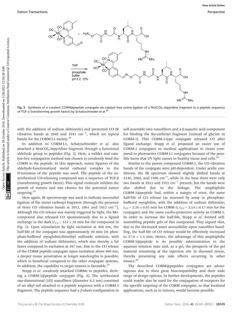
In addition to CORM-L1, Schatzschneider et al. alsoattached a Mo(CO)4-bipyridine fragment through a functionalaldehyde group to peptides (Fig. 3). Here, a milder and cata-lyst-free conjugation method was chosen to covalently bind theCORM to the peptide. In this approach, oxime ligation of thealdehyde-functionalized metal carbonyl complex to theN-terminus of the peptide was used. The peptide of the so-synthesized CO-releasing compound was a sequence of TGF-β(transforming growth factor). This signal molecule inhibits thegrowth of tumors and was chosen for the potential tumortargeting.39
Here again, IR spectroscopy was used to indicate successfulligation of the metal carbonyl fragment (through the presenceof three CO vibration bands at 2015, 1861 and 1812 cm−1).Although the CO release was mainly triggered by light, the Mo-compound also released CO spontaneously due to a ligandexchange in the dark (t1/2 = 123 ± 38 min for the compound inFig. 3). Upon stimulation by light excitation at 468 nm, thehalf-life of the conjugate was approximately 40 min (in phos-phate-buffered myoglobin/dimethyl sulfoxide solution, withthe addition of sodium dithionite), which was thereby a bitfaster compared to excitation at 387 nm. Due to the CO releaseof the CORM peptide conjugate upon excitation above 400 nm,a deeper tissue penetration at longer wavelengths is possible,which is beneficial compared to the other conjugate systems.In addition, the capability to target tumors is favorable.39
Stupp et al. covalently attached CORMs to peptides, deriv-ing a CORM-3@peptide conjugate (Fig. 4). The synthesizedone-dimensional (1D) nanofibers (diameter 8.2 nm) consistedof an alkyl tail attached to a peptide sequence with a CORM-3fragment. The peptide sequence had a β-sheet-configuration to
self-assemble into nanofibers and a β-aspartic acid componentfor binding the Ru-carbonyl fragment (instead of glycine inCORM-3). This CORM-3-type conjugate released CO afterligand exchange. Stupp et al. proposed an easier use ofCORM-3 conjugates in medical applications in tissue com-pared to photoactive CORM-L1 conjugates because of the poss-ible harm that UV light causes to healthy tissue and cells.40
Similar to the parent compound CORM-3, the CO vibrationbands of the conjugate were pH-dependent. Under acidic con-ditions, the IR spectrum showed slightly shifted bands at2140, 2060, and 1986 cm−1, while in the base there were onlytwo bands at 2023 and 1951 cm−1 present, but the bands werealso shifted due to the linkage. The amphiphilicCORM-3@peptide had, within a margin of error, the samehalf-life of CO release (as assessed by assay in phosphate-buffered myoglobin, with the addition of sodium dithionite;t1/2 = 2.16 ± 0.05 min for CORM-3; t1/2 = 2.14 ± 0.17 min for theconjugate) and the same cardio-protective activity as CORM-3.In order to increase the half-life, Stupp et al. formed self-assembling peptide gels of this compound. They argued that,due to the decreased water accessibility upon nanofiber bund-ling, the half-life of CO release would be effectively increasedto 17.8 ± 1.4 min. Hence, the advantage of this amphiphilicCORM-3@peptide is its possible administration in theaqueous solution state and, as a gel, the prospects of the gelmaterial remaining at the injection site in diseased tissue,thereby preventing any side effects occurring in othertissues.40
The described CORM@peptides conjugates are advan-tageous due to their great biocompatibility and their widerange of design options. In further developments, the peptidescould maybe also be used for the conjugation of receptors forthe specific targeting of the CORM conjugate, so that localizedapplications, such as in tumors, would become possible.
Fig. 3 Synthesis of a covalent CORM@peptide conjugate via catalyst-free oxime ligation of a Mo(CO)4-bipyridine fragment to a peptide sequenceof TGF-β (transforming growth factor) by Schatzschneider et al.39
Dalton Transactions Perspective
This journal is © The Royal Society of Chemistry 2016 Dalton Trans., 2016, 45, 18045–18063 | 18049
Ope
n A
cces
s A
rtic
le. P
ublis
hed
on 1
8 O
ctob
er 2
016.
Dow
nloa
ded
on 1
/28/
2022
12:
56:5
8 A
M.
Thi
s ar
ticle
is li
cens
ed u
nder
a C
reat
ive
Com
mon
s A
ttrib
utio
n-N
onC
omm
erci
al 3
.0 U
npor
ted
Lic
ence
.View Article Online

CORMs@polymers
Following the aforementioned covalently CORM@peptidebound bio-conjugates, Hubbell et al. developed the first nano-sized technical polymer conjugate for CORMs. To achieve aslow diffusion of CO to the targeted tissue, polymeric micelleswith a conjugated CORM-3 component were synthesized. TheCO-releasing micelles were built from a triblock copolymer(PEG-bl-OrnRu-bl-nBu) of poly(ethylene glycol), poly[Ru(CO)3Cl(ornithinate acrylamide)] and poly(n-butylacrylamide) (Fig. 5).41
As the PEG-parts were hydrophilic and the nBu-blocks hydro-phobic spherical, micelles with a hydrodynamic diameter in therange of 30–40 nm were formed (Fig. 6).41
The micelles were stable in water as well as serum andreleased CO slower than CORM-3 itself (conditions: myoglobinassay in aqueous PBS buffered myoglobin, sodium dithionite,and 10 mmol L−1 cysteine). CORM-3 released 0.12 mol CO permol CORM over 120 min. From the micelles, only 0.05 mol CO
per mol CORM was released over the same time. The stericbarrier of the PEG shell hampered the diffusion of an exogen-ous release trigger (such as thiol-containing compounds, likecysteine) in the micelles. In the IR spectra, again the typicalthree CO bands were slightly shifted toward 2135, 2049, and1968 cm−1. The micelles showed a reduced toxicity comparedto the parent CORM, owing to the PEG (as assessed by in vitroMTT-assay against THP-1 Blue™ cells) and a high loadingcapacity of 2500 releasable CO molecules per micelle, as deter-mined by myoglobin assay. Furthermore, their size was con-trollable and they were easy to formulate.41 It cannot beexcluded, however, that the micelles slowed the CO diffusionto the outside myoglobin assay or that part of the CO hadalready been lost from the CORM during the micelle prepa-ration. Further, the myoglobin assay commonly leads to lowCO content values due to a number of diverse reasons,especially for slow releasers. Therefore, sometimes the lower COrelease value is an artifact of a slow CO liberation and not an
Fig. 5 Structure of the triblock copolymer PEG-bl-OrnRu-bl-nBu by Hubbell et al. [PEG: poly(ethylene glycol), OrnRu: poly[Ru(CO)3Cl(ornithinateacrylamide)], nBu: poly(n-butylacrylamide)].41
Fig. 4 Synthesis of a covalent CORM@peptide conjugate of an amphiphilic peptide with a CORM-3 fragment by Stupp et al.40
Perspective Dalton Transactions
18050 | Dalton Trans., 2016, 45, 18045–18063 This journal is © The Royal Society of Chemistry 2016
Ope
n A
cces
s A
rtic
le. P
ublis
hed
on 1
8 O
ctob
er 2
016.
Dow
nloa
ded
on 1
/28/
2022
12:
56:5
8 A
M.
Thi
s ar
ticle
is li
cens
ed u
nder
a C
reat
ive
Com
mon
s A
ttrib
utio
n-N
onC
omm
erci
al 3
.0 U
npor
ted
Lic
ence
.View Article Online

incomplete CO release, as was shown by Berends and Kurz in adetailed spectroscopic study on the light-triggered CO releasefrom two manganese PhotoCORMs (including CORM-L1).42
In nearly the same manner, Boyer et al. synthesized thewater-soluble diblock copolymer P(OEGA)-b-P(VB-R-CORM-2)with poly(oligoethylene glycol methyl ether acrylate) as well aspoly(vinylbenzyl chloride) with different thiol moieties and aCORM-2 component (Fig. 7).43 The CO release from thepolymer–Ru-CORM compounds occurred by a ligand exchange.The half-lives were dependent on the bound thiol component,but in general they were slower compared to the parentCORM-2 (as assessed by assay in phosphate-buffered myoglo-bin, with the addition of sodium dithionite). The half-liveswere t1/2 = 180 min for P(OEGA)-b-P(VB-R1-CORM-2), t1/2 =40 min for P(OEGA)-b-P(VB-R2-CORM-2), t1/2 = 90 min forP(OEGA)-b-P(VB-R3-CORM-2), and approximately 0.8 equiva-lents of CO were released.43
The CO-releasing polymer-Ru-CORMs were developed dueto the promising effects of the released CO against the bacte-rium Pseudomonas aeruginosa and the associated possible useof the polymer as an antibiotic. For this possible application,the polymers were tested as an inhibitor of the growth, biofilmformation, and cell viability of the bacterium. The growth ofPseudomonas aeruginosa was in fact reduced by about 90%compared to untreated cultures, with the best results obtainedwith P(OEGA)-b-P(VB-R2-CORM-2). In addition, this positive
result for P(OEGA)-b-P(VB-R2-CORM-2) was supported by theinhibition of biofilm formation, which was also decreased byabout 95%, and the cell viability tests, whereby the number ofcells were reduced by up to 94% through the polymer–Ru-CORMs. All the results were compared to the parent CORM-2,where the better results for the polymer–Ru-CORMs showedthe benefits of CORMs being bound to polymers.43
Following up these good performances, Boyer et al. alsosynthesized micelles of a slightly varied diblock copoly-mer P(OEGA)-b-P(4VP), again with the hydrophilic polymerpoly(oligoethylene glycol methyl ether acrylate) and thehydrophobic polymer poly(4-vinylpyridine) with a CORM-2component (Fig. 8). The aim of using these micelleswas to improve the water solubility, enhance the half-lives, and to utilize the EPR-effect as in the work ofHubbell et al.41,44
The synthesized CORM@polymer was, among other things,analyzed with IR spectroscopy to control the presence ofthe CO vibrational bands (1800–2100 cm−1), which werefound to be comparable to those of free CORM-2 (2136, 2065,2017 cm−1) and the above-mentioned diblock copolymerP(OEGA)-b-P(VB-R-CORM-2).43–45 The CORM conjugates byBoyer et al. released CO through a pH change, which wasdifferent from the cysteine trigger for the CORM@micelles syn-thesized by Hubbell et al. By increasing the pH value from 4 to7 and then up to 9, the half-lives of CO release (as assessed by
Fig. 6 Scheme of the CORM-3@micelle system by Hubbell et al. Reprinted with permission from ref. 41. Copyright 2010 American ChemicalSociety.
Fig. 7 Structure of the diblock copolymer P(OEGA)-b-P(VB-R-CORM-2) synthesized by Boyer et al. [P(OEGA): poly(oligoethylene glycol methylether acrylate), P(VB): poly(vinylbenzyl chloride), R1: 1-thio-β-D-glucose sodium salt, R2: 1-thiolglycerol, R3: 2-diethylaminoethanethiolhydrochloride].43
Dalton Transactions Perspective
This journal is © The Royal Society of Chemistry 2016 Dalton Trans., 2016, 45, 18045–18063 | 18051
Ope
n A
cces
s A
rtic
le. P
ublis
hed
on 1
8 O
ctob
er 2
016.
Dow
nloa
ded
on 1
/28/
2022
12:
56:5
8 A
M.
Thi
s ar
ticle
is li
cens
ed u
nder
a C
reat
ive
Com
mon
s A
ttrib
utio
n-N
onC
omm
erci
al 3
.0 U
npor
ted
Lic
ence
.View Article Online

assay in phosphate-buffered myoglobin, with the addition ofsodium dithionite) also increased from 30 to over 50 and up to60 min, with a release of approximately 0.6 equivalents of COper 4-vinylpyridine unit.44
A PhotoCORM@polymer conjugate system, including thelight-triggered Mn(CO)3-fragment, was synthesized by Kunzet al. to selectively accumulate in tumor tissue due to the EPR-effect. The macromolecular carrier systems used were copoly-mers formed by the copolymerization of methacrylate ormethacrylamide together with a linker unit that carried abis(pyridylmethyl)amine ligand to bind the CORM-fragment(Fig. 9). A second PhotoCORM@polymer conjugate wasextended with a polylactide (PLA) side chain.46
These PhotoCORM@polymer conjugates had a hydro-dynamic diameter of 10 nm and a manganese content of 8.9%without PLA and 2.8% with PLA. Due to their size, their distri-bution within the body is limited to the vascular system. Bothconjugates showed nearly the same three CO vibration bands,i.e., at 2037, 1948, and 1934 cm−1 versus 2036, 1945, and1929 cm−1, which demonstrated that the CORM was still intactafter the reaction. The metal carbonyl in the polymer conju-gate still acted as PhotoCORM and released CO, with a half-lifeof 20 min (as assessed by assay in phosphate-buffered myoglo-bin/dimethyl sulfoxide solution, with the addition of sodiumdithionite). Compound Mn(CO)3@polymer1 had no significantcytotoxicity, whereas Mn(CO)3@polymer2 was toxic, eventhough the individual polymer component parts and theCORM were not (as tested against Hct116 human colon carci-noma and HepG2 human hepatoma cells). These studiesshowed that the design of such polymeric carriers is easilyadaptable to the needs of further applications, but that thecytotoxicity of such conjugates is more complex.46
A benefit shown by the above-mentioned CORM@polymerconjugates are their tremendous design options and therebythe possibility to match the application and the polymer. Inaddition polymers offer the possibility to adapt their size for
use of the EPR-effect to accumulate the conjugates in diseasedtissue. However, one has to pay attention to the toxicity of thepolymer conjugate and the residues after CO release, as shownby Kunz et al.46
CORMs@nanoparticles
As not only polymers are applicable to the EPR-effect, otherresearch groups have focused on nanoparticles as CORM con-jugates. Schatzschneider et al., for example, studied the attach-ment of PhotoCORM-L1 via a copper-catalyzed azide–alkyne1,3-dipolar cycloaddition (CuAAC, “click reaction”) to obtainfunctionalized silicium dioxide nanoparticles (Fig. 10). Themajor benefits of this nano-scale carrier system were thesimple synthesis, controllable size distribution, and biocom-patibility of the silicium dioxide nanoparticles.47
Due to the mild synthesis conditions, the PhotoCORM-L1was stable during the “click reaction”, as verified by IR (CObands at 2050 and 1958 cm−1) and still tested as photoactivein the SiO2 composite material (cf. Fig. 10). The so-decoratedPhotoCORM nanoparticles released 2 to 3 equivalents of COper mole of complex upon irradiation (as assessed by myoglo-bin assay in phosphate-buffered aqueous dimethyl sulfoxidesolution, with the addition of sodium dithionite). However,decoration of the nanoparticle surface gave only 22% surfacefunctionalization due to the limited accessibility of most ofthe surface sites.47
More recently, Schatzschneider et al. attached the [Mn(CO)3(tris(pyrazolyl)methane)]+-fragment to azido-functiona-lized nanodiamonds using the aforementioned CuAAC clickreaction (Fig. 11).48 The advantages of nanodiamonds are agood cellular uptake in order to achieve accumulation in dis-eased tissue and their non-toxicity.49,50 The resulting nano-scale carrier compounds had a content of 0.47 wt% manga-nese and a size of about 10 nm. Although the CORM-fragmentwas still photoactive, quantitative measurement of the COrelease using the myoglobin assay (in phosphate-buffered solu-
Fig. 8 Structure of the diblock copolymer P(OEGA)-b-P(4VP-CORM-2) synthesized by Boyer et al. [P(OEGA): poly(oligoethylene glycol methylether acrylate), P(4VP): poly(4-vinylpyridine)].44
Perspective Dalton Transactions
18052 | Dalton Trans., 2016, 45, 18045–18063 This journal is © The Royal Society of Chemistry 2016
Ope
n A
cces
s A
rtic
le. P
ublis
hed
on 1
8 O
ctob
er 2
016.
Dow
nloa
ded
on 1
/28/
2022
12:
56:5
8 A
M.
Thi
s ar
ticle
is li
cens
ed u
nder
a C
reat
ive
Com
mon
s A
ttrib
utio
n-N
onC
omm
erci
al 3
.0 U
npor
ted
Lic
ence
.View Article Online

tion, with the addition of dimethyl sulfoxide and sodiumdithionite) was not possible due to the formation of cloudydispersions. IR spectroscopy showed that the CO vibration
bands at 2051 and 1961 cm−1 were identical to those of theparent compound CORM-L1 and were not shifted by attach-ment to the nanocarrier.48
Fig. 10 Synthesis of PhotoCORM-L1-functionalized silicium dioxide nanoparticles by Schatzschneider et al.47
Fig. 9 Synthesis of the PhotoCORM Mn(CO)3@polymer1 (a) and Mn(CO)3@polymer2 (b) conjugates by Kunz et al. (HPMA: N-(2-hydroxypropyl)methacrylamide, AIBN: azobisisobutyronitrile, PLA: polylactide).46
Dalton Transactions Perspective
This journal is © The Royal Society of Chemistry 2016 Dalton Trans., 2016, 45, 18045–18063 | 18053
Ope
n A
cces
s A
rtic
le. P
ublis
hed
on 1
8 O
ctob
er 2
016.
Dow
nloa
ded
on 1
/28/
2022
12:
56:5
8 A
M.
Thi
s ar
ticle
is li
cens
ed u
nder
a C
reat
ive
Com
mon
s A
ttrib
utio
n-N
onC
omm
erci
al 3
.0 U
npor
ted
Lic
ence
.View Article Online

In addition Kunz et al. immobilized CORM-3 analogs onthe surface of maghemite (Fe2O3) iron oxide nanoparticles(IONPs) using D/L-dihydroxyphenylalaninato (DOPA) anchoringligands (Fig. 12).51
Maghemite nanoparticles are readily accessible.52 Thedihydroxyphenyl (catecholate) group strongly coordinates to
Fe3+ on the IONP surface. Magnetic IONPs can be heated in analternating magnetic field. This technique is, inter alia,applied in the hyperthermia/thermal ablation treatment ofcancer tissue.53 The idea of combining CORMs with IONPswas that magnetic iron oxide nanoparticles decorated withtemperature-sensitive CORMs on their surface could be trig-gered to release CO by heating of the IONP when stimulated byan external alternating magnetic field (Fig. 13).51
The proof-of-concept study showed that the half-life of COrelease from [RuCl(CO3) (µ-DOPA)]@maghemite nano-particles was halved from 13 ± 2 min to 7 ± 2 min by apply-ing an external alternating magnetic field (31.7 kAm−1, 247kHz, 25 °C, 39.9 mTesla; both at 25 °C solution temperaturein MOPS-buffered myoglobin assay, with the addition ofsodium dithionite). However, the ligand-labile CORM-3analog (as CORM-3 itself)20,23 was already susceptible to COsubstitution by the Na2SO3/protein environment of the myo-globin assay. In addition, the IONPs featured a low watersolubility and were intensely dark colored, which made theCO-release measurement difficult using optical absorption,which is part of the standard protocol for the myoglobinassay.51 A subsequent study addressed this problem using amodified protocol for the myoglobin assay in which theCORM@IONP system was incorporated into polymer beads(see below).
Fig. 12 Synthesis of CORM-functionalized iron oxide nanoparticles(IONPs) using an Fe3+-affine catecholate linker with a CORM-3 analog.The maghemite IONPs had a hydrodynamic diameter of (8 ± 2) nm,which increased to (11 ± 3) nm after functionalization withD/L-dihydroxyphenylalanine. Redrawn from ref. 51 with permission fromThe Royal Society of Chemistry.
Fig. 13 Schematic presentation of triggered CO-release from a temperature-sensitive CORM on the surface of magnetic iron oxide nanoparticles(IONPs) through heating by an external alternating magnetic field. Reprinted from ref. 51 with permission from The Royal Society of Chemistry.
Fig. 11 Synthesis of PhotoCORM-L1-functionalized nanodiamonds (ND) by Schatzschneider et al.48
Perspective Dalton Transactions
18054 | Dalton Trans., 2016, 45, 18045–18063 This journal is © The Royal Society of Chemistry 2016
Ope
n A
cces
s A
rtic
le. P
ublis
hed
on 1
8 O
ctob
er 2
016.
Dow
nloa
ded
on 1
/28/
2022
12:
56:5
8 A
M.
Thi
s ar
ticle
is li
cens
ed u
nder
a C
reat
ive
Com
mon
s A
ttrib
utio
n-N
onC
omm
erci
al 3
.0 U
npor
ted
Lic
ence
.View Article Online

CORMs@nanoparticles are also controllable in size, easyto synthesize and are applicable to the EPR-effect, sameas the CORMs@polymers. However, different to themore often described PhotoCORM conjugate systems, theCORM@magnetic nanoparticle conjugate prepared by Kunzet al. offered a new trigger mechanism, whereby it was the firstknown conjugate to utilize highly localized heating for COrelease, akin to hyperthermia treatment, through a distantalternating magnetic field. This is a benefit for controllabletherapeutic applications as the CO release may be started andstopped at the specific target through an on/off alternatingmagnetic field.
CORM@dendrimers
A metallodendritic PhotoCORM was synthesized by Smithet al. that exhibited potential as a drug carrier in cellularsystems. They made use of their inherent ability to accumulatein tumor tissue due to the EPR-effect. Therefore, the MnBr(CO)3-fragment was attached to a diaminobutane-polypyridyl dendri-meric scaffold. In addition, this strategy does not set small mole-cular degradation products free; after CO release, the remaining
metal fragment is still bound to the macromolecular conjugate,which prevents possible side effects occurring (Fig. 14).54
Depending on the number of attached photoactive CORM-fragments, the CORM-dendrimers released between 8 and 15CO ligands per dendrimer molecule (that is two CO ligandsper CORM unit). The half-life in CO release was 14.5 to16.8 min (as assessed by myoglobin assay in PBS buffereddimethyl sulfoxide solution, with the addition of sodiumdithionite). The metallo-dendrimer compounds were stable inaqueous solution and air in the absence of light, and the CObands in the conjugate were found at 2020, 1920, and1900 cm−1.54
CO@MOFs
Another solid-storage material for CO was synthesized byMetzler-Nolte et al. The group used the biocompatible metal–organic frameworks (MOFs) MIL-88B(Fe) and NH2-MIL-88B(Fe)to adsorb CO at their accessible coordinative unsaturatedmetal sites (CUSs) (Fig. 15). A benefit of the MOFs could betheir so-called breathing effect,55,56 which refers to the revers-ible swelling of the flexible MOF pores due to adsorption of a
Fig. 14 Schematic structure of the PhotoCORM@dendrimer synthesized by Smith et al.54
Dalton Transactions Perspective
This journal is © The Royal Society of Chemistry 2016 Dalton Trans., 2016, 45, 18045–18063 | 18055
Ope
n A
cces
s A
rtic
le. P
ublis
hed
on 1
8 O
ctob
er 2
016.
Dow
nloa
ded
on 1
/28/
2022
12:
56:5
8 A
M.
Thi
s ar
ticle
is li
cens
ed u
nder
a C
reat
ive
Com
mon
s A
ttrib
utio
n-N
onC
omm
erci
al 3
.0 U
npor
ted
Lic
ence
.View Article Online

solvent. This characteristic could potentially be used for con-trolling the opening and closing of the MOF pores and therebyto control the CO release.57
After their microwave-assisted synthesis, the highly porousMOFs were activated at 550 K. Mössbauer spectroscopy showedthat the terminal water molecules at the iron atoms (cf.Fig. 15) were first removed at around 450 K, giving coordina-tively unsaturated FeIII species. Afterwards, the terminal chlor-ide anions were reductively eliminated, generating FeII CUSs(cf. Fig. 15). Following this activation, the CO adsorptionprocess for both MOFs was performed at 98 K. The IR spec-trum of MIL-88B(Fe) showed the typical CO stretchingvibrations at 2181 and 2167 cm−1 due to the different oxi-dation states of iron in the MOF. In comparison, NH2-MIL-88B(Fe) only showed one CO band at 2169 cm−1. The absence ofthe second band was explained by the less-stable FeIII bindingstrength to CO. The amino ligand, as an electron donor,lowers the Lewis acidity of the CUSs and thereby the bindingstrength between the iron species and the adsorbed CO.Probing the CO release with the myoglobin assay (at 37 °C inphosphate-buffered myoglobin, with the addition of sodiumdithionite) showed also a difference for both MOFs. CO-loadedNH2-MIL-88B(Fe) released more CO with a higher half-lifethan MIL-88B(Fe) owing to more activated, accessible CUSs inthe former MOF (0.69 μmol CO per mg NH2-MIL-88B(Fe), t1/2 =76 min versus 0.36 μmol CO per mg MIL-88B(Fe), t1/2 =38 min). This could be explained by the lower binding strengthin NH2-MIL-88B(Fe), which facilitates the CO removal. Also, inboth cases, CO release is accompanied by degradation of theMIL materials.57
CORM@protein cages
Other biocompatible and not cytotoxic (as assessed by in vitroMTT-assay against HEK293/KB-Fluc cells) conjugates, like theabove-mentioned peptides, are self-assembled protein cages.Ueno et al. coordinated photoactive manganese-carbonyl moi-eties into a mutant of the iron storage protein complex ferritinin order to control the light-induced release of CO.58 Similar tothe above-mentioned metallodendritic PhotoCORMs, the IRspectrum of the protein cage showed three CO vibration bandsat 2028, 2011, and 1917 cm−1. The half-life of CO release afterirradiation with visible light at 456 nm was found to be 2.5 ±0.2 min (as assessed by myoglobin assay, PBS buffer, withsodium dithionite added), which is four times faster than thatof CORM-1 under the same conditions (11.4 ± 0.8 min). Inaddition, the cellular uptake of this CORM conjugate was high(0.35% at 1.76 × 10⁻¹⁰ μmol per cell, 0.33% at 1.64 × 10⁻¹⁰
μmol per cell, as measured by ICP/MS with HEK293/KB-Fluccells).58
Encapsulated CORMsCORMs@poly(L-lactide-co-D/L-lactide) (non-wovens)
A different approach related to the conjugation was used bySchiller et al. and the following CORM host systems, wherebythe CORMs were not covalently linked to but were encapsu-lated into the scaffold and thereby the metal fragments remaintrapped in the scaffold after CO release, which avoids toxicside effects. At first Schiller et al. incorporated the photoactiveCORM-1 in biocompatible polymeric non-wovens (Fig. 16).59
By an electrospinning technique, CORM-1 was incorporatedinto the cyto-compatible polymer poly(L-lactide-co-D/L-lactide).Nanofibrous non-wovens with a diameter of about 1 μm and atmost 14.8 wt% embedded, homogenous distributed CORM-1were formed. This CO-releasing material was porous due to amarginal CO release during the synthesis procedure. The COrelease measured by a portable CO detector depended on thewavelength of the light used for irradiation (365 nm: t1/2 =309 s; 480 nm: t1/2 = 1289 s). At least 3.4 μmol of CO per mgsample was released. In experiments against 3T3 mouse fibro-blast cells, the non-wovens were characterized as nontoxic.Furthermore, the leaching of CORM-1 out of the fibers in
Fig. 15 Structure of a µ3-oxo-bridged trinuclear secondary buildingunit of MIL-88B(Fe) and NH2-MIL-88B(Fe). The two terminal aqualigands given with their ellipses are potentially coordinatively unsatu-rated metal sites (CUSs). Generated from the deposited cif-file (CCDC285810, Refcode YEDKOI) for the isostructural Cr compound.
Fig. 16 Scheme of the CORM@non-wovens by Schiller et al. Adaptedfrom ref. 59 with permission from The Royal Society of Chemistry.
Perspective Dalton Transactions
18056 | Dalton Trans., 2016, 45, 18045–18063 This journal is © The Royal Society of Chemistry 2016
Ope
n A
cces
s A
rtic
le. P
ublis
hed
on 1
8 O
ctob
er 2
016.
Dow
nloa
ded
on 1
/28/
2022
12:
56:5
8 A
M.
Thi
s ar
ticle
is li
cens
ed u
nder
a C
reat
ive
Com
mon
s A
ttrib
utio
n-N
onC
omm
erci
al 3
.0 U
npor
ted
Lic
ence
.View Article Online

water as well as during the irradiation was low (0.1–2.4%)owing to the insolubility of CORM-1 in water. However, waterinsoluble CORM-1 is turned water soluble by encapsulation inthe water-soluble polymer frame. The non-wovens are appli-cable in biological systems, such as in skin patches.59
The polymer poly(L-lactide-co-D/L-lactide) and electro-spinning were also utilized by Schiller et al. to encapsulate thePhotoCORM dodecacarbonyl-tetrakis(μ3-propanethiolato) tetra-manganese(I). Again, nanoporous fibers were formed, whichwere slightly larger (1.3–2.1 μm) than formed for theCORM-1@poly(L-lactide-co-D/L-lactide) conjugate. Cytotoxicitytests against 3T3 mouse fibroblast cells showed no cytotoxiceffects of the CORM conjugate. Light-induced measurementswith a LED lamp (14 mW cm−2) and a portable CO detectorshowed a dependency of the CO release on the wavelength ofthe light used for irradiation (365 nm: t30 min = 10.7 μmol ofreleased CO per mg sample; 405 nm: t30 min = 8.1–8.3 μmol ofreleased CO per mg sample). In addition, a laser-coupled glassfiber optical device was used with the CORM conjugatewrapped around the device head, such that different lightintensities (2.1, 14, and 28 mW cm−2) could be applied at405 nm. Variation of the intensity with the fiber optic showedthat the CO release increased with the intensity (after 30 min,at 2.1 mW cm−2: 0.5 μmol of released CO per mg sample;14 mW cm−2: 1.2 μmol; 28 mW cm−2: 2.5 μmol). Thereby, thismethod could allow the application of non-wovens not only forskin patches but also in living cells with high spatialresolution.60
CORM-2@cellulose acetate/PEG (tablets)
Up to now, the therapy of gastrointestinal diseases usingCORMs has been limited due to an insufficient controllableadministration of either external (e.g., light) or internalstimuli, such as enzymes. Meinel et al. described the firstCORM system suitable for oral administration using tablets asan easy and targeted way to deliver encapsulated CORMs(Fig. 17). The tablet core consisted of a citric acid buffer,coated (Eudragit E PO, sodium dodecyl sulfate, stearic acid,and talcum) sodium sulfite crystals, and CORM-2. The corewas encased with a semi-permeable membrane of celluloseacetate/PEG.61
By the diffusion of water through the membrane, first thecoating layer around the sodium sulfite crystals and sub-sequently the crystals themselves dissolved. The releasedsulfite then triggered CO release from CORM-2 through ligandsubstitution. The coating around the Na2SO3 crystals separatesthe sulfite trigger and CORM-2 during storage, so that no COis released before water is added. Only in the presence of waterdoes the sulfite enter into solution, where it interacts with theCORM in a CO-ligand displacement reaction. The system deli-vered CO for up to 10 h, with a nearly linear release kineticbetween 30 and 240 min, as measured by an amperometric COdetection system (aqueous solutions, with the addition ofsodium dithionite and simulated gastric or intestinal fluids).The release was slow due to the controlled flux of water intothe tablet core, which could be controlled by varying the mem-
brane thickness. This tablet system uses the advantage of asuitable CORM/trigger combination along with a preset micro-environment to ensure CORM stability during storage. Waterthen becomes a secondary trigger that activates the actualprimary trigger for CO release. Thereby, the tablet is largelyindependent of the environment within the organism, e.g., thepH value or the ligands, such as in proteins. Further, thetablet also retains the resulting possibly toxic Ru-complex afterCO delivery.61,62
In addition to the synthesis and in vitro analysis, the groupof Meinel et al. also obtained for the first time in vivo data ofthese tablets for the prevention of colitis in mice. For this, thegroup changed the coating of the tablet core and the sodiumsulfite crystals to cellulose acetate butyrate in order to increasethe hydrophobicity of the coating. The tablets were then admi-nistered to mice by oral application, thereby only local andhighly concentrated effects in the gastrointestinal tract wereinduced. For the analysis, the CO-hemoglobin (CO-Hb) for-mation was measured, which started 1–3 h after the adminis-tration of the tablets. The tablets showed protection fromcolitis by a reduced colon damage score and a maximal CO-Hblimit of 1.4% (safety threshold: 5–10% CO-Hb).8,63
MnCORM@Al-MCM-41 nanoparticles
Nanoparticles have high surface areas, may be biocompatible,are easy to synthesize, and can accumulate in tissue due to theEPR-effect. Because of this, Mascharak et al. chose nano-particles of the mesoporous silica Al-MCM-41 as a host for theencapsulation of the PhotoCORM [Mn(pqa)(CO)3]ClO4 (pqa:(2-pyridylmethyl)(2-quinolylmethyl)amine) (Fig. 18).64
By ion-exchange and diffusion over 48 h, the cationicCORM was incorporated into the negatively charged cylindricalpores of the Al-MCM-41 nanoparticles with a load of 2.06 wt%Mn. The strong interactions through the exchange were sup-ported by leaching experiments in PBS (leaching of 2 wt%
Fig. 17 Scheme of the CORM@cellulose acetate/PEG system for oraladministration of a tablet by Meinel et al. Reprinted from ref. 61 withpermission from Elsevier.
Dalton Transactions Perspective
This journal is © The Royal Society of Chemistry 2016 Dalton Trans., 2016, 45, 18045–18063 | 18057
Ope
n A
cces
s A
rtic
le. P
ublis
hed
on 1
8 O
ctob
er 2
016.
Dow
nloa
ded
on 1
/28/
2022
12:
56:5
8 A
M.
Thi
s ar
ticle
is li
cens
ed u
nder
a C
reat
ive
Com
mon
s A
ttrib
utio
n-N
onC
omm
erci
al 3
.0 U
npor
ted
Lic
ence
.View Article Online

CORM after 24 h, 12 wt% after 60 h). The embedding ofCORM again was confirmed by IR spectroscopy, whereby amarginal shift of the CO bands was observed compared to theparent CORM (CORM 2033 and 1928 cm−1; Mn-CORM@Al-MCM-41 2040 and 1947 cm−1). CO release in this Al-MCM-41environment was said to be slower (albeit no times were given)compared to the parent CORM (myoglobin assay, PBS buffer,with the addition of sodium dithionite).64
ALF472@bioMOF-1/MCM-41-SO3H/SBA-15-SO3H
Barea et al. also worked on a diffusion approach into a micro-or mesoporous host like that performed by Mascharaks et al.with the MnCORM@Al-MCM-41 nanoparticles. Barea’sgroup incorporated the photoactive CORM [Mn(tacn)(CO)3]Br(tacn: 1,4,7-triazacyclononane), also known as ALF472, intothree different biocompatible porous inorganic or metal–organic framework (MOF) materials as a potential, controllableon/off-switch release system for the phototherapy of skincancer. By a diffusion process over four days in darkness, thenontoxic, water-soluble, air-stable, cationic CORM wasincorporated into the metal–organic framework bioMOF-1{NH2(CH3)2)2[Zn8(adeninate)4(BPDC)6]·8DMF·11H2O (BPDC =4,4′-biphenyldicarboxylate)} as well as the functionalized
mesoporous silica structures MCM-41-SO3H and SBA-15-SO3Hvia cation exchange in DMF or water (Fig. 19).65
The IR spectrum of the product showed only a marginalchange of the CO bands after the diffusion process (2017 and1895 cm−1 for CORM versus 2030 and 1925 cm−1 for theproduct). As the incorporation proceeded by a cation exchangeprocess, no bromide was detected by EDX. In addition, theCORM load determined by EDX was nearly the same forALF472@bioMOF-1 and ALF472@MCM-41-SO3H (0.25 mmoland 0.22 mmol ALF472+ per gram). ALF472@SBA-15-SO3Hshowed the highest CORM load, with 0.53 mmol ALF472+ pergram. This was also mirrored by the CO-release results (asassessed by myoglobin assay at 37 °C in PBS buffer, with theaddition of sodium dithionite). In MCM-41-SO3H and SBA-15-SO3H, the release of CO was slower compared to that of theparent CORM (0.80 mmol CO per mmol of complex after 24 hfor ALF472; 0.65 mmol CO per mmol of complex after 24 h forMCM-41-SO3H; 0.48 mmol CO per mmol of complex after 24 hfor SBA-15-SO3H). In contrast, ALF472@bioMOF-1 released0.82 mmol CO per mmol of complex but decomposed in water,as shown by the leaching experiments, where 85% of the zincof the MOF and 75% of the manganese of the CORM weredetected in solution after 6 h (ICP-OES). The leaching experi-ments also showed that ALF472@MCM-41-SO3H was the mostpotent material. In water, 80% of the CORM remained withinthe material over a period of three days and only a minordegradation of the mesoporous silica (2.9% silicon after 24 hin solution) was observed. Under physiological conditions,85% of the CORM was leached after 1 h, which is promisingfor its applications.65
Covalently-bound CORM conjugatesin combination with encapsulationPolymer@CORM@iron oxide nanoparticles
A ligand-labile CORM, such as CORM-3 and its analogs, hasalready been shown to be susceptible to CO substitution by theNa2SO3/protein environment of the myoglobin assay.20,23 Also,the IONPs used by Kunz et al. featured low water solubility andare intensely dark colored, which made CO-release measure-
Fig. 19 Scheme of the CORM@porous material system synthesized byBarea et al. Adapted with permission from ref. 65. Copyright 2016American Chemical Society.
Fig. 18 Structure of the incorporated CORM and schematic drawing of the host Al-MCM-41 used by Mascharak et al. Adapted in part from ref. 64with permission of The Royal Society of Chemistry.
Perspective Dalton Transactions
18058 | Dalton Trans., 2016, 45, 18045–18063 This journal is © The Royal Society of Chemistry 2016
Ope
n A
cces
s A
rtic
le. P
ublis
hed
on 1
8 O
ctob
er 2
016.
Dow
nloa
ded
on 1
/28/
2022
12:
56:5
8 A
M.
Thi
s ar
ticle
is li
cens
ed u
nder
a C
reat
ive
Com
mon
s A
ttrib
utio
n-N
onC
omm
erci
al 3
.0 U
npor
ted
Lic
ence
.View Article Online

ment by optical absorption with the myoglobin assaydifficult.51 These problems were addressed by Janiak et al. byencapsulation of the CORM@IONP system into polymer beads(Fig. 20).66
In order to achieve water solubility, CORM@IONPs werecoated with different polymers, e.g., dextrans. Using a 500 kDadextran resulted in core–shell structures. Due to the polymershell, the average diameter of the particles increased from11 ± 3 nm to 85 ± 15 nm. TEM pictures showed that the severalCORM-iron oxide nanoparticles are encapsulated into onepolymer sphere. In addition, the half-life of CO releaseincreased from 9 ± 1 min for CORM@IONP to 32 ± 2 min fordextran@CORM@IONP (both at 37 °C in MOPS-buffered myo-globin, with the addition of sodium dithionite). This isreasonable since the polymer shell is a barrier to the COsubstituting Na2S2O4/myoglobin species used in the assay.Dextran@CORM@IONP showed no significant cytotoxic effectsup to 100 µg mL−1 (with less than 4.77% growth inhibition)against the cell lines tested (i.e., A2780, Cal27, and HEK293).66
However, the now water-soluble polymer-coated iron oxidenanoparticles were still not yet suitable for the measurementof CO release using the myoglobin assay due to their stillstrong absorption in the UV/Vis region, but subsequentembedding of the dextran@CORM@IONP nanoparticles into
alginate spheres allowed for magnetic separation as well as forapplying the myoglobin assay.66
Sodium alginate is an anionic polysaccharide, found in thecell walls of brown algae. It is nontoxic and is already used inbiomedical applications. The linear copolymer consists of β-D-mannuronate and α-L-guluronate blocks or sequences. A gel-likematerial is obtained by ionic cross-linkage of the carboxylateand Ca2+-ions via Coulomb interactions.67,68 Dripping a mixtureof dextran@CORM@IONP and sodium alginate into a solutionof CaCl2 led to stable hollow spheres of the Ca2+ cross-linkedalginate gel containing dextran@CORM@IONP (Fig. 20).66
This alginate shell supported the dextran coating in protect-ing the CORM from rapid ligand-exchange-induced CO-displa-cement reactions, while still allowing for permeation of CO tothe solution. The alginate shell also compartmented thehighly absorbing iron oxide nanoparticles so that they couldbe easily separated magnetically from the myoglobin assaysolution, such that the CO release studies became viablewithout interference in the optical path of the UV cell. Forexample, during the assay, the magnetic alginate particlescould be kept at the bottom of the cuvette using a permanentmagnet outside (Fig. 21).66
The alginate spheres were magnetic, had diameters of1–2 mm, and could be stored over a prolonged period of time
Fig. 21 Alginate@dextran@CORM@IONP spheres (cf. Fig. 20) and their arrangement in magnetic fields. (a) No magnet, (b) round magnet, and (c)bar magnet under the glass vessel. Adapted with permission from ref. 66. Copyright 2015 American Chemical Society.
Fig. 20 Scheme of the synthesis of CORM-functionalized iron oxide nanoparticles (IONPs) (IIa), encapsulated in a Dextran polymer coating (IIb) andcollected into an alginate sphere (IIc). Adapted with permission from ref.66. Copyright 2015 American Chemical Society.
Dalton Transactions Perspective
This journal is © The Royal Society of Chemistry 2016 Dalton Trans., 2016, 45, 18045–18063 | 18059
Ope
n A
cces
s A
rtic
le. P
ublis
hed
on 1
8 O
ctob
er 2
016.
Dow
nloa
ded
on 1
/28/
2022
12:
56:5
8 A
M.
Thi
s ar
ticle
is li
cens
ed u
nder
a C
reat
ive
Com
mon
s A
ttrib
utio
n-N
onC
omm
erci
al 3
.0 U
npor
ted
Lic
ence
.View Article Online

in 3-(N-morpholino)propanesulfonic acid (MOPS) buffer. Thehalf-life of CO release increased to 172 ± 27 min at 37 °Cwithout and to 65 ± 5 min with the application of a magneticAC-field (from 9 ± 1 min for CORM@IONP). From the tempera-ture-variable kinetic measurements, an activation energy of78 kJ mol−1 was determined for the CO release from the IONP-surface-bound CORM-3 analog in the alginate@dextranpolymer shell.66
In a follow-up study, oxime-based CORMs69 were similarlycovalently immobilized on IONPs, followed by theoximeCORM@IONP encapsulation in dextran and alginate(Fig. 22).70
The half-life of CO release from the composite materialalginate@dextran@oximeCORM@IONP was 346 ± 83 min at37 °C compared to the parent oximeCORM of 16 ± 1 min (asassessed by myoglobin assay, MOPS buffer, with the additionof sodium dithionite). Thus, the polymer coating again pre-sented a remarkable stabilization with respect to a rapid CO-displacement reaction by Na2S2O4/protein during the myoglo-bin assay. Local magnetic heating of the IONP by applicationof an external alternating magnetic field (31.7 kAm−1, 247kHz, 39.9 mTesla) decreased the half-life of CO release to 153 ±27 min, but the solution temperature was not affected (37 °C).The activation energy for the CO release fromalginate@dextran@oximeCORM@IONPs using an Arrheniusplot gave a value of 62(17) kJ mol−1.70
Conclusion/personal view
CORMs are typically small metal carbonyl molecules. In CORMconjugates, these CORMs have been covalently bound to pep-tides, polymers, nanoparticles, dendrimers, and protein cagesor have been incorporated into non-wovens, tablets, or metal–organic frameworks to improve their stability, to target themto special tissues, to avoid possible side effects, to use theEPR-effect, or to allow for special triggers.
In summary, all the described CORM conjugates, either withthe CORM covalently bound or incorporated, show interestingoptions for biological applications. They are biocompatible andnot cytotoxic or at least are less toxic than the parent CORMcompounds. Although it has to be noted that for the presentedconjugates, different divergent cytotoxicity tests and cell lineswere used. Indeed these in vitro data of the CO release are onlyavailable for some conjugates and these results could differfrom the in vivo profiles. For application of the conjugates,additional in vivo data should be analyzed and compared to thein vitro results as has appeared to have been done for the firsttime by the group of Meinel et al.63 The same as CORMs insolution, these conjugates play a key role in the pharmacoki-netic parameters. They are prodrugs and their action is comple-tely dependent on the in vivo constraints. In addition, it isnecessary to analyze the remaining (metal) fragments or resi-dues after CO release in order to detect if there are any sideeffects and to avoid them in future carrier designs.62,71,72
So far, the absence of side effects seems to be a benefit ofthe incorporated CORMs, where the CORM moieties after COrelease are trapped in the system and cannot affect the tissue.This is not achieved by the exposed CORMs in the conjugates.There, it still has to be analyzed if the potentially cytotoxicruthenium metal moiety reaches the tissue and acts toxic.
For the CO release measurements, most research groupsused a myoglobin assay to show the increased half-lives of COrelease due to the covalent binding or incorporation of theCORMs into the conjugate. One problem that has emerged inthe last few years with this method is the influence of dithio-nite on the CO release. With dithionite, CO release occurswithout the stimulation of a trigger, which alters the results ofthe half-life as well as the amount of released CO calculatedafterwards.20 To overcome this problem, commercial CO detec-tors, where the sensor part is brought in contact with theCORM system, could be used to follow the CO release. First inthe work of Meinel et al.61 and later also in the work of Schilleret al.,59 the disassembly of such a commercial CO detector(Ei207D from Ei Electronics, Shannon, Ireland) was described,whereby the amperometric CO sensing part was achieved in aclosed Erlenmeyer flask, with the rest of the CO detectorremaining outside (connected by a cable to the sensor).Thereby, the CO release into the headspace (gas space) of theflask could be measured directly above the solution or solidCORM system.61 For focused light irradiation, the use of alaser-coupled fiber optical device could be advanced in the invivo applications of PhotoCORMs.60
Many of the described conjugates are geared toward theenhanced permeability and retention (EPR) effect. The dia-meter of nanoparticles and the size of polymers can easily becontrolled and adapted. Thereby, it may be possible toaccumulate the CORM conjugates in the targeted tumor tissuedue to the EPR-effect and then to release the CO there if a suit-able trigger is available (see below).73
Although the design of all the conjugates seems to be prom-ising, the CORMs at large still show problems for the thera-peutic application of CO, irrespective of their assembly into a
Fig. 22 Schematic synthesis of the composite materialalginate@dextran@oximeCORM@IONP, starting with immobilization ofthe oximeCORM on the maghemite nanoparticle surface, followed byencapsulating in dextran (molar mass 500 kDa) and alginate. Adaptedfrom ref. 70 with permission from The Royal Society of Chemistry.
Perspective Dalton Transactions
18060 | Dalton Trans., 2016, 45, 18045–18063 This journal is © The Royal Society of Chemistry 2016
Ope
n A
cces
s A
rtic
le. P
ublis
hed
on 1
8 O
ctob
er 2
016.
Dow
nloa
ded
on 1
/28/
2022
12:
56:5
8 A
M.
Thi
s ar
ticle
is li
cens
ed u
nder
a C
reat
ive
Com
mon
s A
ttrib
utio
n-N
onC
omm
erci
al 3
.0 U
npor
ted
Lic
ence
.View Article Online

conjugate. For the light-triggered CORMs (and conjugates),such as CORM-L1, the trigger excitation with wavelengthsunder 500 nm (i.e., the blue spectral region) has only a lowtissue penetration depth. For CORMs below the skin or insidetissue wavelengths of about 750 nm (i.e., the red spectralregion), it would be highly desirable to trigger the release ofCO without the harmful effects of UV radiation.74 However,longer wavelengths are not yet suitable for CORMs, and alsocannot be adjusted through a conjugation of the CORMs.Exceptions to the case are the CORMs@non-wovens syn-thesized by Schiller et al., because they are designed for asurface application, e.g., to only act on the skin as patches,thereby a deep tissue penetration would not be necessary.
For most of the described conjugates, the release of CO at aspecific target is still a problem, especially if they are used forlocal applications, such as in tumor tissues. Here, the tabletsof Meinel et al. can be emphasized, because of their preciseapplication for gastrointestinal diseases. Most of the CORMconjugates are more suitable for systemic applications, such asagainst inflammations, which need no specific targeting. Anadvantage of the solvent- or pH-triggered CORM conjugates(e.g., CORM-3) would then be the presumably longer lasting ordelayed-release effect of CO, because the trigger needs todiffuse to or into the conjugate and will thereby trigger therelease of CO over an extended time, thus avoiding burstreleases of the parent CORM.
A common problem to all CORMs, irrespective if free orpart of a conjugate, is the steady, spontaneous, low back-ground release of CO, which cannot be completely stopped,even if no trigger seems to be present. It could be supposedthat this background release is triggered by oxidation pro-cesses. Metal carbonyl-based CORMs are intrinsically thermo-dynamically unstable toward O2 and other oxidants, whichcould lead to this unavoidable, steady, low CO release.15
Indeed, for a tissue-targeted therapeutic application, it ishighly desirable that the release only starts at this targettissue. For this, an on/off-switch of the CORM conjugatesystem would be essential. Yet, it may be difficult to controlthese triggers from outside an organism in a medicinal appli-cation. The trigger of a localized heating of CORM@iron oxidenanoparticles through an alternating magnetic AC-field wasthus developed in this direction. However, even this systemhad the problem of a low background release of CO throughthe use of a temperature-sensitive CORM-3 derivative. The con-trolled release is also important for the treatment of diseaseswhere a complete, immediate release of CO would be necess-ary, such as in tumor tissues.
Elaborate designs of CORM conjugates have been achieved,but more research is necessary to design CORM systems thathave the possibility of a controllable on/off-switch for COrelease. Only if this can be achieved may other measures, forexample the addition of receptors to the conjugate for aspecific targeting, seem plausible and sensible.
Further developments for the incorporation of CORMscould be MOFs with switchable linkers, which are able to“open/close” the MOF pores and channels by a trigger and
thereby perhaps achieve a complete, controllable on/off-switchfor the CO release.75 As described above, MOFs that exhibit thebreathing effect55,56 have already been used for the conju-gation of CO by the group of Metzler-Nolte et al.57
References
1 C. C. Romão, W. A. Blättler, J. D. Seixas andG. J. L. Bernardes, Chem. Soc. Rev., 2012, 41, 3571–3583.
2 C. G. Douglas, J. S. Haldane and J. B. Haldane, J. Physiol.,1912, 44, 274–304.
3 J. B. Haldane, Biochem. J., 1927, 21, 1068–1075.4 T. Sjöstrand, Nature, 1949, 164, 580–581.5 L. Wu and R. Wang, Pharmacol. Rev., 2005, 57, 585–630.6 A. K. Mustafa, M. M. Gadalla and S. H. Snyder, Sci.
Signaling, 2009, 68, 1–8.7 T. R. Johnson, B. E. Mann, J. E. Clark, R. Foresti,
C. J. Green and R. Motterlini, Angew. Chem., Int. Ed., 2003,42, 3722–3729.
8 R. Motterlini and L. E. Otterbein, Nat. Rev. Drug Discovery,2010, 9, 728–743.
9 S. W. Ryter, J. Alam and A. M. K. Choi, Physiol. Rev., 2006,86, 583–650.
10 R. Motterlini, J. E. Clark, R. Foresti, P. Sarathchandra,B. E. Mann and C. J. Green, Circ. Res., 2002, 90, e17–e24.
11 J. E. Clark, P. Naughton, S. Shurey, C. J. Green,T. R. Johnson, B. E. Mann, R. Foresti and R. Motterlini,Circ. Res., 2003, 93, e2–e8.
12 J. Niesel, A. Pinto, H. W. P. N’Dongo, K. Merz, I. Ott,R. Gust and U. Schatzschneider, Chem. Commun., 2008, 15,1798–1800.
13 U. Schatzschneider, Inorg. Chim. Acta, 2011, 374, 19–23.14 S. Romanski, B. Kraus, U. Schatzschneider, J.-M. Neudörfl,
S. Amslinger and H.-G. Schmalz, Angew. Chem., Int. Ed.,2011, 50, 2392–2396.
15 J. D. Seixas, A. Mukhopadhyay, T. Santos-Silva,L. E. Otterbein, D. J. Gallo, S. S. Rodrigues, B. H. Guerreiro,A. M. L. Gonçalves, N. Penacho, A. R. Marques,A. C. Coelho, P. M. Reis, M. J. Romão and C. C. Romão,Dalton Trans., 2013, 42, 5985–5998.
16 G. L. Bannenberg and H. L. Vieira, Epxert Opin. Ther. Pat.,2009, 19, 663–682.
17 P. C. Kunz, H. Meyer, A. Schmidt and C. Janiak, Nano-SizedCarriers for Carbon Monoxide Releasing Molecules, inEUROBIC 11, ed. J. M. Gonzaléz-Pérez, A. Matilla-Hernández and J. Niclós-Gutiérrez, Medimond, Bologna,Italy, 2013, p. 25–30.
18 T. R. Johnson, B. E. Mann, I. P. Teasdale, H. Adams,R. Foresti, C. J. Green and R. Motterlini, Dalton Trans.,2007, 1500–1508.
19 J. D. Seixas, M. F. A. Santos, A. Mukhopadhyay,A. C. Coelho, P. M. Reis, L. F. Veiros, A. R. Marques,N. Penacho, A. M. L. Gonçalves, M. J. Romão,G. J. L. Bernardes, T. Santos-Silva and C. C. Romão, DaltonTrans., 2015, 44, 5058–5075.
Dalton Transactions Perspective
This journal is © The Royal Society of Chemistry 2016 Dalton Trans., 2016, 45, 18045–18063 | 18061
Ope
n A
cces
s A
rtic
le. P
ublis
hed
on 1
8 O
ctob
er 2
016.
Dow
nloa
ded
on 1
/28/
2022
12:
56:5
8 A
M.
Thi
s ar
ticle
is li
cens
ed u
nder
a C
reat
ive
Com
mon
s A
ttrib
utio
n-N
onC
omm
erci
al 3
.0 U
npor
ted
Lic
ence
.View Article Online

20 S. McLean, B. E. Mann and R. K. Poole, Anal. Biochem.,2012, 427, 36–40.
21 V. S. Stoll and J. S. Blanchard, Methods Enzymol., 2009, 463,43–56.
22 S. H. Heinemann, T. Hoshi, M. Westerhausen andA. Schiller, Chem. Commun., 2014, 50, 3644–3660.
23 T. Santos-Silva, A. Mukhopadhyay, J. D. Seixas,G. J. L. Bernandes, C. C. Romão and M. J. Romao, J. Am.Chem. Soc., 2011, 133, 1192–1195.
24 L. Yuan, W. Y. Lin, L. Tan, K. B. Zheng and W. M. Huang,Angew. Chem., Int. Ed., 2013, 52, 1628–1630.
25 B. W. Michel, A. R. Lippert and C. J. Chang, J. Am. Chem.Soc., 2012, 134, 15668–15671.
26 M. Klein, U. Neugebauer, A. Gheisari, A. Malassa,T. M. Jazzazi, F. Froehlich, M. Westerhausen, M. Schmittand J. Popp, J. Phys. Chem. A, 2014, 118, 5381–5390.
27 H. J. Vreman and D. K. Stevenson, Anal. Biochem., 1988,168, 31–81.
28 M. Balazy and H. Jiang, Acta Haematol., 2000, 103, 78–83.29 R. Motterlini, B. E. Mann and R. Foresti, Expert Opin.
Invest. Drugs, 2005, 14, 1305–1318.30 R. Mottelini, B. E. Mann, T. R. Johnson, J. E. Clark,
R. Foresti and C. J. Green, Curr. Pharm. Des., 2003, 9, 2525–2539.
31 S. García-Gallego and G. J. L. Bernardes, Angew. Chem., Int.Ed., 2014, 53, 9712–9721.
32 U. Schatzschneider, Br. J. Pharmacol., 2015, 172, 1638–1650.
33 J. N. Talbert and J. M. Goddard, Colloids Surf., B, 2012, 93,8–19.
34 Y. Matsumura and H. Maeda, Cancer Res., 1986, 46, 6387–6392.
35 H. Maeda, J. Wu, T. Sawa, Y. Matsumura and K. Hori,J. Controlled Release, 2000, 65, 271–284.
36 L. D. D’Andrea, A. Romanelli, R. Di Stasi and C. Pedone,Dalton Trans., 2010, 39, 7625–7635.
37 E. Neuse, Met.-Based Drugs, 2008, 2008, 1–19.38 H. Pfeiffer, A. Rojas, J. Niesel and U. Schatzschneider,
Dalton Trans., 2009, 4292–4298.39 H. Pfeiffer, T. Sowik and U. Schatzschneider, J. Organomet.
Chem., 2013, 734, 17–24.40 J. B. Matson, M. J. Webber, V. K. Tamboli, B. Weber and
S. I. Stupp, Soft Matter, 2012, 8, 6689–6692.41 U. Hasegawa, A. J. van der Vlies, E. Simeoni, C. Wandrey and
J. A. Hubbell, J. Am. Chem. Soc., 2010, 132, 18273–18280.42 H.-M. Berends and P. Kurz, Inorg. Chim. Acta, 2012, 380,
141–147.43 D. Nguyen, T.-K. Nguyen, S. A. Rice and C. Boyer,
Biomacromolecules, 2015, 16, 2776–2786.44 D. Nguyen, N. N. M. Adnan, S. Oliver and C. Boyer,
Macromol. Rapid Commun., 2016, 37, 739–744.45 B. F. G. Johnson, R. D. Johnston and J. Lewis, J. Chem. Soc.
A, 1969, 792–797.46 N. E. Brückmann, M. Wahl, G. J. Reiß, M. Kohns,
W. Wätjen and P. C. Kunz, Eur. J. Inorg. Chem., 2011, 29,4571–4577.
47 G. Dördelmann, H. Pfeiffer, A. Birkner andU. Schatzschneider, Inorg. Chem., 2011, 50, 4362–4367.
48 G. Dördelmann, T. Meinhardt, T. Sowik, A. Krueger andU. Schatzschneider, Chem. Commun., 2012, 48, 11528–11530.
49 O. Faklaris, V. Joshi, T. Irinopoulou, P. Tauc, M. Sennour,H. Girard, C. Gesset, J.-C. Arnault, A. Thorel, J.-P. Boudou,P. A. Curmi and F. Treussart, ACS Nano, 2009, 3, 3955–3962.
50 V. Vaijayanthimala and H.-C. Chang, Nanomedicine, 2009,4, 47–55.
51 P. C. Kunz, H. Meyer, J. Barthel, S. Sollazzo, A. M. Schmidtand C. Janiak, Chem. Commun., 2013, 49, 4896–4898.
52 G. A. van Ewijk, G. J. Vroege and A. P. J. Philip, J. Magn.Magn. Mater., 1999, 201, 31–33.
53 A. J. Cole, V. C. Yang and A. E. David, Trends Biotechnol.,2011, 29, 323–332.
54 P. Govender, S. Pai, U. Schatzschneider and G. S. Smith,Inorg. Chem., 2013, 52, 5470–5478.
55 C. Serre, C. Mellot-Draznieks, S. Surblé, N. Audebrand,Y. Filinchuk and G. Férey, Science, 2007, 315, 1828–1831.
56 G. Férey and C. Serre, Chem. Soc. Rev., 2009, 38, 1380–1399.57 M. Ma, H. Noei, B. Mienert, J. Niesel, E. Bill, M. Muhler,
R. A. Fischer, Y. Wang, U. Schatzschneider and N. Metzler-Nolte, Chem. – Eur. J., 2013, 19, 6785–6790.
58 K. Fujita, Y. Tanaka, S. Abe and T. Ueno, Angew. Chem., Int.Ed., 2016, 55, 1056–1060.
59 C. Bohlender, S. Gläser, M. Klein, J. Weisser, S. Thein,U. Neugebauer, J. Popp, R. Wyrwa and A. Schiller, J. Mater.Chem. B, 2014, 2, 1454–1463.
60 S. Gläser, R. Mede, H. Görls, S. Seupel, C. Bohlender,R. Wyrwa, S. Schirmer, S. Dochow, G. U. Reddy, J. Popp,M. Westerhausen and A. Schiller, Dalton Trans., 2016, 45,13222–13233.
61 C. Steiger, T. Lühmann and L. Meinel, J. Controlled Release,2014, 189, 46–53.
62 I. C. Winburn, K. Gunatunga, R. D. McKernan,R. J. Walker, I. A. Sammut and J. C. Harrison, Basic Clin.Pharmacol. Toxicol., 2012, 111, 31–41.
63 C. Steiger, K. Uchiyama, T. Takagi, K. Mizushima,Y. Higashimura, M. Gutmann, C. Hermann, S. Botov,H.-G. Schmalz, Y. Naito and L. Meinel, J. Controlled Release,2016, 239, 128–136.
64 M. A. Gonzales, H. Han, A. Moyes, A. Radinos, A. J. Hobbs,N. Coombs, S. R. J. Oliver and P. K. Mascharak, J. Mater.Chem. B, 2014, 2, 2107–2113.
65 F. J. Carmona, S. Rojas, P. Sánchez, H. Jeremias,A. R. Marques, C. C. Romão, D. Choquesillo-Lazarte,J. A. R. Navarro, C. R. Maldonado and E. Barea, Inorg.Chem., 2016, 55, 6525–6531.
66 H. Meyer, F. Winkler, P. Kunz, A. M. Schmidt,A. Hamacher, M. U. Kassack and C. Janiak, Inorg. Chem.,2015, 54, 11236–11246.
67 K. I. Draget, Alginates, in Handbook of hydrocolloidsm, ed.G. O. Phillips and P. A. Williams, 2009, pp. 807–828.
68 S. N. Pawar and K. J. Edgar, Biomaterials, 2012, 33, 3279–3305.
Perspective Dalton Transactions
18062 | Dalton Trans., 2016, 45, 18045–18063 This journal is © The Royal Society of Chemistry 2016
Ope
n A
cces
s A
rtic
le. P
ublis
hed
on 1
8 O
ctob
er 2
016.
Dow
nloa
ded
on 1
/28/
2022
12:
56:5
8 A
M.
Thi
s ar
ticle
is li
cens
ed u
nder
a C
reat
ive
Com
mon
s A
ttrib
utio
n-N
onC
omm
erci
al 3
.0 U
npor
ted
Lic
ence
.View Article Online

69 L. Oresmaa, H. Tarvainen, K. Machal and M. Haukka,Dalton Trans., 2012, 41, 11170–11175.
70 H. Meyer, M. Brenner, S.-P. Höfert, T.-O. Knedel,P. C. Kunz, A. M. Schmidt, A. Hamacher, M. U. Kassackand C. Janiak, Dalton Trans., 2016, 45, 7605–7615.
71 H. Song, H. Zhao, Y. Qu, Q. Sun, F. Zhang, Z. Du, W. Liang,Y. Qi and P. Yang, J. Periodontal Res., 2011, 46, 48–57.
72 M. Desmard, K. S. Davidge, O. Bouvet, D. Morin, D. Roux,R. Foresti, J. D. Ricard, E. Denamur, R. K. Poole,
P. Montravers, R. Motterlini and J. Boczkowski, FASEB J.,2009, 23, 1023–1031.
73 H. Maeda, Bioconjugate Chem., 2010, 21, 797–802.74 F. Vasefi, N. MacKinnon and D. L. Farkas, Hyperspectral
and Multispectral Imaging in Dermatology, in Imaging inDermatology, ed. M. R. Hamblin, P. Avci and G. K. Gupta,Academic Press, Elsevier, 2016, p. 187.
75 S. Castellanos, F. Kapteijn and J. Gascon, CrystEngComm,2016, 18, 4006–4012.
Dalton Transactions Perspective
This journal is © The Royal Society of Chemistry 2016 Dalton Trans., 2016, 45, 18045–18063 | 18063
Ope
n A
cces
s A
rtic
le. P
ublis
hed
on 1
8 O
ctob
er 2
016.
Dow
nloa
ded
on 1
/28/
2022
12:
56:5
8 A
M.
Thi
s ar
ticle
is li
cens
ed u
nder
a C
reat
ive
Com
mon
s A
ttrib
utio
n-N
onC
omm
erci
al 3
.0 U
npor
ted
Lic
ence
.View Article Online