Co-ordination of the Cardiac Cycle SBI3U. The heart is made of cardiac muscle. When the cells...
12
Co-ordination of the Cardiac Cycle SBI3U
-
Upload
willa-baker -
Category
Documents
-
view
215 -
download
0
Transcript of Co-ordination of the Cardiac Cycle SBI3U. The heart is made of cardiac muscle. When the cells...
Co-ordination of the Cardiac CycleSBI3U

• The heart is made of cardiac muscle.
• When the cells receive an electrical impulse they contract - causing a heartbeat.
• Cardiac muscle is myogenic - it can contract on its own, without needing nerve impulses.

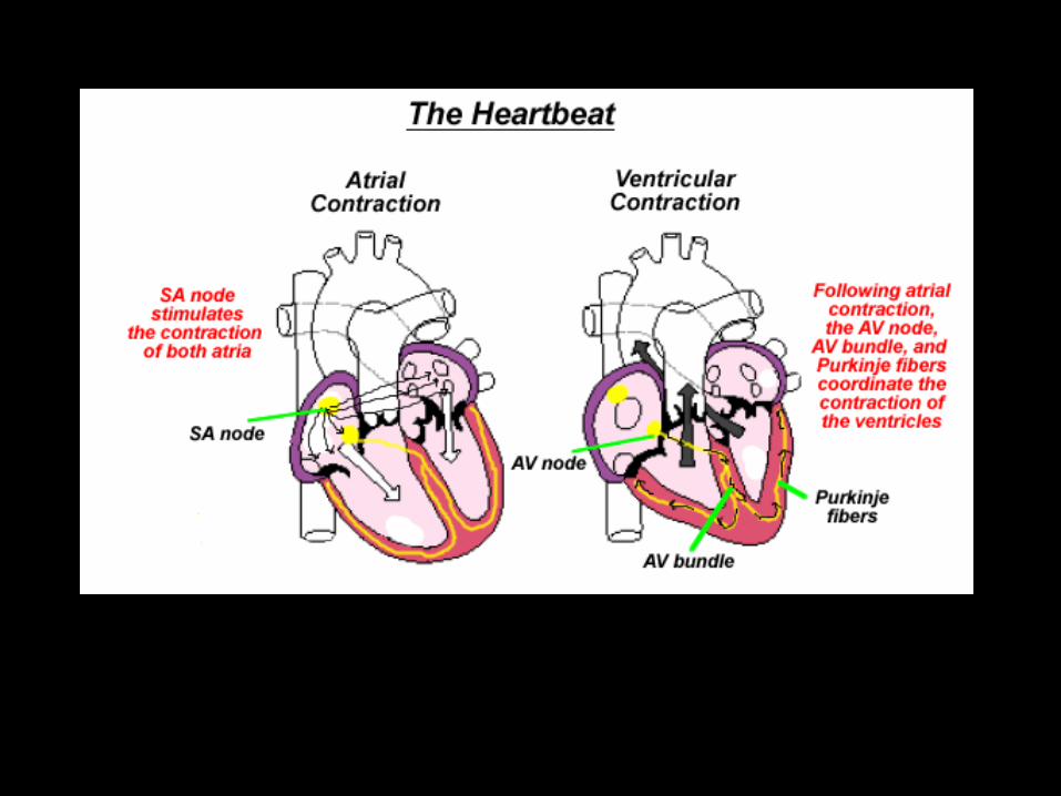

Sinoatrial node (SA node)
• This specialized node is found on the upper inside wall of the right atrium.
• The SA node is known as the pacemaker of the heart and initiates a heartbeat every 0.85 seconds.
• This signal travels across the atria causing them to contract and load the ventricles with blood.
• Ventricles are electrically insulated from atria - so they don’t contract yet.
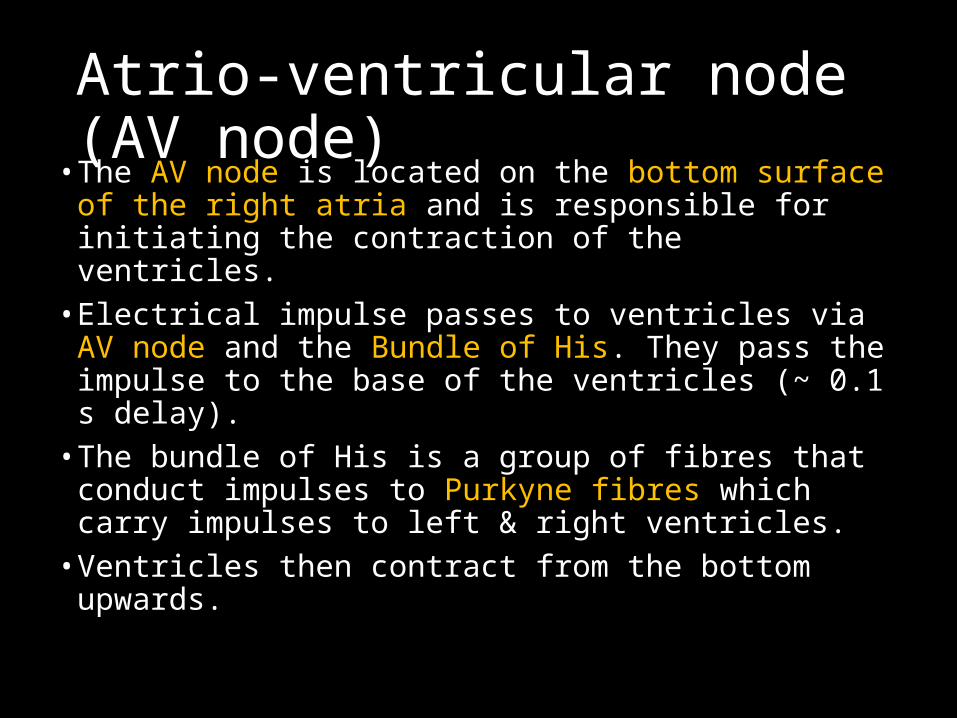
Atrio-ventricular node (AV node)
• The AV node is located on the bottom surface of the right atria and is responsible for initiating the contraction of the ventricles.
• Electrical impulse passes to ventricles via AV node and the Bundle of His. They pass the impulse to the base of the ventricles (~ 0.1 s delay).
• The bundle of His is a group of fibres that conduct impulses to Purkyne fibres which carry impulses to left & right ventricles.
• Ventricles then contract from the bottom upwards.

No impulse
• Cardiac muscle relaxes = diastole

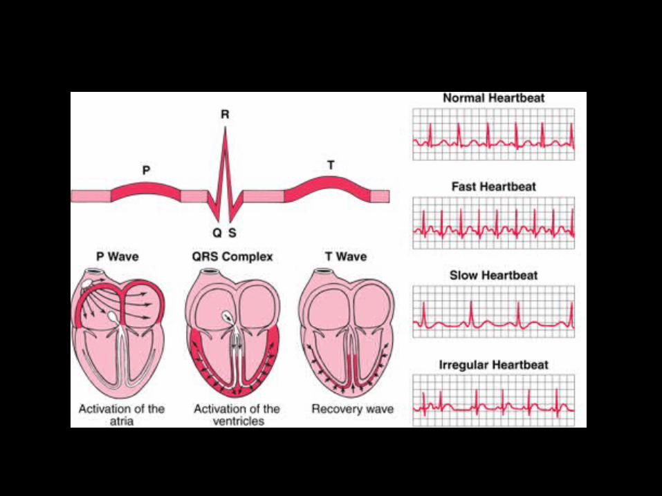
Electrocardiograph
P = atrial systole
QRS = ventricular systole
T = ventricular diastole
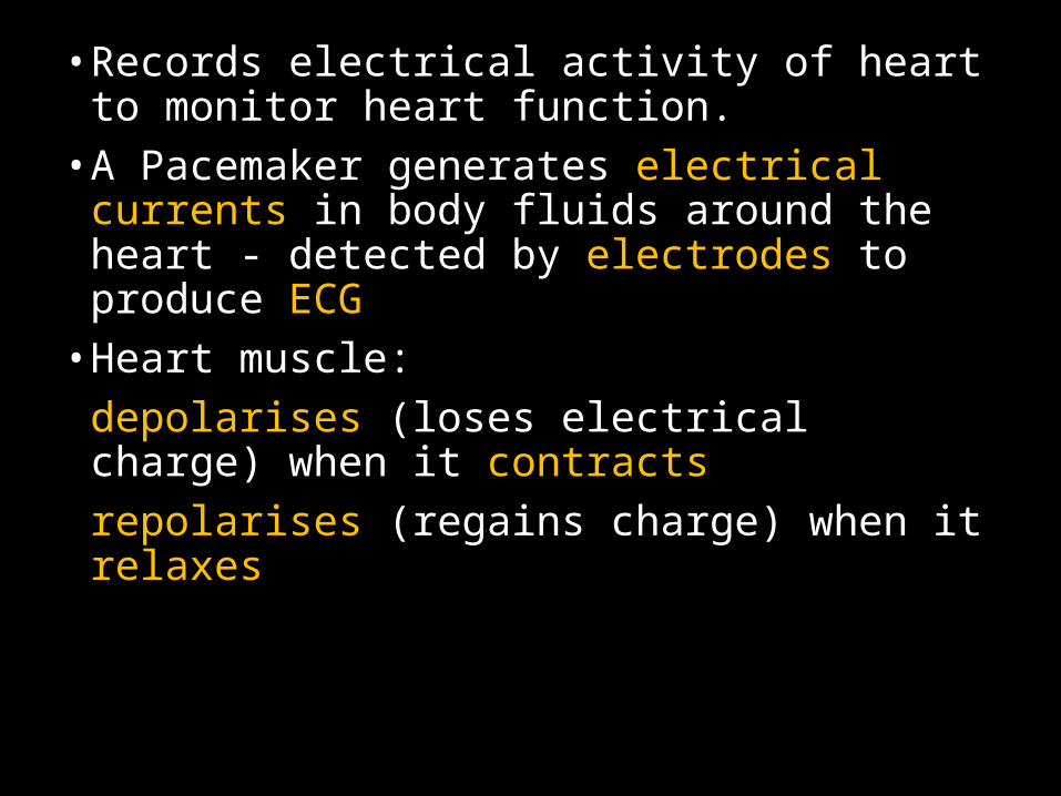
• Records electrical activity of heart to monitor heart function.
• A Pacemaker generates electrical currents in body fluids around the heart - detected by electrodes to produce ECG
• Heart muscle:depolarises (loses electrical charge) when it contractsrepolarises (regains charge) when it relaxes

Ventricular fibrillation is an abnormal heart rhythm that is disorganized and irregular.
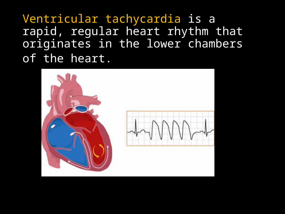
Ventricular tachycardia is a rapid, regular heart rhythm that originates in the lower chambers of the heart.