CNS Pathology Fall 2009 Final. INFLAMMATORY DISEASE OF CNS.
106
CNS Pathology CNS Pathology Fall 2009 Fall 2009 Final Final
-
Upload
jeremy-paul -
Category
Documents
-
view
223 -
download
0
Transcript of CNS Pathology Fall 2009 Final. INFLAMMATORY DISEASE OF CNS.

CNS PathologyCNS Pathology
Fall 2009Fall 2009
FinalFinal

INFLAMMATORY INFLAMMATORY DISEASE OF CNSDISEASE OF CNS

MeningitisMeningitis
1.1. Inflammation fo the meningeal coverings Inflammation fo the meningeal coverings of the brain and spinal cordof the brain and spinal cord
2.2. Can be caused by Can be caused by 1.1. Bacteria, virus and other organisms via Bacteria, virus and other organisms via
blood or lymphblood or lymph2.2. Trauma, pentrating wounds or adjacent Trauma, pentrating wounds or adjacent
structures infectedstructures infected
3.3. Bacterial is most common (can cause Bacterial is most common (can cause hydrocephalus)hydrocephalus)

Pathogens causing MeningitisPathogens causing Meningitis
______________________________________ Chronic meningitisChronic meningitis Often associated with AIDS and immunodepressant drug Often associated with AIDS and immunodepressant drug
therapytherapy
______________________________________ Viral meningitis can be caused by mumps, poliovirus and herpes Viral meningitis can be caused by mumps, poliovirus and herpes
simplexsimplex
______________________________________ Most commonMost common Bacteria release toxins that destroy meningeal cells stimulating Bacteria release toxins that destroy meningeal cells stimulating
immune & inflammatory reactionsimmune & inflammatory reactions

Pathogens causing MeningitisPathogens causing Meningitis
FungiFungi Chronic meningitisChronic meningitis Often associated with AIDS and immunodepressant drug Often associated with AIDS and immunodepressant drug
therapytherapy
VirusVirus Viral meningitis can be caused by mumps, poliovirus and herpes Viral meningitis can be caused by mumps, poliovirus and herpes
simplexsimplex
BacteriaBacteria Most commonMost common Bacteria release toxins that destroy meningeal cells stimulating Bacteria release toxins that destroy meningeal cells stimulating
immune & inflammatory reactionsimmune & inflammatory reactions

Acute Meningitis Acute Meningitis Clinical SymptomsClinical Symptoms
FeverFever HeadacheHeadache Stiff neckStiff neck VomitingVomiting Changes in LOCChanges in LOC Severely ill in 24 hoursSeverely ill in 24 hours RashRash Chronic symptoms are Chronic symptoms are
the same but occur over the same but occur over weeksweeks

Diagnosis of MeningitisDiagnosis of Meningitis Brain CTBrain CT
Rule out contraindications to do a spinal tapRule out contraindications to do a spinal tap
Spinal tapSpinal tap LP to remove CSF to send to labLP to remove CSF to send to lab
Sometimes MRI is used Sometimes MRI is used Is most sensitive modality for demonstrating pia and Is most sensitive modality for demonstrating pia and
arachnoid arachnoid
Treatment includes:Treatment includes: antibiotics and if secondary to encephalitis: antiviral antibiotics and if secondary to encephalitis: antiviral
drugsdrugs

Radiographic AppearanceRadiographic Appearance
Initially meninges Initially meninges show vascular show vascular congestion, edema congestion, edema and minute and minute hemorrhageshemorrhages
MRI and CT scans MRI and CT scans could appear normal could appear normal if appropriate therapy if appropriate therapy is done right awayis done right away Meningitis as a result of a Staph infection

EncephalitisEncephalitis
Infection of the brain tissue that is viralInfection of the brain tissue that is viral May occur subsequent to chickenpox, small May occur subsequent to chickenpox, small
pox, influenza and measlespox, influenza and measles May be caused by mosquitoes and herpesMay be caused by mosquitoes and herpes
Survival rates depend of cause of the Survival rates depend of cause of the disease (can be fatal)disease (can be fatal) 30% of cases in children30% of cases in children When caused by herpes it is often fatalWhen caused by herpes it is often fatal
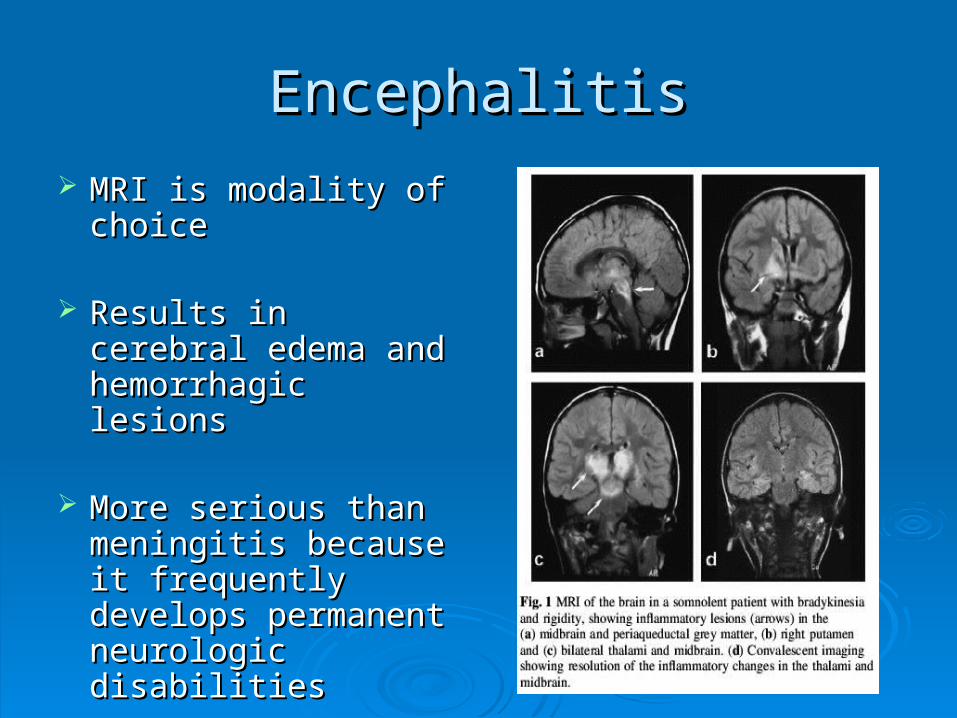
EncephalitisEncephalitis
MRI is modality of MRI is modality of choicechoice
Results in cerebral Results in cerebral edema and edema and hemorrhagic lesionshemorrhagic lesions
More serious than More serious than meningitis because it meningitis because it frequently develops frequently develops permanent neurologic permanent neurologic disabilitiesdisabilities

Encephalitis:Encephalitis:Symptoms and TreatmentSymptoms and Treatment
Symptoms:Symptoms: HeadacheHeadache
MalaiseMalaise
ComaComa
FeverFever
SeizuresSeizures
Treatment:Treatment: Treated with antiviral Treated with antiviral
medicationsmedications
Herpes induced is Herpes induced is treated with Acyclovirtreated with Acyclovir
• Interferes with DNA Interferes with DNA synthesis and inhibits synthesis and inhibits viral replicationviral replication

CONGENITAL CONGENITAL DISEASES OF CNSDISEASES OF CNS

Spinal BifidaSpinal Bifida Is a congenital diseaseIs a congenital disease
Bony neural arch that not completely closedBony neural arch that not completely closed
Most common in lumbar regionMost common in lumbar region May or may not herniate through openingMay or may not herniate through opening
Can range in risk from treatable to life threateningCan range in risk from treatable to life threatening
Can be diagnosed in utero Can be diagnosed in utero With amniocentesisWith amniocentesis UltrasoundUltrasound Elevated beta fetoprotein in mother’s bloodElevated beta fetoprotein in mother’s blood

Types of Spinal BifidaTypes of Spinal Bifida
________________________________ Only the meninges protrudeOnly the meninges protrude Local defect of bone & duraLocal defect of bone & dura
________________________________ Protrusion of spinal cordProtrusion of spinal cord
________________________________
Protrusion of meninges and Protrusion of meninges and spinal cord into the skin of the spinal cord into the skin of the backback
Most seriousMost serious
________________________________ No protrusion of spinal No protrusion of spinal
contentscontents Least severeLeast severe

Types of Types of Spinal BifidaSpinal Bifida
MeningoceleMeningocele Only the meninges Only the meninges
protrudeprotrude Local defect of bone & duraLocal defect of bone & dura
MyeloceleMyelocele Protrusion of spinal cordProtrusion of spinal cord
MeningomeloceleMeningomelocele Protrusion of meninges and Protrusion of meninges and
spinal cord into the skin of spinal cord into the skin of the backthe back
Most seriousMost serious
Spinal bifida occultaSpinal bifida occulta No protrusion of spinal No protrusion of spinal
contentscontents Least severeLeast severe

Radiographic AppearanceRadiographic Appearance
Can be demonstrated Can be demonstrated with CT, MRI and with CT, MRI and myelographymyelography Prenatally with Prenatally with
ultrasound (in utero)ultrasound (in utero)
Large bony defectsLarge bony defects
Herniated spinal Herniated spinal contentscontents
Meningomyelocele
Meningocele
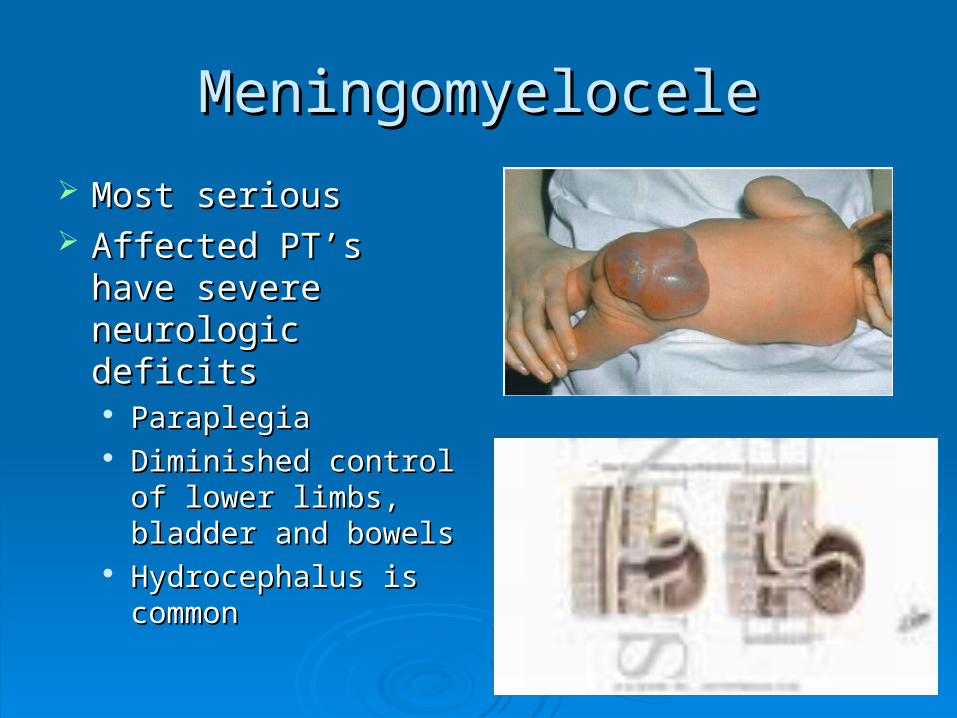
MeningomyeloceleMeningomyelocele
Most serious Most serious Affected PT’s have Affected PT’s have
severe neurologic severe neurologic deficitsdeficits ParaplegiaParaplegia Diminished control of Diminished control of
lower limbs, bladder lower limbs, bladder and bowelsand bowels
Hydrocephalus is Hydrocephalus is commoncommon

Spinal Bifida ImagingSpinal Bifida Imaging

Spinal Bifida TreatmentSpinal Bifida Treatment
Can be surgically repairedCan be surgically repaired Neurological damage is permanent still and cannot be Neurological damage is permanent still and cannot be
reversedreversed
Most measures are supportive rather than Most measures are supportive rather than correctivecorrective Physical therapyPhysical therapy Physical supportsPhysical supports BracesBraces SplintsSplints

CRANIAL AND SPINAL CRANIAL AND SPINAL FRACTURESFRACTURES

Cranial FracturesCranial Fractures Cerebral fractures usually occurs to Cerebral fractures usually occurs to
fractures of the calvaria of the skullfractures of the calvaria of the skull
3 types of cranial fractures3 types of cranial fractures• _____________- straight and sharply defined_____________- straight and sharply defined
Is 80% of all cranial fracturesIs 80% of all cranial fractures
• _____________- curvilinear density_____________- curvilinear density• _____________- Air fluid levels are indicative _____________- Air fluid levels are indicative
Hard to diagnosis radiographicallyHard to diagnosis radiographically

Cranial FracturesCranial Fractures Cerebral fractures usually occurs to Cerebral fractures usually occurs to
fractures of the calvaria of the skullfractures of the calvaria of the skull
3 types of cranial fractures3 types of cranial fractures• LinearLinear- straight and sharply defined- straight and sharply defined
Is 80% of all cranial fracturesIs 80% of all cranial fractures
• Depressed-Depressed- curvilinear density curvilinear density• BasilarBasilar- Air fluid levels are indicative - Air fluid levels are indicative
Hard to diagnosis radiographicallyHard to diagnosis radiographically

Cranial FracturesCranial Fractures
Location of FX is more important that the Location of FX is more important that the extent of the FXextent of the FX If FX crosses artery a bleed can occur If FX crosses artery a bleed can occur
causing a hematomacausing a hematoma
Fx that enters mastoid air cells or sinus can Fx that enters mastoid air cells or sinus can cause an infection that can result incause an infection that can result in• Meningitis Meningitis • EncephalitisEncephalitis

Linear FracturesLinear Fractures Non branching lines that Non branching lines that
are intensely radiolucentare intensely radiolucent
Vascular markings are Vascular markings are occasionally mistaken for occasionally mistaken for fracturesfractures
Fracture appears more Fracture appears more translucent and translucent and transverses the full transverses the full thickness of skullthickness of skull
SuturesSutures
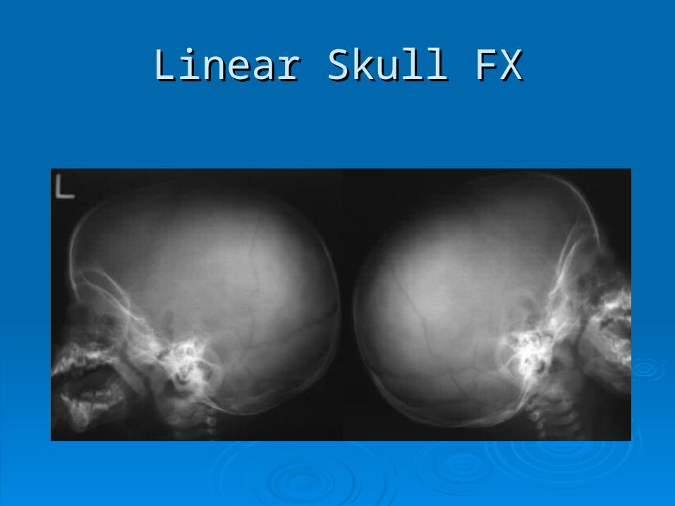
Linear Skull FXLinear Skull FX

Depressed FractureDepressed Fracture The fractured edges The fractured edges
overlapoverlap
Usually caused by a high Usually caused by a high velocity impact with a velocity impact with a small objectsmall object
Can cause bleeding into Can cause bleeding into subarachnoid spacesubarachnoid space
Best demonstrated with Best demonstrated with CR tangential to the FXCR tangential to the FX

Depressed Skull FXDepressed Skull FX

Basilar FractureBasilar Fracture Very difficult to demonstrate with x-rayVery difficult to demonstrate with x-ray
Air fluid levels in sphenoid sinusesAir fluid levels in sphenoid sinuses Clouding of mastoid air cellsClouding of mastoid air cells
Often X-table lateral is done to demonstrate thisOften X-table lateral is done to demonstrate this CT & MRI are most often used for this type CT & MRI are most often used for this type

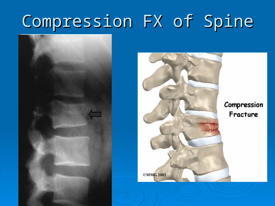
Compression Fracture of spineCompression Fracture of spine
Most frequent type of injury involving Most frequent type of injury involving vertebral bodyvertebral body
Generally occurs in T and L-spineGenerally occurs in T and L-spine T11- T12 and T12 – L1T11- T12 and T12 – L1
Damage is usually limited to the upper Damage is usually limited to the upper portion of the vertebral body, particularly to portion of the vertebral body, particularly to the anterior marginthe anterior margin
Compression FX of SpineCompression FX of Spine

Compression FX of SpineCompression FX of Spine

Hangman’s FractureHangman’s Fracture FX of the arch of the 2FX of the arch of the 2ndnd c-spine vertebrae c-spine vertebrae
Usually accompanied by anterior subluxation of the 2Usually accompanied by anterior subluxation of the 2ndnd and 3and 3rdrd cervical vertebrae cervical vertebrae
Sometimes called traumatic spondylosisSometimes called traumatic spondylosis
Resulting from acute hyperextension of the head & neckResulting from acute hyperextension of the head & neck
Originally seen commonly in hangingsOriginally seen commonly in hangings Now seen more for MVANow seen more for MVA

Hangman’s FractureHangman’s Fracture

Hangman’s FractureHangman’s Fracture

Jefferson’s FractureJefferson’s Fracture Comminuted FX of the ring of the atlasComminuted FX of the ring of the atlas
First described as a “burst FX” First described as a “burst FX” Generally occurs as a result of severe axial force Generally occurs as a result of severe axial force
such as a MVAsuch as a MVA
With this FX particular attn needs to be paid to With this FX particular attn needs to be paid to the transverse longitudinal ligament by reviewing the transverse longitudinal ligament by reviewing lateral masses on the open mouth odontoidlateral masses on the open mouth odontoid
MRI is preferred method for this ligamentMRI is preferred method for this ligament

Jefferson’s FractureJefferson’s Fracture

Jefferson’s Jefferson’s Fracture Fracture

TRAUMATIC DISEASETRAUMATIC DISEASE

Cerebral ContusionCerebral Contusion Is an injury to the brain tissue caused by a Is an injury to the brain tissue caused by a
movement of the brain within the calvaria movement of the brain within the calvaria after blunt traumaafter blunt trauma
Occurs when brain contacts rough skull Occurs when brain contacts rough skull surfaces such as orbital floor and petrous surfaces such as orbital floor and petrous ridgesridges

CT appearance of CT appearance of Cerebral ContusionCerebral Contusion
CT scans appear as low density areas of CT scans appear as low density areas of edema and tissue necrosisedema and tissue necrosis
When IV contrast is used it will enhance When IV contrast is used it will enhance several weeks after injuryseveral weeks after injury
Plays an important role in diagnosisPlays an important role in diagnosis

MR of Cerebral ContusionMR of Cerebral Contusion
Cerebral edema causes high signal Cerebral edema causes high signal intensity on T2 scansintensity on T2 scans
T1 scans may produce high signal regionsT1 scans may produce high signal regions
Diagnosis can also include CT, MRI and Diagnosis can also include CT, MRI and PETPET

Cerebral Cerebral ContusionContusion
Clinical symptoms:Clinical symptoms: DrowsinessDrowsiness ConfusionConfusion AgitationAgitation HemiparesisHemiparesis Unequal pupil sizeUnequal pupil size
Treatment:Treatment: PT is hospitalizedPT is hospitalized
• Prevent shockPrevent shock
If there is swelling If there is swelling medication is given to medication is given to decrease cranial decrease cranial pressurepressure
• Control edemaControl edema• Drainage of hematomaDrainage of hematoma
Surgery is usually not Surgery is usually not necessarynecessary

Cerebral ContusionCerebral Contusion
HematomasHematomas
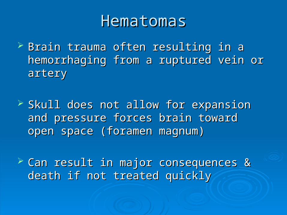
Brain trauma often resulting in a hemorrhaging Brain trauma often resulting in a hemorrhaging from a ruptured vein or arteryfrom a ruptured vein or artery
Skull does not allow for expansion and pressure Skull does not allow for expansion and pressure forces brain toward open space (foramen forces brain toward open space (foramen magnum)magnum)
Can result in major consequences & death if not Can result in major consequences & death if not treated quicklytreated quickly

Epidural HematomasEpidural Hematomas
Highest mortality relate of the hematomasHighest mortality relate of the hematomas Even when treated quickly mortality rate is 30%Even when treated quickly mortality rate is 30%
Results from a torn artery and its branchesResults from a torn artery and its branches Most often occurs from a FX of the temporal boneMost often occurs from a FX of the temporal bone 80% of cases conventional radiograph shows fracture80% of cases conventional radiograph shows fracture
Usually meningeal artery with blood pooling Usually meningeal artery with blood pooling between bones of the skull & dura materbetween bones of the skull & dura mater

Epidural HematomaEpidural Hematoma
Usually a shift of midlineToward opposite side
CT shows increased density
Emergency surgicaldecompression is required to relieve cranial pressure

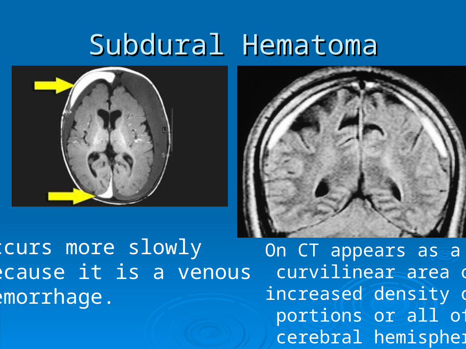
Subdural HematomasSubdural Hematomas
Between the dura mater & arachnoid Between the dura mater & arachnoid meningeal layersmeningeal layers Caused by blunt trauma to frontal or occipital Caused by blunt trauma to frontal or occipital
lobes and can tear subdural veinslobes and can tear subdural veins
Pushes brain away from skull across Pushes brain away from skull across midline (including ventricles)midline (including ventricles)
Subdural HematomaSubdural Hematoma
Occurs more slowlyBecause it is a venousHemorrhage.
On CT appears as a curvilinear area of Iincreased density on portions or all of the cerebral hemispheres
Subdural HematomasSubdural Hematomas
Subacute stage (up to several days)Subacute stage (up to several days) Appears on CT as a decreased density or Appears on CT as a decreased density or
isodense fluid collectionisodense fluid collection
In chronic state (2-3 weeks)In chronic state (2-3 weeks) The surface of the hematoma becomes The surface of the hematoma becomes
concaveconcave Delayed coma con occurDelayed coma con occur

Symptoms of HematomasSymptoms of Hematomas
HeadachesHeadaches
AgitationAgitation
DrowsinessDrowsiness
Gradual radiograph deficitsGradual radiograph deficits

Treatment of HematomasTreatment of Hematomas
In small hematomas without inclination to In small hematomas without inclination to rebleedrebleed
Severe casesSevere cases
Less invasive treatment may includeLess invasive treatment may include

Degenerative DiseasesDegenerative Diseases
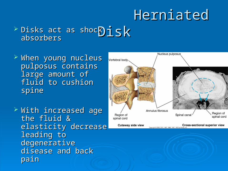
Herniated DiskHerniated Disk Disks act as shock Disks act as shock absorbersabsorbers
When young nucleus When young nucleus pulposus contains pulposus contains large amount of fluid large amount of fluid to cushion spineto cushion spine
With increased age With increased age the fluid & elasticity the fluid & elasticity decrease leading to decrease leading to degenerative disease degenerative disease and back painand back pain

Herniated DiskHerniated Disk May result from either degenerative disease or May result from either degenerative disease or
traumatrauma
A weakened or torn annulus is subject to ruptureA weakened or torn annulus is subject to rupture Nucleus pulposus protrudes & compresses spinal Nucleus pulposus protrudes & compresses spinal
nerve rootsnerve roots Can prolapse in any direction, sometimes without painCan prolapse in any direction, sometimes without pain When it projects posteriorly there is pain and When it projects posteriorly there is pain and
weakening of muscles supplied by those nervesweakening of muscles supplied by those nerves Most commonly occurs is lower cervical & lumbarMost commonly occurs is lower cervical & lumbar
• Lumbar: Most at L4-L5 and L5 – S1Lumbar: Most at L4-L5 and L5 – S1• Cervical: Most at C6 – C7Cervical: Most at C6 – C7• Thoracic: T9-T12Thoracic: T9-T12

Herniated DiskHerniated Disk

Herniated DiskHerniated Disk
MRI is modality of choiceMRI is modality of choice CT and Myelography can also be usedCT and Myelography can also be used

Symptoms of Herniated DiskSymptoms of Herniated Disk
Sudden weak & severe onset of painSudden weak & severe onset of pain
Compression of nerve roots in C-spine:Compression of nerve roots in C-spine:
Compression in lumbar in L-spine:Compression in lumbar in L-spine:

Treatment: Herniated DiskTreatment: Herniated Disk
Conservative treatmentConservative treatment
Surgical interventionSurgical intervention
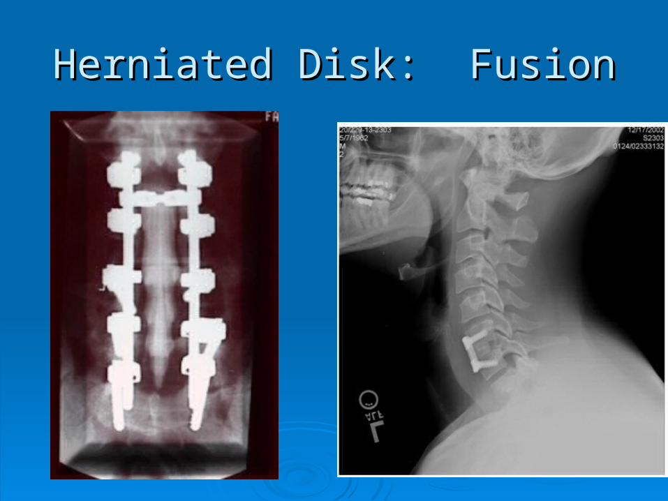
Herniated Disk: FusionHerniated Disk: Fusion
Brain & Spinal Brain & Spinal TumorsTumors

Spinal TumorsSpinal Tumors
Primary tumors as less common is spinal Primary tumors as less common is spinal cord than those of the braincord than those of the brain
Divided into extradural and intraduralDivided into extradural and intradural Intradural further divided intoIntradural further divided into
• Intramedullary (within spinal cord)Intramedullary (within spinal cord) Most common are: Astrocytoma & EpenymomaMost common are: Astrocytoma & Epenymoma
• Extramedullary (outside spinal cord)Extramedullary (outside spinal cord) Most common types of primary spinal neoplasm's Most common types of primary spinal neoplasm's
(>60%) are: Meningiomas and Neurofibromas(>60%) are: Meningiomas and Neurofibromas

Symptoms of Spinal TumorsSymptoms of Spinal Tumors
ExtramedullaryExtramedullary
Similar symptoms as a Similar symptoms as a herniated nucleus herniated nucleus pulposuspulposus
Compress nerve roots Compress nerve roots leading to pain and leading to pain and muscle weaknessmuscle weakness
IntramedullaryIntramedullary
Can cause progressive Can cause progressive paraparesisparaparesis
Sensory lossSensory loss

Extramedullary Spinal TumorsExtramedullary Spinal Tumors
NeurofibromaMeningioma

Intramedullary Spinal tumorsIntramedullary Spinal tumors
Astrocytoma Ependymoma

Imaging of Spinal TumorsImaging of Spinal Tumors
MRI is the modality of choiceMRI is the modality of choice
Conventional radiographyConventional radiography Can demonstrate bony destructionCan demonstrate bony destruction Widening of the vertebral pediclesWidening of the vertebral pedicles CT myelo may be necessary to identify CT myelo may be necessary to identify
extradural tumorsextradural tumors

Treatment of Spinal TumorsTreatment of Spinal Tumors
Both intramedullary and extramedullary Both intramedullary and extramedullary can be removed surgicallycan be removed surgically 50% of patients who have surgery experience 50% of patients who have surgery experience
a reverse of clinical anomaliesa reverse of clinical anomalies
In cases where surgery is contraindicatedIn cases where surgery is contraindicated Radiation therapy is the primary means of Radiation therapy is the primary means of
treating a tumortreating a tumor

Brain TumorsBrain Tumors
Gliomas acct for 50% of all brain tumorsGliomas acct for 50% of all brain tumors
Meningiomas are the most frequently Meningiomas are the most frequently occurring nonglial tumorsoccurring nonglial tumors
All tumors have greater incidence in malesAll tumors have greater incidence in males
Interfere with circulation of the CSF Interfere with circulation of the CSF causing a hydrocephaluscausing a hydrocephalus
Brain TumorsBrain Tumors In children 20% of all tumors are brain In children 20% of all tumors are brain
tumorstumors Most common are astrocytomas, Most common are astrocytomas,
medulloblastomas, glioblastomas and medulloblastomas, glioblastomas and craniopharyngliomascraniopharyngliomas• 30% of primary ped. tumors are medulloblastoma30% of primary ped. tumors are medulloblastoma
In adults most prevalent are:In adults most prevalent are: Astrocytomas, glioblastomas, metastatic Astrocytomas, glioblastomas, metastatic
tumors and menigiomastumors and menigiomas
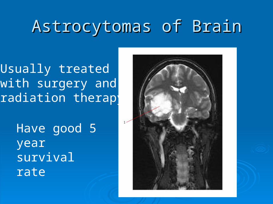
Astrocytomas of BrainAstrocytomas of Brain
Usually treated with surgery and radiation therapy
Have good 5 year survival rate

Ependymoma of BrainEpendymoma of Brain
Usually treated with surgical removal

Medulloblastomas of BrainMedulloblastomas of Brain

Craniopharyngliomas of BrainCraniopharyngliomas of Brain
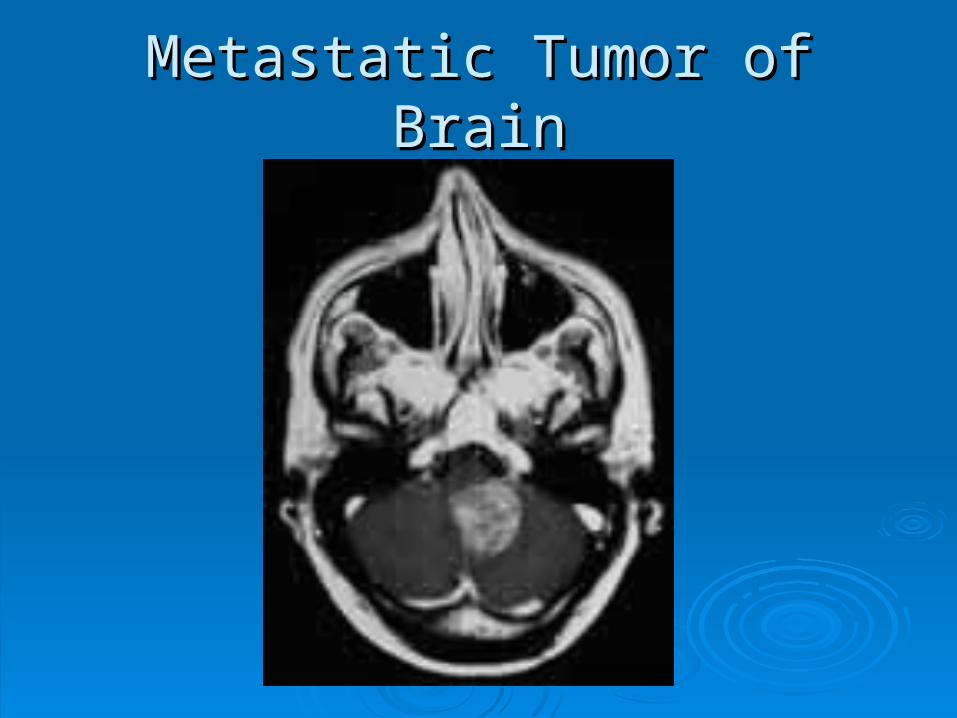
Metastatic Tumor of BrainMetastatic Tumor of Brain

Meningiomas of BrainMeningiomas of Brain
Usually benign
More frequent in women
Rare in children
Less common to see in brain than spinal cord

Symptoms of Brain TumorsSymptoms of Brain Tumors
HeadacheHeadache Nausea and VomitingNausea and Vomiting LethargyLethargy SeizuresSeizures ParalysisParalysis AphasiaAphasia BlindnessBlindness DeafnessDeafness Abnormal changes in personality & behaviorAbnormal changes in personality & behavior

Treatment of Brain TumorsTreatment of Brain Tumors
Surgical resectionSurgical resection Radiation therapyRadiation therapy
Survival rate for surgery & Radiation therapy Survival rate for surgery & Radiation therapy combined is 80% over a 5 year periodcombined is 80% over a 5 year period
Rate of survival decrease to 3% over a Rate of survival decrease to 3% over a 10 year period10 year period

HydrocephalusHydrocephalus
Can be congenital or acquiredCan be congenital or acquired
Refers to an excessive amount of fluid in the Refers to an excessive amount of fluid in the ventriclesventricles
Two typesTwo types Non- communicatingNon- communicating
CommunicatingCommunicating

HydrocephalusHydrocephalus
Non-communicatingNon-communicating Can be congenitalCan be congenital Can be from tumor Can be from tumor
growthgrowth Trauma (hemorrhage)Trauma (hemorrhage) InflammationInflammation
CommunicatingCommunicating Can come with Can come with
increased cranial increased cranial pressurepressure
Raised intrathoracic Raised intrathoracic pressure impairing pressure impairing venous flowvenous flow
Inflammation from Inflammation from meningitismeningitis
Subarachnoid Subarachnoid hemorrhagehemorrhage

Radiographic AppearanceRadiographic Appearance
Generalized enlargement of the ventricular systemGeneralized enlargement of the ventricular system
PA radiograph can reveal separation of the suturesPA radiograph can reveal separation of the sutures
CT clearly demonstrates ventricular dilatation CT clearly demonstrates ventricular dilatation
MRI is more specific in demonstrating the underlying MRI is more specific in demonstrating the underlying cause of obstruction or in excluding obstructioncause of obstruction or in excluding obstruction
Ultrasound is useful in utero and in infantsUltrasound is useful in utero and in infants Sound waves transverse open fontanelsSound waves transverse open fontanels

HydrocephalusHydrocephalus

HydrocephalusHydrocephalus

Hydrocephalus Clinical SymptomsHydrocephalus Clinical Symptoms
The cranial size is The cranial size is enlargedenlarged
Scalp veins distendedScalp veins distended Skin of scalp thin, Skin of scalp thin,
fragile and shinyfragile and shiny Neck muscles Neck muscles
underdevelopedunderdeveloped Severe casesSevere cases
Orbital roofs are Orbital roofs are depresseddepressed
Eyes displaced Eyes displaced downwardsdownwards
•In adults •ALOC•Ataxia•Incontinence•Decreased intellectual •capabilities

Treatment of HydrocephalusTreatment of Hydrocephalus Placement of a shuntPlacement of a shunt
Internal jugular, heart or Internal jugular, heart or peritoneumperitoneum
Contains one way valve to Contains one way valve to prevent backflow of blood prevent backflow of blood into ventriclesinto ventricles
Radiographs taken to Radiographs taken to verify shunt placementverify shunt placement
CT or MRI done to CT or MRI done to evaluate success of evaluate success of treatmenttreatment Ventricularjugular Shunt
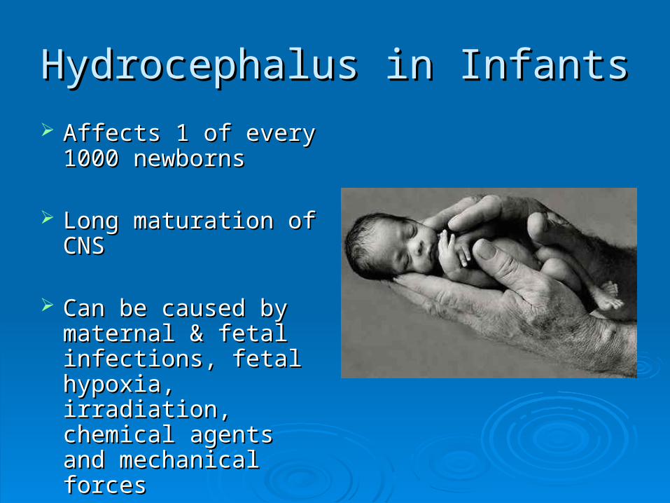
Hydrocephalus in InfantsHydrocephalus in Infants
Affects 1 of every Affects 1 of every 1000 newborns1000 newborns
Long maturation of Long maturation of CNSCNS
Can be caused by Can be caused by maternal & fetal maternal & fetal infections, fetal infections, fetal hypoxia, irradiation, hypoxia, irradiation, chemical agents and chemical agents and mechanical forcesmechanical forces

Hydrocephalus In UteroHydrocephalus In Utero
X-ray used to be taken for fetal age and X-ray used to be taken for fetal age and positionposition
With hydrocephalic fetus- hard to deliver With hydrocephalic fetus- hard to deliver vaginallyvaginally
Pelvimetry was ordered to determine Pelvimetry was ordered to determine measurements of inlet and outletmeasurements of inlet and outlet Very uncomfortable Very uncomfortable Three exposuresThree exposures

Fetal HydrocephalusFetal Hydrocephalus
CommunicatingCommunicating The flow of CSF is free The flow of CSF is free
between ventricles & between ventricles & subarachnoid space subarachnoid space about cauda equinaabout cauda equina
Infants head is normal Infants head is normal size but there is size but there is bulging of the frontal bulging of the frontal fontanellesfontanelles
Caused by poor Caused by poor absorption of CSFabsorption of CSF
Non-communicatingNon-communicating Obstruction between Obstruction between
ventricles and cauda ventricles and cauda equinaequina
Most common form of Most common form of obstructive obstructive hydrocephalus is from hydrocephalus is from abnormalities between abnormalities between the 3the 3rdrd and 4 and 4thth ventriclesventricles

Multiple SclerosisMultiple Sclerosis
Chronic progressive disease of the Chronic progressive disease of the nervous systemnervous system Affects women more than men at approx 20-Affects women more than men at approx 20-
40 years of age40 years of age
There is no cure and it s origin is unknownThere is no cure and it s origin is unknown Treatment only slows the processTreatment only slows the process Some research indicates it may come from Some research indicates it may come from
herpes or retrovirusherpes or retrovirus Appears more in temperate climants than Appears more in temperate climants than
tropical climatestropical climates

Multiple SclerosisMultiple Sclerosis Demyelination of the myelin sheath covering Demyelination of the myelin sheath covering
nervous tissue of spinal cord & white matter nervous tissue of spinal cord & white matter within the brainwithin the brain
It has episodes of relapses and remissionIt has episodes of relapses and remission
Eventually leads to neurological damageEventually leads to neurological damage Impairment of nerve conductionImpairment of nerve conduction
Patients life is not shortenedPatients life is not shortened Quality of life is diminishedQuality of life is diminished

Symptoms Of Multiple SclerosisSymptoms Of Multiple Sclerosis
Difficulty speaking Difficulty speaking clearlyclearly
Bladder dysfunctionBladder dysfunction
Muscle impairmentMuscle impairment
Loss of balanceLoss of balance
Poor coordination Poor coordination
TremorsTremors Muscle weakness Muscle weakness
Double visionDouble vision
Nystagmus (rapid eye Nystagmus (rapid eye movement)movement)
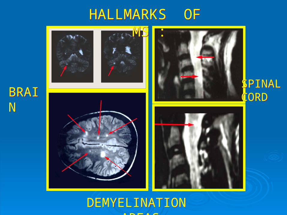
DEMYELINATION AREAS
DEMYELINATION AREAS
BRAINBRAIN
SPINAL CORDSPINAL CORD
HALLMARKS OF MS :
HALLMARKS OF MS :

Imaging of Multiple SclerosisImaging of Multiple Sclerosis
Scars from areas of Scars from areas of demyelinated nervesdemyelinated nerves
Sclerotic lesions Sclerotic lesions throughout nervous systemthroughout nervous system
Called MS plaquesCalled MS plaques
MRI is modality of choiceMRI is modality of choice Contrast enhanced can Contrast enhanced can
differentiate active differentiate active inflammation from older inflammation from older brain plaquesbrain plaques
Functional MRI assesses Functional MRI assesses alterations in normal CSF alterations in normal CSF functionfunction

Multiple Sclerosis: MRIMultiple Sclerosis: MRI

CT imaging of Multiple SclerosisCT imaging of Multiple Sclerosis
CT shows old inactive diseaseCT shows old inactive disease Well defined areas of decreased attenuationWell defined areas of decreased attenuation
With contrast, in an acute phaseWith contrast, in an acute phase Shows a mixture of decreased density (old)Shows a mixture of decreased density (old) Enhancing regions (active)Enhancing regions (active)

Treatment for MSTreatment for MS Immunosuppressive Immunosuppressive
agentsagents Limit the autoimmune Limit the autoimmune
attackattack
AntiviralAntiviral Slows the progress of the Slows the progress of the
diseasedisease
Beta interferonBeta interferon Immunomodulatory agents Immunomodulatory agents
that reduce the severity of that reduce the severity of the attacksthe attacks
Given subcutaneouslyGiven subcutaneously
Corticosteroids (short Corticosteroids (short term)term)
Shortens the symptomatic Shortens the symptomatic periodsperiods
Delays progression of Delays progression of diseasedisease
Reduces frequency of Reduces frequency of attacksattacks
Regular exerciseRegular exercise Reduces spasms and Reduces spasms and
increases ROMincreases ROM
Cerebrovascular Accident (CVA)Cerebrovascular Accident (CVA)
Is an atherosclerotic disease affecting blood Is an atherosclerotic disease affecting blood supply to the brainsupply to the brain
33rdrd leading cause of death in U.S. leading cause of death in U.S. 2 types of stroke:2 types of stroke:
Ischemic and HemorrhagicIschemic and Hemorrhagic Both CT and MRI distinguish between the two Both CT and MRI distinguish between the two
typestypes MRI is especially sensitive to infarction within hours of MRI is especially sensitive to infarction within hours of
onsetonset CT, at times appears negative for a day or soCT, at times appears negative for a day or so
Carotid duplex and MRA are also useful in the Carotid duplex and MRA are also useful in the diagnosis of a strokediagnosis of a stroke

Ischemic StrokeIschemic Stroke Blood clot blocks a blood vessel in the brainBlood clot blocks a blood vessel in the brain Is the majority of strokesIs the majority of strokes
Two types:Two types: Thrombosis of cerebral arteryThrombosis of cerebral artery
• Blood clot that blocks a blood vesselBlood clot that blocks a blood vessel Embolism of the brainEmbolism of the brain
• Is a mass of undissolved matter (solid, liquid or gas) present Is a mass of undissolved matter (solid, liquid or gas) present in a blood vessel brought there by blood currentin a blood vessel brought there by blood current
Diagnosed with CT and MRIDiagnosed with CT and MRI Angiography can be used if other modalities are Angiography can be used if other modalities are
questionablequestionable

Symptoms of Thrombotic Symptoms of Thrombotic Ischemic StrokeIschemic Stroke
Symptoms come on over hours to daysSymptoms come on over hours to days ConfusionConfusion HemiplegiaHemiplegia AphasiaAphasia
May be preceded by a temporary episode of May be preceded by a temporary episode of nerurologic dysfunction called transient Ischemic nerurologic dysfunction called transient Ischemic attack (TIA)attack (TIA) Includes hemiparesis, monocular blindness- clears up Includes hemiparesis, monocular blindness- clears up
in about 2 hoursin about 2 hours

Ischemic Stroke: from EmbolismIschemic Stroke: from Embolism
Sudden onset of symptoms without warningSudden onset of symptoms without warning
Mortality rate is 20%Mortality rate is 20%
Prognosis depends on location, extent, age, and Prognosis depends on location, extent, age, and general healthgeneral health Complete recovery is rareComplete recovery is rare Deficits remaining after 6 months are likely to be Deficits remaining after 6 months are likely to be
permanentpermanent
TreatmentTreatment Bed rest Bed rest Clot blockers within 3 hours (recombinant tissue Clot blockers within 3 hours (recombinant tissue
plasminogen activator (rtPA)plasminogen activator (rtPA)

Ischemic StrokeIschemic Stroke

Imaging of Ischemic StrokeImaging of Ischemic Stroke Non-contrast CT scans are most commonly Non-contrast CT scans are most commonly
usedused
MRI is also excellent for imagingMRI is also excellent for imaging
CT, MRA and US may offer info regarding CT, MRA and US may offer info regarding patency in the brain and carotid arteriespatency in the brain and carotid arteries
PET may be used in the future to identify PET may be used in the future to identify decreased Oxygen flow and consumption within decreased Oxygen flow and consumption within the brainthe brain
Hemorrhagic StrokeHemorrhagic Stroke Occurs from a weakening in the diseased blood Occurs from a weakening in the diseased blood
vesselvessel Typically weakened from atherosclerosis from Typically weakened from atherosclerosis from
hypertensionhypertension
Sudden and often lethal because it comes on so Sudden and often lethal because it comes on so suddenlysuddenly
Accounts for 10-15% of all CVA’s Accounts for 10-15% of all CVA’s
Two types:Two types: Subarachnoid and IntracerebralSubarachnoid and Intracerebral

Hemorrhagic StrokeHemorrhagic Stroke
Most occur in the cerebrum and bleed into Most occur in the cerebrum and bleed into lateral ventriclelateral ventricle
Most often preceded by an intense headache Most often preceded by an intense headache and vomitingand vomiting
LOC follows in minutes and leads to LOC follows in minutes and leads to contralateral hemiplegia or deathcontralateral hemiplegia or death
Prognosis is poorPrognosis is poor 35% die day after stroke35% die day after stroke 15% die within a few weeks, usually from another 15% die within a few weeks, usually from another
vessel rupturevessel rupture

Imaging of Hemorrhagic StrokesImaging of Hemorrhagic Strokes
CT is modality of choiceCT is modality of choice Can demonstrate high density blood in the Can demonstrate high density blood in the
subarachnoid space in more than 95% of subarachnoid space in more than 95% of casescases
Can demonstrate aneurysms greater than Can demonstrate aneurysms greater than 3mm3mm
With contrast is contraindicated because With contrast is contraindicated because surgeon will not operate without an angiogramsurgeon will not operate without an angiogram
MRI is relatively insensitive for MRI is relatively insensitive for subarachnoid bleedssubarachnoid bleeds

Treatment ofTreatment ofHemorrhagic StrokesHemorrhagic Strokes
SurgerySurgery Preceded by a surgical angiogramPreceded by a surgical angiogram
If surgical intervention is postponed so will If surgical intervention is postponed so will the angiogramthe angiogram

Hemorrhagic StrokeHemorrhagic Stroke

Pathology Summary and Pathology Summary and Modality of ChoiceModality of Choice
Pathology Summary: Central Nervous Pathology Summary: Central Nervous SystemSystem
Pathology Imaging Modalities of Pathology Imaging Modalities of Choice Additive or Subtractive Choice Additive or Subtractive PathologyPathology
Hydrocephalus Hydrocephalus CT, MRI, sonography in the neonateCT, MRI, sonography in the neonate
MeningitisMeningitis MRIMRI
EncephalitisEncephalitis MRIMRI
Brain abscessBrain abscess CT, MRICT, MRI
Herniated nucleus pulposusHerniated nucleus pulposus MRI, CT, myelographyMRI, CT, myelography
Cervical spondylosisCervical spondylosis Radiography SubtractiveRadiography Subtractive Multiple sclerosisMultiple sclerosis
MRIMRI CVACVA
MRI, CT, sonography, PETMRI, CT, sonography, PET
GliomaGlioma MRI, CTMRI, CT
MedulloblastomaMedulloblastoma MRI, CTMRI, CT
MeningiomaMeningioma CT, MRICT, MRI
Pituitary adenomaPituitary adenoma CT, MRICT, MRI
CraniopharyngiomaCraniopharyngioma CTCT
Acoustic neuromaAcoustic neuroma MRIMRI
Spinal tumorSpinal tumor MRI, radiography, CT, myelographyMRI, radiography, CT, myelography
Both Metastases from other sitesBoth Metastases from other sites MRI, radiography, CTSubtractiveMRI, radiography, CTSubtractive