Clostridium Spores Ribbon-like Appendages · of Clostridium sp. NI is oval. Atypical spore (Fig. 1)...
14
JOURNAL OP BACTERIOLOGY, Mar., 1967, p. 1160-1173 Copyright @ 1967 American Society for Microbiology Vol. 93, No. 3 Printed in U.S.A. Clostridium Spores with Ribbon-like Appendages L. J. RODE, MARGARET A. CRAWFORD, AND M. GLENN WILLIAMS Department of Microbiology anid Electron Microscope Laboratory, The University of Texas, Austin, Texas Received for publication 15 December 1966 Spores of Clostridium sp. Ni are characterized by numerous broad ribbon-like appendages attached to one end. The appendages are two to three times the length of the spore and, at their maximal dimension, may be two-thirds the width of the spore. They are attached to the spore body by a common trunk which is continu- ous with the outer spore coat. Each appendage is a multilayered structure and is enclosed in an amorphous material. Details of spore and appendage formation are described, and appendage ultrastructural features are presented. The function of the appendages is not known. Bacterial spores commonly exhibit surfaces which are sculptured and irregular (2, 3, 16). Although ribbed surface structures have been described (1, 3, 7, 8), neither marked spore protuberances nor appendages had been reported prior to the recent papers by Krasil'nikov, Duda, and Sokolov (10-12). These Russian workers described for soil clostridia elaborate spore protuberances which were sufficiently prominent and varied to be considered taxonomically significant. More recently, Hodgkiss and Ordal (6) described multiple tubular appendages radiating from the surface of Clostridium botu- linum type E spores. Although the Clostridium which we have ex- amined in the present work was obtained from V. I. Duda, it appears that this organism (Clos- tridium sp. N1) may not have been included in the survey accounts published by the Russian microbiologists. At any rate, we are not aware of an existing published description of this unusual sporeformer. MATERLALS AND METHODS Organism. Clostridium sp. Ni was obtained in December 1965 from V. I. Duda, Moscow State Uni- versity, Moscow, USSR. It was characterized by him as an anaerobe which grows well on ordinary bac- teriological media, with the production of spores possessing appendages. Data on culture source, isola- tion procedures, and species designation have not been available. Growth and spore production. Brain Heart Infusion (Difco), pH 7.4, supplemented with 500 ,ug of sodium thioglycolate per ml and 1.5% agar (Difco), was used for growth and spore production. Petri plates were surface-inoculated and incubated at 30 C in desiccators over wet oats to provide the anaerobic environment. Sporulation was abundant in 6 days. Spore crops, free from vegetative cells, were obtained by conventional procedures of differential centrifuga- tion and repeated washing with demineralized water (15). Spores were stored in demineralized water at 4 C. Germination did not occur under these condi- tions. Spore treatmenits. In certain cases, spore suspen- sions were treated to achieve particular objectives prior to electron microscope study. Appendages were removed from free spores by sonic treatment with a Branson-Sonifier (model S-75). The spores were chilled in an ice bath during succes- sive 30-sec treatments until the desired results were obtained. Preliminary appraisal of appendage re- moval was by phase-contrast microscopy; conclusive appraisal was by electron microscopy. Spores, stripped of appendages and coat structures, were prepared by treatment with 0.1% sodium hypo- chlorite for periods up to 30 min at 37 C. A different effect of sodium hypochlorite on Bacillus megaterium spores has been described (16). The effects of the proteolytic enzyme, Pronase, and of lysozyme and of ribonuclease on free spores, in particular on the integrity of the appendages, were studied in phosphate buffer (0.016 M, pH 7). In each case, enzyme concentrations were 50 ,ug/ml and incu- bation was at 37 C for periods up to 6 hr. Appraisals of enzyme effects were by phase-contrast and electron microscopy. Specimeni preparationi for electron microscopy. Specimens were examined as carbon replicas, as thin sections, or directly with a negative stain. For preparation of replicas, spore specimens were placed on squares of freshly cleaved mica, allowed to dry, and shadowed with platinum at 35 to 45°. The specimens were then coated with carbon, and the carbon films, removed from the mica by flotation onto water, were treated overnight with 0.5% sodium hypo- chlorite to remove the cellular material. After thorough washing with demineralized water, the car- bon replica films (replicas) were transferred to copper grids for examination (16). Specimens for thin sectioning were, in most cases, fixed for 1 to 3 hr at room temperature in 2% glu- 1160 on May 17, 2019 by guest http://jb.asm.org/ Downloaded from
Transcript of Clostridium Spores Ribbon-like Appendages · of Clostridium sp. NI is oval. Atypical spore (Fig. 1)...
JOURNAL OP BACTERIOLOGY, Mar., 1967, p. 1160-1173Copyright @ 1967 American Society for Microbiology
Vol. 93, No. 3Printed in U.S.A.
Clostridium Spores with Ribbon-like AppendagesL. J. RODE, MARGARET A. CRAWFORD, AND M. GLENN WILLIAMS
Department of Microbiology anid Electron Microscope Laboratory, The University of Texas, Austin, Texas
Received for publication 15 December 1966
Spores of Clostridium sp. Ni are characterized by numerous broad ribbon-likeappendages attached to one end. The appendages are two to three times the lengthof the spore and, at their maximal dimension, may be two-thirds the width of thespore. They are attached to the spore body by a common trunk which is continu-ous with the outer spore coat. Each appendage is a multilayered structure and isenclosed in an amorphous material. Details of spore and appendage formationare described, and appendage ultrastructural features are presented. The functionof the appendages is not known.
Bacterial spores commonly exhibit surfaceswhich are sculptured and irregular (2, 3, 16).Although ribbed surface structures have beendescribed (1, 3, 7, 8), neither marked sporeprotuberances nor appendages had been reportedprior to the recent papers by Krasil'nikov, Duda,and Sokolov (10-12). These Russian workersdescribed for soil clostridia elaborate sporeprotuberances which were sufficiently prominentand varied to be considered taxonomicallysignificant. More recently, Hodgkiss and Ordal(6) described multiple tubular appendagesradiating from the surface of Clostridium botu-linum type E spores.
Although the Clostridium which we have ex-amined in the present work was obtained fromV. I. Duda, it appears that this organism (Clos-tridium sp. N1) may not have been included inthe survey accounts published by the Russianmicrobiologists. At any rate, we are not awareof an existing published description of thisunusual sporeformer.
MATERLALS AND METHODS
Organism. Clostridium sp. Ni was obtained inDecember 1965 from V. I. Duda, Moscow State Uni-versity, Moscow, USSR. It was characterized by himas an anaerobe which grows well on ordinary bac-teriological media, with the production of sporespossessing appendages. Data on culture source, isola-tion procedures, and species designation have notbeen available.
Growth and spore production. Brain Heart Infusion(Difco), pH 7.4, supplemented with 500 ,ug of sodiumthioglycolate per ml and 1.5% agar (Difco), wasused for growth and spore production. Petri plateswere surface-inoculated and incubated at 30 C indesiccators over wet oats to provide the anaerobicenvironment. Sporulation was abundant in 6 days.Spore crops, free from vegetative cells, were obtained
by conventional procedures of differential centrifuga-tion and repeated washing with demineralized water(15). Spores were stored in demineralized water at4 C. Germination did not occur under these condi-tions.
Spore treatmenits. In certain cases, spore suspen-sions were treated to achieve particular objectivesprior to electron microscope study.
Appendages were removed from free spores bysonic treatment with a Branson-Sonifier (model S-75).The spores were chilled in an ice bath during succes-sive 30-sec treatments until the desired results wereobtained. Preliminary appraisal of appendage re-moval was by phase-contrast microscopy; conclusiveappraisal was by electron microscopy.
Spores, stripped of appendages and coat structures,were prepared by treatment with 0.1% sodium hypo-chlorite for periods up to 30 min at 37 C. A differenteffect of sodium hypochlorite on Bacillus megateriumspores has been described (16).The effects of the proteolytic enzyme, Pronase, and
of lysozyme and of ribonuclease on free spores, inparticular on the integrity of the appendages, werestudied in phosphate buffer (0.016 M, pH 7). In eachcase, enzyme concentrations were 50 ,ug/ml and incu-bation was at 37 C for periods up to 6 hr. Appraisalsof enzyme effects were by phase-contrast and electronmicroscopy.
Specimeni preparationi for electron microscopy.Specimens were examined as carbon replicas, as thinsections, or directly with a negative stain.
For preparation of replicas, spore specimens wereplaced on squares of freshly cleaved mica, allowed todry, and shadowed with platinum at 35 to 45°. Thespecimens were then coated with carbon, and thecarbon films, removed from the mica by flotation ontowater, were treated overnight with 0.5% sodium hypo-chlorite to remove the cellular material. Afterthorough washing with demineralized water, the car-bon replica films (replicas) were transferred to coppergrids for examination (16).
Specimens for thin sectioning were, in most cases,fixed for 1 to 3 hr at room temperature in 2% glu-
1160
on May 17, 2019 by guest
http://jb.asm.org/
Dow
nloaded from
APPENDAGES OF CLOSTRIDIUM SPORES
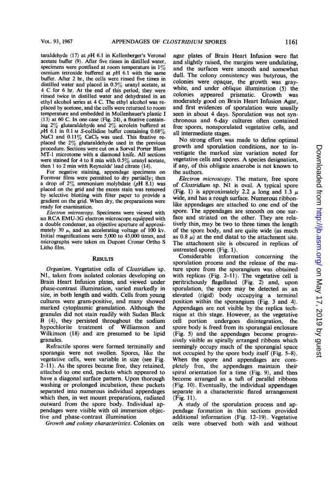
taraldehyde (17) at pH 6.1 in Kellenberger's Veronalacetate buffer (9). After five rinses in distilled water,specimens were postfixed at room temperature in 1%osmium tetroxide buffered at pH 6.1 with the samebuffer. After 2 hr, the cells were rinsed five times indistilled water and placed in 0.5% uranyl acetate, at4 C for 6 hr. At the end of this period, they wererinsed twice in distilled water and dehydrated in anethyl alcohol series at 4 C. The ethyl alcohol was re-placed by acetone, and the cells were returned to roomtemperature and embedded in Mollenhauer's plastic I(13) at 60 C. In one case (Fig. 24), a fixative contain-ing 2% glutaraldehyde and 2% acrolein buffered atpH 6.1 in 0.1 M S-collidine buffer containing 0.68%NaCl and 0.11% CaCl2 was used. This fixative re-placed the 2% glutaraldehyde used in the previousprocedure. Sections were cut on a Sorval Porter BlumMT-1 microtome with a diamond knife. All sectionswere stained for 4 to 8 min with 0.5% uranyl acetate,then 1 to 2 min with Reynolds' lead citrate (14).
For negative staining, appendage specimens onFormvar films were permitted to dry partially; thena drop of 2% ammonium molybdate (pH 8.1) wasplaced on the grid and the excess stain was removedby selective blotting with filter paper to provide agradient on the grid. When dry, the preparations wereready for examination.
Electron microscopy. Specimens were viewed withan RCA EMU-3G electron microscope equipped witha double condenser, an objective aperture of approxi-mately 30 ,u, and an accelerating voltage of 100 kv.Initial magnifications were 5,000 to 43,000 times, andmicrographs were taken on Dupont Cronar Ortho SLitho film.
RESULTS
Organism. Vegetative cells of Clostridium sp.NI, taken from isolated colonies developing onBrain Heart Infusion plates, and viewed underphase-contrast illumination, varied markedly insize, in both length and width. Cells from youngcultures were gram-positive, and many showedmarked cytoplasmic granulation. Although thegranules did not stain readily with Sudan BlackB (4), they persisted throughout the sodiumhypochlorite treatment of Williamson andWilkinson (18) and are presumed to be lipidgranules.
Refractile spores were formed terminally andsporangia were not swollen. Spores, like thevegetative cells, were variable in size (see Fig.2-11). As the spores became free, they retained,attached to one end, packets which appeared tohave a diagonal surface pattern. Upon thoroughwashing or prolonged incubation, these packetsseparated into numerous individual appendageswhich then, in wet mount preparations, radiatedoutward from the spore body. Individual ap-pendages were visible with oil immersion objec-tive and phase-contrast illumination
Growth and colony characteristics. Colonies on
agar plates of Brain Heart Infusion were flatand slightly raised, the margins were undulating,and the surfaces were smooth and somewhatdull. The colony consistency was butyrous, thecolonies were opaque, the growth was gray-white, and under oblique illumination (5) thecolonies appeared prismatic. Growth wasmoderately good on Brain Heart Infusion Agar,and first evidences of sporulation were usuallyseen in about 4 days. Sporulation was not syn-chronous and 6-day cultures often containedfree spores, nonsporulated vegetative cells, andall intermediate stages.No strong effort was made to define optimal
growth and sporulation conditions, nor to in-vestigate the marked size variation noted forvegetative cells and spores. A species designation,if any, of this obligate anaerobe is not known tothe authors.
Electron microscopy. The mature, free sporeof Clostridium sp. NI is oval. A typical spore(Fig. 1) is approximately 2.2 ,u long and 1.3 ,uwide, and has a rough surface. Numerous ribbon-like appendages are attached to one end of thespore. The appendages are smooth on one sur-face and striated on the other. They are rela-tively thin, may be two to three times the lengthof the spore body, and are quite wide (as muchas 0.8 ,u) at the end distal to the attachment site.The attachment site is obscured in replicas ofuntreated spores (Fig. 1).
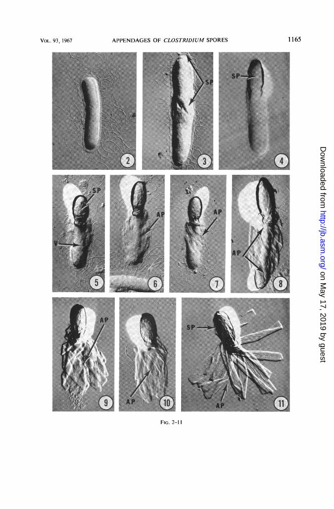
Considerable information concerning thesporulation process and the release of the ma-ture spore from the sporangium was obtainedwith replicas (Fig. 2-11). The vegetative cell isperitrichously flagellated (Fig. 2) and, uponsporulation, the spore may be detected as anelevated (rigid) body occupying a terminalposition within the sporangium (Fig. 3 and 4).Appendages are not visible by the replica tech-nique at this stage. However, as the vegetativecell portion undergoes disintegration, thespore body is freed from its sporangial enclosure(Fig. 5) and the appendages become progres-sively visible as spirally arranged ribbons whichseemingly occupy much of the sporangial spacenot occupied by the spore body itself (Fig. 5-8).When the spore and appendages are com-pletely free, the appendages maintain theirspiral orientation for a time (Fig. 9), and thenbecome arranged as a tuft of parallel ribbons(Fig. 10). Eventually, the individual appendagesseparate in a characteristic flared arrangement(Fig. 11).A study of the sporulation process and ap-
pendage formation in thin sections providedadditional information (Fig. 12-19). Vegetativecells were observed both with and without
VOL. 93,1967 1161
on May 17, 2019 by guest
http://jb.asm.org/
Dow
nloaded from

RODE, CRAWFORD, AND WILLIAMS
*: ::*. .:: :
...
*.:
.. ...
::;.: ;::;
...X..., ...-
'. :!
... .. ":: ..::
'..> .,
.: '.'.-.
*: ::
':
;',
sa.. ..,, .|l
..
,, :.
;'.''.'' '..
FIG. 1. Typical mature free spore of Clostridium sp. Nl from a 7-day culture. The spore (SP) surface is rough,and numerous ribbon-like appendages extend from a common origin at one end. The appendages are relativelysmooth (SM) on one surface, but appear striated (ST) on the other. Replica. X 15,000.
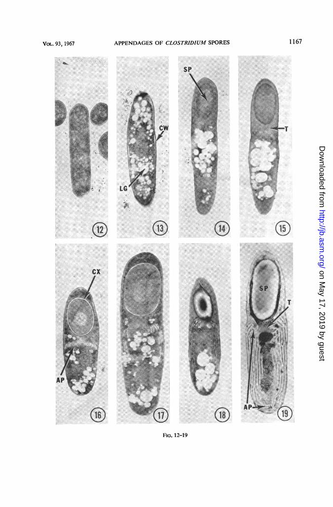
"lipid granules" (Fig. 12 and 13). No appendageswere visible in sporangia at very early stages ofsporulation, prior to coat formation (Fig. 14).However, coincident with early coat formation,an appendage trunk was formed (Fig. 15), andtrom this trunk appendages originated and pro-gressively occupied more and more of the sporan-gial space distal to the spore body as the cortexwas laid down and the spore continued to de-
velop (Fig. 16, 17 and 18). Finally, upon matur-ity, the spore body and its appendages occupied avery significant proportion of the sporangialspace (Fig. 19). The appendages within the spor-angium were initially oriented upward towardthe spore body from their trunk origin, but thenassumed a spiralling, downward direction.Multiple layers of appendages were visible inthin sections, and the appendages were enclosed
1162 J. BACTERIOL.
XI
AF
on May 17, 2019 by guest
http://jb.asm.org/
Dow
nloaded from
APPENDAGES OF CLOSTRIDIUM SPORES
in an electron transparent substance whichseparated them from one another (Fig. 19; seebelow).
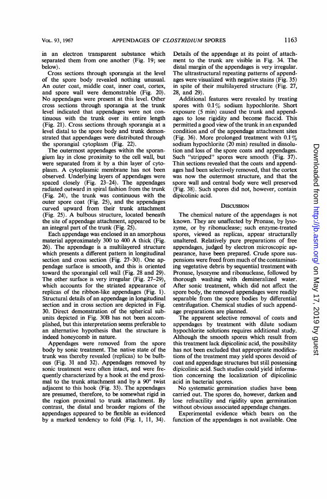
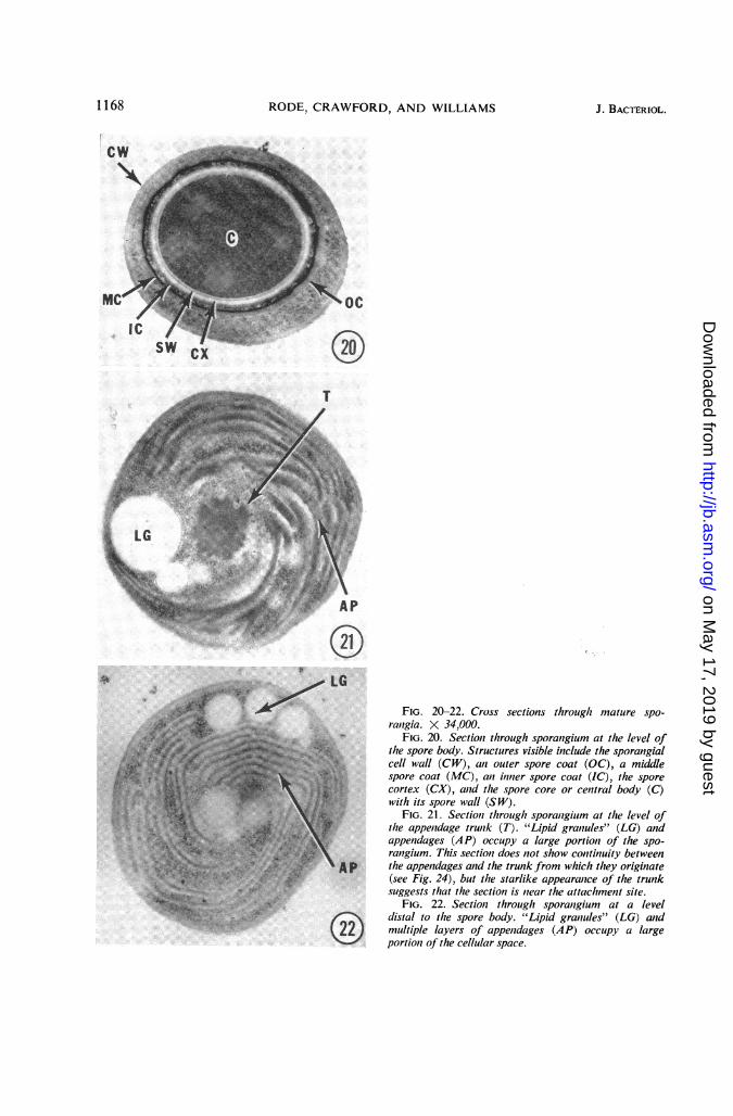
Cross sections through sporangia at the levelof the spore body revealed nothing unusual.An outer coat, middle coat, inner coat, cortex,and spore wall were demonstrable (Fig. 20).No appendages were present at this level. Othercross sections through sporangia at the trunklevel indicated that appendages were not con-tinuous with the trunk over its entire length(Fig. 21). Cross sections through sporangia at alevel distal to the spore body and trunk demon-strated that appendages were distributed throughthe sporangial cytoplasm (Fig. 22).The outermost appendages within the sporan-
gium lay in close proximity to the cell wall, butwere separated from it by a thin layer of cyto-plasm. A cytoplasmic membrane has not beenobserved. Underlying layers of appendages werespaced closely (Fig. 23-24). The appendagesradiated outward in spiral fashion from the trunk(Fig. 24), the trunk was continuous with theouter spore coat (Fig. 25), and the appendagescurved upward from their trunk attachment(Fig. 25). A bulbous structure, located beneaththe site of appendage attachment, appeared to bean integral part of the trunk (Fig. 25).Each appendage was enclosed in an amorphous
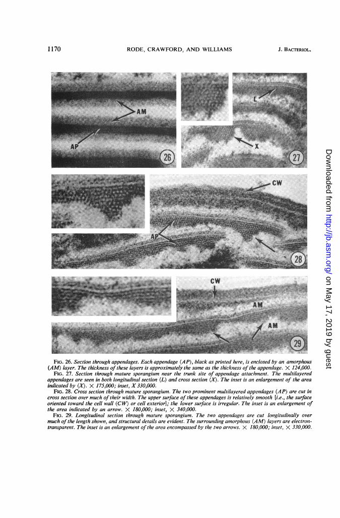
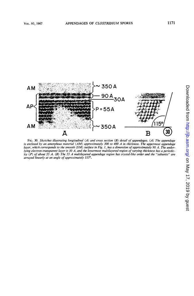
material approximately 300 to 400 A thick (Fig.26). The appendage is a multilayered structurewhich presents a different pattern in longitudinalsection and cross section (Fig. 27-30). One ap-pendage surface is smooth, and this is orientedtoward the sporangial cell wall (Fig. 28 and 29).The other surface is very irregular (Fig. 27-29),which accounts for the striated appearance ofreplicas of the ribbon-like appendages (Fig. 1).Structural details of an appendage in longitudinalsection and in cross section are depicted in Fig.30. Direct demonstration of the spherical sub-units depicted in Fig. 30B has not been accom-plished, but this interpretation seems preferable toan alternative hypothesis that the structure isindeed honeycomb in nature.Appendages were removed from the spore
body by sonic treatment. The native state of thetrunk was thereby revealed (replicas) to be bulb-ous (Fig. 31 and 32). Appendages removed bysonic treatment were often intact, and were fre-quently characterized by a hook at the end proxi-mal to the trunk attachment and by a 900 twistadjacent to this hook (Fig. 33). The appendagesare presumed, therefore, to be somewhat rigid inthe region proximal to trunk attachment. Bycontrast, the distal and broader regions of theappendages appeared to be flexible as evidencedby a marked tendency to fold (Fig. 1, 11, 34).
Details of the appendage at its point of attach-ment to the trunk are visible in Fig. 34. Thedistal margin of the appendages is very irregular.The ultrastructural repeating patterns of append-ages were visualized with negative stains (Fig. 35)in spite of their multilayered structure (Fig. 27,28, and 29).
Additional features were revealed by treatingspores with 0.1% sodium hypochlorite. Shortexposure (5 min) caused the trunk and append-ages to lose rigidity and become flaccid. Thispermitted a good view of the trunk in an expandedcondition and of the appendage attachment sites(Fig. 36). More prolonged treatment with 0.1 %sodium hypochlorite (20 min) resulted in dissolu-tion and loss of the spore coats and appendages.Such "stripped" spores were smooth (Fig. 37).Thin sections revealed that the coats and append-ages had been selectively removed, that the cortexwas now the outermost structure, and that thespore wall and central body were well preserved(Fig. 38). Such spores did not, however, containdipicolinic acid.
DIscussIoNThe chemical nature of the appendages is not
known. They are unaffected by Pronase, by lyso-zyme, or by ribonuclease; such enzyme-treatedspores, viewed as replicas, appear structurallyunaltered. Relatively pure preparations of freeappendages, judged by electron microscopic ap-pearance, have been prepared. Crude spore sus-pensions were freed from much of the contaminat-ing vegetative debris by sequential treatment withPronase, lysozyme and ribonuclease, followed bythorough washing with demineralized water.After sonic treatment, which did not affect thespore body, the removed appendages were readilyseparable from the spore bodies by differentialcentrifugation. Chemical studies of such append-age preparations are planned.The apparent selective removal of coats and
appendages by treatment with dilute sodiumhypochlorite solutions requires additional study.Although the smooth spores which result fromthis treatment lack dipicolinic acid, the possibilityhas not been excluded that appropriate modifica-tions of the treatment may yield spores devoid ofcoat and appendage structures but still possessingdipicolinic acid. Such studies could yield informa-tion concerning the localization of dipicolinicacid in bacterial spores.No systematic germination studies have been
carried out. The spores do, however, darken andlose refractility and rigidity upon germinationwithout obvious associated appendage changes.
Experimental evidence which bears on thefunction of the appendages is not available. One
1163VOL. 93, 1967
on May 17, 2019 by guest
http://jb.asm.org/
Dow
nloaded from
RODE, CRAWFORD, AND WILLIAMS
might speculate that the appendages facilitatespore dissemination in nature, assist in spore nu-trition during formation, or have no function andresult from a deranged metabolism. It seemsfruitless to speculate on these and other possi-bilities until supporting evidence is available.
ACKNOWLEDGMENTSWe thank Orville Wyss for obtaining Clostridium
sp. NI while on a trip to Moscow, and V. I. Dudafor supplying the culture. We thank, also, DianeYolton for capable technical assistance, LeodociaPope for Fig. 37, and Pauline West for Fig. 30.
This investigation was supported by Public HealthService Grant Al 07582-01 from the National Insti-tute of Allergy and Infectious Diseases and by Con-tract Nonr 375(12) from the Office of Naval Research.
LITERATURE CITED1. BRADLEY, D. E., AND D. J. WILLIAMS. 1957. An
electron microscope study of the spores of somespecies of the genus Bacillus using carbon repli-cas. J. Gen. Microbiol. 17:75-79.
2. FITZ-JAMES, P. C. 1959. Morphology of spores ofBacillus apiarius Katznelson. J. Bacteriol. 78:765-768.
3. FITZ-JAMES, P. C., AND I. E. YOUNG. 1959. Cyto-logical comparison of spores of different strainsof Bacillus megaterium. J. Bacteriol. 78:755-764.
4. HARTMAN, T. 1940. The use of sudan black B asa bacterial fat strain. Stain Technol. 15:23-28.
5. HENRY, B. S. 1933. Dissociation in the genusBrucella. J. Infect. Diseases 52:374-402.
6. HODGKISS, W., AND Z. J. ORDAL. 1966. Mor-phology of the spore of some strains of Clos-tridium botulinum type E. J. Bacteriol. 91:2031-2036.
7. HOLBERT, P. E. 1960. An effective method of pre-paring sections of Bacillus polymyxa sporangiaand spores for electron microscopy. J. Biophys.Biochem. Cytol. 7:373-376.
8. HOOF, A. VAN DEN, AND S. ANINGA. 1956. Anelectron microscope study on the shape of thespores of Bacillus polymyxa. Antonie vanLeeuwenhoek J. Microbiol. Serol. 22:327-330.
9. KELLENBERGER, E., A. R. RYTER, AND J. SECHAUD.1958. Electron microscope study of DNA-containing plasms. II. Vegetative and maturephage DNA as compared with normal bacterialnucleoids in different physiological states. J.Biophys. Biochem. Cytol. 4:671-678.
10. KRASIL'NIKOV, N. A., V. I. DUDA, AND A. A.SOKOLOV. 1963. Outward growths on spores ofanaerobic bacteria of thegenus Clostridium. Proc.Acad. Sci. USSR Microbiol. Sect. 152:735-736.
11. KRASIL'NIKOV, N. A., V. I. DUDA, AND A. A.SOKOLOV. 1964. New type of sporulation inanaerobic bacteria. Proc. Acad. Sci. USSR159:434-435.
12. KRASIL'NIKOV, N. A., V. I. DUDA, AND A. A.SOKOLOV. 1964. Protrusions on the surface ofspores of anaerobic bacteria of the genusClostridium. Mikrobiologiya 33:454-458.
13. MOLLENHAUER, H. H. 1964. Plastic embeddingmixture for use in electron niicroscopy. StainTechnol. 39:111-114.
14. REYNOLDS, E. S. 1963. The use of lead citrate athigh pH as an electron-opaque stain in electronmicroscopy. J. Cell Biol. 17:208-212.
15. RODE, L. J., AND J. W. FosTER. 1960. Mechanicalgermination of bacterial spores. Proc. Natl.Acad. Sci. U.S. 46:118-128.
16. RODE, L. J., AND M. G. WILLIAMS. 1966. Utilityof sodium hypochlorite for ultrastructure studyof bacterial spore integuments. J. Bacteriol.92:1772-1778.
17. SABATINI, D. D., F. MILLER, AND R. J. BARRNETrr.1964. Aldehyde fixation for morphological andenzyme histochemical studies with the electronmicroscope. J. Histochem. Cytochem. 12:57-71.
18. WILLIAMSON, D. H., AND J. F. WILKINSON. 1958.The isolation and estimation of the poly-3-hydroxybutyrate inclusions of Bacillus species.J. Gen. Microbiol. 19:198-209.
FIG. 2-11. Replica series, showing sequential steps in the development of the free spore of Clostridium sp. Ni.Culture age variedfrom 6 days (Fig. 2-6) to 7 days (Fig. 7-11). Replicas. X 7,000.
FIG. 2. Vegetative cell with peritrichous flagella.FIG. 3. Early indication ofspore (SP) formation (arrows).FIG. 4. Sporangium with maturing spore (SP); the spore body is rigid. This is indicated by the prominent shladow
it casts compared with that cast by the vegetative portion of the sporangium. Spores at this stage are refractileunder phase-contrast illumination.
FIG. 5. Spore body (SP) is free and it has a rough surface; the vegetative (V) portion of the cell is undergoinglysis and disintegration.
FIG. 6-7. First evidence ofspirally arranged ribbon-like appendages (AP) in the disintegrating vegetative cell.FIG. 8. Spore body with exposed spiral ofattached appendages (AP, arrows).FIG. 9. Free spore with some appendages (AP) still spirally arranged; last vestiges of vegetative cell have dis-
appeared.FIG. 10. Free spore with appendages (AP) appearing now as a tuft of parallel ribbons.FIG. 11. Free spore with ribbon-like appendages (AP) now separated and flared. The connection between ap-
pendages and spore body (SP) is obscured.
1164 J. BACTERIOL.
on May 17, 2019 by guest
http://jb.asm.org/
Dow
nloaded from
APPENDAGES OF CLOSTRIDIUM SPORES
(.g
FIG. 2-11
1165VOL.93, 1967
on May 17, 2019 by guest
http://jb.asm.org/
Dow
nloaded from

RODE, CRAWFORD, AND WILLIAMS J. BACTERIOL.
FIG. 12-19. Thin section series showing sequential steps in spore and appendage formation by Clostridium sp
NJ. Culture age variedfrom 5 days (Fig. 12-15) to 6 days (Fig. 16-19). X 15,000.FIG. 12. Vegetative cell lacking inclusion bodies.FIG. 13. Vegetative cell with lipid granules (LG) and partially separated cell wall (CW).FIG. 14. Sporangium with an early, immature spore (SP); no evidence of appendages.FIG. 15. Sporangium with immature spore; the spore coat(s) and the trunk (T) from which the appendages
originate have formed, and early appendage formation proximal to the trunk is faintly visible.FIG. 16. Sporangium with maturing spore; the cortex (CX) has developed and appendages (AP) are forming
near the trunk.FIG. 17-18. Later stages; the appendages now extend progressively farther into the vegetative cytoplasm.FIG. 19. Sporangium with mature spore body (SP) and appendages (AP) extending from their trunk (T) origin.
These structures occupy a large portioni of the sporangial space.
1166
on May 17, 2019 by guest
http://jb.asm.org/
Dow
nloaded from
APPENDAGES OF CLOSTRIDIUM SPORES
SP
.t)
LGI
#01,
(12 (15)
CX
M.di
FIG. 12-19
1167VoL. 93, 1967
MI
so.!wz!. -i:F,:.'f4 "..6 '. .':1.4v
on May 17, 2019 by guest
http://jb.asm.org/
Dow
nloaded from
RODE, CRAWFORD, AND WILLIAMS
NIC ScSW )
d l d ~~~~~~~~ani. X 34!,0.
]~~~~~~~~~~~~~~h spr bod. Strcue visibl inld the sporangiatX~~~ ~ ~ ~ ~ ~ ~ ~~~cl wal (CW) an oue spr coa (O) amiddle?}l
X~~~~ ~ ~ ~ ~ ~ ~~~~pr coa (MC) an inne spr coa (iC) th sporetl ...........
i~~~~ ~ ~ ~ ~ ~ ~~~~cre (C) an th spr core or ceiitral body (C)
_lw i s w (SW).
ef_=>.~ ~ ~ ~ ~~~~ ~agim This sectio dosntso oniut ewe,,5~~~~~~~ P th apedgs an th trn rmwihte rgnt
sugsstattescin snah attahmn sie
WM_,
4 X ~~~~~~~~~FIG.20-22. Cross sections throuigh mature spo-¢tstS;w ~~~rcitigia. X 34,000.43 0 FIG. 20. Sectiont through sporangium at the level of
dsa tothe spore body.St ructur esvisible include thesporangialZWjW 9'Y X L L ~~~~~~~cellwall (CW), ani outer spore coat (OC), a middle_il'a,''>ie->9F.''l,.. ~~spore coat (MC), ani itiner spore coat (IC), the spore
,'1 E : , t \ :; wi~~~cotex t(5CpX), aid lte t(sepore core or cenitral body (C)
,i;#!|4a\; } ,,: < :.S: ~FIG. 21. Sectiost througlt sporantgium at thze level of't2''Jt%>5Ot tFg Xs.* thae appentdage trunik (T). "ii riue"(G n
(22 mutpelappendages (AP) occupy a largeportion of the spo--__po2LT$rangium. This sectioeu does not show continuity between
cs.....X the appenidages and the trunk from which they originate>-3f<,t--9+Xes;_a 2. e}-''2r(see Fig. 24), but the starlike appearanice of the trunk,. =_;e5;<so. $ +} r ~~~suggests that the section is niear the attaclhment site.w ,: ; ,RN ~~~~~~FIG.22. Sectionz throuigh sporanigium at a level
: Xi,.T.}.t dl~~~dstal to the spore body. "Lipid graniules" (LG) and- t22 ) ~~~multiple layers of appenidages (AP) occtupy a large
_ portionz of tlte cellular space.
1168 J. BACTERIOL.
on May 17, 2019 by guest
http://jb.asm.org/
Dow
nloaded from

APPENDAGES OF CLOSTRIDIUM SPORES
oc-0 TI
FIG. 23. Longitudinal section through sporangium. The relationship of the multiple layers of appendages (AP)to the cell wall (CW) is apparent. The unlabeled arrow points in the general direction of the spore body and trunk.X 87,000.
FIG. 24. Cross section through sporangium at the level of the appendage trunk (T). The appendages, 15 to 20in number, radiate outward from the trunk in spiral fashion. X 40,000.
FIG. 25. Longitudinal section which shows the relationship of the appendage trunk to the spore body. The trunk(T) is continuous with the outer coat (OC) of the spore body. The appendages (AP) originate at the trunk and areoriented upwards near their origin. The trunk has a bulblike protuberance (B) beneath the site of appendage at-tachment. X 47,000.
1169VOL. 93, 1967
on May 17, 2019 by guest
http://jb.asm.org/
Dow
nloaded from
RODE, CRAWFORD, AND WILLIAMS
FIG. 26. Section through appendages. Each appendage (AP), black as printed here, is enclosed by an amorphous(AM) layer. The thickness of these layers is approximately the same as the thickness of the appendage. X 124,000.
FIG. 27. Section through mature sporangium near the trunk site of appendage attachment. Tlte multilayeredappendages are seen in both longitudinal section (L) and cross section (X). The inset is an enlargement of the areaindicated by (X). X 175,000; inset, X 330,000.
FIG. 28. Cross section through mature sporangium. The two prominent multilayered appendages (AP) are cut incross section over much of their width. The upper surface of these appendages is relatively smooth [i.e., the surfaceoriented toward the cell wall (CW) or cell exterior]; the lower surface is irregular. The inset is an enilargement ofthe area indicated by an arrow. X 180,000; inset, X 340,000.
FIG. 29. Longitudinal section through mature sporangium. The two appendages are cut longitudinally overmuch of the length shown, and structural details are evident. The surrounding amorphous (AM) layers are electron-transparent. The inset is an enlargement of the area encompassed by the two arrows. X 180,000; inset, X 330,000.
1170 J. BACTERIOL.
on May 17, 2019 by guest
http://jb.asm.org/
Dow
nloaded from
APPENDAGES OF CLOSTRIDIUM SPORES 1171
AM
AP. oWP - A4
AM~~~~~~~~~~~/~~~~0.OR
AM 7, 350AB
FIG. 30. Sketches illustrating longitudinal (A) and cross section (B) detail of appendages. (A) The appendageis enclosed by an amorphous material (AM) approximately 300 to 400 A in thickness. The uppermost appendagelayer, which corresponds to the smooth (SM) surface in Fig. 1, has a dimension ofapproximately 90 A. The under-lying electron-transparent layer is 30 A, and the lowermost multilayered region of varying thickness has a periodic-ity (P) of about 55 A. (B) The 55 A multilayered appendage region has crystal-like order and the "subunits" are
arrayed linearly at an angle of approximately 115°.
VOL. 93, 1967
on May 17, 2019 by guest
http://jb.asm.org/
Dow
nloaded from

RODE, CRAWFORD, AND WILLIAMS
.. «n.
;Jle<SsRr
.^ ^ .,... .. S_ ].i,*h w..... . ?g w m* _! {Xz .Z_ . > & .;>'' ' tj ....... ' t.. , t_ .
E_......... )w. ^, s_ .. . , :.: . * . .
W::. _
_-x:: ::......- 2" '; ": .. ..E :,'{,''.' ,5,., ... ,; , ... .. . . aS . , . .. , :
_Y''''' '.' > -\.:
,-xe, ;, ,,,., , , : : r <1 \\ ._ ... . ,, ' . '' ' . ;,: . : . :7 '. z | ]B," w'_.::',, _'., .e . WO. .wratit..w7M'':.aS '=. f . d. E.>9 - : * S- < -x, ,R,;
AT
(3.
FIG. 31-35
1172 J. BACTERIOL.
orz:---,. 7
on May 17, 2019 by guest
http://jb.asm.org/
Dow
nloaded from
APPENDAGES OF CLOSTRIDIUM SPORES
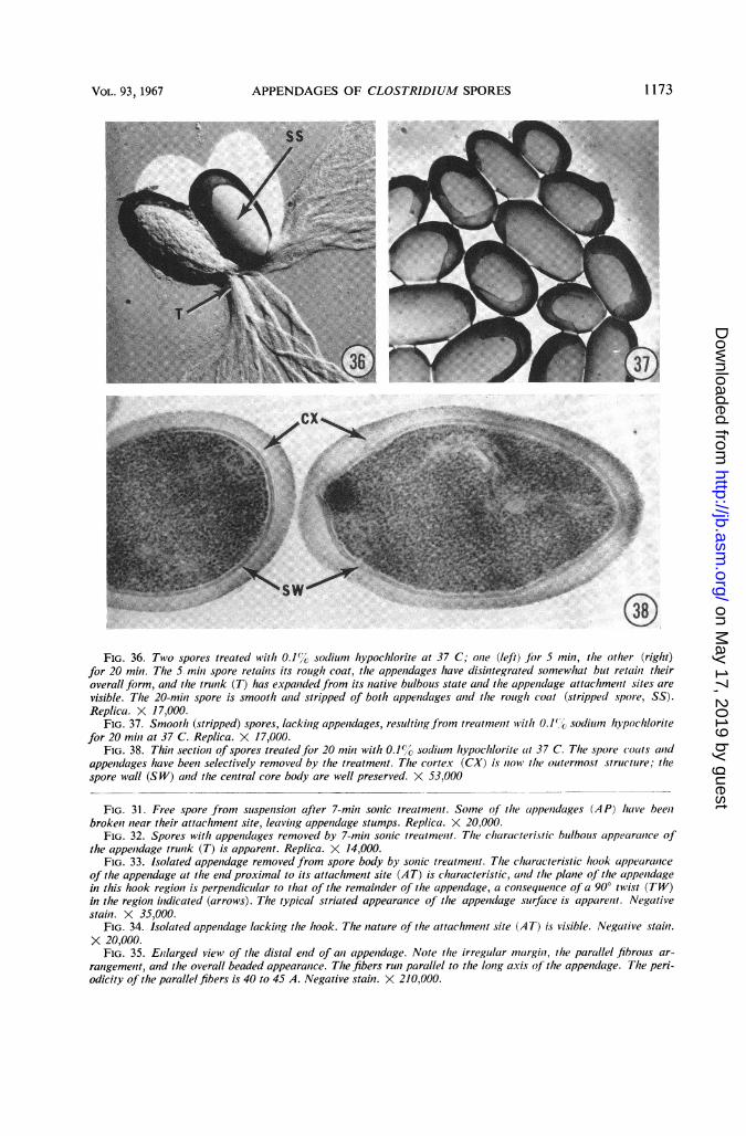
FIG. 36. Two spores treated with 0.1%1' soclium hypochlorite at 37 C; oiie (left) fbr 5 min, the other (right)for 20 miii. The 5 miii spore retains its rough coat, the appendages have disiiitegrated somewhat but retail? theiroverall form, and the trunk (T) has expandedfrom its native bulbous state and the appeiiclage attachment sites arevisible. The 20-mimi spore is smooth aiid stripped of both appendages and the rough coat (stripped spore, SS).Replica. X 17,000.
FIG. 37. Smooth (stripped) spores, lackilig appeuidages, resulting from treat/nelit with 0.1%', sodium hypochloritefor 20 mini at 37 C. Replica. X 17,000.
FIG. 38. Thin section of spores treatedfor 20 miii with 0.1%" sodium hypochlorite at 37 C. The spore (oats anidappenidages have beeii selectively removed by the treatment. The cortex (CX) is now the outermost structure; thespore wall (SW) and the central core body are well preserved. X 53,000
FIG. 31. Free spore from suspension after 7-mimi sonic treatmemit. Some of the cappenidages (AP) have beeiibrokeii near their attachmemit site, leavimig appemidage stumps. Replica. X 20,000.
FIG. 32. Spores with appendages removed by 7-min sonic treatmemit. Th1e characteristic bulbous appearclaice ofthe appemidage trumik (T) is apparent. Replica. X 14,000.
FIG. 33. Isolated appendage removed from spore body by son/ic treatmemit. The characteristic hook appearaniceof the appendage at the end proximal to its attachmenit site (AT) is characteristic, aiid the plane of the appemidagein this hook region is perpendicuilar to that of the remain7der of the appenidage, a comisequemice of a 90° twist (TW)in tlte regioni imidicated (arrows). The typical striated appearance of the appendage surface is apparenit. Negativestaimi. X 35,000.
FIG. 34. Isolated appenidage lackilig the hook. Tlhe nature of the attachmemit site (AT) is visible. Negative staimi.X 20,000.
FIG. 35. Enilarged view of the distal eiid of ali appendage. Note the irregular mnargimi, tle parallel fibrous ar-ramigememit, and the overall beaded appearance. Tlhe fibers run parallel to the lobg axis of the appenidage. The peri-odicity of the parallel fibers is 40 to 45 A. Negative stain. X 210,000.
1173VOL. 93, 1967
-' ':.._.:
.::
... cx z:-.:,.
;21
on May 17, 2019 by guest
http://jb.asm.org/
Dow
nloaded from