Clostridium difficile Infection: molecular and...
48
Universidade de Lisboa Faculdade de Ciências Departamento de Biologia Vegetal Clostridium difficile Infection: molecular and epidemiological study Joana Vanessa Duarte Isidro Dissertação orientada pela Doutora Mónica Oleastro (INSA) e pelo Professor Doutor Manuel Gomes (FCUL) Mestrado em Biologia Molecular e Genética 2015
-
Upload
trannguyet -
Category
Documents
-
view
233 -
download
1
Transcript of Clostridium difficile Infection: molecular and...
Universidade de Lisboa
Faculdade de Ciências
Departamento de Biologia Vegetal
Clostridium difficile Infection: molecular and epidemiological study
Joana Vanessa Duarte Isidro
Dissertação orientada pela Doutora Mónica Oleastro (INSA) e pelo
Professor Doutor Manuel Gomes (FCUL)
Mestrado em Biologia Molecular e Genética
2015


The work presented in this dissertation was conducted in the National Reference Laboratory
for Gastrointestinal Infections, Department of Infectious Diseases, of the National Health Institute
Doutor Ricardo Jorge, Lisbon, Portugal.

Clostridium difficile Infection: molecular and epidemiological study
I
Table of Contents
Acknowledgements ..................................................................................................................... III
List of Tables ............................................................................................................................... IV
List of Figures .............................................................................................................................. IV
List of Symbols and Abbreviations ................................................................................................ V
Abstract ..................................................................................................................................... VII
Keywords .................................................................................................................................. VIII
Resumo ....................................................................................................................................... IX
Palavras-chave ............................................................................................................................ XI
Publications ............................................................................................................................... XII
I - Introduction ............................................................................................................................. 1
1. Clostridium difficile as the etiologic agent of pseudomembranous colitis ................................. 1
2. Transmission and pathogenesis .................................................................................................. 1
3. C. difficile changing epidemiology ............................................................................................... 2
4. Risk factors for CDI ...................................................................................................................... 3
5. Diagnosis and treatment ............................................................................................................. 3
6. C. difficile virulence factors ......................................................................................................... 4
6.1. Pathogenicity Locus ............................................................................................................. 4
6.2. Binary toxin .......................................................................................................................... 6
7. Antimicrobial susceptibility of C. difficile .................................................................................... 6
7.1. Antibiotics and mechanisms of resistance .......................................................................... 7
8. Aims and objectives ..................................................................................................................... 8
II - Methods ................................................................................................................................. 8
1. Samples and data collection ........................................................................................................ 8
2. C. difficile culture ......................................................................................................................... 8
3. Antibiotic susceptibility testing by Etest ..................................................................................... 9
4. Antibiotic susceptibility testing by agar dilution ......................................................................... 9
5. Molecular characterization of strains ......................................................................................... 9
5.1. Pathogenicity factors ......................................................................................................... 12
5.1.1. Toxin typing ................................................................................................................... 12
5.1.2. tcdC analysis .................................................................................................................. 12
5.2. Ribotyping.......................................................................................................................... 12
Clostridium difficile Infection: molecular and epidemiological study
II
5.3. Mechanisms of antimicrobial resistance ........................................................................... 13
5.3.1. ermB, tetM and catD ..................................................................................................... 13
5.3.2. gyrA, gyrB and rpoB ....................................................................................................... 13
5.3.3. Penicillin-binding proteins ............................................................................................. 13
6. Statistical analysis ...................................................................................................................... 13
III – Results ................................................................................................................................ 14
1. Patients’ characteristics ............................................................................................................ 14
2. Molecular characterization of strains ....................................................................................... 15
2.1. Ribotypes ........................................................................................................................... 15
2.2. Toxin typing and tcdC mutations ....................................................................................... 16
3. Antimicrobial susceptibility ....................................................................................................... 17
4. Mechanisms of antimicrobial resistance ................................................................................... 19
4.1. ermB, tetM and catD ......................................................................................................... 19
4.2. GyrA and GyrB mutations .................................................................................................. 20
4.3. RpoB mutations ................................................................................................................. 20
4.4. PBP mutations ................................................................................................................... 21
IV – Discussion ........................................................................................................................... 22
V – Conclusion ............................................................................................................................ 27
VI - References ........................................................................................................................... 28

Clostridium difficile Infection: molecular and epidemiological study
III
Acknowledgements
First of all, I want to express my gratitude to Mónica Oleastro, my advisor at INSA, for the
privilege of working under her guidance in this project, for the tireless support and motivation, for
always taking my suggestions into account and doing everything within her power so I could
accomplish my objectives, and for improving my scientific and critical reasoning with the immense
knowledge transmitted. At last but not least, I want to thank the everyday enthusiasm, the kindness
and understanding that go far beyond the scientific field.
I am extremely thankful to Cláudia Júlio for being the first person to receive and supervise
me in INSA, making possible everything else that followed, including this dissertation.
To my advisor in FCUL, Professor Manuel Gomes, I thank the constant availability to answer
all of my questions (even the most insignificant ones), the precious help with the statistical analysis
and the suggestions and dissertation review.
To Andrea Santos, who I also consider as an (informal) advisor at INSA, I want to thank the
constant support and availability, the never-ending patience to teach me and clarify all of my doubts
and the theoretical and practical knowledge she passed onto me and without which I couldn’t have
possibly done this work.
I am grateful to Lúcia Reis and João Carlos Rodrigues for welcoming me in their laboratory
and for being constantly ready to help.
I am thankful to Leonor Silveira for welcoming me in her laboratory, the place where I wrote
the majority of this dissertation, and for accompanying and cheering up these months of work with
our conversations and peaceful silences.
I also want to express my gratitude to all the other people, including Maria Raquel Rocha and
Rui Matias, that, in some way, contributed to my integration in the everyday routine and brought joy
to my days in INSA.
I want to thank Rafael for being by my side in every moment, for the motivation, for listening
to my concerns and helping me face the problems calmly, as only he can do. Above all, for making
me believe and strive for a better tomorrow.
To my brother, my always and forever friend, I am grateful for the concern and everyday
interest in this work, for the talks and laughs, for the patience, kindness and love.
To my parents, to whom I owe much of who I am, I want to thank the unconditional support
and love and for allowing me to dream unrestrictedly and get to where I am today. I thank to my
mom, in particular, for always being there in the difficult moments, for believing in me, for the
infinite optimism and, most of all, for the irreplaceable friendship and love.
Finally, to all my family and friends, thank you for constantly helping me remember the most
important things in life.
Clostridium difficile Infection: molecular and epidemiological study
IV
List of Tables
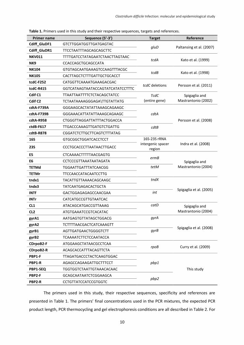
Table 1. Primers used in this study and their respective sequences, targets and references. ............. 10
Table 2. Primer pairs used in this study, final concentration used in PCR mixture, amplicon size, PCR
annealing temperature and electrophoresis conditions. ...................................................................... 11
Table 3. Characteristics of the patients diagnosed with CDI included in this study. ............................ 14
Table 4. Distribution of toxin genes profiles, tcdC deletions and mutations and the most common RTs
in each genotype. .................................................................................................................................. 16
Table 5. Strains susceptibility, geometric mean MIC and number of strains with resistant to MXF,
VAN, MTZ, RIF and TGC. ........................................................................................................................ 17
Table 6. MTZ susceptibility of strains from RT027 vs strains from other ribotypes. ............................ 18
Table 7. Comparison of MICs of IMP determined by Etest with MICs determined by the agar dilution
method. ................................................................................................................................................. 18
Table 8. Distribution of resistance determinants profiles and common PCR RTs associated. ............. 19
Table 9. Predicted amino acid substitutions in GyrA and GyrB associated with MXF resistance and
common PCR RTs associated. ................................................................................................................ 20
Table 10. Predicted amino acid substitutions in RpoB found in 21 RIF-resistant strains and common
PCR RTs associated. ............................................................................................................................... 21
Table 11. Predicted amino acid substitutions found in five IMP-resistant strains, their respective RTs
and MICs of IMP. ................................................................................................................................... 21
List of Figures
Figure 1. C. difficile pathogenicity locus (PaLoc) (a) and binary toxin locus (CDT locus) (b) (adapted
from Rupnik et al., 2009). ........................................................................................................................ 4
Figure 2. Frequencies of PCR ribotypes - RT; number of strains; % (Other – Includes 73 RTs with 4 or
less strains). ........................................................................................................................................... 15
Figure 3. Localization of the amino acid substitutions (in black) found in PBP1 of five IMP-resistant
strains (the conserved motifs are highlighted in grey; S – amino acid sequence in the IMP-susceptible
strains). .................................................................................................................................................. 21

Clostridium difficile Infection: molecular and epidemiological study
V
List of Symbols and Abbreviations
Δ deletion
% percentage
°C degree Celsius
aa amino acid residue
bp base pair
CFU colony-forming unit
h hour
kb kilo base pair
kDa kilodalton
L liter
min minute
mg milligram
mL milliliter
mm millimeter
no. number
V volt
μL microliter
μM micromolar
CA Community-associated
CDI Clostridium difficile infection
CDT Clostridium difficile transferase
CO-HCFA Community onset health-care facility-associated
DNA Deoxyribonucleic acid
EUCAST European Committee on Antimicrobial Susceptibility Testing
HO-HCFA Health-care onset health-care facility-associated
IMP Imipenem
INSARJ Portuguese National Institute of Health Dr. Ricardo Jorge
MIC Minimum inhibitory concentration
MLSB Macrolide, Lincosamide and Streptogramin B
MLST Multi-locus sequence typing
MTZ Metronidazole
MXF Moxifloxacin
PCR Polymerase chain reaction
PBP Penicillin-binding protein
QRDR Quinolone resistance-determining region
RIF Rifampicin
RNA Ribonucleic acid
RT PCR Ribotype
TGC Tigecycline
VAN Vancomycin
WGS Whole-genome sequencing

Clostridium difficile Infection: molecular and epidemiological study
VI
Clostridium difficile Infection: molecular and epidemiological study
VII
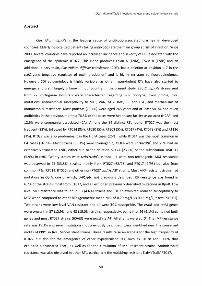
Abstract
Clostridium difficile is the leading cause of antibiotic-associated diarrhea in developed
countries. Elderly hospitalized patients taking antibiotics are the main group at risk of infection. Since
2000, several countries have reported an increased incidence and severity of CDI associated with the
emergence of the epidemic RT027. This clone produces Toxin A (TcdA), Toxin B (TcdB) and an
additional binary toxin, Clostridium difficile transferase (CDT), has a deletion at position 117 in the
tcdC gene (negative regulator of toxin production) and is highly resistant to fluoroquinolones.
However, CDI epidemiology is highly variable, as other hypervirulent RTs have also started to
emerge, and is still largely unknown in our country. In the present study, 286 C. difficile strains sent
from 22 Portuguese hospitals were characterized regarding PCR ribotype, toxin profile, tcdC
mutations, antimicrobial susceptibility to MXF, VAN, MTZ, IMP, RIF and TGC, and mechanisms of
antimicrobial resistance. Most patients (73.4%) were aged ≥65 years and at least 54.9% had taken
antibiotics in the previous months; 76.5% of the cases were healthcare facility-associated (HCFA) and
12.6% were community-associated (CA). Among the 84 distinct RTs found, RT027 was the most
frequent (22%), followed by RT014 (8%), RT020 (5%), RT203 (5%), RT017 (4%), RT078 (3%) and RT126
(3%). RT027 was also predominant in the HCFA cases (26%), while RT014 was the most common in
CA cases (16.7%). Most strains (96.1%) were toxinogenic, 31.8% were cdtA/cdtB+ and 29% had an
extensively truncated TcdC, either due to the deletion Δ117A (23.1%) or the substitution 184C→T
(5.9%) in tcdC. Twenty strains were tcdA-/tcdB+. In total, 11 were non-toxinogenic. MXF-resistance
was observed in 95 (33.8%) strains, mainly from RT027 (62/95) and RT017 (9/95) but also from
common RTs (RT014, RT020) and other non-RT027 cdtA/cdtB+ strains. Most MXF-resistant strains had
mutations in GyrA, one of which, D-81→N, not previously described. RIF-resistance was found in
6.7% of the strains, most from RT017, and all exhibited previously described mutations in RpoB. Low
level MTZ-resistance was found in 13 (4.6%) strains and RT027 exhibited reduced susceptibility to
MTZ when compared to other RTs (geometric mean MIC of 0.79 mg/L vs 0.16 mg/L; t test, p<0.01).
Two strains were low-level VAN-resistant and all were TGC-susceptible. The ermB and tetM genes
were present in 37 (12.9%) and 43 (15.0%) strains, respectively, being that 26 (9.1%) contained both
genes and most RT027 strains (60/63) were ermB-/tetM-. All strains were catD-. The IMP-resistance
rate was 19.3% and seven mutations (not previously described) were identified near the conserved
motifs of PBP1 in five IMP-resistant strains. These results raise awareness for the high frequency of
RT027 but also for the emergence of other hypervirulent RTs, such as RT078 and RT126 that
exhibited a truncated TcdC, as well as for the circulation of MXF-resistant strains. Antimicrobial
resistance was also observed in other RTs, particularly the multidrug resistant TcdA-/TcdB+ RT017.
Clostridium difficile Infection: molecular and epidemiological study
VIII
Keywords:
Clostridium difficile infection, epidemiology, PCR ribotype 027, antimicrobial susceptibility, resistance
mechanisms
Clostridium difficile Infection: molecular and epidemiological study
IX
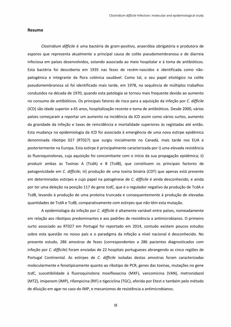
Resumo
Clostridium difficile é uma bactéria de gram-positivo, anaeróbia obrigatória e produtora de
esporos que representa atualmente a principal causa de colite pseudomembranosa e de diarreia
infeciosa em países desenvolvidos, estando associada ao meio hospitalar e à toma de antibióticos.
Esta bactéria foi descoberta em 1935 nas fezes de recém-nascidos e identificada como não-
patogénica e integrante da flora colónica saudável. Como tal, o seu papel etiológico na colite
pseudomembranosa só foi identificado mais tarde, em 1978, na sequência de múltiplos trabalhos
conduzidos na década de 1970, quando esta patologia se tornou mais frequente devido ao aumento
no consumo de antibióticos. Os principais fatores de risco para a aquisição da infeção por C. difficile
(ICD) são idade superior a 65 anos, hospitalização recente e toma de antibióticos. Desde 2000, vários
países começaram a reportar um aumento na incidência da ICD assim como vários surtos, aumento
da gravidade da infeção e taxas de reincidência e mortalidade superiores às registadas até então.
Esta mudança na epidemiologia da ICD foi associada à emergência de uma nova estirpe epidémica
denominada ribotipo 027 (RT027) que surgiu inicialmente no Canadá, mais tarde nos EUA e
posteriormente na Europa. Esta estirpe é principalmente caracterizada por i) uma elevada resistência
às fluoroquinolonas, cuja aquisição foi concomitante com o início da sua propagação epidémica; ii)
produzir ambas as Toxinas A (TcdA) e B (TcdB), que constituem os principais factores de
patogenicidade em C. difficile; iii) produção de uma toxina binária (CDT) que apenas está presente
em determinadas estirpes e cujo papel na patogénese de C. difficile é ainda desconhecido; e ainda
por ter uma deleção na posição 117 do gene tcdC, que é o regulador negativo da produção de TcdA e
TcdB, levando à produção de uma proteína truncada e consequentemente à produção de elevadas
quantidades de TcdA e TcdB, comparativamente com estirpes que não têm esta mutação.
A epidemiologia da infeção por C. difficile é altamente variável entre países, nomeadamente
em relação aos ribotipos predominantes e aos padrões de resistência a antimicrobianos. O primeiro
surto associado ao RT027 em Portugal foi reportado em 2014, contudo existem poucos estudos
sobre esta questão no nosso país e o paradigma da infeção a nível nacional é desconhecido. No
presente estudo, 286 amostras de fezes (correspondentes a 286 pacientes diagnosticados com
infeção por C. difficile) foram enviadas de 22 hospitais portugueses abrangendo as cinco regiões de
Portugal Continental. As estirpes de C. difficile isoladas destas amostras foram caracterizadas
molecularmente e fenotipicamente quanto ao ribotipo de PCR, genes das toxinas, mutações no gene
tcdC, suscetibilidade à fluoroquinolona moxifloxacina (MXF), vancomicina (VAN), metronidazol
(MTZ), imipenem (IMP), rifampicina (RIF) e tigeciclina (TGC), aferida por Etest e também pelo método
de diluição em agar no caso do IMP, e mecanismos de resistência a antimicrobianos.
Clostridium difficile Infection: molecular and epidemiological study
X
A maioria dos pacientes (73.4%) tinha ≥65 anos e pelo menos 54.9% tinha tomado
antibióticos nos três meses precedentes ao diagnóstico de ICD; 76.5% dos casos foram associados ao
meio hospitalar e 12.6% foram associados à comunidade. Entre os 84 diferentes ribotipos
encontrados no total das estirpes, o RT027 foi o mais frequente (22%), seguido do RT014 (8%), RT020
(5%), RT203 (5%), RT017 (4%), RT078 (3%) e RT126 (3%). O RT027 foi também o mais frequente entre
as estirpes de casos associados ao meio hospitalar (26%), ao passo que o RT014 foi o mais comum
nos casos associados à comunidade (16.7%). A maioria das estirpes eram toxinogénicas, 31.8% eram
também cdtA/cdtB+ e 29% continham um TcdC extensamente truncado com apenas 61 ou 65
resíduos de aminoácidos (comparativamente com o TcdC não-truncado de 232 resíduos), quer
devido à deleção Δ117A (23.1%), típica do RT027, quer devido à substituição 184C→T (5.9%),
presente noutros ribotipos hipervirulentos, como RT078 e RT126, que levam à inserção de codões
stop prematuros. Vinte estirpes eram TcdA-/TcdB+, 12 das quais pertencentes ao RT017, sendo que
10 dessas estirpes continham uma deleção de cerca de 1800 pb em tcdA e em duas não houve
amplificação em tcdA, indicando uma deleção mais extensa no gene. No total, 11 estirpes eram não-
toxinogénicas. Observou-se resistência à MXF em 95 (33.8%) estirpes, a maioria das quais
pertencente aos RT027 (62/95) e RT017 (9/95), contudo também foram identificadas estirpes de
outros ribotipos comuns (RT014, RT020) e outros cinco ribotipos CDTA/CDTB+ que não o RT027. A
maioria das estirpes resistentes à MXF continha mutações em GyrA, nomeadamente as já descritas T-
82→I e D-426→N e uma ainda não descrita anteriormente, D-81→N, encontrada numa estirpe do
RT020. A resistência à RIF foi observada em 6.7% das estirpes, sendo a maioria do RT017. Em 14
estirpes resistentes à RIF, 13 continham as mutações H-502→N e R-505→K em RpoB (já
anteriormente descritas), e uma continha apenas a mutação R-505→K. Observou-se um baixo nível
de resistência ao MTZ (3-16 mg/L) em 13 estirpes (4.6%), sete das quais pertencentes ao RT027. Para
além disso, o RT027 registou uma suscetibilidade reduzida ao MTZ comparativamente às estirpes de
outros ribotipos (média geométrica da concentração mínima inibitória = 0.79 mg/L vs 0.16 mg/L;
teste t, p<0.01), como já tinha sido observado anteriormente noutros estudos. Apenas duas estirpes
exibiram um baixo nível de resistência à VAN, sendo as restantes sensíveis, e todas foram sensíveis à
TGC. Os genes ermB e tetM, que conferem resistência à clindamicina/eritromicina e tetraciclina,
respetivamente, foram encontrados em 37 (12.9%) e 43 (15.0%) estirpes, respetivamente, sendo que
26 estirpes (9.1%) continham ambos os genes e a maioria das estirpes do RT027 (60/63) não
continham nenhum deles. O gene catD, que confere resistência ao cloranfenicol, não foi encontrado
em nenhuma estirpe. A taxa de resistência ao IMP, quando determinada pelo método de diluição em
agar, foi de 19.3%. Em cinco estirpes resistentes ao IMP foram encontradas sete mutações distintas
não descritas anteriormente (E-520→K, K-524→E, L-543→H, K-553→T, A-555→T, M-557→I e T-
674→I). Todas estas mutações encontram-se localizadas próximo dos motivos conservados da PBP1

Clostridium difficile Infection: molecular and epidemiological study
XI
e não foram encontradas nas estirpes sensíveis, sendo provável que contribuam para o fenótipo de
resistência observado. Contudo são necessários mais estudos que demonstrem que as mutações são
funcionais.
Os resultados aqui apresentados alertam para a elevada frequência do clone epidémico
RT027 nos hospitais portugueses comparativamente com os restantes ribotipos e também com os
resultados de outros estudos europeus recentes que descrevem uma frequência inferior deste
ribotipo e inclusivamente a diminuição da sua incidência. Como tal, é necessário aplicar medidas de
prevenção da transmissão de C. difficile no contexto hospitalar, nomeadamente através do
diagnóstico e tratamento de todos os pacientes com ICD. Contudo, a grande variedade de ribotipos
encontrados sugere que existem outras vias de transmissão de C. difficile para além do contacto com
o meio hospitalar, pelo que outras potenciais fontes devem ser analisadas futuramente,
nomeadamente alimentos e animais, uma vez que a transmissão zoonótica já foi anteriormente
descrita para o RT078. A emergência de resistência à MFX noutros ribotipos além do RT027,
nomeadamente ribotipos comummente encontrados, incluindo RT014 e RT020, ou hipervirulentos,
em que a proteína reguladora da expressão das toxinas, TcdC, está truncada, nomeadamente RT078
e RT126, sugere que o potencial epidémico observado em RT027 em resultado destes factores pode
também surgir nestas estirpes emergentes. Finalmente, dada a frequência de estirpes
multirresistentes observada, das quais o RT017 é o caso mais relevante, este estudo contribui para
alertar para a necessidade de uma utilização adequada dos agentes antimicrobianos.
Palavras-chave:
Infeção por Clostridium difficile, epidemiologia, ribotipo 027, suscetibilidade aos antimicrobianos,
mecanismos de resistência
Clostridium difficile Infection: molecular and epidemiological study
XII
Publications
The present study contributed to the publication of the following works:
Article
Santos, A., Isidro, J., Júlio, C., Oleastro, M. (2015). Clostridium difficile: diversidade genética e perfis
de suscetibilidade aos antimicrobianos. Boletim Epidemiológico Observações. Janeiro-março, 4(11),
15-18.
Poster
Isidro, J., Santos, A., Júlio, C., Boaventura, L., Diogo, J., Faustino, A., Oleastro, M. (2014, December).
Genótipos e perfis de suscetibilidade a antimicrobianos de estirpes de Clostridium difficile isoladas de
hospitais portugueses. Poster session presented at the Congresso Nacional VIH, Doenças Infeciosas e
Microbiologia Clínica, Lisbon, Portugal.
Oral Communication
Isidro, J. (2014, December). Genótipos e perfis de suscetibilidade a antimicrobianos de estirpes de
Clostridium difficile isoladas de hospitais portugueses. Best of Posters session at the Congresso
Nacional VIH, Doenças Infeciosas e Microbiologia Clínica, Lisbon, Portugal.
Clostridium difficile Infection: molecular and epidemiological study
1
I - Introduction
1. Clostridium difficile as the etiologic agent of pseudomembranous colitis
Clostridium difficile is the leading cause of nosocomial antibiotic-associated infectious
diarrhea and pseudomembranous colitis in developed countries (Rupnik, et al., 2009). This gram-
positive, spore-forming obligate anaerobic bacterium was first isolated in 1935 from the stools of
neonates as part of the normal colonic flora and hence thought to be non-pathogenic. At the time,
Hall and O’Toole (1935) named the bacterium Bacillus difficilis due to its difficult isolation and slow
growth in culture.
C. difficile infection (CDI) is a colonic disease caused by the action of C. difficile toxins on the
inner surface of the colon which, in severe forms of the disease, can result in pseudomembranous
colitis. This inflammatory condition, characterized by the formation of inflammatory lesions and
pseudomembranes in the colon in response to the toxins (Rupnik, et al., 2009), was first described in
1893. However, the later discovery of C. difficile in 1935 didn’t lead to its immediate association with
the disease since it was thought to be non-pathogenic and the interest on this infection only arose
after the 1950s, when pseudomembranous colitis became a common disease due to the rise in
antibiotic use. The subsequent publication of various independent studies in the 1970s finally
converged into the identification of C. difficile as the etiologic agent of pseudomembranous colitis in
1978 (Bartlett, 1994).
2. Transmission and pathogenesis
C. difficile spores are the main route for the spreading of CDI due to their high resistance to
cleaning measures and disinfection agents, such as alcohol-based antiseptics that are largely used by
healthcare workers as a substitute for the conventional hand washing, allowing spores to persist in
the environment for many months or years. The main route of transmission of C. difficile is the direct
contact between individuals, either direct contact with an infected patient (symptomatic or
asymptomatic) or with a healthcare professional. Alternatively, C. difficile may be acquired from
contaminated surfaces or objects in healthcare settings (Cohen et al., 2010).
Although healthcare facilities have been considered the main source of C. difficile, in two
recent studies investigating ward-based transmission by multi-locus sequence typing (MLST) and
whole-genome sequencing (WGS), only 25% and 19% of the isolates, respectively, could be linked to
a hospital contact. These results thus point to the existence of other routes of transmission and
reservoirs of the microorganism outside the hospital setting (Bauer and Kuijper, 2015).
Clostridium difficile Infection: molecular and epidemiological study
2
C. difficile enters the host via the fecal-oral route following the ingestion of the gastric acid-
resistant spores that later germinate into vegetative cells in the intestines. Antibiotic treatments may
disrupt the normal colonic flora and, hence, a C. difficile strain that is resistant to the antibiotic has a
selective advantage and is more likely to cause infection. When the treatment stops and the level of
antibiotic diminish, the microbiota remains unbalanced for an undetermined period of time during
which infection is possible either by a susceptible or a resistant strain. In such depleted microbiota
conditions, C. difficile is able to proliferate and produce its two main virulence factors, toxin A and
toxin B that consequently trigger the infection by disruption of tight junctions and loosening of the
epithelial barrier of the colon. The clinical outcomes of CDI can range from an asymptomatic
colonization to diarrhea, abdominal pain, and fever and, in more severe states of the disease,
pseudomembranous colitis, ileus, toxic megacolon, bowel perforation, sepsis, shock, and death
(Rupnik, et al., 2009).
3. C. difficile changing epidemiology
CDI rates and disease severity have been increasing since 2000, mainly in elderly patients
with a recent hospitalization or living in a long-term care facility. Carriage rates of C. difficile in
healthy adults is about 5-15% and can reach more than 80% and 50% in healthy infants and residents
in long-term care facilities, respectively (Surawicz et al., 2013).
In 2002, several hospitals in Quebec, Canada, registered a major increase in the incidence of
CDI with a concomitant increase in disease morbidity and mortality. Notably, the incidence increased
from 35.6 cases per 100 000 population in 1991 to 156.3 per 100 000 in 2003 and was even greater
in patients aged 65 years or more, increasing from 102.0 to 866.5 cases per 100 000 (Pépin et al.,
2004). The same increase in incidence, severity and mortality occurred in the US, with outbreaks
reported in several states. Soon after, the first reports arose in Europe, with particularly severe
outbreaks in the UK that led to a six-fold increase in CDI-related mortality from 499 deaths in 1999 to
3393 in 2006 (Clements et al., 2010; Kelly and LaMont, 2008). Since then, several European countries
have registered the same pattern in C. difficile epidemiology with increasing rates of infection
(Freeman et al., 2010).
These North American and European outbreaks were associated with the emergence of a
new epidemic, hypervirulent C. difficile strain, categorized as group BI by restriction endonuclease
analysis, North American pulse-field type 1 by pulse-field gel electrophoresis and as 027 by PCR
ribotype (BI/NAP1/027) (Warny et al., 2005) that will be, from here on, simply referred to as ribotype
(RT) 027. Notably, in 2008, RT027 was detected in 16 European countries and by 2009 it had already
been responsible for outbreaks in at least 11 of those countries (Freeman et al., 2010). In 2014, the
Clostridium difficile Infection: molecular and epidemiological study
3
first outbreak was reported in Portugal with a CDI-attributable mortality rate of 11.3% and a crude
mortality rate at 6 months of 64.2%. However, since data on CDI in Portugal is still scarce, the
previous occurrence of RT027 in Portuguese hospitals is unknown (Oleastro et al., 2014).
The epidemic RT027 produces both toxins A and B, carries a mutation in the negative
regulator of toxins expression (TcdC) and produces an additional binary toxin (CDT) (Rupnik et al.,
2009). Furthermore, this spread of the epidemic RT027 is concomitant with an acquired resistance to
fluoroquinolones, such as moxifloxacin (MXF), presumably due to selection pressure since this was
the most prescribed class of antibiotics in the US, in 2002 (Pépin et al., 2005).
Nonetheless, the increase in CDI rates can’t be attributed to RT027 only since its prevalence
is decreasing in some countries (Bauer et al., 2011) and other RTs, such as RT078 in Europe, and
RT244 in Australia, have also been associated with outbreaks and severe cases (Rupnik et al., 2009;
Lim et al., 2014).
4. Risk factors for CDI
The use of antibiotics is the main risk factor for CDI since C. difficile can only colonize the gut
if the normal microbiota is disrupted. Other risk factors include advanced age (≥65 years), recent
hospitalization (because it increases the risk of exposure to the microorganism), use of medications
that reduce gastric acid (like proton-pump inhibitors), and severe comorbidities (Surawicz et al.,
2013).
Elderly hospitalized patients taking antibiotics constitute the main group at risk of infection.
However, there has been an increase in the incidence of infection in populations previously thought
to be at low risk, namely pregnant women and healthy and younger people in the community with
no previous contact either with the hospital environment or with antibiotics. This change towards an
increase in community-associated cases raises the need to consider CDI as a possibility when
diagnosing any symptomatic patient and not only those initially at risk (Rupnik et al., 2009) and also
demonstrates that the epidemiology of CDI is constantly changing.
5. Diagnosis and treatment
Since C. difficile can be carried asymptomatically by healthy individuals and this carriage may
be increased in patients taking antibiotics, only the stools from patients with diarrhea should be
tested for the presence of the microorganism.
The best standard laboratory method for CDI diagnosis is yet to be established. Firstly, C.
difficile culture alone cannot be used as a diagnosis test since only toxigenic strains cause disease and
Clostridium difficile Infection: molecular and epidemiological study
4
this method does not distinguish toxigenic from non-toxigenic strains. Secondly, the two reference
methods, C. difficile cytotoxin neutralization assay and toxigenic culture, are time consuming
techniques that require specialized staff and equipment. As such, diagnostic testing has evolved to
yield a faster diagnosis. A two or three-stepwise screening algorithm is recommended in which a first
positive test must be followed by one or two confirmatory tests, e.g., the stools can be tested with
an enzyme immunoassay to detect glutamate dehydrogenase (GDH), thus confirming the presence of
C. difficile, followed by an enzyme immunoassay to detect free Toxins A and B or, preferentially, due
to higher sensitivity, a nucleic acid amplification test to detect the toxins genes, thus confirming the
presence of a toxigenic strain (Rupnik et al., 2009; Surawicz et al., 2013).
Disease treatment is stratified according to disease severity. Briefly, patients with mild-to-
moderate disease should be treated with metronidazole (MTZ) and severe disease with vancomycin
(VAN); in cases of severe and complicated disease, surgical therapy should also be considered in
addition to antibiotic therapy; fecal microbiota transplant is an alternative treatment in cases of
recurrent infection and ineffective antibiotic therapies (Surawicz et al., 2013).
6. C. difficile virulence factors
6.1. Pathogenicity Locus
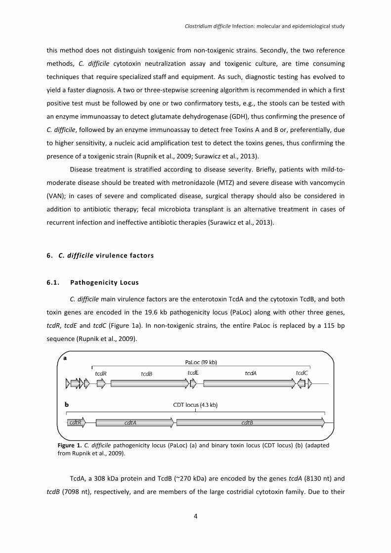
C. difficile main virulence factors are the enterotoxin TcdA and the cytotoxin TcdB, and both
toxin genes are encoded in the 19.6 kb pathogenicity locus (PaLoc) along with other three genes,
tcdR, tcdE and tcdC (Figure 1a). In non-toxigenic strains, the entire PaLoc is replaced by a 115 bp
sequence (Rupnik et al., 2009).
TcdA, a 308 kDa protein and TcdB (~270 kDa) are encoded by the genes tcdA (8130 nt) and
tcdB (7098 nt), respectively, and are members of the large costridial cytotoxin family. Due to their
Figure 1. C. difficile pathogenicity locus (PaLoc) (a) and binary toxin locus (CDT locus) (b) (adapted from Rupnik et al., 2009).
Clostridium difficile Infection: molecular and epidemiological study
5
close proximity, high sequence similarity (66%), functional homology and common conserved
domains, it has been proposed that these two genes may have arisen through gene duplication.
TcdA and TcdB are both intracellular bacterial toxins that glucosylate small GTPases. After
entering the intoxicated cell through receptor-mediated endocytosis, these toxins are able to
hydrolyze UDP-glucose in the cytosol and transfer the released sugar moiety to the GTP-binding
domain of small GTPases, such as Rho, Rac and Cdc42 that are implicated in the regulation of the
actin cytoskeleton, hence inactivating these small regulatory proteins. This leads to loss of cell
structural integrity and destruction of the intestinal epithelium through disruption of the actin
cytoskeleton and tight junctions, resulting in fluid accumulation and inflammatory responses (Hunt
and Ballard, 2013; Rupnik et al., 2009; Voth and Ballard, 2005).
Several conserved functional domains have been identified in TcdA and TcdB. Namely, the N-
terminal enzymatic domain, responsible for the glycosyltransferase activity; a cysteine protease
domain that carries out the autoproteolytic processing that releases the enzymatic domain of the
toxin in the cytosol after receptor-mediated endocytosis; a hydrophobic region (near the middle of
the toxins) associated with membrane insertion and translocation; and the C-terminal, receptor-
binding domain, composed of combined repetitive oligopeptide repeats (CROPs) known to bind
glycans (Hunt and Ballard, 2013; Voth and Ballard, 2005).
The CROP region is more extensive in TcdA, however it is unknown how this difference
interferes in the toxins tropism. In fact, there are variations in cell targeting between the two toxins,
as TcdB intoxicates a broader range of cell types. In fact, TcdB, that was initially thought to be
secondary in C. difficile disease, is a more potent cytotoxin than TcdA and has been shown to be
sufficient per se to cause the same disease pattern when compared with both toxins simultaneously,
which indicates that TcdB may also function as an enterotoxin (Hunt and Ballard, 2013).
TcdA and TcdB production occurs during late log and stationary phases and their production
depends on the strain and environmental factors (nutrients available, temperature, etc.), being
regulated by other genes, namely tcdR and tcdC, both located in the PaLoc (Rupnki et al, 2009).
TcdC is an anti-sigma factor expressed during exponential growth that appears to negatively
regulate TcdA and TcdB expression through disruption of TcdR interaction with RNA polymerase. This
protein (232 aa, 25.5 kDa), encoded by the tcdC gene (696 nt), has been given much attention since
several variations identified in the tdcC sequence are related to increased virulence in some C.
difficile strains, namely the epidemic RT027 known to produce increased amounts of TcdA and TcdB
(Hunt and Ballard, 2013; Warny et al., 2005). These epidemic strains carry an 18 bp deletion and a
single nucleotide deletion at base 117 that induces a frameshift resulting in a premature stop codon
that truncates TcdC, consequently yielding a protein with 65 residues. Other deletions and mutations
have also been identified in non-RT027, namely 39 bp and 36 bp deletions and a nonsense mutation
Clostridium difficile Infection: molecular and epidemiological study
6
(transition C to T) in position 184 that also leads to a truncated TcdC (Curry et al., 2007; Hunt and
Ballard, 2013).
6.2. Binary toxin
Some specific strains of C. difficile also produce an additional toxin, the binary toxin CDT that
belongs to the family of binary ADP-ribosylating toxins. It is composed of two independent
components, the proteins CDTa and CDTb, whose respective genes, cdtA and cdtB, are located in the
CDT locus that also contains the regulatory protein gene, cdtR, located upstream of cdtA and cdtB
(Figure 1b). Such as the PaLoc, the CdtLoc also presents variability between strains, ranging from a
complete locus to its complete absence, in which case it is replaced by a 68 bp insert (Hunt and
Ballard, 2013).
Regarding the CDT mechanism of action, it has been proposed that CDTb, the binding
component of the binary toxin, binds to a receptor on the surface of host cells, and then translocates
the enzymatic component, CDTa, into the cytosol. There, CDTa ADP-ribosylates actin causing
depolymerization of the actin cytoskeleton which results in the formation of long microtubule
protrusions. Electron microscopy studies have shown that this protrusions increase the adherence of
bacteria to the surface of epithelial cells (Gerding et al., 2014).
Nevertheless, the role of CDT as a virulence factor in CDI is still unclear. Until recently, its
pathogenic potential had been disregarded since many disease-causing strains do not produce CD;
however this view began to change (Hunt and Ballard, 2013) as, for instance, CDT-producing strains,
including RT027 and RT078, among others, have been associated with an increased fatality rate
(Bacci et al., 2011).
7. Antimicrobial susceptibility of C. difficile
Antimicrobial stewardship has been proven effective in the prevention of CDI. Clindamycin,
cephalosporins and fluoroquinolones are the antibiotics that pose the greater risk for infection.
However, virtually any antibiotic can cause the disease (Surawicz et al., 2013) and the risk is further
enhanced if the strain is resistant to the antibiotics used (Rupnik et al., 2009). This has been
particularly evident with the epidemic RT027 whose emergence was concomitant with the
acquirement of resistance to fluoroquinolones. In a study conducted in Canada (Pépin et al., 2005),
the use of this class of antibiotics was identified as the most important risk factor for the
development of CDI. In a study including strains from thirteen European countries, several non-027
Clostridium difficile Infection: molecular and epidemiological study
7
RTs, namely the hypervirulent RT078, showed resistance to fluoroquinolones (Spigaglia et al., 2008),
raising awareness for the emergence of new hypervirulent strains.
Overall, and since antimicrobial resistance varies widely between countries (Huang et al.,
2009), it is important to monitor the susceptibility of C. difficile strains to antibiotics, particularly to
those used in CDI treatment.
7.1. Antibiotics and mechanisms of resistance
Resistance to clyndamicin and/or erythromycin is the most common phenotype in C. difficile.
Resistance to these MLSB antibiotics (inhibitors of protein synthesis) in C. difficile is generally due to
the presence of the erythromycin ribosomal methylase gene B (ermB) that methylates the 23S
ribosomal RNA, thus altering the antibiotic binding site (Huang et al., 2009).
Resistance to tetracycline, a protein synthesis inhibitor as well, has also been observed in C.
difficile strains with varying rates between countries. This phenotype is often due to the presence of
the tetM gene that confers ribosomal protection and is carried by a Tn5397 or a Tn916-like
conjugative element (Huang et al., 2009). Resistance to chloramphenicol in C. difficile is generally
also mediated by a transposon-associated resistance determinant, the catD gene that encodes a
chloramphenicol acetyltransferase responsible for inactivating the antibiotic (Spigaglia et al., 2011).
Alternatively, mutations in some antimicrobial target genes are involved in the resistance to
fluoroquinolones and rifampicin (RIF). Fluoroquinolones are a family of broad-spectrum antibiotics
(including MXF) that inhibit DNA synthesis and, as stated previously, are associated with increased
risk for CDI. The principal mechanism of resistance in C. difficile is the substitution of amino acids
residues in the quinolone resistance-determining region (QRDR) of the target enzymes, specifically
the DNA gyrase subunits GyrA or GyrB (Huang et al., 2009). RIF is an inhibitor of RNA synthesis that
has been proposed to treat relapses of CDI. However, resistance to this antibiotic has already been
described in C. difficile and is associated with point mutations in rpoB, the gene encoding the beta
subunit of RNA polymerase (Curry et al., 2009).
Resistance to imipenem (IMP), a carbapenem that binds to penicillin-binding proteins (PBPs)
inhibiting cell wall synthesis (Papp-Wallace et al., 2011), has also been described in C. difficile isolates
with varying rates (7.41% to 25%), much dependent on the antimicrobial susceptibility method used
(Kim et al., 2010; Freeman et al., 2015). However, no resistance mechanism has been described yet.
Resistance to MTZ and VAN in C. difficile is rarely found and the underlying mechanisms are
still unknown. However, the emergence of reduced susceptibility to MTZ has been reported in some
frequent RTs, such as RT027, compared to other less common RTs, as well as in recent isolates vs
historic isolates (Freeman et al., 2010; Freeman et al., 2015).
Clostridium difficile Infection: molecular and epidemiological study
8
8. Aims and objectives
CDI is a major concern in developed countries. However, the differences in surveillance
systems, infection ascertainment and laboratory diagnosis between and within countries have been
limiting the understanding of CDI epidemiology. The present dissertation concerns the study of the
epidemiology of CDI in Portugal, where few studies have been conducted on this topic, aiming to a
better understanding of this infection. The main objectives of this work are: i) the molecular and
phenotypic characterization of C. difficile strains, namely through identification of the predominant
RTs, comparison of the prevalence of RTs identified in hospital settings vs community setting and
detection and characterization of pathogenicity factors; ii) analysis of antimicrobial susceptibility
patterns and identification of mechanisms of resistance; iii) analysis of patients’ epidemiological data
to identify potential risk factors.
II - Methods
1. Samples and data collection
From September 2012 to May 2015, diarrheal faecal samples belonging to patients aged
above 2 years, exhibiting typical CDI symptoms and with a positive test for the presence of C. difficile
toxins and/or GDH were sent to the National Reference Laboratory of the Portuguese National
Institute of Health Dr. Ricardo Jorge (INSARJ) from 22 different Portuguese hospitals from the five
regions of Continental Portugal. Each sample was accompanied by a short questionnaire on the
patient’s clinical and epidemiological data, including age, sex, place (hospital or community) and time
of symptoms onset, and antibiotics consumption within the three months prior to diagnosis of CDI.
According to time of symptoms onset and history of hospitalization, CDI cases were classified as
health-care onset health-care facility-associated (HO-HCFA), community onset health-care facility-
associated (CO-HCFA), community-associated (CA) or unknown, as described by Surawicz and
colleagues (2013).
2. C. difficile culture
Faecal samples were treated with ethanol in a 1:1 proportion, vortexed and left to rest for 1
hour to select the C. difficile spores. Roughly 1-2 drops were then cultured on the selective and
chromogenic medium chromID™ C. difficile agar (bioMérieux) and incubated at 37°C for 48 h, under
anaerobic atmosphere generated with AnoxomatTM (MART Microbiology BV) with catalyst. C. difficile
Clostridium difficile Infection: molecular and epidemiological study
9
colonies were identified based on the typical morphology (5-7 mm in diameter, flat with a
filamentous edge) and black colour.
3. Antibiotic susceptibility testing by Etest
Minimum inhibitory concentrations (MICs) of MXF, VAN, MTZ, IMP, RIF and tigecycline (TGC)
were determined by Etest (bioMérieux) on Brucella Agar (BA) with 5% Sheep Blood, Hemin and
Vitamin K1 (BD, BBL™). Briefly, one colony from each cultured sample was selected and cultured on
brain-heart infusion (BHI) broth and incubated anaerobically at 37°C for 48 hours. Broth cultures
were then adjusted with sterile broth, to an opacity equivalent to the 1 McFarland (3x10⁸ CFU/mL)
and inoculated by flooding onto prereduced BA plates, according to the manufacturer’s instructions.
After the surface was completely dry, an Etest strip was applied to each plate. The plates were
incubated anaerobically at 37°C for 48 h. The MICs were read where growth intersects the strip. The
breakpoints used were > 4 mg/L for MXF, > 2 mg/L for VAN and MTZ, > 8 mg/L for IMP, > 0.004 mg/L
for RIF, and > 0.25 mg/L for TGC, as stipulated by the EUCAST guidelines. (European Committee on
Antimicrobial Susceptibility Testing, 2015, http://www.eucast.org/clinical_breakpoints/). Two strains
(RT027 and RT001) belonging to the ECDC collection, gently given to INSARJ by Dr Ed Kuijper from
Leiden University Medical Centre, were used as controls.
4. Antibiotic susceptibility testing by agar dilution
To confirm the susceptibility to IMP, some strains were also tested by the agar dilution
method on Wilkins-Chalgren (Oxoid) agar, as described by Freeman et al. (2005). Procedure was
according to the EUCAST recommendations. Briefly, Wilkins-Chalgren plates were prepared with
three distinct IMP concentrations, namely 8, 16, and 32 mg/L, plus drug-free control plates. Selected
strains were raised in BHI broth anaerobically at 37°C for 48h. Broth cultures were adjusted to a 0.5
McFarland standard (≈ 108 CFU/mL) and further diluted 1:10 with sterile broth in a 96-well microtiter
plate to obtain a concentration of approximately 107 CFU/mL. Subsequently, to yield a cell density of
around 104 CFU/spot, 1 μl of the suspension was inoculated per spot. Plates were incubated
anaerobically at 37°C and read after 48 h. The breakpoint used was the same as for the Etest method
(> 8 mg/L).
5. Molecular characterization of strains
DNA was extracted with NucliSENS® easyMAG® (bioMérieux) using the Generic 2.0.1
protocol. The strains were characterized by PCR assays, PCR ribotyping and nucleotide sequencing.
Clostridium difficile Infection: molecular and epidemiological study
10
Table 1. Primers used in this study and their respective sequences, targets and references.
The primers used in this study, their respective sequences, specificity and references are
presented in Table 1. The primers’ final concentrations used in the PCR mixtures, the expected PCR
product length, PCR thermocycling and gel electrophoresis conditions are all described in Table 2. For
Primer name Sequence (5’-3’) Target Reference
Cdiff_GluDF1 GTCTTGGATGGTTGATGAGTAC gluD Paltansing et al. (2007)
Cdiff_GluDR1 TTCCTAATTTAGCAGCAGCTTC
NKV011 TTTTGATCCTATAGAATCTAACTTAGTAAC tcdA Kato et al. (1999)
NK9 CCACCAGCTGCAGCCATA
NK104 GTGTAGCAATGAAAGTCCAAGTTTACGC tcdB Kato et al. (1998)
NK105 CACTTAGCTCTTTGATTGCTGCACCT
tcdC-F252 CATGGTTCAAAATGAAAGACGAC tcdC deletions Persson et al. (2011)
tcdC-R415 GGTCATAAGTAATACCAGTATCATATCCTTTC
Cdif C1 TTAATTAATTTTCTCTACAGCTATCC TcdC (entire gene)
Spigaglia and Mastrantonio (2002) Cdif C2 TCTAATAAAAGGGAGA\TTGTATTATG
cdtA-F739A GGGAAGCACTATATTAAAGCAGAAGC
cdtA
Persson et al. (2008)
cdtA-F739B GGGAAACATTATATTAAAGCAGAAGC
cdtA-R958 CTGGGTTAGGATTATTTACTGGACCA
ctdB-F617 TTGACCCAAAGTTGATGTCTGATTG cdtB cdtB-R878 CGGATCTCTTGCTTCAGTCTTTATAG
16S GTGCGGCTGGATCACCTCCT 16S-23S rRNA intergenic spacer
region Indra et al. (2008)
23S CCCTGCACCCTTAATAACTTGACC
E5 CTCAAAACTTTTTAACGAGTG ermB
Spigaglia and Mastrantonio (2004)
E6 CCTCCCGTTAAATAATAGATA
TETMd TGGAATTGATTTATCAACGG tetM
TETMr TTCCAACCATACAATCCTTG
tndx1 TACATTGTTAAAACAGCAAGC tndX
Spigaglia et al. (2005) tndx3 TATCAATGAGACACTGCTA
INTf GACTGGAGAGAGCCAACGAA int
INTr CATCATGCCGTTGTAATCAC
CL1 ATACAGCATGACCGTTAAAG catD Spigaglia and Mastrantonio (2004) CL2 ATGTGAAATCCGTCACATAC
gyrA1 AATGAGTGTTATAGCTGGACG gyrA
Spigaglia et al. (2008) gyrA2 TCTTTTAACGACTCATCAAAGTT
gyrB1 AGTTGATGAACTGGGGTCTT gyrB
gyrB2 TCAAAATCTTCTCCAATACCA
CDrpoB2-F ATGGAAGCTATAACGCCTCAA rpoB Curry et al. (2009)
CDrpoB2-R ACAGCACCATTTACAGTTCTA
PBP1-F TTAGATGACCCTACTCAAGTGGAC
pbp1
This study
PBP1-R AGAGCCAGAAGATTGCTTTCCT
PBP1-SEQ TGGTGGTCTAATTGTAAACACAAC
PBP2-F GCAGCAATAATCTCGGAAGCA pbp2
PBP2-R CCTGTTATCCATCCGTGGTC
Clostridium difficile Infection: molecular and epidemiological study
11
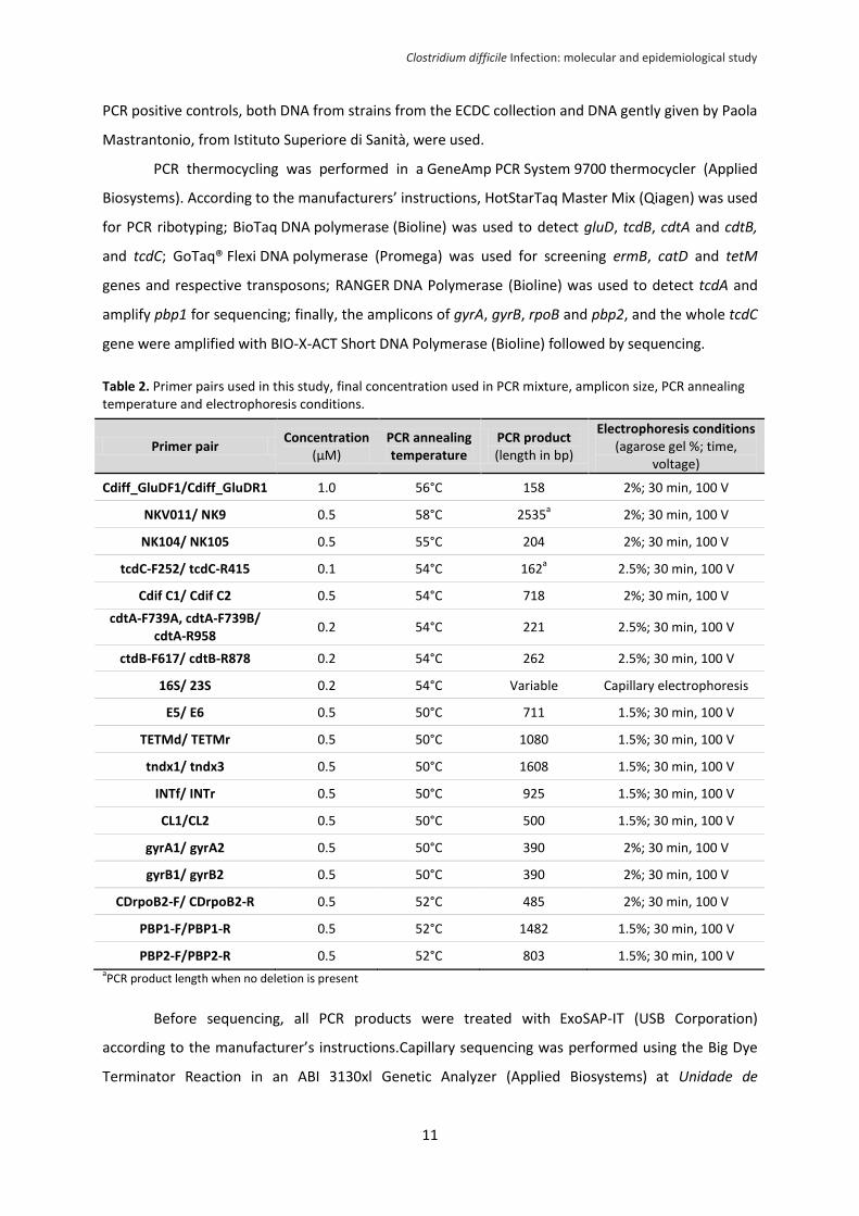
PCR positive controls, both DNA from strains from the ECDC collection and DNA gently given by Paola
Mastrantonio, from Istituto Superiore di Sanità, were used.
PCR thermocycling was performed in a GeneAmp PCR System 9700 thermocycler (Applied
Biosystems). According to the manufacturers’ instructions, HotStarTaq Master Mix (Qiagen) was used
for PCR ribotyping; BioTaq DNA polymerase (Bioline) was used to detect gluD, tcdB, cdtA and cdtB,
and tcdC; GoTaq® Flexi DNA polymerase (Promega) was used for screening ermB, catD and tetM
genes and respective transposons; RANGER DNA Polymerase (Bioline) was used to detect tcdA and
amplify pbp1 for sequencing; finally, the amplicons of gyrA, gyrB, rpoB and pbp2, and the whole tcdC
gene were amplified with BIO-X-ACT Short DNA Polymerase (Bioline) followed by sequencing.
Table 2. Primer pairs used in this study, final concentration used in PCR mixture, amplicon size, PCR annealing temperature and electrophoresis conditions.
aPCR product length when no deletion is present
Before sequencing, all PCR products were treated with ExoSAP-IT (USB Corporation)
according to the manufacturer’s instructions.Capillary sequencing was performed using the Big Dye
Terminator Reaction in an ABI 3130xl Genetic Analyzer (Applied Biosystems) at Unidade de
Primer pair Concentration
(µM) PCR annealing temperature
PCR product (length in bp)
Electrophoresis conditions (agarose gel %; time,
voltage)
Cdiff_GluDF1/Cdiff_GluDR1 1.0 56°C 158 2%; 30 min, 100 V
NKV011/ NK9 0.5 58°C 2535a
2%; 30 min, 100 V
NK104/ NK105 0.5 55°C 204 2%; 30 min, 100 V
tcdC-F252/ tcdC-R415 0.1 54°C 162a
2.5%; 30 min, 100 V
Cdif C1/ Cdif C2 0.5 54°C 718 2%; 30 min, 100 V
cdtA-F739A, cdtA-F739B/ cdtA-R958
0.2 54°C 221 2.5%; 30 min, 100 V
ctdB-F617/ cdtB-R878 0.2 54°C 262 2.5%; 30 min, 100 V
16S/ 23S 0.2 54°C Variable Capillary electrophoresis
E5/ E6 0.5 50°C 711 1.5%; 30 min, 100 V
TETMd/ TETMr 0.5 50°C 1080 1.5%; 30 min, 100 V
tndx1/ tndx3 0.5 50°C 1608 1.5%; 30 min, 100 V
INTf/ INTr 0.5 50°C 925 1.5%; 30 min, 100 V
CL1/CL2 0.5 50°C 500 1.5%; 30 min, 100 V
gyrA1/ gyrA2 0.5 50°C 390 2%; 30 min, 100 V
gyrB1/ gyrB2 0.5 50°C 390 2%; 30 min, 100 V
CDrpoB2-F/ CDrpoB2-R 0.5 52°C 485 2%; 30 min, 100 V
PBP1-F/PBP1-R 0.5 52°C 1482 1.5%; 30 min, 100 V
PBP2-F/PBP2-R 0.5 52°C 803 1.5%; 30 min, 100 V

Clostridium difficile Infection: molecular and epidemiological study
12
Tecnologia e Inovação (Departamento de Genética Humana, National Institute of Health Dr. Ricardo
Jorge, Lisbon, Portugal). The corresponding amino acid sequences were obtained with the ExPASy
Translate tool (http://web.expasy.org/tools/translate/) and further analyzed and aligned with wild-
type sequences obtained in the NCBI database, using the Multalin software
(http://multalin.toulouse.inra.fr/multalin/).
5.1. Pathogenicity factors
5.1.1. Toxin typing
The primer pair NKV011/NK9 was used to identify both the tcdA gene and the deletions in its
repeating regions, resulting in a 2535 bp amplicon when no deletion is present, and ≈1800 bp or
≈700 bp amplicons when deletions are present. For detection of tcdB gene, the primer pair
NK104/NK105 was used to amplify a 204 bp amplicon.
The binary toxin, CDT, was identified through amplification of a 221 bp amplicon of the cdtA
gene using the primers cdtA-F739A, cdtA-F739B, and cdtA-R958, and amplification of a 262 bp
segment of cdtB with the primers ctdB-F617 and cdtB-R878, as described by Persson et al. (2008)
(Table 1). Only the strains with both amplicons were considered cdtA/cdtB+.
5.1.2. tcdC analysis
The primer pair tcdC-F252 and tcdC-R415 was used to identify the internal in-frame deletions
of the tcdC gene, as described by Persson et al. (2011) (Table 1). Non-deleted strains show a 162 bp
amplicon, whereas amplicons of 144, 126 and 108 bp are obtained in result of deletions of 18, 39 and
54 bp, respectively. Strains in which a deletion was detected were selected for amplification of the
entire tcdC gene using the primers C1 and C2, followed by sequencing of the purified DNA fragment
with both primers in order to confirm the dimension of the deletion and identify other mutations.
5.2. Ribotyping
RTs were determined by capillary gel electrophoresis-based PCR ribotyping using the
fluorescein-labelled forward primer 16S and the non-labelled reverse primer 23S, as described by
Indra et al. (2008) (Table 1). The PCR RTs were determined by uploading the data to the Webribo
database (https://webribo.ages.at/).
Clostridium difficile Infection: molecular and epidemiological study
13
5.3. Mechanisms of antimicrobial resistance
5.3.1. ermB, tetM and catD
The ermB, tetM and catD genes were detected by PCR amplification using the primer pairs
E5-E6, TETMd-TETMr and CL1-CL2, respectively. To determine the element harboring the tetM gene
in tetM+ strains, the primer pairs INT1-INT2 and tndx1-tndx2 were used to detect the int gene,
marker for the Tn916-like elements, and the tndX gene, marker for the Tn5397-like elements,
respectively.
5.3.2. gyrA, gyrB and rpoB
Mutations conferring resistance to MXF were identified using the primer pairs gyrA1-gyrA2
and gyrB1-gyrB2 to amplify the QRDRs of gyrA and gyrB, respectively, followed by nucleotide
sequencing of the purified PCR product using the forward primers, gyrA1 and gyrB1, respectively.
The primers CDrpoB2-F and CDrpoB2-F were used to amplify a region of the rpoB gene
already known to contain mutations associated with RIF resistance (Curry et al., 2009). The resulting
PCR products were purified and sequenced using the forward primer CDrpoB2-F.
5.3.3. Penicillin-binding proteins
To identify mutations that confer resistance to IMP in C. difficile, two genes encoding PBPs
were selected for analysis. Primers were designed using the NCBI Primer-BLAST tool
(http://www.ncbi.nlm.nih.gov/tools/primer-blast/index.cgi?LINK_LOC=BlastHome) based on the
genes CD630_07810 (2694 bp; NCBI Gene ID: 4915584) and CD630_12290 (1665 bp; NCBI Gene ID:
4914157) from the genome of C. difficile strain 630 (GenBank accession no. NC_009089.1), encoding
for pbp1 and pbp2, respectively. Primer design was aimed to amplify a region of the genes that
included the consensus sequences encoding the penicillin-binding motifs SXXK, SXN and KT/SG, since
mutations in, or adjacent to, these regions have already been reported to be related to carbapenems
resistance in S. pneumoniae (Davies et al., 2008). For quality assurance, the primers were blasted
against other C. difficile genomes in the NCBI database.
6. Statistical analysis
Patients’ data and the results from the characterization of the strains were recorded and
analysed with a MS EXCEL™ spreadsheet. Student’s t test was done with OpenEpi
(http://www.OpenEpi.com).
Clostridium difficile Infection: molecular and epidemiological study
14
III – Results
1. Patients’ characteristics
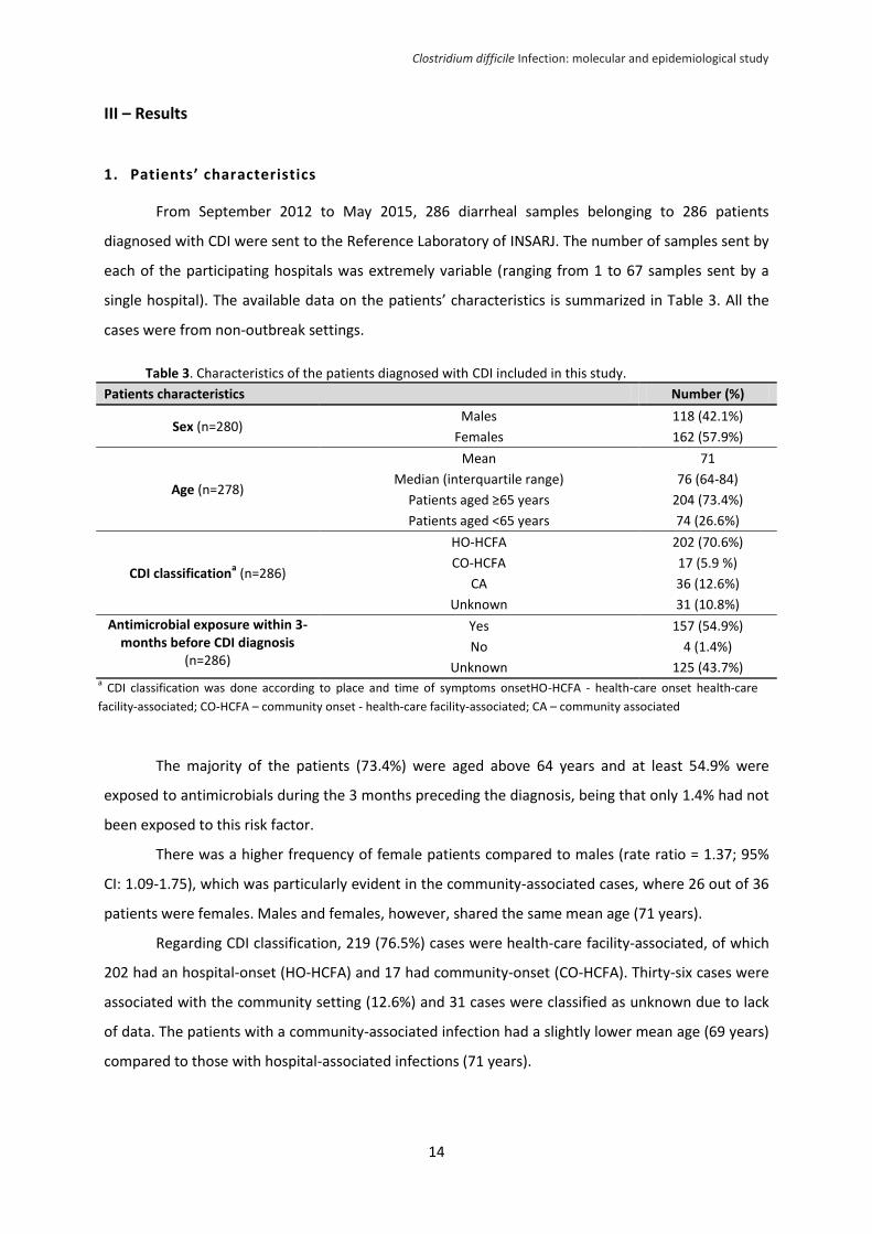
From September 2012 to May 2015, 286 diarrheal samples belonging to 286 patients
diagnosed with CDI were sent to the Reference Laboratory of INSARJ. The number of samples sent by
each of the participating hospitals was extremely variable (ranging from 1 to 67 samples sent by a
single hospital). The available data on the patients’ characteristics is summarized in Table 3. All the
cases were from non-outbreak settings.
Table 3. Characteristics of the patients diagnosed with CDI included in this study.
Patients characteristics Number (%)
Sex (n=280) Males 118 (42.1%)
Females 162 (57.9%)
Age (n=278)
Mean 71
Median (interquartile range) 76 (64-84)
Patients aged ≥65 years 204 (73.4%)
Patients aged <65 years 74 (26.6%)
CDI classificationa (n=286)
HO-HCFA 202 (70.6%)
CO-HCFA 17 (5.9 %)
CA 36 (12.6%)
Unknown 31 (10.8%)
Antimicrobial exposure within 3-months before CDI diagnosis
(n=286)
Yes 157 (54.9%)
No 4 (1.4%)
Unknown 125 (43.7%)
The majority of the patients (73.4%) were aged above 64 years and at least 54.9% were
exposed to antimicrobials during the 3 months preceding the diagnosis, being that only 1.4% had not
been exposed to this risk factor.
There was a higher frequency of female patients compared to males (rate ratio = 1.37; 95%
CI: 1.09-1.75), which was particularly evident in the community-associated cases, where 26 out of 36
patients were females. Males and females, however, shared the same mean age (71 years).
Regarding CDI classification, 219 (76.5%) cases were health-care facility-associated, of which
202 had an hospital-onset (HO-HCFA) and 17 had community-onset (CO-HCFA). Thirty-six cases were
associated with the community setting (12.6%) and 31 cases were classified as unknown due to lack
of data. The patients with a community-associated infection had a slightly lower mean age (69 years)
compared to those with hospital-associated infections (71 years).
a CDI classification was done according to place and time of symptoms onsetHO-HCFA - health-care onset health-care
facility-associated; CO-HCFA – community onset - health-care facility-associated; CA – community associated
Clostridium difficile Infection: molecular and epidemiological study
15
2. Molecular characterization of strains
All isolates were genetically confirmed to be C. difficile by PCR detection of the gluD gene,
which encodes the highly conserved C. difficile-specific glutamate dehydrogenase (Carman et al.,
2012), using the previously published primers GluDF1 and GluDR1.
2.1. Ribotypes
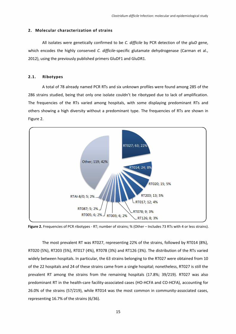
A total of 78 already named PCR RTs and six unknown profiles were found among 285 of the
286 strains studied, being that only one isolate couldn’t be ribotyped due to lack of amplification.
The frequencies of the RTs varied among hospitals, with some displaying predominant RTs and
others showing a high diversity without a predominant type. The frequencies of RTs are shown in
Figure 2.
The most prevalent RT was RT027, representing 22% of the strains, followed by RT014 (8%),
RT020 (5%), RT203 (5%), RT017 (4%), RT078 (3%) and RT126 (3%). The distribution of the RTs varied
widely between hospitals. In particular, the 63 strains belonging to the RT027 were obtained from 10
of the 22 hospitals and 24 of these strains came from a single hospital; nonetheless, RT027 is still the
prevalent RT among the strains from the remaining hospitals (17.8%; 39/219). RT027 was also
predominant RT in the health-care facility-associated cases (HO-HCFA and CO-HCFA), accounting for
26.0% of the strains (57/219), while RT014 was the most common in community-associated cases,
representing 16.7% of the strains (6/36).
Figure 2. Frequencies of PCR ribotypes - RT; number of strains; % (Other – Includes 73 RTs with 4 or less strains).
Clostridium difficile Infection: molecular and epidemiological study
16
2.2. Toxin typing and tcdC mutations
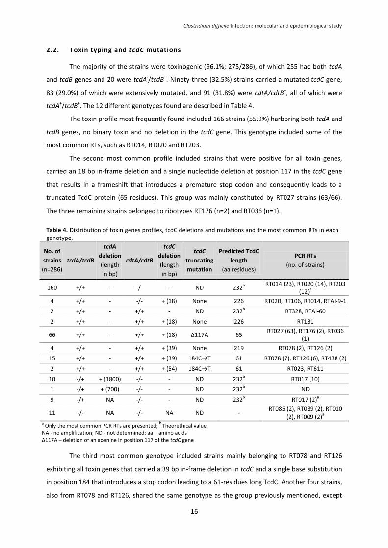
The majority of the strains were toxinogenic (96.1%; 275/286), of which 255 had both tcdA
and tcdB genes and 20 were tcdA-/tcdB+. Ninety-three (32.5%) strains carried a mutated tcdC gene,
83 (29.0%) of which were extensively mutated, and 91 (31.8%) were cdtA/cdtB+, all of which were
tcdA+/tcdB+. The 12 different genotypes found are described in Table 4.
The toxin profile most frequently found included 166 strains (55.9%) harboring both tcdA and
tcdB genes, no binary toxin and no deletion in the tcdC gene. This genotype included some of the
most common RTs, such as RT014, RT020 and RT203.
The second most common profile included strains that were positive for all toxin genes,
carried an 18 bp in-frame deletion and a single nucleotide deletion at position 117 in the tcdC gene
that results in a frameshift that introduces a premature stop codon and consequently leads to a
truncated TcdC protein (65 residues). This group was mainly constituted by RT027 strains (63/66).
The three remaining strains belonged to ribotypes RT176 (n=2) and RT036 (n=1).
Table 4. Distribution of toxin genes profiles, tcdC deletions and mutations and the most common RTs in each genotype.
No. of
strains
(n=286)
tcdA/tcdB
tcdA
deletion
(length
in bp)
cdtA/cdtB
tcdC
deletion
(length
in bp)
tcdC
truncating
mutation
Predicted TcdC
length
(aa residues)
PCR RTs
(no. of strains)
160 +/+ - -/- - ND 232b RT014 (23), RT020 (14), RT203
(12)a
4 +/+ - -/- + (18) None 226 RT020, RT106, RT014, RTAI-9-1
2 +/+ - +/+ - ND 232b RT328, RTAI-60
2 +/+ - +/+ + (18) None 226 RT131
66 +/+ - +/+ + (18) Δ117A 65 RT027 (63), RT176 (2), RT036
(1)
4 +/+ - +/+ + (39) None 219 RT078 (2), RT126 (2)
15 +/+ - +/+ + (39) 184C→T 61 RT078 (7), RT126 (6), RT438 (2)
2 +/+ - +/+ + (54) 184C→T 61 RT023, RT611
10 -/+ + (1800) -/- - ND 232b RT017 (10)
1 -/+ + (700) -/- - ND 232b ND
9 -/+ NA -/- - ND 232b RT017 (2)
a
11 -/- NA -/- NA ND - RT085 (2), RT039 (2), RT010
(2), RT009 (2)a
a Only the most common PCR RTs are presented;
b Theorethical value
NA - no amplification; ND - not determined; aa – amino acids Δ117A – deletion of an adenine in position 117 of the tcdC gene
The third most common genotype included strains mainly belonging to RT078 and RT126
exhibiting all toxin genes that carried a 39 bp in-frame deletion in tcdC and a single base substitution
in position 184 that introduces a stop codon leading to a 61-residues long TcdC. Another four strains,
also from RT078 and RT126, shared the same genotype as the group previously mentioned, except
Clostridium difficile Infection: molecular and epidemiological study
17
for the fact that, even though they exhibited the 39 bp in-frame deletion, they didn’t have the
truncating mutation in tcdC, 184C→T, which probably leads to the TcdC with 219 residues.
The fourth most common genotype was exclusively composed of RT017 strains that were
tcdA- (1800 bp deletion), tcdB+, cdtA/cdtB- and had no deletion in tcdC. Other two strains from this
ribotype yielded no tcdA amplification indicating a larger deletion in the gene. Notably, these two
RT017 strains were MXF-susceptible, contrasting with the remaining RT017 strains, suggesting they
belong to a different clonal lineage, in accordance to their different hospital origin.
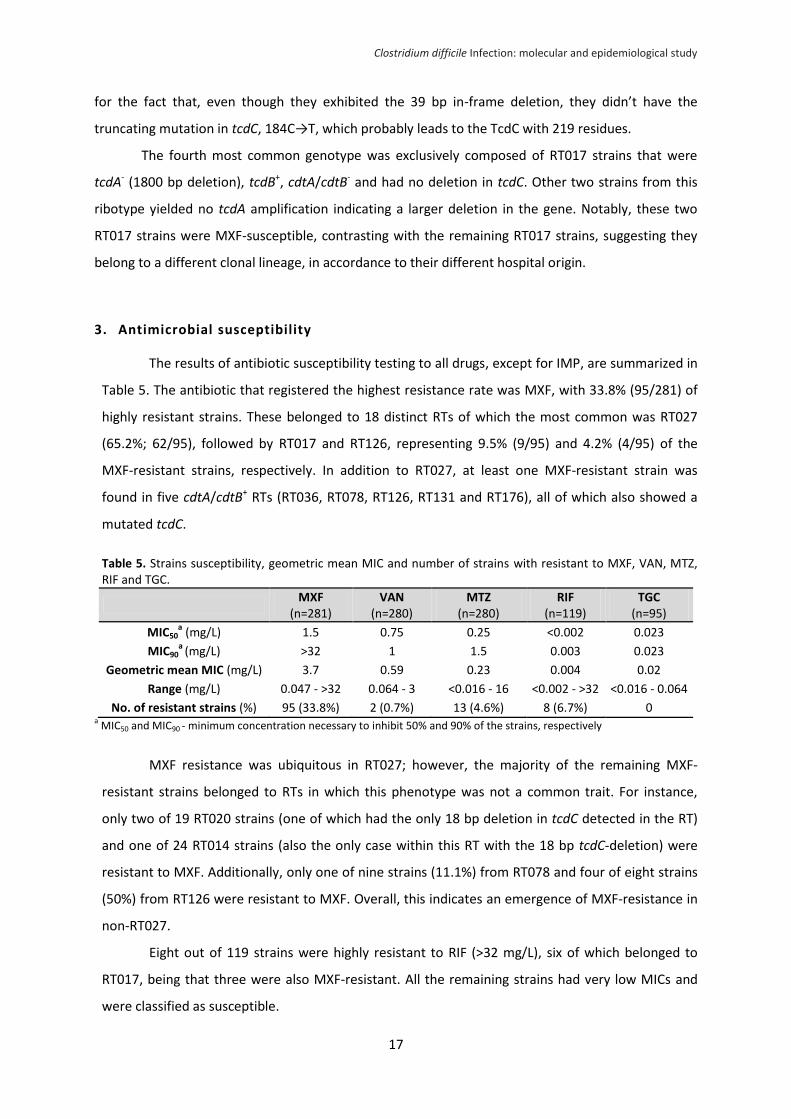
3. Antimicrobial susceptibility
The results of antibiotic susceptibility testing to all drugs, except for IMP, are summarized in
Table 5. The antibiotic that registered the highest resistance rate was MXF, with 33.8% (95/281) of
highly resistant strains. These belonged to 18 distinct RTs of which the most common was RT027
(65.2%; 62/95), followed by RT017 and RT126, representing 9.5% (9/95) and 4.2% (4/95) of the
MXF-resistant strains, respectively. In addition to RT027, at least one MXF-resistant strain was
found in five cdtA/cdtB+ RTs (RT036, RT078, RT126, RT131 and RT176), all of which also showed a
mutated tcdC.
Table 5. Strains susceptibility, geometric mean MIC and number of strains with resistant to MXF, VAN, MTZ, RIF and TGC.
MXF
(n=281) VAN
(n=280) MTZ
(n=280) RIF
(n=119) TGC
(n=95)
MIC50a (mg/L) 1.5 0.75 0.25 <0.002 0.023
MIC90a
(mg/L) >32 1 1.5 0.003 0.023
Geometric mean MIC (mg/L) 3.7 0.59 0.23 0.004 0.02
Range (mg/L) 0.047 - >32 0.064 - 3 <0.016 - 16 <0.002 - >32 <0.016 - 0.064
No. of resistant strains (%) 95 (33.8%) 2 (0.7%) 13 (4.6%) 8 (6.7%) 0 a MIC50 and MIC90 - minimum concentration necessary to inhibit 50% and 90% of the strains, respectively
MXF resistance was ubiquitous in RT027; however, the majority of the remaining MXF-
resistant strains belonged to RTs in which this phenotype was not a common trait. For instance,
only two of 19 RT020 strains (one of which had the only 18 bp deletion in tcdC detected in the RT)
and one of 24 RT014 strains (also the only case within this RT with the 18 bp tcdC-deletion) were
resistant to MXF. Additionally, only one of nine strains (11.1%) from RT078 and four of eight strains
(50%) from RT126 were resistant to MXF. Overall, this indicates an emergence of MXF-resistance in
non-RT027.
Eight out of 119 strains were highly resistant to RIF (>32 mg/L), six of which belonged to
RT017, being that three were also MXF-resistant. All the remaining strains had very low MICs and
were classified as susceptible.

Clostridium difficile Infection: molecular and epidemiological study
18
MTZ-resistance was observed in 13 strains, of which the majority were low-level resistant
(3-6 mg/L), seven belonged to RT027 and one to RT017 (Table 6). Notably, the MIC50, MIC90 and
geometric mean MIC of RT027 strains were superior to those observed in other RTs. The difference
between means, tested after logarithmic transformation of the values for normalization, was
statistically significant (t-test, P<0.01) which, overall, points to a reduced susceptibility to MTZ in
RT027 strains.
Table 6. MTZ susceptibility of strains from RT027 vs strains from other ribotypes.
RT027 (n=61) Other RTs (n=219)
MIC50a (mg/L) 1.0 0.19
MIC90a (mg/L) 3.0 0.75
Geometric mean MIC (mg/L) 0.79b
0.16b
Range (mg/L) 0.016 - 16 <0.016 - 6 a
MIC50 and MIC90 - minimum concentration necessary to inhibit 50% and 90% of the strains, respectively
b Difference between means
is statistically significant (t-test, P<0.01)
Almost all strains (99.3%) were susceptible to VAN and only two isolates (RT001 and RTAI-
58) showed a low-level resistance to this antibiotic (3 mg/L). The MICs of VAN for RT027 strains
were similar to those observed in the remaining RTs (MIC50=0.5 vs 0.75 mg/L, for RT027 vs other
RTs; MIC90= 1.0 mg/L and geometric mean MIC= 0.6 mg/L for both groups).
All strains were highly susceptible to TGC, with very low MICs (Table 5).
IMP susceptibility determined by Etest yielded unclear results since almost every strain
exhibited macrocolonies inside the inhibition, which would indicate an IMP-resistant phenotype and
hence suggested the existence of heterogeneous resistance. However, the colonies only appeared at
48h of growth, but not at 24h, and the selection of these hypothetically resistant colonies and
subsequent Etest didn’t result in homogeneous resistance but instead yielded the same
heterogeneous resistance appearance with macrocolonies inside the inhibition ellipse. As such, the
reference agar dilution method was applied to a subset of 57 strains to test the hypothetical
resistance and compare the results to those obtained by Etest (Table 7). Since only the
concentrations of 8, 16 and 32 mg/L were tested, it was not possible to ascertain the MICs of the
susceptible strains, which are simply described as ≤8 mg/L.
Table 7. Comparison of MICs of IMP determined by Etest with MICs determined by the agar dilution method.
IMP MICs determined by Etest (n=110) IMP MICs determined by agar
dilution (n=57) Considering colonies
inside ellipse Ignoring colonies
inside ellipse
MIC50 (mg/L) 8 4 8
MIC90 (mg/L) >32 >32 16
Geometric mean MIC (mg/L) 8.68 4.55 ND
Range (mg/L) 0.19 - >32 0.19 - >32 ≤8 - 32
No. of resistant strains (%) 53 (48.2%) 22 (20.0%) 11 (19.3%) ND – not determined
Clostridium difficile Infection: molecular and epidemiological study
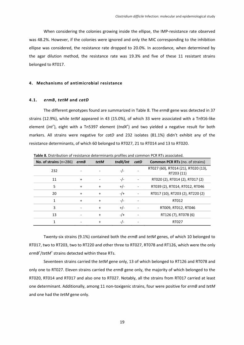
19
When considering the colonies growing inside the ellipse, the IMP-resistance rate observed
was 48.2%. However, if the colonies were ignored and only the MIC corresponding to the inhibition
ellipse was considered, the resistance rate dropped to 20.0%. In accordance, when determined by
the agar dilution method, the resistance rate was 19.3% and five of these 11 resistant strains
belonged to RT017.
4. Mechanisms of antimicrobial resistance
4.1. ermB, tetM and catD
The different genotypes found are summarized in Table 8. The ermB gene was detected in 37
strains (12.9%), while tetM appeared in 43 (15.0%), of which 33 were associated with a Tn916-like
element (int+), eight with a Tn5397 element (tndX+) and two yielded a negative result for both
markers. All strains were negative for catD and 232 isolates (81.1%) didn’t exhibit any of the
resistance determinants, of which 60 belonged to RT027, 21 to RT014 and 13 to RT020.
Table 8. Distribution of resistance determinants profiles and common PCR RTs associated.
No. of strains (n=286) ermB tetM tndX/int catD Common PCR RTs (no. of strains)
232 - - -/- - RT027 (60), RT014 (21), RT020 (13),
RT203 (11)
11 + - -/- - RT020 (2), RT014 (2), RT017 (2)
5 + + +/- - RT039 (2), RT014, RT012, RT046
20 + + -/+ - RT017 (10), RT203 (2), RT220 (2)
1 + + -/- - RT012
3 - + +/- - RT009, RT012, RT046
13 - + -/+ - RT126 (7), RT078 (6)
1 - + -/- - RT027
Twenty-six strains (9.1%) contained both the ermB and tetM genes, of which 10 belonged to
RT017, two to RT203, two to RT220 and other three to RT027, RT078 and RT126, which were the only
ermB+/tetM+ strains detected within these RTs.
Seventeen strains carried the tetM gene only, 13 of which belonged to RT126 and RT078 and
only one to RT027. Eleven strains carried the ermB gene only, the majority of which belonged to the
RT020, RT014 and RT017 and also one to RT027. Notably, all the strains from RT017 carried at least
one determinant. Additionally, among 11 non-toxigenic strains, four were positive for ermB and tetM
and one had the tetM gene only.
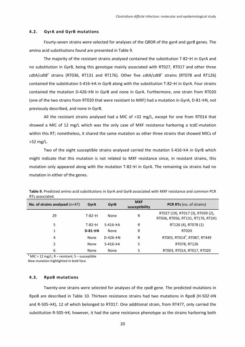
Clostridium difficile Infection: molecular and epidemiological study
20
4.2. GyrA and GyrB mutations
Fourty-seven strains were selected for analyses of the QRDR of the gyrA and gyrB genes. The
amino acid substitutions found are presented in Table 9.
The majority of the resistant strains analysed contained the substitution T-82→I in GyrA and
no substitution in GyrB, being this genotype mainly associated with RT027, RT017 and other three
cdtA/cdtB+ strains (RT036, RT131 and RT176). Other five cdtA/cdtB+ strains (RT078 and RT126)
contained the substitution S-416→A in GyrB along with the substitution T-82→I in GyrA. Four strains
contained the mutation D-426→N in GyrB and none in GyrA. Furthermore, one strain from RT020
(one of the two strains from RT020 that were resistant to MXF) had a mutation in GyrA, D-81→N, not
previously described, and none in GyrB.
All the resistant strains analysed had a MIC of >32 mg/L, except for one from RT014 that
showed a MIC of 12 mg/L which was the only case of MXF resistance harboring a tcdC-mutation
within this RT; nonetheless, it shared the same mutation as other three strains that showed MICs of
>32 mg/L.
Two of the eight susceptible strains analysed carried the mutation S-416→A in GyrB which
might indicate that this mutation is not related to MXF resistance since, in resistant strains, this
mutation only appeared along with the mutation T-82→I in GyrA. The remaining six strains had no
mutation in either of the genes.
Table 9. Predicted amino acid substitutions in GyrA and GyrB associated with MXF resistance and common PCR RTs associated.
No. of strains analysed (n=47) GyrA GyrB MXF
susceptibility PCR RTs (no. of strains)
29 T-82→I None R RT027 (19), RT017 (3), RT039 (2),
RT036, RT056, RT131, RT176, RT241
5 T-82→I S-416→A R RT126 (4), RT078 (1)
1 D-81→N None R RT020
4 None D-426→N R RT003, RT014a, RT087, RT449
2 None S-416→A S RT078, RT126
6 None None S RT003, RT014, RT017, RT020 a MIC = 12 mg/L; R – resistant; S – susceptible
New mutation highlighted in bold face.
4.3. RpoB mutations
Twenty-one strains were selected for analyses of the rpoB gene. The predicted mutations in
RpoB are described in Table 10. Thirteen resistance strains had two mutations in RpoB (H-502→N
and R-505→K), 12 of which belonged to RT017. One additional strain, from RT477, only carried the
substitution R-505→K; however, it had the same resistance phenotype as the strains harboring both
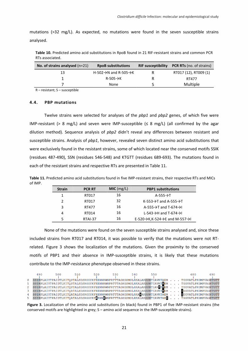
Clostridium difficile Infection: molecular and epidemiological study
21
mutations (>32 mg/L). As expected, no mutations were found in the seven susceptible strains
analysed.
Table 10. Predicted amino acid substitutions in RpoB found in 21 RIF-resistant strains and common PCR RTs associated.
R – resistant; S – susceptible
4.4. PBP mutations
Twelve strains were selected for analyses of the pbp1 and pbp2 genes, of which five were
IMP-resistant (> 8 mg/L) and seven were IMP-susceptible (≤ 8 mg/L) (all confirmed by the agar
dilution method). Sequence analysis of pbp2 didn’t reveal any differences between resistant and
susceptible strains. Analysis of pbp1, however, revealed seven distinct amino acid substitutions that
were exclusively found in the resistant strains, some of which located near the conserved motifs SSIK
(residues 487-490), SSN (residues 546-548) and KTGTT (residues 689-693). The mutations found in
each of the resistant strains and respective RTs are presented in Table 11.
Table 11. Predicted amino acid substitutions found in five IMP-resistant strains, their respective RTs and MICs of IMP.
Strain PCR RT MIC (mg/L) PBP1 substitutions
1 RT017 16 A-555→T
2 RT017 32 K-553→T and A-555→T
3 RT477 16 A-555→T and T-674→I
4 RT014 16 L-543→H and T-674→I
5 RTAI-37 16 E-520→K,K-524→E and M-557→I
None of the mutations were found on the seven susceptible strains analysed and, since these
included strains from RT017 and RT014, it was possible to verify that the mutations were not RT-
related. Figure 3 shows the localization of the mutations. Given the proximity to the conserved
motifs of PBP1 and their absence in IMP-susceptible strains, it is likely that these mutations
contribute to the IMP-resistance phenotype observed in these strains.
No. of strains analysed (n=21) RpoB substitutions RIF susceptibility PCR RTs (no. of strains)
13 H-502→N and R-505→K R RT017 (12), RT009 (1)
1 R-505→K R RT477
7 None S Multiple
Figure 3. Localization of the amino acid substitutions (in black) found in PBP1 of five IMP-resistant strains (the conserved motifs are highlighted in grey; S – amino acid sequence in the IMP-susceptible strains).
Clostridium difficile Infection: molecular and epidemiological study
22
IV – Discussion
The present study gives a comprehensive view of the current paradigm of CDI in Portugal
concerning the prevalent RTs, main virulence factors and the rates of antimicrobial resistance and
the associated mechanisms of resistance. It should however be noted that this study is based on a
convenience sample of the hospitals involved, which during different time periods voluntarily agreed
to contribute to the surveillance of CDI, and therefore it is not representative of our country.
Various studies have reported an increased incidence of CDI in female patients. For instance,
in a study conducted in Australia, a higher relative risk was attributed to females compared to males
(relative risk = 1.27, P=0.01) (Mitchell et al., 2012) and a recent study in the USA (Lessa et al., 2015)
also reports an increased incidence in females (rate ratio = 1.26; 95% CI, 1.25-1.27). Furthermore, a
surveillance report in the UK describes an infection rate of 25.4 vs 18.3 cases per 100 000 population,
for females vs males (Public Health England, 2015). In the present study, there was also a higher
number of female infected patients compared to males (57.9% vs 42.1%) but this should not be
interpreted as a tendency for higher incidence in females, as it is not significantly different from the
sex ratio in the elderly population, as reckoned by the 2011 Portuguese national census
(http://censos.ine.pt/xportal/xmain?xpid=CENSOS&xpgid=censos2011_apresentacao),where woman
aged above 65 years old comprised 58.1% of the population.
C. difficile is an intensively typed pathogen, with a wide range of methods applied to
understand its epidemiology. The emergence of the so-called ‘hypervirulent’ C. difficile types has
intensified the challenge of CDI epidemiology. In 2005, in a study conducted by the European Study
Group of C. difficile including strains from 14 countries, the most common RTs identified were RT001
(13%) and RT014 (9%), being that RT027 represented only 6% of the strains (Freeman et al., 2010).
Later, another study of C. difficile epidemiology in Europe, based upon a network of 106 laboratories
in 34 countries, identified 65 different RTs, of which RT014/020, RT001 and RT078 were the most
prevalent and RT027 only represented 5% of the strains (Bauer et al., 2011). In contrast, RT027 was
the most prevalent (28.1%) in the US hospitals between 2011 and 2013, followed by RT014/020
(12.4%) and RT106 (6.1%) (Tickler et al., 2014) In a more recent pan-European study involving 22
European countries (including Portugal) RT027 was also predominant accounting for 12% of the
strains, followed by RT001/072 (9%), RT078, and RT014 (both 8%) (Freeman et al., 2015). Similarly, in
the present study, the most prevalent was RT027, with a considerably high frequency (22%),
followed by RT014, RT020 and RT203. Notably, there was a high variety of RTs among our samples
(78 known RTs in 286 samples) compared to other studies, namely, Tickler et al. (2014) that
identified 29 RTs in 508 samples and Freeman et al. (2015) with 65 RTs in 953 samples.
Clostridium difficile Infection: molecular and epidemiological study
23
Kuehne et al. (2014) created isogenic toxin mutants and observed an increased virulence in
TcdA+TcdB-CDT+ strains when compared to TcdA+TcdB-CDT-. Also the infection with strains only
producing CDT caused the death of three out of nine animals and, in another study, five patients
infected with naturally occurring TcdA-TcdB-CDT+ strains showed typical disease symptoms (Eckert et
al., 2015), indicating a potential role for CDT in virulence. In the present study, 91 (31.8%) of the
strains studied carried the binary toxin, of which RT027 was the predominant type; other strains
belonged to ribotypes RT036, RT126, RT176 and the emergent RT078, all of which exhibited at least
one MXF-resistant strain. Unfortunately, disease severity could not be ascertained since detailed
clinical data of the patients was not available. Nonetheless, given that the emergence of the
epidemic RT027 was concomitant with its acquired resistance to fluoroquinolones (Pepin et al., 2005)
and that these other RTs also carry the binary toxin and a truncated TcdC, it is legitimate to speculate
about the epidemic potential of these also hypervirulent strains if their fluoroquinolones-resistance
rates increase.
Due to the frequent association between the in-frame deletions and the truncating
mutations in tcdC, studies using whole-gene sequencing to identify the truncating mutations have
only focused on the strains harboring an in-frame deletion. However, Curry et al. (2007) have
identified a strain with the Δ117A mutation but not the 18 bp deletion, meaning that there’s the
possibility that some strains with a truncating mutation might have been missed in the present study,
since a single base deletion wouldn’t be identifiable through the amplicon size. Nevertheless, an
extensively truncated TcdC (61 or 65 residues) was identified in 83 strains, all of which were also
cdtA/cdtB+, with RT027 accounting for 75.9%. Since the TcdC truncation has been associated with an
increased production of both toxins TcdA and TcdB in RT027 (Warny et al., 2005), other RTs with a
truncated TcdC, such as RT078 and RT126, should be tested by an enzymatic immunoassay to
confirm if they share the same phenotype. Furthermore, an in-frame deletion without a truncating
mutation was identified in 10 strains; since four of those belonged to RT078 and RT126 (which are
mainly associated with a truncated TcdC) this might indicate that the deletion precedes the
appearance of the truncating mutation. This trait was also found in strains cdtA/cdtB+ from RT131
and, notably, in cdtA/cdtB- strains, namely the common RT014 and RT020. Remarkably, the tcdC-
mutated RT014 strain was the only case of MXF-resistance within that RT. Along with the results that
indicate the emergence of fluoroquinolones-resistance in non-RT027, it is legitimate to speculate
that other RTs might evolve to a more virulent state (MXF-resistance and truncated TcdC), similarly
to what happened with RT027.
C. difficile has been found in both companion and farm animals, with RT078 being the most
prevalent in calves and pigs. In fact, in the Netherlands, human and pig strains from RT078 have been
demonstrated to be genetically related, with high degree of homology (Knetsch et al., 2014). It has
Clostridium difficile Infection: molecular and epidemiological study
24
also been reported that RT078 affects younger patients and is more frequently community-
associated when compared to other RTs (Rupnik et al., 2009). Alternatively, Janezic et al. (2012)
showed that RT014/020 were the most commonly found in humans, animals and environmental
samples. In the present study, RT014, RT020 and RT078 were the 2nd, 3rd and 6th most commonly
identified, being that RT014 was also the predominant type in community-associated cases. Overall,
this indicates a potential zoonotic transmission of the pathogen, particularly when considering that
many people in Portugal live in more rural communities, namely the high-risk elderly population that
is often in contact with domestic animals. Also, given the high variability of RTs identified (namely
between and within hospitals) it is tempting to suggest that transmission routes other than the
contact between individuals must be contributing to the high diversity observed and, as such, it
would be useful to identify the prevalent RTs in animal, food and environmental samples. However,
such study is yet to be conducted in Portugal.
The overall MXF-resistance rate observed was 33.8% and was mainly associated with the
RT027 and RT017. In the pan-European study (Freeman et al., 2015) the overall MXF-resistance rate
was 39.99%, although varying between countries, and was mainly associated with RT027, RT018,
RT356 and RT017. Similarly, other studies (several European countries, US, Canada and China) have
also reported variable rates of resistance and also associated this phenotype also with RT027 and
RT017, as well as with RT001 and RT106 (Huang et al., 2009), in accordance to the results presented
here. All the mutations found in GyrA and GyrB had already been described in other studies, except
for the mutation D-81→N in GyrA, found in a strain from RT020, associated with a MIC >32 mg/L. In
accordance to previous studies, alterations in GyrA were more common than in GyrB. Moreover, the
GyrB mutation S-416→A had already been described in susceptible strains and, hence, is not
associated with MXF resistance (Huang et al., 2009).
In the recent pan-European study, the overall RIF-resistance rate was 13.4%; however the
rates were extremely variable between countries (from 0 to 62.4%) (Freeman et al., 2015). Our rate
(6.7%), although lower than the overall European rate, was superior to those observed in the
majority of the countries. Curiously, they associated the RIF-resistance mostly with RT018 and RT027,
while in the present study it was ubiquitous among RT017 and was not detected in RT027. The two
mutations detected in RpoB had already been described in RIF-resistant strains (Huang et al., 2009;
Spigaglia et al., 2011). The mutation R-505→K alone induced the same resistance level as when both
mutations were simultaneously present (>32 mg/L), in accordance to the intermediate RIF-resistance
previously found in strains where only the mutation H-502→N was present (Huang et al., 2009).
In this study, the distribution of the resistance determinants ermB and tetM was highly
variable between and within RTs. Nonetheless, almost all RT027 strains were negative for both
determinants, which is in accordance with the description of clindamycin as a protective antibiotic
Clostridium difficile Infection: molecular and epidemiological study
25
against CDI caused by RT027 (Huang et al., 2009). Moreover, a recent study showed that RT027
ermB+ strains displayed a reduced fitness in vitro when compared to susceptible strains, which might
be the explanation behind the absence of ermB observed in this ribotype (Wasels et al., 2013).
Both these determinants were found in 50% of non-toxigenic strains; therefore it should be
noted that these generally unnoticed strains might be a pool of resistance determinants
transmittable to toxigenic strains. Although high-level resistance to erythromycin and clindamycin is
generally associated with the presence of ermB, other mechanisms have been described, such as
mutations in 23S rDNA, which were not evaluated in the present study (Huang et al., 2009). In
accordance, the minimum MLSB-resistance rate that can be deduced from the ermB screening
performed in this study is very low (12.9%) when compared to what has been reported in most
countries (Huang et al., 2009). Freeman et al. (2015) reported an overall clindamycin-resistance rate
of 49.6% in Europe, being that most countries had a rate of at least 20% (including the Portuguese
strains). These results indicate that our real MLSB-resistance rate must be higher than the one
deduced from the frequency of the ermB gene and, therefore, all strains must be tested to identify
all the resistant strains. The rate of tetracycline-resistance deduced from the presence of the tetM
gene (15.0%) was above the rates reported in most studies (<10%). Nevertheless, it is also possible
that this figure may be underestimated since other mechanisms of tetracycline-resistance have been
identified (Huang et al., 2009).
Low-level resistance to MTZ was observed in a few strains, mostly from RT027. Moreover,
RT027 strains displayed higher MICs compared to the remaining RTs, which goes in accordance to the
already reported reduced susceptibility in this RT, namely, among European strains (Freeman et al.,
2010; Freeman et al., 2015). This phenotype is probably due to the selective pressure of the MTZ
usage in CDI treatment. Accordingly, reduced MTZ-susceptibility was described in RT001, RT106 and
RT027 strains, in the UK, which were the predominant RTs detected in the country (Freeman et al.,
2010). As such, it seems that this phenomenon occurs in the predominant RTs, like RT027 in our
country, and, hence, the MTZ-susceptibility pattern of other emerging ribotypes should also be
monitored. To date, failure of CDI therapy with metronidazole has been associated with low dosage
and/or poor pharmacokinetics and the contribution of the in vitro reduced susceptibility of the strain
remains to be determined (Kuijper and Wilcox, 2008).
In concordance to what has been seen in various studies (Huang et al., 2009; Freeman et al.,
2010; Freeman et al., 2015), almost every strain was susceptible to VAN and all were susceptible to
TGC. Furthermore, the presence of tetM didn’t seem to have any effect on the susceptibility to TGC
since the MICs observed were all very low.
In a study conducted in Sweden (Norén et al., 2010), IMP-susceptibility testing by Etest has
led to the assumption of a false high level of IMP-resistance due to the appearance of colonies inside
Clostridium difficile Infection: molecular and epidemiological study
26
the inhibition zone at 48h (but not at 24h), being that the real levels of resistance were determined
by agar dilution, similarly to what was reported here. The authors stated a possible explanation that
also matches our results: the late germination of spores (previously present in the inoculum) inside
the inhibition zone that would be facilitated by the degradation of the antibiotic. As such, IMP
susceptibility determined by Etest should be cautiously evaluated to prevent false results. In the pan-
European study, the IMP-resistance rate, determined by agar dilution, was 7.4%, on the whole, and
16.7% in the Portuguese strains (Freeman et al., 2015), which is similar to the rate observed here
(19.3%).
In gram-positive bacteria, such as Enterococcus faecalis and Streptococcus pneumoniae,
resistance to carbapenems is generally due to amino acid substitutions in PBPs or acquisition of a
novel carbapenem-resistant PBP (Ono et al., 2005; Davies et al., 2008; Sauvage et al., 2008; Papp-
Wallace et al., 2011). As such, it was thought that IMP-resistance in C. difficile could also be due to
altered PBPs, which led to the identification of seven distinct mutations in one of the pbp genes
analysed in IMP-resistant strains. The low number of strains analysed doesn’t confirm the association
between the mutations and the IMP-resistant phenotype; however, the fact that two of the
mutations were present in resistant strains from RT017 and RT014 but not in the susceptible strains
from the same RTs supports this hypothesis. Nevertheless, further studies are needed on this subject
in order to demonstrate that these mutations are functional.
Spigaglia et al. (2011) characterized a set of European multidrug-resistant isolates mainly
belonging to RT001, RT017 and RT012. Similarly, in other studies, these RTs were also associated with
multidrug-resistance (Huang et al., 2009). Notably, in this study, the most prominent multidrug-
resistant RT was also RT017, in which most strains (7/12) were resistant to MXF, RIF and harbored
both ermB and tetM genes; one was also low-level MTZ-resistant and at least five were IMP-
resistant.
RT017 is one of the predominant RTs in Asian countries but only accounted for 1.8% of the
strains in Europe (Freeman et al., 2015). In Poland (from 2004 to 2006) and Bulgaria (from 2008 to
2012), however, RT017 has been emerging, being nowadays identified as the predominant RT (King
et al., 2015), and accounted for 4% of the toxigenic strains in another European study (Bauer et al.,
2011). Similarly, RT017 was the fifth most commonly found type (4%) in the present study. This
multidrug-resistant TcdA-/TcdB+ RT has been associated with outbreaks and a similar disease pattern
when compared to cases of CDI with TcdA+/TcdB+ strains, and has also been found in farm animals
(Freeman et al, 2010). In fact, it has been suggested that TcdA-TcdB+ strains are associated with a
more severe disease than strains with both toxins and that this might be explained by an altered
substrate specificity of the TcdB produced by the toxin variant strains, presumably to compensate for
Clostridium difficile Infection: molecular and epidemiological study
27
the lack of TcdA (Pruitt and Lacy, 2012). Overall, these results highlight the importance of Toxin B in
CDI pathogenesis and raise awareness for a further spread of RT017.
V – Conclusion
The data presented in this study shows that CDI in the samples from Portuguese hospitals is
mainly associated with the epidemic RT027 whose prevalence was significant even when compared
to other common RTs. This indicates a wide spread of this strain in the healthcare setting and, as
such, demands that more patients must be tested and treated for CDI. Patients with an undiagnosed,
self-resolved CDI constitute a major source of contamination since they are left in contact with other
individuals. Therefore all CDI cases should be identified for an effective reduction of CDI incidence.
In community-associated cases, however, RT014 was the predominant type, which, along
with the high frequency of this RT in the hospital isolates and the high diversity of RTs identified in
both hospital- and community-associated cases, indicates that other sources of infection outside the
hospital-setting must be contributing to the observed variety of strains. As such, more studies should
be conducted to evaluate the prevalence of C. difficile in samples collected from other potential
sources, namely, food, environment (e.g. water) and, importantly, domestic animals, which might
constitute an important reservoir of C. difficile and antimicrobial-resistance determinants.
Multidrug-resistance was observed in a significant number of strains and, importantly, MXF-
resistance seemed to be emerging in other common RTs other than RT027, raising awareness for the
potential spread of these strains, specially the hypervirulent RTs harboring the binary toxin and a
mutated TcdC. The reduced susceptibility to MTZ observed in RT027 is also a cause of concern since
it is still unknown how this might affect the efficacy of the CDI treatment with this antibiotic.
Overall, these results demand the application of an improved antibiotic stewardship in the
prescription of antibiotics to both humans and animals for the prevention of CDI and avoidance of
further acquisition of antimicrobial-resistance in C. difficile.
In conclusion, this study constitutes the first epidemiological and molecular study including C.
difficile isolates from several Portuguese hospitals, comprising a general view of the current
paradigm of CDI in Portugal. It might also be the foundation for further and wider studies on this
subject and provide data for comparison with future studies in order to understand how the
epidemiology of CDI is evolving in our country.

Clostridium difficile Infection: molecular and epidemiological study
28
VI - References
Bacci, S., Molbak, K., Kjeldsen, M., Olsen, K. (2011). Binary toxin and death after Clostridium difficile infection. Emerg Infect Dis. 17, 976-82.
Bartlett, J. (1994). Clostridium difficile: history of its role as an enteric pathogen and the current state of knowledge about the organism. Clin Infect Dis. 18(Suppl 4), S265–72.
Bauer, M., Notermans, D., van Benthem, B., Brazier, J., Wilcox, M., Rupnik, M., Monnet, D., van Dissel, J., Kuijper, E., ECDIS Study Group (2011). Clostridium difficile infection in Europe: a hospital-based survey. Lancet. 377, 63–73.
Bauer, M. and Kuijper, E. (2015). Potential Sources of Clostridium difficile in Human Infection. Infect Dis Clin N Am. 29(1), 29-35.
Carman, R., Wickham, K., Chen, L., Lawrence, A., Boone, J., Wilkins, T., Kerkering, T., Lyerly, D. (2012). Glutamate dehydrogenase is highly conserved among Clostridium difficile ribotypes. J Clin Microbiol. 50, 1425–6.
Clements, A., Magalhaes, R., Tatem, A., Paterson, D., Riley, T. (2010). Clostridium difficile PCR ribotype 027: assessing the risks of further worldwide spread. Lancet Infect Dis 10, 395-404.
Cohen, S., Gerding, D., Johnson, S., Kelly, C., Loo, V., McDonald, L., Pepin, J., Wilcox, M., Society for Healthcare Epidemiology of America; Infectious Diseases Society of America. (2010). Clinical practice guidelines for Clostridium difficile infection in adults: 2010 update by the society for healthcare epidemiology of America (SHEA) and the infectious diseases society of America (IDSA). Infect Control Hosp Epidemiol 31, 431–55.
Curry, S., Marsh, J., Muto, C., O'Leary, M., Pasculle, A., Harrison, L. (2007). tcdC genotypes associated with severe TcdC truncation in an epidemic clone and other strains of Clostridium difficile. J Clin Microbiol 45, 215–21.
Curry, S., Marsh, J., Shutt, K., Muto, C., O’Leary, M., Saul, M., Pasculle, A., Harrison, L. (2009). High frequency of rifampin resistance identified in an epidemic Clostridium difficile clone from a large teaching hospital. Clin Infect Dis 48, 425–9.
Davies, T., Shang, W., Bush, K., Flamm, R. (2008). Activity of doripenem and comparator β-lactams against US clinical isolates of Streptococcus pneumoniae with defined mutations in the penicillin-binding domains of pbp1a, pbp2b and pbp2x. J Antimicrob Chemother 61, 751–3.
Eckert, C., Emirian, A., Le Monnier, A., Cathala, L., De Montclos, H., Goret, J., Berger, P., Petit, A., De Chevigny, A., Jean-Pierre, H., Nebbad, B., Camiade, S., Meckenstock, R., Lalande, V., Marchandin, H., Barbut, F. (2015). Prevalence and pathogenicity of binary toxin-positive Clostridium difficile strains that do not produce toxins A and B. New Microbe and New Infect 3, 12–7.
European Committee for Antimicrobial Susceptibility Testing (EUCAST) of the European Society for Clinical Microbiology and Infectious Diseases (ESCMID). (2000). EUCAST Definitive Document E. Def 3.1: Determination of minimum inhibitory concentrations (MICs) of antibacterial agents by agar dilution. Clin Microbiol Infec 6, 509-15.
Freeman, J., Stott, J., Baines, S., Fawley, W., Wilcox, M. (2005). Surveillance for resistance to metronidazole and
vancomycin in genotypically distinct and UK epidemic Clostridium difficile isolates in a large teaching hospital. J Antimicrob Chemother 56, 988–9.
Freeman, J., Bauer, M., Baines, S., Corver, J., Fawley, W., Goorhuis, B., Kuijper, E., Wilcox, M. (2010). The
changing epidemiology of Clostridium difficile infections. Clin Microbiol Rev 23, 529-49.
Freeman, J., Vernon, J., Morris, K., Nicholson, S., Todhunter, S., Longshaw, C., Wilcox, M. (2015). Pan-European longitudinal surveillance of antibiotic resistance among prevalent Clostridium difficile ribotypes. Clin Microbiol Infect. 21(3), 248-e9.
Gerding, D., Johnson, S., Rupnik, M., Aktories, K. (2014). Clostridium difficile binary toxin CDT: Mechanism, epidemiology, and potential clinical importance. Gut Microbes. 5(1), 15–27.
Hall, I.C. and O'Toole, E. (1935). Intestinal flora in new-born infants: with a description of a new pathogenic anaerobe, Bacillus difficilis. Am J Dis Child. 49, 390-402.
Clostridium difficile Infection: molecular and epidemiological study
29
Huang, H., Weintraub, A., Fang, H., Nord, C. (2009). Antimicrobial resistance in Clostridium difficile. Int J Antimicrob Agents. 34, 516–22.
Hunt, J. and Ballard, J. (2013). Variations in virulence and molecular biology among emerging strains of Clostridium difficile. Microbiol Mol Biol Rev. 77(4), 567-81.
Indra, A., Huhulescu, S., Schneeweis, M., Hasenberger, P., Kernbichler, S., Fiedler, A., Wewalka, G., Allerberger, F., Kuijper, E. (2008). Characterization of Clostridium difficile isolates using capillary gel electrophoresis-based PCR ribotyping. J Med Microbiol. 57, 1377–82.
Janezic, S., Ocepek, M., Zidaric, V., Rupnik, M. (2012). Clostridium difficile genotypes other than ribotype 078 that are prevalent among human, animal and environmental isolates. BMC Microbiol. 12, 48.
Kato, H., Kato, N., Watanabe, K., Iwai, N., Nakamura, H., Yamamoto, T., Suzuki, K., Kim, S., Chong, Y., Wasito, E. (1998). Identification of toxin A-negative, toxin B-positive Clostridium difficile by PCR. J Clin Microbiol. 36, 2178-82.
Kato, H., Kato, N., Katow, S., Maegawa, T., Nakamura, S., Lyerly, D. (1999). Deletions in the repeating sequences of the toxin A gene of toxin A-negative, toxin B-positive Clostridium difficile strains. FEMS Microbiol Lett. 175, 197–203.
Kelly, C. and LaMont, J. (2008). Clostridium difficile – more difficult than ever. N Engl J Med. 359, 1932–40.
Knetsch, C., Connor, T., Mutreja, A., van Dorp, S., Sanders, I. M., Browne, H., Harris, D., Lipman, L., Keessen, E., Corver, J., Kuijper, E., Lawley, T. (2014). Whole genome sequencing reveals potential spread of Clostridium difficile between humans and farm animals in the Netherlands, 2002 to 2011. Euro Surveill. 19(45), 30-41.
Kim, H., Jeong, S., Roh, K., Hong, S., Kim, J., Shin, M., Kim, M., Shin, H., Uh, Y., Lee, H. (2010). Investigation of toxin gene diversity, molecular epidemiology, and antimicrobial resistance of Clostridium difficile isolated from 12 hospitals in South Korea. Korean J Lab Med. 30(5), 491-7.
Kuehne, S., Collery, M., Kelly, M., Cartman, S., Cockayne, A., Minton, N. (2014). Importance of toxin A, toxin B, and CDT in virulence of an epidemic Clostridium difficile strain. J Infect Dis. 209, 83–6.
Kuijper, E. and Wilcox, M. (2008). Decreased effectiveness of metronidazole for the treatment of Clostridium difficile infection? Clin Infect Dis. 47, 63–5.
Lessa, F., Mu, Y., Bamberg, W., Beldavs, Z., Dumyati, G., Dunn, J., Farley, M., Holzbauer, S., Meek, J., Phipps, E., Wilson, L., Winston, L., Cohen, J., Limbago, B., Fridkin, S., Gerding, D., McDonald, L. (2015). Burden of Clostridium difficile infection in the United States. N Engl J Med. 372, 825–34.
Lim, S., Stuart, R., Mackin, K., Carter, G., Kotsanas, D., Francis, M., Easton, M., Dimovski, K., Elliott, B., Riley, T., Hogg, G., Paul, E., Korman, T., Seemann, T., Stinear, T., Lyras, D., Jenkin, G. (2014). Emergence of a ribotype 244 strain of Clostridium difficile associated with severe disease and related to the epidemic ribotype 027 strain. Clin Infect Dis. 58(12), 1723–30.
Mitchell, B., Wilson, F., McGregor, A. (2012). An increase in community onset Clostridium difficile infection: a population-based study, Tasmania, Australia. Healthcare Infection. 17, 127-32.
Norén, T., Alriksson, I., Andersson, J., Unemo, M. (2010). False resistance to imipenem observed for clinical Clostridium difficile isolates using Etest is partly explained by late sporulation. Poster presented at Örebro University Hospital, Örebro, Sweden, 2010.
Oleastro, M., Coelho, M., Gião, M., Coutinho, S., Mota, S., Santos, A., Rodrigues, J., Faria, D. (2014). Outbreak of Clostridium difficile PCR ribotype 027 - the recent experience of a regional hospital. BMC Infect Dis. 14, 209.
Ono, S., Muratani, T., Matsumoto, T. (2005). Mechanisms of resistance to imipenem and ampicillin in Enterococcus faecalis. Antimicrob Agents Chemother. 49, 2954–8.
Paltansing, S., van den Berg, R., Guseinova, R., Visser, C., van der Vorm, E., Kuijper, E. (2007). Characteristics and incidence of Clostridium difficile-associated disease in The Netherlands, 2005. Clin Microbiol Infect. 13, 1058–64.
Papp-Wallace, K., Endimiani, A., Taracila, M., Bonomo, R. (2011). Carbapenems: past, present, and future. Antimicrob Agents Chemother. 55, 4943–60.

Clostridium difficile Infection: molecular and epidemiological study
30
Pépin, J., Valiquette, L., Alary, M., Villemure, P., Pelletier, A., Forget, K., Pépin, K., Chouinard, D. (2004). Clostridium difficile–associated diarrhea in a region of Quebec from 1991 to 2003: a changing pattern of disease severity. CMAJ. 171, 466–72.
Pépin, J., Saheb, N., Coulombe, M., Alary, M., Corriveau, M., Authier, S., Leblanc, M., Rivard, G., Bettez,
M., Primeau, V., Nguyen, M., Jacob, C., Lanthier, L. (2005). Emergence of fluoroquinolones as the
predominant risk factor for Clostridium difficile–associated diarrhea: a cohort study during an epidemic in Quebec. Clin Infect Dis. 41, 1254–60.
Persson, S., Torpdahl, M., Olsen, K. (2008). New multiplex PCR method for the detection of Clostridium difficile toxin A (tcdA) and toxin B (tcdB) and the binary toxin (cdtA/cdtB) genes applied to a Danish strain collection. Clin Microbiol Infect. 14, 1057-64.
Persson, S., Jensen, J., Olsen, K. (2011). Multiplex PCR method for detection of Clostridium difficile tcdA, tcdB, cdtA, and cdtB and internal in-frame deletion of tcdC. J Clin Microbiol. 49, 4299–300.
Pruitt, R. and Lacy, D. (2012). Toward a structural understanding of Clostridium difficile toxins A and B. Front Cell Infect Microbiol. 2: 28.
Public Health England. Voluntary surveillance of Clostridium difficile, England, Wales and Northern Ireland: 2014. Infection report 9 (Number 21) Published electronically on 19 June 2015. https://www.gov.uk/government/uploads/system/uploads/attachment_data/file/436569/hpr2115_cdff.pdf Accessed 19 August 2015.
Rupnik, M., Wilcox, M., Gerding, D. (2009). Clostridium difficile infection: new developments in epidemiology and pathogenesis. Nat Rev Microbiol. 7, 526–36.
Sauvage, E., Kerff, F., Terrak, M., Ayala, J., Charlier, P. (2008). The penicillin-binding proteins: structure and role in peptidoglycan biosynthesis. FEMS Microbiol Rev. 32, 234–58.
Spigaglia, P. and Mastrantonio, P. (2002). Molecular analysis of the pathogenicity locus and polymorphism in the putative negative regulator of toxin production (TcdC) among Clostridium difficile clinical isolates. J Clin Microbiol. 40, 3470–5.
Spigaglia, P. and Mastrantonio, P. (2004). Comparative analysis of Clostridium difficile clinical isolates belonging to different genetic lineages and time periods. J Med Microbiol. 53, 1129–36.
Spigaglia, P., Carucci, V., Barbanti, F., Mastrantonio, P. (2005). ErmB determinants and Tn916-like elements from clinical isolates of Clostridium difficile. Antimicrob Agents Chemother. 49, 2550–3.
Spigaglia, P., Barbanti, F., Mastrantonio, P., Brazier, J., Barbut, F., Delmée, M., Kuijper, E., Poxton, I.; European Study Group on Clostridium difficile. (2008). Fluoroquinolone resistance in Clostridium difficile isolates from a prospective study of C. difficile infections in Europe. J Med Microbiol. 57, 784–9.
Spigaglia, P., Barbanti, F., Mastrantonio, P.; European Study Group on Clostridium difficile. (2011). Multidrug resistance in European Clostridium difficile clinical isolates. J Antimicrob Chemother. 66, 2227–34.
Surawicz, C., Brandt, L., Binion, D., Ananthakrishnan, A., Curry, S., Gilligan, P., McFarland, L., Mellow, M., Zuckerbraun, B. (2013). Guidelines for diagnosis, treatment, and prevention of Clostridium difficile infections. Am J Gastroenterol. 108, 478-98.
Tickler, I., Goering, R., Whitmore, J., Lynn, A., Persing, D., Tenover, F. (2014). Strain types and antimicrobial resistance patterns of Clostridium difficile isolates from the United States, 2011 to 2013. Antimicrob Agents Chemother. 58, 4214-8.
Voth, D. and Ballard, J. (2005). Clostridium difficile toxins: mechanism of action and role in disease. Clin Microbiol Rev. 18, 247–63.
Warny, M., Pepin, J., Fang, A., Killgore, G., Thompson, A., Brazier, J., Frost, E., McDonald, L. (2005). Toxin production by an emerging strain of Clostridium difficile associated with outbreaks of severe disease in North America and Europe. Lancet. 366, 1079-84.
Wasels, F., Spigaglia, P., Barbanti, F., Mastrantonio, P. (2013). Clostridium difficile erm(B)-containing elements and the burden on the in vitro fitness. J Med Microbiol. 62, 1461–7.