CloneSelect Single-Cell Printer Series - Paralab
8
CloneSelect Single-Cell Printer Series Eyewitness proof of monoclonality The CloneSelect™ Single-Cell Printer™ Series deposit single cells gently and with high efficiency using a patented, inkjet-like disposable, one-way dispensing cartridge. Sort cells using high-res imaging in either brightfield or optional fluorescence and capture 5 images per each single cell deposit. Prove monoclonality, improve your efficiency, maintain and enrich your viability, and prevent cross-contamination. KEY FEATURES • Cell isolation proof captured for 96- or 384-well plate formats • Clonal outgrowth improved up to 8x • Cells stay healthy and clean with sterile, disposable microfluidic cartridge • Cells sorted in brightfield or fluorescence
Transcript of CloneSelect Single-Cell Printer Series - Paralab

CloneSelect Single-Cell Printer SeriesEyewitness proof of monoclonality
The CloneSelect™ Single-Cell Printer™ Series deposit single cells gently and with high efficiency using a patented, inkjet-like disposable, one-way dispensing cartridge. Sort cells using high-res imaging in either brightfield or optional fluorescence and capture 5 images per each single cell deposit. Prove monoclonality, improve your efficiency, maintain and enrich your viability, and prevent cross-contamination.
KEY FEATURES• Cell isolation proof captured for
96- or 384-well plate formats
• Clonal outgrowth improved up to 8x
• Cells stay healthy and clean with sterile, disposable microfluidic cartridge
• Cells sorted in brightfield or fluorescence

Figure 1. How the single cell isolation and deposition works.
Droplet with a single cell
A single cell is detected. The droplet with a single cell is ejected while the vacuum
is inactive.
It is then deposited into the well.
Droplet with more than one cell
The system detects > 1 cell in the nozzle.
The droplet with > 1 cell is ejected and immediately vacuumed into
the waste. No cells are deposited.
Droplet without a cell
No cells are detected in the nozzle. The empty droplet is ejected and immediately deflected into waste.
No cells are deposited.
Working principles of the CloneSelect Single-Cell Printer Series

Disposable cartridge at the heart of the CloneSelect Single-Cell Printer SeriesThe core of all the CloneSelect Single-Cell Printer systems is the patented single-cell isolating cartridge technology, which enables automated deposition of single cells into standard 96- and 384-well plate formats. The system uses an inkjet-like principle featuring a sterile and disposable one-way flow cartridge (Figure 2).
Up to 80 µL of cell sample at concentrations ranging from 0.5 to 1 x 10⁶ cells/mL can be hand pipetted into a cartridge. Its silicon microfluidic chip generates gently-dispensed microdroplet that contain single cells. A single cartridge is sufficient to deposit single cells into wells of several microplates. After use, these cartridges can easily be discarded, preventing cross contamination and time associated with handling and sterilizing reusable components.
A high magnification and large depth-of-field camera captures a sequence of events to determine the cell number in each droplet. When multi-cell or zero cell events are automatically detected, the droplets are immediately siphoned away with a vacuum. Once a single cell event with desired and preset size and roundness is observed, a fast shutter mechanism closes access to the vacuum, enabling droplets containing a single cell to be deposited into the microplate (Figure 1).
Captured proof of cell isolation The cell isolation cartridge is imaged at the nozzle using a high depth-of-field camera which ensures cells appear in focus. An intelligent image analysis algorithm detects single cell events accurately and on the fly. A sequence of five images are captured to track a single cell as it descends down the nozzle tip and into the microplate, providing added evidence of monoclonality over a single snapshot (Figure 3A).
A B
Figure 2. The disposable, sterile cartridge for the Single-Cell Printer Series.
Figure 3. (A) For each single cell a sequence of 5 images are captured at the cartridge nozzle, providing evidence of single cell deposition. These images describe the path taken by a single cell prior to and following ejection from the nozzle tip. Images 1-3 show the cell approaching the nozzle. Image 4 shows the detection of a single cell (inner circle) and the absence of any cells in the vicinity (outer circle). Finally, Image 5 shows the nozzle after droplet ejection to provide evidence that the single cell was expelled from the nozzle. (B) The software interface for cell selection is shown. Users can select cells based on the cell size and morphology, ensuring that non-desired objects (debris, dividing cells, unhealthy cells) are not deposited into the wells.

Improved single-cell isolation efficiencyCurrent workflows for single cell isolation within cell line development have major drawbacks such as inefficient single cell deposition and a lack of image evidence for single cell-containing wells.
Limiting dilution, the standard technique most often used to isolate single cells, relies on statistical probability to favor single cell deposition. The best approaches using limiting dilution yield only 30%, single cell-containing wells with a majority of void and doublet wells. CloneSelect Single-Cell Printer Series significantly improves the efficiency by vacuuming the droplets identified as no cells or multiple cells to waste.
Isolate more viable clones with confidenceAnother drawback of current methods is low cell viability post sorting and isolation. For example, it is common to observe <25% cell viability with flow cytometry-based methods.
The CloneSelect’s unique microfluidics-based cell isolation technology is as gentle as pipetting. The cell-containing microdroplets are dispensed slowly and delicately in the absence of any pressure, electric fields, or lasers observed in other technologies. Additionally, the ability to screen cell populations by gating for size and roundness offers the potential to exclude imperfect non-viable cells, making the CloneSelect Single-Cell Printer Series performance on par with limiting dilution when it comes to isolating healthy cells.
After being deposited into microplates, the single cells are monitored and imaged for several days using the CloneSelect™ Imager to follow the growth characteristics. The cells remain viable and form monoclonal colonies within days, indicating high cell viability after deposition (Figure 5).
Figure 4. Traditional methods of single cell isolation are compared with that of the CloneSelect Single-Cell Printer Series. The Single-Cell Printer Series provide significant advantages over those techniques in efficiency of deposition and viability of clones.
* Based on predicted values. For limiting dilution, the single cell deposit efficiency was determined based on theoretically seeding 0.5 cells/well. For flow cytometry, viabilities can vary significantly but is usually reported between 5-50%. Thus, a 25% average viability was chosen.
A
B
C
CloneSelect Single-Cell Printer Series*
Limiting Dilution*
Flow cytometry*
1 cell, viable clone
0 cell
>1 cell
1 cell, non-viable clone
CloneSelect Single-Cell Printer Series*
Limiting Dilution*
Flow cytometry*
1 cell, viable clone
0 cell
>1 cell
1 cell, non-viable clone
Methods for single cell sorting
CloneSelect™ Single-CellPrinter™ Series
Limiting Dilution Flow Cytometry
n 1 cell, viable clone n 1 cell, non-viable clone n >1 cell n 0 cell
D
We
lls in
96
-we
ll p
late

A
Figure 5. (A) Following single-cell deposition with the CloneSelect Single-Cell Printer system, the single cells remain viable and form a monoclonal colony within a span of several days. Example image of a single cell deposited with c.sight and imaged on the CloneSelect Imager for several days post deposition. (B) Single cell colonies (shown in green) derived from limiting dilution (bottom panel) are compared to equivalent colonies from the c.sight (top panel) by imaging on day 6 with the CloneSelect Imager. A representative 384-well plate imaged on the CloneSelect Imager is shown for limiting dilution and c.sight. A total of 13 single-cell clones resulted from limiting dilution, while 167 colonies resulted from single-cell printing. The higher efficiency of single-cell deposition along with similar viabilities allow researchers to characterize more clones per plate, significantly increasing throughput and reducing time. (C) The viabilities of single-cell wells obtained from different cell types are compared. Single-Cell Printer Series can be used to isolate and deposit a wide variety of cells, resulting in high viability. Note that cell viability can vary based on cell type and sample preparation, cell media, gating parameters, and other external factors.
CloneSelect™ Single-Cell Printer™ c.sight™ system
B
Limiting Dilution Method
Day 0 Day 1 Day 4 Day 6
Day 10
C
Via
bili
ty o
f clo
ne
s d
eri
ved
fr
om
a s
ing
le c
ell
100%
80%
60%
40%
20%
0%CHO-K1 HEK293 L929
Improve clonal outgrowth up to 8x
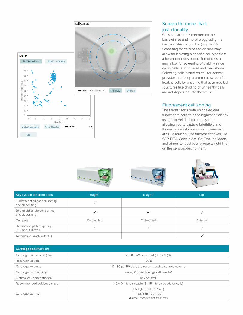
Screen for more than just clonalityCells can also be screened on the basis of size and morphology using the image analysis algorithm (Figure 3B). Screening for cells based on size may allow for isolating a specific cell type from a heterogeneous population of cells or may allow for screening of viability since dying cells tend to swell and then shrivel. Selecting cells based on cell roundness provides another parameter to screen for healthy cells by ensuring that asymmetrical structures like dividing or unhealthy cells are not deposited into the wells.
Fluorescent cell sortingThe f.sight™ sorts both unlabeled and fluorescent cells with the highest efficiency using a novel dual camera system allowing you to capture brigthfield and fluorescence information simultaneously at full resolution. Use fluorescent dyes like GFP, FITC, Calcein AM, CellTracker Green, and others to label your products right in or on the cells producing them.
Key system differentiators f.sight™ c.sight™ scp™
Fluorescent single cell sorting and depositing ü
Brightfield single cell sorting and depositing ü ü ü
Computer Embedded Embedded External
Destination plate capacity (96- and 384-well)
1 1 2
Automation ready with API ü
Cartridge specifications
Cartridge dimensions (mm) ca. 8.8 (W) x ca. 16 (H) x ca. 5 (D)
Reservoir volume 100 µl
Cartridge volumes 10–80 µL, 50 µL is the recommended sample volume
Cartridge compatibility water, PBS and cell growth media*
Optimal cell concentration 1e6 cells/mL
Recommended cell/bead sizes 40x40 micron nozzle (5–35 micron beads or cells)
Cartridge sterility
UV light (CWL 254 nm)
TSE/BSE free: Yes
Animal component free: Yes
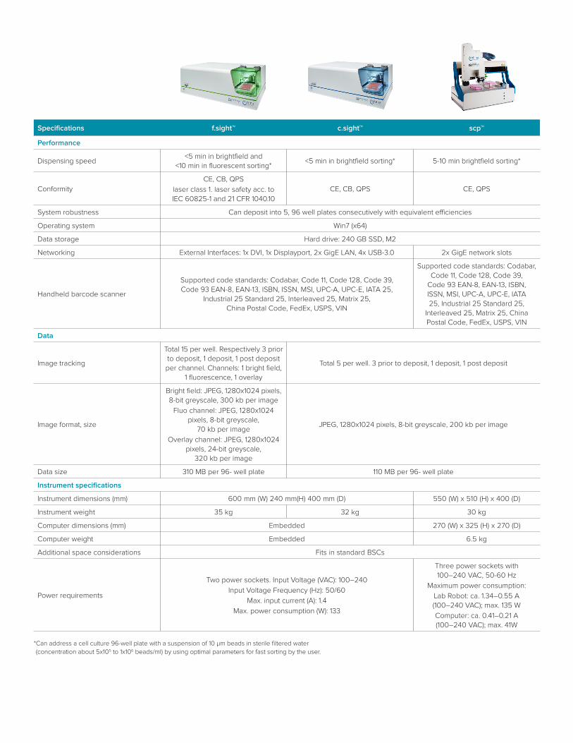
Specifications f.sight™ c.sight™ scp™
Performance
Dispensing speed<5 min in brightfield and
<10 min in fluorescent sorting*<5 min in brightfield sorting* 5-10 min brightfield sorting*
ConformityCE, CB, QPS
laser class 1. laser safety acc. to IEC 60825-1 and 21 CFR 1040.10
CE, CB, QPS CE, QPS
System robustness Can deposit into 5, 96 well plates consecutively with equivalent efficiencies
Operating system Win7 (x64)
Data storage Hard drive: 240 GB SSD, M2
Networking External Interfaces: 1x DVI, 1x Displayport, 2x GigE LAN, 4x USB-3.0 2x GigE network slots
Handheld barcode scanner
Supported code standards: Codabar, Code 11, Code 128, Code 39, Code 93 EAN-8, EAN-13, ISBN, ISSN, MSI, UPC-A, UPC-E, IATA 25,
Industrial 25 Standard 25, Interleaved 25, Matrix 25, China Postal Code, FedEx, USPS, VIN
Supported code standards: Codabar, Code 11, Code 128, Code 39,
Code 93 EAN-8, EAN-13, ISBN, ISSN, MSI, UPC-A, UPC-E, IATA 25, Industrial 25 Standard 25,
Interleaved 25, Matrix 25, China Postal Code, FedEx, USPS, VIN
Data
Image tracking
Total 15 per well. Respectively 3 prior to deposit, 1 deposit, 1 post deposit per channel. Channels: 1 bright field,
1 fluorescence, 1 overlay
Total 5 per well. 3 prior to deposit, 1 deposit, 1 post deposit
Image format, size
Bright field: JPEG, 1280x1024 pixels, 8-bit greyscale, 300 kb per image
Fluo channel: JPEG, 1280x1024 pixels, 8-bit greyscale,
70 kb per image
Overlay channel: JPEG, 1280x1024 pixels, 24-bit greyscale,
320 kb per image
JPEG, 1280x1024 pixels, 8-bit greyscale, 200 kb per image
Data size 310 MB per 96- well plate 110 MB per 96- well plate
Instrument specifications
Instrument dimensions (mm) 600 mm (W) 240 mm(H) 400 mm (D) 550 (W) x 510 (H) x 400 (D)
Instrument weight 35 kg 32 kg 30 kg
Computer dimensions (mm) Embedded 270 (W) x 325 (H) x 270 (D)
Computer weight Embedded 6.5 kg
Additional space considerations Fits in standard BSCs
Power requirements
Two power sockets. Input Voltage (VAC): 100–240
Input Voltage Frequency (Hz): 50/60
Max. input current (A): 1.4
Max. power consumption (W): 133
Three power sockets with 100–240 VAC, 50-60 Hz
Maximum power consumption:
Lab Robot: ca. 1.34–0.55 A (100–240 VAC); max. 135 W
Computer: ca. 0.41–0.21 A (100–240 VAC); max. 41W
* Can address a cell culture 96-well plate with a suspension of 10 µm beads in sterile filtered water (concentration about 5x10⁵ to 1x10⁶ beads/ml) by using optimal parameters for fast sorting by the user.

Contact Us
Phone: +1-800-635-5577Web: www.moleculardevices.comEmail: [email protected] our website for a current listing of worldwide distributors.
The trademarks used herein are the property of Molecular Devices, LLC or their respective owners. Specifications subject to change without notice. Patents: www.moleculardevices.com/productpatents FOR RESEARCH USE ONLY. NOT FOR USE IN DIAGNOSTIC PROCEDURES.
©2019 Molecular Devices, LLC 5/19 2127BPrinted in USA
Specifications f.sight™ c.sight™ scp™
Instrument specifications, continued
Light sources
LED source 1: white, 1 Watt
LED source 2: red, 3 Watt
Laser source 1: 473 nm, 100 mW, class 3B
f.sight is declared laser class 1 for standard operation
LED source 1: white, 1 Watt
LED source 2: red, 3 Watt
LED source 1: white, 5 Watt
LED source 2: blue, 3 Watt
Fluorescence detection
Excitation: 473 nm
Emission: 525/20 nm
Laser power: 1- 100 mW (freely tunable)
Exposure Times: 0.1 – 20 ms (freely tunable)
Fluorophores tested and compatible: GFP, Cell-Tracker Green, FITC,
Calcein-AM, DyLight
LED source 1: white, 1 Watt
LED source 2: red, 3 Watt
LED source 1: white, 5 Watt
LED source 2: blue, 3 Watt
Environmental conditionsTemperature: 10°–30° Celsius
Humidity: 10–80% (noncondensing)
Cell camera
8-bit grayscale
Type: CMOS
Sensor: monochrome
Image resolution1280x1024 (bright field camera) and
1936x1216 (fluorescence camera)1280x1024 1280–1024 pixels
Magnification and numerical aperture 10x (0.14 NA) 11x (0.01 NA) 11x (0.01 NA)
Microplate compatibility
SBS or SLAS standard cell culture well plates (96- and 384-well format*)
* May be customizable with additional formats (6-well, 24-well, slides)
Not compatible with:
deep well plates, or skirt-less/frameless plates.
Not compatible with:
skirt-less/frameless plates.
Disposable compatibility SCP cartridges and Eppendorf 200 mL pipette tips and Mettler Toledo adapter enabling most tip compatibility.
Instrument sterility Resistant against 70% Ethanol, Resistant against UV (254 nm CWL)
Component sterilityMetal piston guide, tubing (agitation and shutter), and Luer-Lock connectors are resistant to ethanol
and isopropanol. Piston guide and tubing can be autoclaved (121°C).
Available from Molecular Devices in North America, for all other countries please contact Cytena directly [email protected].