Duke Masters Of Minimally Invasive Thoracic Surgery Minimally Invasive Ivor Lewis Esophagectomy

Clinical StudySialendoscopy and Combined Minimally Invasive Treatment forLarge Parotid Stones
Jan Rotnágl, Šárka Zavázalová, Olexii Vorobiov, and Jaromír Astl
Otorhinolaryngology and Maxillofacial Surgery Department of the 3rd Faculty of Medicine of Charles University andthe Military University Hospital Prague, Prague, Czech Republic
Correspondence should be addressed to Jan Rotnagl; [email protected]
Received 5 August 2016; Revised 18 September 2016; Accepted 18 October 2016
Academic Editor: Francesco Doglietto
Copyright © 2016 Jan Rotnagl et al. This is an open access article distributed under the Creative Commons Attribution License,which permits unrestricted use, distribution, and reproduction in any medium, provided the original work is properly cited.
Sialendoscopy (SE) represents nowadays one of the standard diagnostic and therapeutic procedures in the treatment of majorsalivary glands lithiasis. We know from experience that it is successful only in small percentage of patients, when used inmonotherapy. However, it represents an indispensable part of all of the combined minimally invasive gland-preserving treatmenttechniques, the success rate ofwhich is around 90%. In thiswork, we focused on the role of sialendoscopy in the treatment of patientswith larger inflamed fixed stones in glandula parotis. We conducted a total of 364 sialendoscopy procedures in 332 patients on oursite. We have confirmed lithiasis as a cause of salivary gland obstruction in 246 (74%) patients. In 9 patients there was larger, single,or multiple inflamed fixed lithiasis of glandula parotis. In this subgroup of patients endoscopically assisted sialolithectomy fromexternalmini-incision has become themethod of choice. In 9 of the 9 (100%) cases we have achieved complete elimination of stones,and in 8 of the 9 (89%) cases we have achieved complete elimination of complaints. Sialoendoscopically assisted sialolithectomyof glandula parotis from external mini-incision has proved to be highly effective technique to eliminate stones with minimalcomplications.
1. Introduction
Since the 90s of the 20th century, a dynamic developmentof minimally invasive gland-preserving techniques in thetreatment of obstructivemajor salivary gland diseases is seen,particularly sialolithiasis causing obstruction in up to 80%of cases [1–4]. In this contemporary trend, sialendoscopyplays a pivotal role. While being successful in monotherapyin treatment of lithiasis only in a small percentage of patients[5], however, its combination with other minimally invasivetechniques is raising the overall success of gland-preservingtreatment that is ranging between 80 and 90%, dependingon the affected gland, location and size of the stone, andthe technique used [3, 5]. In small, loosen stones in sub-mandibular and parotid glands, themethod of the first choiceis endoscopic extraction [1, 3–7]. Larger stones whose sizedoes not allow intraductal manipulation are in the case ofsubmandibular gland indicated for intraoral sialolithectomy,which is preferred over ESWL (extracorporeal shockwave
lithotripsy) [3, 5, 7–9]. On the contrary, larger stones up to thesize of 7mm located in the parotid gland are preferably solvedby ESWL, with lower success rate—about 60%—of completeelimination. Additional 20–30% of patients reach alleviationof symptoms [3, 10–12]. Due to the fact that sialendoscopyis relatively new method, more detailed processing of thelong-term effect of gland-preserving treatment is still missing[3]. However, the individual techniques, their limits, andsuccessfulness are already pretty well mapped. Here we quoteour result of relatively less traditional technique, endoscop-ically assisted sialolithectomy from external mini-incision,indication of which applies only to a narrow range of patients[3, 13, 14]. Most of the authors reserve this method for casesof failure of other minimally invasive techniques [3, 4, 12–18].In our case it involved 9 of the 246 (3.7%) subjects from thetotal number of patients affected by lithiasis. In the subgroupof patients with lithiasis in glandula parotis, the studied groupincluded 9 of the 32 (28%) subjects.
Hindawi Publishing CorporationBioMed Research InternationalVolume 2016, Article ID 1354202, 5 pageshttp://dx.doi.org/10.1155/2016/1354202
2 BioMed Research International
Patientchronic sialoadenitis with obstructive symptoms
Clinical examination&
US
SE
US+ US−
US+ SE+ US− SE+ US+ SE− US− SE−
SE success SE failureSE success SE failure
ESWL ESWLunavailable
ESWLsuccess
ESWLfailure
ESWLpartial success
Combinedapproach
US navigatedsialolithectomy
Superficialparotidectomy
Success Failure
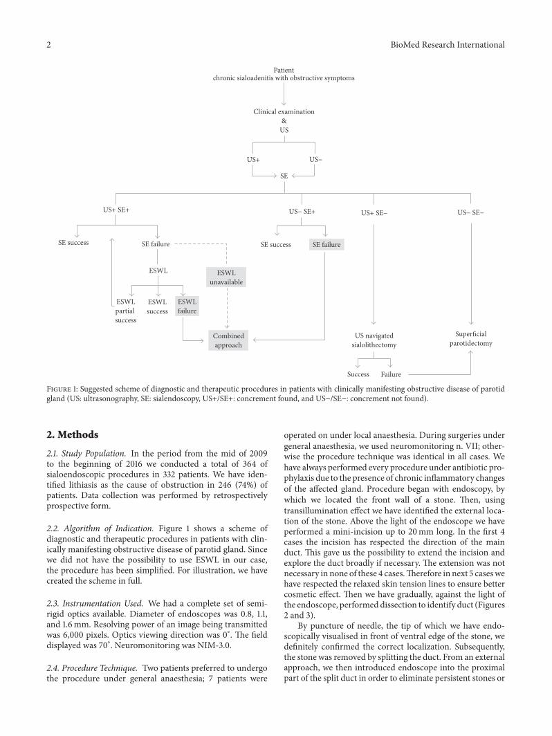
Figure 1: Suggested scheme of diagnostic and therapeutic procedures in patients with clinically manifesting obstructive disease of parotidgland (US: ultrasonography, SE: sialendoscopy, US+/SE+: concrement found, and US−/SE−: concrement not found).
2. Methods
2.1. Study Population. In the period from the mid of 2009to the beginning of 2016 we conducted a total of 364 ofsialoendoscopic procedures in 332 patients. We have iden-tified lithiasis as the cause of obstruction in 246 (74%) ofpatients. Data collection was performed by retrospectivelyprospective form.
2.2. Algorithm of Indication. Figure 1 shows a scheme ofdiagnostic and therapeutic procedures in patients with clin-ically manifesting obstructive disease of parotid gland. Sincewe did not have the possibility to use ESWL in our case,the procedure has been simplified. For illustration, we havecreated the scheme in full.
2.3. Instrumentation Used. We had a complete set of semi-rigid optics available. Diameter of endoscopes was 0.8, 1.1,and 1.6mm. Resolving power of an image being transmittedwas 6,000 pixels. Optics viewing direction was 0∘. The fielddisplayed was 70∘. Neuromonitoring was NIM-3.0.
2.4. Procedure Technique. Two patients preferred to undergothe procedure under general anaesthesia; 7 patients were
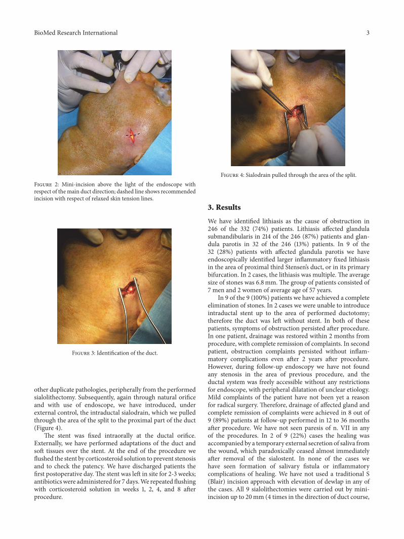
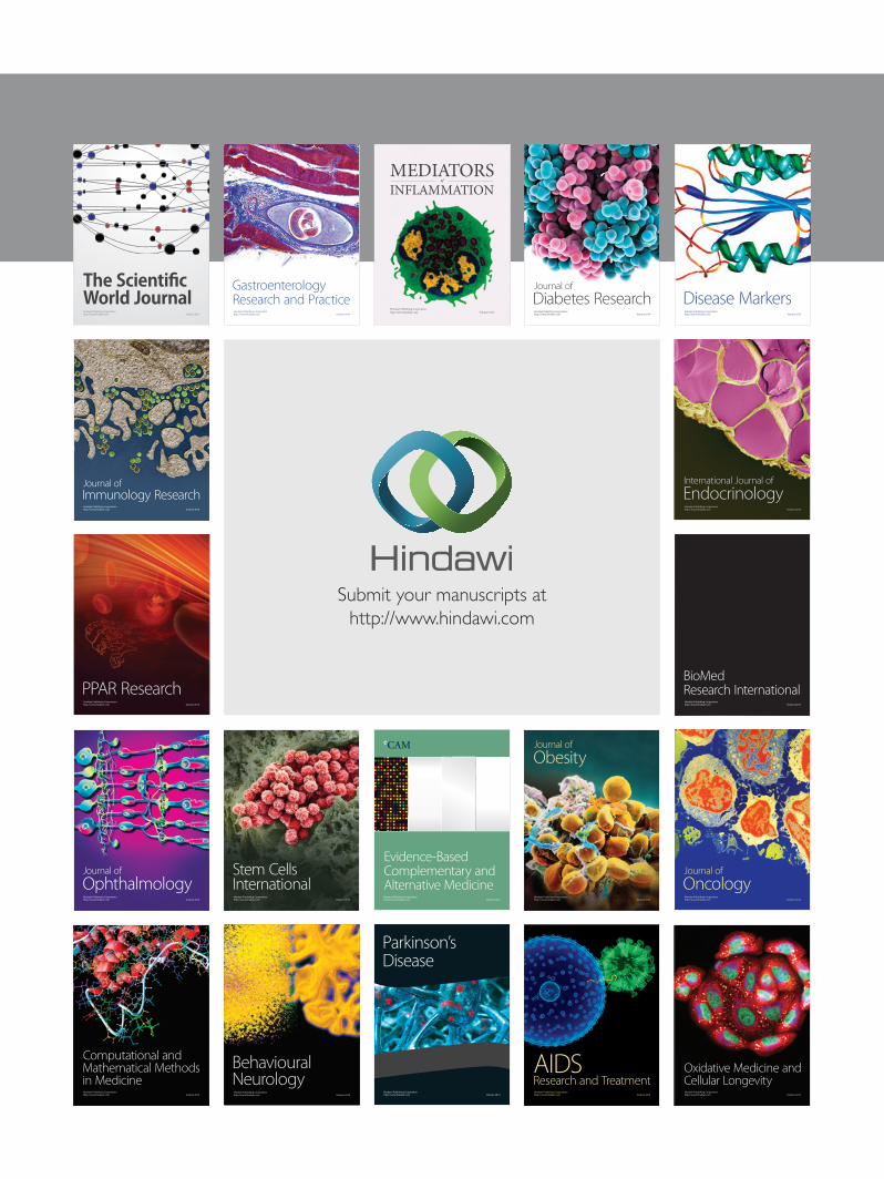
operated on under local anaesthesia. During surgeries undergeneral anaesthesia, we used neuromonitoring n. VII; other-wise the procedure technique was identical in all cases. Wehave always performed every procedure under antibiotic pro-phylaxis due to the presence of chronic inflammatory changesof the affected gland. Procedure began with endoscopy, bywhich we located the front wall of a stone. Then, usingtransillumination effect we have identified the external loca-tion of the stone. Above the light of the endoscope we haveperformed a mini-incision up to 20mm long. In the first 4cases the incision has respected the direction of the mainduct. This gave us the possibility to extend the incision andexplore the duct broadly if necessary. The extension was notnecessary in none of these 4 cases.Therefore in next 5 caseswehave respected the relaxed skin tension lines to ensure bettercosmetic effect. Then we have gradually, against the light ofthe endoscope, performeddissection to identify duct (Figures2 and 3).
By puncture of needle, the tip of which we have endo-scopically visualised in front of ventral edge of the stone, wedefinitely confirmed the correct localization. Subsequently,the stone was removed by splitting the duct. From an externalapproach, we then introduced endoscope into the proximalpart of the split duct in order to eliminate persistent stones or
BioMed Research International 3
Figure 2: Mini-incision above the light of the endoscope withrespect of themain duct direction; dashed line shows recommendedincision with respect of relaxed skin tension lines.
Figure 3: Identification of the duct.
other duplicate pathologies, peripherally from the performedsialolithectomy. Subsequently, again through natural orificeand with use of endoscope, we have introduced, underexternal control, the intraductal sialodrain, which we pulledthrough the area of the split to the proximal part of the duct(Figure 4).
The stent was fixed intraorally at the ductal orifice.Externally, we have performed adaptations of the duct andsoft tissues over the stent. At the end of the procedure weflushed the stent by corticosteroid solution to prevent stenosisand to check the patency. We have discharged patients thefirst postoperative day.The stent was left in site for 2-3 weeks;antibioticswere administered for 7 days.We repeated flushingwith corticosteroid solution in weeks 1, 2, 4, and 8 afterprocedure.
Figure 4: Sialodrain pulled through the area of the split.
3. Results
We have identified lithiasis as the cause of obstruction in246 of the 332 (74%) patients. Lithiasis affected glandulasubmandibularis in 214 of the 246 (87%) patients and glan-dula parotis in 32 of the 246 (13%) patients. In 9 of the32 (28%) patients with affected glandula parotis we haveendoscopically identified larger inflammatory fixed lithiasisin the area of proximal third Stensen’s duct, or in its primarybifurcation. In 2 cases, the lithiasis was multiple. The averagesize of stones was 6.8mm.The group of patients consisted of7 men and 2 women of average age of 57 years.
In 9 of the 9 (100%) patients we have achieved a completeelimination of stones. In 2 cases we were unable to introduceintraductal stent up to the area of performed ductotomy;therefore the duct was left without stent. In both of thesepatients, symptoms of obstruction persisted after procedure.In one patient, drainage was restored within 2 months fromprocedure, with complete remission of complaints. In secondpatient, obstruction complaints persisted without inflam-matory complications even after 2 years after procedure.However, during follow-up endoscopy we have not foundany stenosis in the area of previous procedure, and theductal system was freely accessible without any restrictionsfor endoscope, with peripheral dilatation of unclear etiology.Mild complaints of the patient have not been yet a reasonfor radical surgery. Therefore, drainage of affected gland andcomplete remission of complaints were achieved in 8 out of9 (89%) patients at follow-up performed in 12 to 36 monthsafter procedure. We have not seen paresis of n. VII in anyof the procedures. In 2 of 9 (22%) cases the healing wasaccompanied by a temporary external secretion of saliva fromthe wound, which paradoxically ceased almost immediatelyafter removal of the sialostent. In none of the cases wehave seen formation of salivary fistula or inflammatorycomplications of healing. We have not used a traditional S(Blair) incision approach with elevation of dewlap in any ofthe cases. All 9 sialolithectomies were carried out by mini-incision up to 20mm (4 times in the direction of duct course,

4 BioMed Research International
5 times in the direction of skin fissility), without the need toexpand the operational approach.
4. Discussion
Sialendoscopy, as part of minimally invasive treatment ofstones inmajor salivary glands, significantly raised the gland-preserving treatment success rate in the past 25 years. Withstones in the parotid gland, which do not allow primarilyendoscopic removal ormechanical intraductal fragmentationdue to their size, individual sites slightly differ in preferenceof treatment techniques. Larger stones up to 7mm are usuallypreferentially solved by ESWL with reported success rate incomplete elimination reaching 60%. In another roughly 30%of patients, their clinical complaints are only alleviated. Inpart of these patients, the partial success of ESWL can becombined retroactively with endoscopic additional removalof residual stone fragments [3, 10, 11]. Recently, there havebeen also several newer studies published on the use of intra-ductal laser lithotripsy. Particularly, the use of Thulium-YAGlaser in combination with subsequent endoscopic extractionof fragments had been evaluated as relatively safe techniqueof sialolithectomy. Successfulness in relation with ESWLwas comparable in the parotid gland treatment and eventwice higher in the submandibular salivary gland treatment[19]. Despite these optimistic results, the long-term reservedattitude towards this technique does not change very muchbecause of existing risks of iatrogenic damage to soft tissues.
Even today, intraductal laser lithotripsy used to treatsialolithiasis does not belong to commonly used methods inthe world’s leading medical centres [1, 4].The same applies tothe combined sialoendoscopically assisted sialolithectomy ofparotid gland which is, due to the need for external incisionand relative risk for facial nerve, reserved only for cases offailure of other techniques [3, 4, 12–18].
In our case, this technique became a method of choicein 9 patients. Nationwide unavailability of ultrasound-guidedESWL contributed to it, too. In available references inliterature, individual authors also show results for similarlysmall groups of patients, confirming rarity of the indication.Nahlieli et al. described as one of the first authors the useof combined external approach in 12 patients [14]. He alsoproposed indication criteria for this procedure as follows:location of the stone in the posterior one-third of Stensen’sduct, small duct diameter, and impacted stone larger than5mm, and intraparenchymal stones. 9 of the 12 (75%) stoneswere successfully removed. In 7 of the 12 patients glandfunctioned normally [14]. McGurk et al. described the useof external approach in 8 patients; from this number, sevenpatients suffered from lithiasis and one from stenosis. In all7 cases, the stone was removed successfully. In one case,duct ligature has been performed due to the impossibilityto reconstruct the affected duct. Function of the affectedgland has been preserved in 75% of patients [13]. Kochet al. described his experience with this technique in 19patients, 17 of which suffered from lithiasis. The treatmentwas successful in 89.5% and in 94% in lithiasis, respectively.In two cases it was necessary to perform parotidectomydue to the impossibility to perform duct reconstruction.
The conditions of successful treatment achievement werethe possibility to reach the pathology endoscopically, thepossibility to reconstruct the duct, and the ability to recoverthe function of the affected gland [17]. Marchal described 37patients with refractory stones larger than 6mm, and withduct stenosis.The treatment was successful in 92% of patients[20]. Konstantinidis et al. described similar experiences in12 patients with lithiasis. Sialolithectomy has been successfulin 100% of cases. Intraductal stent was used in 9 of the 12patients. The follow-up endoscopy discovered mild stenosiswithout clinical manifestation in 7 of the 12 cases; the follow-up scintigraphy showed normal function in 11 of the 12 cases[18]. Mikolajczak et al. [16] and Kopec et al. [12] come upwithsimilar results. On the contrary, for example, Numminen etal. report successful combined treatment in 6 of the 8 patientswithout the use of stent [15].
Upon failure of previous therapy, or in unavailability ofESWL, this method can be the only treatment option beforeradical resection. Understandable condition for success is theendoscopic accessibility of ventral edge of the stone [17]. Inour group, all of the 9 patients were affected by lithiasis whichmeets this condition. Stones were successfully removed in9 of the 9 (100%) cases, and the physiological function ofthe gland was restored in 8 of the 9 (89%) patients. In onepatient, mild problems with obstruction persisted but havenot required radical treatment. The follow-up endoscopy,however, has not proved stenosis in site of the surgery. Fromour experience, stenting of the duct after the performedsurgery did not appear essential for the preservation of ductpatency. Nevertheless, good long-term results in 7 of the9 patients, who had the postoperative stent, allow us torather recommend its use. But we have also noticed probableobstructive action of the stent for peripheral part of glandthat resulted in external secretion from the wound in 2 cases.Removal of the stent and compression resulted, in both of thecases, in almost immediate cessation of the external secretion.From the long-term perspective, this fact had no effect onthe overall favourable healing and physiological function ofthe gland. Endoscopically assisted sialolithectomy can be per-formed under general and/or local anaesthesia, depending onthe surgical approach. Majority of procedures referred to inliterature are performed under general anaesthesia [12, 14–18]. In all cases of our group, mini-incision proved to besufficient surgical approach, allowing the procedure to beperformed under a local anaesthesia. General anaesthesiaprovides the surgeonwith greater convenience and possibilityto use neuromonitoring. On the contrary, performing theprocedure under local anaesthesia makes this proceduremore acceptable in the eyes of patients, and less serious, andit can be performed as an ambulatory procedure. Since inour group we have not noticed any complication resultingfrom the use of local anaesthesia, in our view it can berecommended for well-selected patients.
5. Conclusion
Sialoendoscopically assisted glandula parotis sialolithectomyfrom external mini-incision proved to be highly successfultechnique to eliminate stones with minimal complications.

BioMed Research International 5
In larger fixed stones in glandula parotis, refractory to othertherapies, and/or in unavailability of ESWL, this techniquecan be recommended as a suitable alternative to minimallyinvasive treatment that saves the patient from radical resec-tion. The only condition for success is the ability to reach thestone with endoscope.
Our results show that, in properly selected patients, thisprocedure is very well tolerated even under local anaesthesia,which further increases its attractiveness.
Competing Interests
The authors declare that there is no conflict of interestsregarding the publication of this paper.
Acknowledgments
This work is supported by project of Ministry of Defenceof the Czech Republic, Military University Hospital Prague(ZRVOMO 1012).
References
[1] P. Capaccio, S. Torretta, F. Ottavian, G. Sambataro, and L. Pig-nataro, “Modern management of obstructive salivary diseases,”Acta Otorhinolaryngologica Italica, vol. 27, no. 4, pp. 161–172,2007.
[2] M. P. Escudier andM.McGurk, “Symptomatic sialoadenitis andsialolithiasis in the English population, an estimate of the costof hospital treatment,” British Dental Journal, vol. 186, no. 9, pp.463–466, 1999.
[3] H. Iro, J. Zenk, M. P. Escudier et al., “Outcome of minimallyinvasive management of salivary calculi in 4,691 patients,” TheLaryngoscope, vol. 119, no. 2, pp. 263–268, 2009.
[4] O. Nahlieli, H. Iro, M. Mcgurk, and J. Zenk, Modern Manage-ment Preserving the Salivary Glands, Isradon Publishing House,Herzeliya, Israel, 2007.
[5] J. Zenk, M. Koch, N. Klintworth et al., “Sialendoscopy in thediagnosis and treatment of sialolithiasis: a study on more than1000 patients,” Otolaryngology—Head and Neck Surgery, vol.147, no. 5, pp. 858–863, 2012.
[6] P. Capaccio, A. Bottero, M. Pompilio, and F. Ottaviani, “Con-servative transoral removal of hilar submandibular salivarycalculi,” Laryngoscope, vol. 115, no. 4, pp. 750–752, 2005.
[7] M. McGurk, M. P. Escudier, and J. E. Brown, “Modern manage-ment of salivary calculi,” British Journal of Surgery, vol. 92, no.1, pp. 107–112, 2005.
[8] J. Zenk, J. Constantinidis, B. Al-Kadah, and H. Iro, “Transoralremoval of submandibular stones,”Archives of Otolaryngology—Head and Neck Surgery, vol. 127, no. 4, pp. 432–436, 2001.
[9] J. Zenk, F. Gottwald, A. Bozzato, and H. Iro, “Submandibularsialoliths. Stone removal with organ preservation,” HNO, vol.53, no. 3, pp. 243–249, 2005.
[10] M. P. Escudier, J. E. Brown, N. A. Drage, and M. McGurk,“Extracorporeal shockwave lithotripsy in the management ofsalivary calculi,” British Journal of Surgery, vol. 90, no. 4, pp.482–485, 2003.
[11] J. Zenk, A. Bozzato, M. Winter, F. Gottwald, and H. Iro, “Extra-corporeal shock wave lithotripsy of submandibular stones:
evaluation after 10 years,” Annals of Otology, Rhinology andLaryngology, vol. 113, no. 5 I, pp. 378–383, 2004.
[12] T. Kopec, W. Szyfter, and M. Wierzbicka, “Sialoendoscopyand combined approach for the management of salivary glandstones,” European Archives of Oto-Rhino-Laryngology, vol. 270,no. 1, pp. 219–223, 2013.
[13] M. McGurk, A. D. MacBean, K. F. M. Fan, C. Sproat, and C.Darwish, “Endoscopically assisted operative retrieval of parotidstones,”British Journal of Oral andMaxillofacial Surgery, vol. 44,no. 2, pp. 157–160, 2006.
[14] O. Nahlieli, D. London, A. Zagury, and E. Eliav, “Combinedapproach to impacted parotid stones,” Journal of Oral andMaxillofacial Surgery, vol. 60, no. 12, pp. 1418–1423, 2002.
[15] J. Numminen, S. Sillanpaa, J. Virtanen, M. Sipila, and M.Rautiainen, “Retrospective analysis of a combined endoscopicand transcutaneous technique for the management of parotidsalivary gland stones,” ORL. Oto-Rhino-Laryngology and itsRelated Specialties, vol. 76, no. 5, pp. 282–287, 2014.
[16] S. Mikolajczak, M. Bremke, D. Beutner, and J. C. Luers, “Com-bined endoscopic and transcutaneous approach for immobileparotid stones,” Acta Oto-Laryngologica, vol. 135, no. 1, pp. 85–89, 2015.
[17] M. Koch, H. Iro, and J. Zenk, “Combined endoscopic-transcutaneous surgery in parotid gland sialolithiasis and otherductal diseases: reporting medium- to long-term objective andpatients’ subjective outcomes,” European Archives of Oto-Rhino-Laryngology, vol. 270, no. 6, pp. 1933–1940, 2013.
[18] I. Konstantinidis, A. Chatziavramidis, I. Iakovou, and J. Con-stantinidis, “Long-term results of combined approach inparotid sialolithiasis,” European Archives of Oto-Rhino-Lary-ngology, vol. 272, no. 11, pp. 3533–3538, 2015.
[19] M. Durbec, E. Dinkel, S. Vigier, F. Disant, F. Marchal, andF. Faure, “Thulium-YAG laser sialendoscopy for parotid andsubmandibular sialolithiasis,” Lasers in Surgery and Medicine,vol. 44, no. 10, pp. 783–786, 2012.
[20] F. Marchal, “A combined endoscopic and external approachfor extraction of large stones with preservation of parotid andsubmandibular glands,” Laryngoscope, vol. 117, no. 2, pp. 373–377, 2007.
Submit your manuscripts athttp://www.hindawi.com
Stem CellsInternational
Hindawi Publishing Corporationhttp://www.hindawi.com Volume 2014
Hindawi Publishing Corporationhttp://www.hindawi.com Volume 2014
MEDIATORSINFLAMMATION
of
Hindawi Publishing Corporationhttp://www.hindawi.com Volume 2014
Behavioural Neurology
EndocrinologyInternational Journal of
Hindawi Publishing Corporationhttp://www.hindawi.com Volume 2014
Hindawi Publishing Corporationhttp://www.hindawi.com Volume 2014
Disease Markers
Hindawi Publishing Corporationhttp://www.hindawi.com Volume 2014
BioMed Research International
OncologyJournal of
Hindawi Publishing Corporationhttp://www.hindawi.com Volume 2014
Hindawi Publishing Corporationhttp://www.hindawi.com Volume 2014
Oxidative Medicine and Cellular Longevity
Hindawi Publishing Corporationhttp://www.hindawi.com Volume 2014
PPAR Research
The Scientific World JournalHindawi Publishing Corporation http://www.hindawi.com Volume 2014
Immunology ResearchHindawi Publishing Corporationhttp://www.hindawi.com Volume 2014
Journal of
ObesityJournal of
Hindawi Publishing Corporationhttp://www.hindawi.com Volume 2014
Hindawi Publishing Corporationhttp://www.hindawi.com Volume 2014
Computational and Mathematical Methods in Medicine
OphthalmologyJournal of
Hindawi Publishing Corporationhttp://www.hindawi.com Volume 2014
Diabetes ResearchJournal of
Hindawi Publishing Corporationhttp://www.hindawi.com Volume 2014
Hindawi Publishing Corporationhttp://www.hindawi.com Volume 2014
Research and TreatmentAIDS
Hindawi Publishing Corporationhttp://www.hindawi.com Volume 2014
Gastroenterology Research and Practice
Hindawi Publishing Corporationhttp://www.hindawi.com Volume 2014
Parkinson’s Disease
Evidence-Based Complementary and Alternative Medicine
Volume 2014Hindawi Publishing Corporationhttp://www.hindawi.com