Minimally Invasive Thyroid Surgery - Free...
22
Minimally Invasive Thyroid Surgery Vanella S ¹* ; Crocco A²; Michele S 2 ; Simone M 2 ; Chetta N¹; Di Meo G¹; Logrieco G¹ ¹General and Oncologic Surgery II, Ecclesiastical Institution Regional Hospital “F. Miulli”, Acquaviva delle Fonti, Italy ²General Oncologic Surgery, Oncologic Institute “Giovanni Paolo II” Bari, Italy *Correspondence to: Vanella S, MD, PhD, General and Oncologic Surgery II, Ecclesiastical Institution Regional Hos- pital “F. Miulli”, Acquaviva delle Fonti, Italy Email: [email protected] Chapter 2 Thyroid Disorders Keywords: Endoscopic Thyroidectomy; Minimally Invasive Video-Assisted Thyroidectomy; Transoral Thyroidec- tomy; Transoral Thyroidectomy; Bilateral Axillo-Breast Approach (BA-BA); Retroauricolar Thyroidectomy; Robotic Thyroidectomy 1. Introduction The trans cervical approach established by Kocher is the most widely used for thyroi- dectomy [1]. Advancements in operative technique have led to decreased morbidity, mortality, and surgery through ever-shrinking cervical incisions [2]. Despite these improvements, studies have demonstrated that there can be a significant negative impact on patient quality of life as a result of a visible cervical scar [3]. Moreover, it is not only the severity or length of the scar but the mere presence of one that leads to this finding [4]. International communities, most notably in Asia, have made significant strides in remote access and minimally invasive thyroidectomy while demonstrating safety profiles similar to those through a traditional anterior cervical inci- sion. Minimally invasive surgery has been adopted for thyroid surgery since Gagner in 1996 [5]. For over two decades, various approaches for endoscopic thyroidectomies have been pro- posed, with some becoming popular to date. The rationale for these procedures was to reduce, prevent, or eliminate scarring on the neck by moving to other areas with less pain, less bleed- ing and a faster recovery period [6-9].
Transcript of Minimally Invasive Thyroid Surgery - Free...
Minimally Invasive Thyroid SurgeryVanella S¹*; Crocco A²; Michele S2; Simone M2; Chetta N¹; Di Meo G¹; Logrieco G¹
¹General and Oncologic Surgery II, Ecclesiastical Institution Regional Hospital “F. Miulli”, Acquaviva delle Fonti,
Italy
²General Oncologic Surgery, Oncologic Institute “Giovanni Paolo II” Bari, Italy
*Correspondence to: Vanella S, MD, PhD, General and Oncologic Surgery II, Ecclesiastical Institution Regional Hos-
pital “F. Miulli”, Acquaviva delle Fonti, Italy
Email: [email protected]
Chapter 2
Thyroid Disorders
Keywords: Endoscopic Thyroidectomy; Minimally Invasive Video-Assisted Thyroidectomy; Transoral Thyroidec-tomy; Transoral Thyroidectomy; Bilateral Axillo-Breast Approach (BA-BA); Retroauricolar Thyroidectomy; Robotic Thyroidectomy
1. Introduction
The trans cervical approach established by Kocher is the most widely used for thyroi-dectomy [1].
Advancements in operative technique have led to decreased morbidity, mortality, and surgery through ever-shrinking cervical incisions [2]. Despite these improvements, studies have demonstrated that there can be a significant negative impact on patient quality of life as a result of a visible cervical scar [3]. Moreover, it is not only the severity or length of the scar but the mere presence of one that leads to this finding [4]. International communities, most notably in Asia, have made significant strides in remote access and minimally invasive thyroidectomy while demonstrating safety profiles similar to those through a traditional anterior cervical inci-sion.
Minimally invasive surgery has been adopted for thyroid surgery since Gagner in 1996 [5]. For over two decades, various approaches for endoscopic thyroidectomies have been pro-posed, with some becoming popular to date. The rationale for these procedures was to reduce, prevent, or eliminate scarring on the neck by moving to other areas with less pain, less bleed-ing and a faster recovery period [6-9].
2
ww
w.openaccessebooks.comThyroid Disorders
Vane
lla S
These techniques include endoscopic or robotic breast, bilateral axillobreast, and axil-lary incisions, as well as the retroauricolar approach [10-12]. More recently, transoral thy-roidectomy has become a favored approach as it provides midline access to the thyroid with minimal extra-cervical tissue dissection and no cutaneous scar [13-18].
2. Minimally Invasive Video-Assisted Thyroidectomy
As a matter of fact, contemporary high-volume Thyroid Surgery Centers report morbid-ity and mortality rates below 1% even in elderly patients with massive goiters and upper airway obstruction [19,20]. Having secured the feasibility of the thyroidectomy, surgeons shifted their attention towards the optimization of cosmetic results and diminishment of hospitalization duration as well as of postoperative pain. Gagner et al reported the first endoscopic parathyroi-dectomy in 1996 [5]. Subsequently, several techniques of minimally invasive thyroidectomy were developed. Since Bellantone et al [21] and Miccoli et al [22] in 1999 described the feasi-bility of adapting video-assisted parathyroidectomy technique [23] to thyroid surgery for small follicular nodules, minimally invasive video-assisted thyroidectomy (MIVAT) was adopted all over the world [24-26].
Some techniques which require the use of endoscope [5,21,24,27-31] and others which do not need its use [32] have been proposed.
The different techniques implying the use of the endoscope can be classified in purely endoscopic [5,27-29] and video-assisted [21,24,31] procedures. In the totally endoscopic tech-niques, surgical dissection is completely carried out under endoscopic vision. This requires a continuous CO2 insufflation [5,29-30].
As reported by Gagner and coworkers [5, 33], CO2 neck insufflation may cause hyper-capnia, respiratory acidosis, and subcutaneous emphysema. In order to avoid these complica-tions, the use of a wall lifter allowed Huscher and coworkers [34] to perform thyroid lobec-tomy with lower pressure levels of CO2 insufflation (6 mm Hg). A less extensive use of CO2 insufflation was also advocated by Miccoli et al [30] for video-assisted parathyroid surgery.
The totally endoscopic approaches have as a major limit the more difficult dissection in comparison with the conventional techniques, mainly in case of extra-cervical accesses. They are difficult to be reproduced in different settings, especially by not skilled endoscopic surgeons, and they are technically demanding. Indeed, the totally endoscopic procedures en-countered only limited diffusion [35].
On the contrary, minimally invasive video-assisted Parathyroidectomy (MIVAP) and minimally invasive video-assisted thyroidectomy (MIVAT), short after their introduction dur-ing the late 1990s [21,30], gained a quite large worldwide diffusion, maybe because its mini-

3
Thyroid Disorders
mally invasive nature offers advantages over its conventional counterpart, by combining the bene-fits related to the endoscopic magnification with those due to its close similarity with traditional surgery.
The same intervention can be performed through a very minimal skin incision thanks to the use of the endoscope [21,24,35,36]. The excellent visualization, due to the 2 to 3 fold en-doscopic magnification, allows an easy and prompt identification of all anatomical structures.
Its low invasiveness and the similarity with the conventional procedure render this ap-proach feasible also under loco-regional anesthesia (cervical block) [37], showing the best results in patients with relative contraindications for general anesthesia such as pregnant pa-tients with papillary thyroid carcinoma (PTC), because the strong patient’s motivation plays a relevant role in the feasibility of the procedure.
2.1. Surgical technique
Most part of surgical instruments necessary for MIVAT is usually available in almost all operating rooms, and it is not a source of additional costs. The only instruments not commonly used for a conventional thyroidectomy are small dedicated tools (2-3 mm in diameter) neces-sary for dissection (ad hoc designed spatulas and spatula-shaped aspirator) which, however, are reusable.
Ultrasound knife system showed to be very useful in this kind of operation in reducing the operative time [38-40].
The operation is usually performed under general anesthesia, with the patient supine and the neck not hyperextended. The operating surgeon stands on the right side of the patient. An assistant is necessary to hold the retractors and stands above the patient’s head. The camera can be held either by another assistant or by a nurse [41].
The monitor is positioned at the head of the patient in front of the surgeon. The absence of any external support allows modulating and changing the position of the endoscope in relationship to the different step of dissection. This represents an important advantage of the video assisted procedure over purely endoscopic techniques. The tip of the endoscope is usu-ally oriented towards the head of the patient, but it can be changed to expose and explore the upper mediastinum when, for example, a concomitant central compartment lymphadenectomy is required [42].
A 1.5-2 cm horizontal skin incision is made 1 cm below the cricoid cartilage in the central neck (figure 1). Two small retractors (army-navy type) can easily expose the midline, which is then longitudinally incised for 2 to 3 cm. The thyroid lobe is bluntly dissected away from the strap muscles. A 30°, 5 mm or 7 mm video scope is inserted. The thyrotracheal groove

4
Thyroid Disorders
Figure 1: Skin markers in Minimally Invasive Video-Assisted thyroidectomy (MIVAT)
is dissected under videoscopic vision using small (2 mm in diameter) instruments.
An ultrasonic dissector can be used for hemostasis. The upper pedicle is dissected by retracting the thyroid lobe downward and medially, until the external branch of the superior laryngeal nerve is identified (Figure 2).
Figure 2: Left upper pole dissection and external branch of superior laryngeal nerve identification during MIVAT
The thyroid lobe is then retracted medially and the recurrent laryngeal nerve is identified (Figure 3). During endoscopic operations, the authors use the posterior lobe of the thyroid as a landmark beneath which the nerve lies. The superior parathyroid gland can be then dissected by using the ultrasonic device. Once the thyroid lobe is completely freed, it can be extracted by rotating its upper pole. The lobe is further freed from the trachea by dissecting the ligament of Berry. The laryngeal nerve should be checked again to avoid injury before the final step. No drainage is left in the neck, and the midline is closed by a single stitch.
Figure 3: Left recurrent laryngeal nerve and left superior parathyroid gland identification during MIVAT
5
Thyroid Disorders
In the most recent reports [43,44], a trend toward a slight enlargement of the skin inci-sion was noted, leading to 3–3.5-cm wounds, but this was mainly due to the effort of expand-ing the indications for MIVAT to larger-sized nodules and higher-volume glands [44-46].
For a surgeon who approaches the MIVAT technique, the learning curve takes a longer time than with conventional surgery, having to gain confidence with a smaller surgical incision and with the use of endoscopic instruments [36,47].
Lombardi et al. report that 10 patients represent the early stage of the learning process, 30 pa-tients are the minimal number of cases in order to move on to a higher level and performing at least 100 times the same procedure allows a reduction of the complication rate [47].
2.2 Indications
An accurate patients’ selection plays a key role to ensure the success of MIVAT.
Initially, this technique was indicated for surgical treatment of benign nodule smaller than 3.5 cm or differentiated low-risk carcinomas up to 2 cm, in a gland with total volume lesser than 25 mL and in absence of thyroiditis or lymph nodes involvement and prior neck surgery. [45,47-49],
Nowadays, preoperatively estimated thyroid volume represents the only selection pa-rameter in benign pathology. Instead, in suspected or proven malignancy, only accurate clini-cal staging can determine its indication to a minimally invasive procedure.
As the technique has been adopted worldwide, its indications expanded from the initial benign thyroid disease to low-risk and intermediate-risk carcinoma: some studies compar-ing the results of MIVAT and conventional thyroidectomy in terms of adequacy of surgical resection showed that MIVAT is safe and effective for the treatment of small papillary thyroid carcinoma (PTC) and that its oncological effectiveness is similar to traditional thyroidectomy [50].
Instead, the possibility to perform a concomitant lymphadenectomy during a video-assisted thyroidectomy for PTC has been demonstrated [51-53]: Bellantone et al. and Lom-bardi et al. in early 2000s, standardized a video-assisted lymph-node dissection (VALD) of the central compartment [52,53].
Also, Lomardi et al. evaluated the feasibility of minimally invasive video-assisted func-tional lateral neck dissection (VALNED) in patients with PTC with encouraging results [54].
Patients carrying RET oncogene mutation for familial forms of medullary thyroid carci-noma but not even expressing the disease (absence of detectable nodules and basal/stimulated calcitonin in the normal range) are excellent candidates for MIVAT [55].

6
Thyroid Disorders
Based on the surgeons’ experience, patients with previous contralateral video-assisted neck surgery or thyroiditis can be selected for MIVAT. Some authors demonstrated that in selected patients with Graves’ disease, MIVAT is feasible and can be performed safely with results comparable with open surgery [56]. Bellantone et al. purposed MIVAT also in case of nodule >35 mm in diameter.
In Italy, approximately only 20 % of the patients can take advantage of this approach [57,58], but the number increases significantly in the USA experience, where approximately 30 % of the patients can undergo MIVAT, according to several reports [45,58]. This difference could be consistent with the differences of thyroid size existing between endemic and nonen-demic goiter regions [59].
2.3 Safety and Effectiveness
In the past 20 years, several overviews and systematic reviews discussed different as-pects of MIVAT [60-64].
Minimally invasive surgery is not free from complications, which are the same as in tra-ditional surgery, including major complications, such as permanent recurrent laryngeal nerve (RLN) dysfunction and expanding hematoma, and minor complications as temporary RLN dysfunction, temporary hypocalcemia, temporary superior laryngeal nerve dysfunction, cel-lulitis, nonsurgical hematoma, seroma, postoperative pain and hypertrophic scar.
The literature is almost unanimous in affirming the similarity of MIVAT and CT (con-ventional technique) in terms of complications [47].
To date, the advantages in favor of MIVAT have an historical perspective: in the first years, optimization of cosmetic results was the main concern reported in literature [36,65,67,69,74].
These favorable results are strengthened by conclusions of Dionigi and colleagues, which showed improved results of MIVAT compared with conventional technique by consid-ering overall wound morbidity [75]. It can be explained with the minimal dissection of sub-platysmal space needed during MIVAT, which could be a risk factor of seroma arising and, as a consequence, surgical site infection SSI and impairment of cosmetic result.
Afterward, the minimal invasiveness was related to the decrease of surgical stress and reduced requirement of analgesic drugs [66-69]. This effect is confirmed also in specific pain evaluation biochemical patterns [48-69]. Increase in operative time can be easily solved by ascending learning curve [48].
Another important interest for MIVAT is the reduced incidence of voice and swallow-ing symptoms, frequently complained after thyroidectomy [49,71-73], possibly due to image

7
Thyroid Disorders
magnification that allows the external branch of superior laryngeal nerve visualization, general reduction in surgical dissection and, therefore, reduced adhesions in the surgical site [74].
In a 2017 review, Lombardi et al conclude that, in selected cases and in experienced centers, MIVAT can be considered the standard treatment, having proven to be a feasible, prac-tical, safe and effective surgical option [47].
3. NOTES and TOETVA
The concept of thyroid NOTES has been developed by Witzel et al. via the floor of the mouth [76]. Karakas et al. [77] natural orifice tranlumenal endoscopic surgery introduced another refined technique for the parathyroid called trans oral partial Parathyroidectomy (TOPP).
The combination with sublingual and vestibular approach was first introduced by Ben-hidjeb et al. [78] and was called transoral video-assisted thyroidectomy (TOVAT). Another technique was performed by Wilhelm et al. similar to TOVAT but the name was changed to endoscopic minimally invasive thyroidectomy (eMIT) [79,80].
The sublingual route, including combination, has been critiqued and has experienced decreasing popularity due to the difficulty of the technique and its high complication rate [81-85].
The oral vestibular approach was first described by Richmon et al. By robotic-assist-ed approach (TRAT). Nakajo et al. performed transoral video-assisted neck surgery by (TO-VANS).
The transoral endoscopic thyroidectomy vestibular approach (TOETVA) was performed by Anuwong [86].
3.1 Procedure
The patient is positioned supine under naso- or oro-tracheal intubation. Three incisions are then made in the oral vestibule to accommodate central and lateral trocars. The central incision is 1.5 cm in length and is placed beyond the cranial aspect of the buccal-mandibular frenulum, while two stab incisions are made at the lateral most aspect of the oral commissure in the mucosal border to avoid mental nerve injury and instrument collision. A subplatysmal pocket is then developed with use of mechanical dilators.
Two small incisions (5mm) are made laterally on the labial mucosa, near the commis-sures, almost at the transition between the mucosa and the vermilion of the lip, the 5mm trocars are introduced. CO2 is injected at a pressure from 6mmHg to 9 mmHg in a flow rate of 15 L/min. An endoscope is then inserted through the central trochar 6mmHg, and the subplatysmal
8
Thyroid Disorders
flap is then fully elevated to the level of the sternal notch inferiorly.
CO2 insufflation is utilized to maintain this working space and the midline raphe is iden-tified, divided, and the ipsilateral strap muscles are elevated.
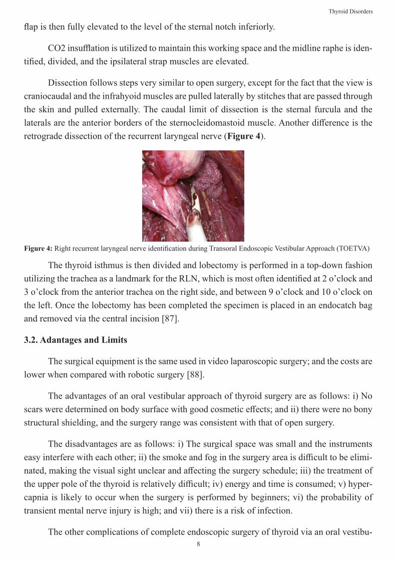
Dissection follows steps very similar to open surgery, except for the fact that the view is craniocaudal and the infrahyoid muscles are pulled laterally by stitches that are passed through the skin and pulled externally. The caudal limit of dissection is the sternal furcula and the laterals are the anterior borders of the sternocleidomastoid muscle. Another difference is the retrograde dissection of the recurrent laryngeal nerve (Figure 4).
Figure 4: Right recurrent laryngeal nerve identification during Transoral Endoscopic Vestibular Approach (TOETVA)
The thyroid isthmus is then divided and lobectomy is performed in a top-down fashion utilizing the trachea as a landmark for the RLN, which is most often identified at 2 o’clock and 3 o’clock from the anterior trachea on the right side, and between 9 o’clock and 10 o’clock on the left. Once the lobectomy has been completed the specimen is placed in an endocatch bag and removed via the central incision [87].
3.2. Adantages and Limits
The surgical equipment is the same used in video laparoscopic surgery; and the costs are lower when compared with robotic surgery [88].
The advantages of an oral vestibular approach of thyroid surgery are as follows: i) No scars were determined on body surface with good cosmetic effects; and ii) there were no bony structural shielding, and the surgery range was consistent with that of open surgery.
The disadvantages are as follows: i) The surgical space was small and the instruments easy interfere with each other; ii) the smoke and fog in the surgery area is difficult to be elimi-nated, making the visual sight unclear and affecting the surgery schedule; iii) the treatment of the upper pole of the thyroid is relatively difficult; iv) energy and time is consumed; v) hyper-capnia is likely to occur when the surgery is performed by beginners; vi) the probability of transient mental nerve injury is high; and vii) there is a risk of infection.
The other complications of complete endoscopic surgery of thyroid via an oral vestibu-

9
Thyroid Disorders
lar approach are primarily the same as endoscopic thyroidectomy via other approaches [89], including hypercapnia, subcutaneous emphysema, subcutaneous ecchymosis, incision infec-tion, skin perforation, air embolism and others [90].
The endoscope is located at the lip-side of the mandible, the range of activities is wide [91]. This is beneficial to the exposure and treatment of the upper pole of the thyroid and Del-phian lymph nodes. The incision may be extended along the two sides of the vestibular sulcus, which facilitates the complete removal of relatively large specimens (specimens >4.0 cm can be completely removed). The brightness and visual field of the surgery area are good when the 1.0 cm endoscope is used.
Limited by the mouth width and mental nerve, the three trocars are relatively crowded and the chopsticks effect is significant.
As for the oral vestibular approach, leakage of CO2 may easily occur during the sur-gery.
However, it appeared that there was no association with CO2 flow rate when it was <11 l/min. When the surgery time was <180 min and the inflation pressure of CO2 was <6 mmHg, hypercapnia almost no longer occurred [92,93].
Based on the studies by Zhao et al [89] and Wang et al [91], inflation combined with suspension technologies were adopted, and the surgery space could be maintained if the skin was pulled up and suspended on the head rack following two needles of suture with No. 2-0 thread at the hyoid level and the cricoid cartilage level in the middle line of the neck.
It was determined in domestic and foreign autopsy studies that the mental foramen lo-cated in the buccal side of the first premolar had an average distance of 3.5 cm to the middle line. If the surgery channel on both sides moved into the labial side of canine teeth, within a 2.5 cm distance to the middle line, the probability of mental nerve injury may have been ef-fectively decreased [94]. Thus, if it is possible to dissect the mental nerve first and produce a 0.5-cm instrument channel at the labial side of canine, the incidences of transient numbness of lower lip may be significantly decreased.
Due to the size of the incision (approximately 1.5cm), sometimes it is necessary to frag-ment the gland in order to extract it, which can compromise an adequate anatomopathological evaluation, such as margins, capsular invasion, and tumor size.
In patients who have the upper pedicle in a very high position, dissection may be more challenging. Dissection can also be problematic if the tumor is located in the upper pole, since manipulation of this area is essential to expose the recurrent laryngeal nerve in TOETVA, and the seizure of this pole can cause tumor rupture.

10
Thyroid Disorders
To prevent infection during the perioperative period, the solutions were as follows: i) Perform supragingival scaling three days prior to the surgery; ii) gargle collutory of chlor-hexidine and metronidazole compound three times per day; iii) intravenous bolus of cefazolin sodium 30 min prior to anesthesia iv) the oral cavity could be insulated with a rubber barrier during the surgery; and v) the mental region and superior part neck could be dressed with an elastic face mask to prevent subcutaneous dropsy when the surgery is completed [89].
3.3 Oncological Considerations
It was considered that the indications of complete endoscopic radical surgery of thyroid cancer via an oral vestibular approach are as follows: i) Patients who have a strong demand for cosmetic salvage and refuse any visible scars on the surface of body; ii) patients with cicatricle diathesis; iii) patients with differentiated thyroid cancer had below T2, thyroid volumes are ap-proximately normal and no clinical cervical lymph node metastasis.
Complete endoscopic surgery may be conducted in a caudocranial direction or cranio-caudal direction. The most common surgery direction for complete endoscopic thyroidectomy with incision out of neck was performed with a caudocranial view and there are two problems cannot be overcome when this was applied in the differentiated thyroid carcinoma: i) Scars remained on the chest wall, breast or axillary and ii) due to the shielding of the clavicle and sternum, the lymph nodes could not be sufficiently exposed in clearing the lymph nodes in the VI region [95].
The lymph nodes of regions VI, IV and the inferior part of III may be dissected by simple endoscopic surgery via an oral approach; however, the lymph nodes in region II and su-perior part of region III could not be touched [96]. If the lymph nodes in region II and superior part of region III require treatment, approaches combined with chest wall or axillaries should be used [97].
4. Robotic Thyroidectomy
The improvements of technology in the last few years led to the introduction of devices and equipment that profoundly changed the surgical approaches for thyroidectomy. Robot-assisted surgery is developing as an important contribution to neck surgery. The Da Vinci robot system overcame the previous endoscopic thyroid surgery limitations such as video camera platform instability, straight endoscopic instruments, two-dimensional imaging by providing a three-dimensional magnified view, decreased tremor, and superior range of motion [98] with excellent patient-derived (pain, cosmetic satisfaction) and short term oncologic outcomes [99]. Prof. W. Y. Chung, from South Corea, was the first who performed a robot-assisted trans axil-lary thyroidectomy in 2007 using the Da Vinci robot [100]. Since that different approaches to robotic thyroidectomy have been developed such as the retroauricolar or “facelift” approach

11
Thyroid Disorders
and the newest transoral technique. Each approaches has its own advantages and limitations. The selection of technique generally depends on surgeon’s expertise and preference.
4.1 Trans-axillary Approach
The patient, under general anesthesia, is placed in the supine position with slight exten-sion of the neck. The arm of axillary access, that is chosen according to the thyroid lobe to be removed in case of lobectomy and to the larger nodule if total thyroidectomy has to be per-formed, is raised upon the head ensuring the absence of extra tension to avoid brachial plexus injury. The procedure is divided into three steps: trans-axillary subcutaneous tunnel creation (working space formation), docking stage and console time (thyroidectomy). Once a 4-5 cm skin incision following the lateral edge of major pectoralis muscle is made, a subcutaneous flap is prepared using laparoscopic instruments with a 30° camera, from the axilla to the ante-rior neck area preserving the pectoralis fascia to prevent postoperative chest pain due to adhe-sions. An external retractor is placed to keep the working space exposed. The dissection con-tinues until the sternal and clavicular heads of the sternocleidomastoideum muscle are exposed (SCM) avoiding external jugular vein injuries or thermal burns on the overlying skin; then it follows the opening of the avascular plane between the two SCM branches. Once encountered the strap muscles, the thyroid can be exposed. The Da Vinci robot is docked controlaterally to the axillary; it is possible to use a three or four robotic arms techniques with a further indepen-dent incision at the inferior part of the axilla. A 30° optic, the Harmonic scalpel, the Maryland forceps and the ProGasp are used during the operation that proceeds in the same manners or the conventional open thyroidectomy (figure 5) starting with the dissection of the superior pole with its vessels. The inferior thyroid artery (ITA), the recurrent laryngeal nerve (RLN) and the parathyroid glands are identified subsequently once the inferior pole is released: the thyroid lobe is then dissected from the trachea, resected with the isthmus and extracted through the axillary skin incision. Contralateral lobectomy proceeds with the same method: the identifica-tion of contralateral RLN is the real difficult time of procedure. Once specimen is removed, the surgical field is checked for hemostasis; a suction drain could be inserted through the separated
Figure 5: Left lobe dissection during robotic thyroidectomy

12
Thyroid Disorders
incision in the axilla if present. The axillary incision scar is completely covered [101,102].
4.2 Bilateral Axillo-Breast Approach (BABA)
The robotic trans-axillary thyroidectomy (TAA) can be performed using also an alterna-tive bilateral axillo-breast approach (BABA). It is currently one of the most popular remote-access thyroidectomy techniques in the world, particularly in Korea [103]. Using a midline approach, BABA provides a symmetrical view to both thyroid lobes for optimal visualization and dissection of vital structures. Such midline access also allows a familiar operation process to surgeons. The surgical procedures used in the original BABA robotic thyroidectomy have been described in detail by Lee et al. [104]. The working space formation step consists in a supra-areolar skin incision followed by the creation of a subcutaneous tunnel along the tangen-tial lines directed towards the neck. A plane is created over the chest and neck in the marked area. A similar procedure is repeated on the opposite side. This is a blind procedure and one should be careful not to go too deep into the breast parenchyma or too superficial into the der-mis which causes bruising. Two 10 mm ports, are placed over bilateral supra-areolar margins, serve as the camera and main working port and CO2 insufflation started. Another 10 mm port is placed at axillary skin fold on the side of surgeon which serves as second working port. Finally, a 5 mm port is placed at the opposite axillary skin fold. Then it follows Robot dock-ing and flap dissection in the infra-clavicular and anterior chest area. After raising flap, mid-line division is extended from the suprasternal notch inferiorly to the thyroid cartilage notch superiorly as in conventional open surgery. Dissection is continued until the identification of trachea, and here, the isthmus is identified, divided and dissected off anterior tracheal surface. The thyroid lobe is detached from the strap muscle on the lateral side and medially from the trachea and the cricoid cartilage. The operation proceeds with the dissection of the upper and lower pole and the identification and preservation of RLN and parathyroid glands (PTG). For total thyroidectomy, the same procedure is repeated on the opposite side. Postoperative bleed-ing and hematoma, RLN injury, permanent or transient ipocalcemia, tracheal and esophageal lesions can occur in robotic thyroidectomy as well as in the conventional cervical approach. Specific complications of TAA and BABA robotic thyroidectomy due to the need of skin flap dissection such as thermal burns, brachial plexus injury, jugular vein lesions and anterior chest paresthesia must be also taken into account.
4.3 Retroauricolar Approach
The retroauricolar or “facelift” approach was first described by Terris to avoid the com-plications associated with the trans-axillary techniques most partly due to the challenges of the distance between the access site and the thyroid gland, and the unfamiliar vector of approach to the thyroid compartment.
The patient, under general anesthesia, is positioned supine with the head turned slightly

13
Thyroid Disorders
(30°) away from the side of the procedure. An incision is made in the post-auricular sulcus and continued within the occipital hairline. A musculocutaneous flap is raised superficial to the greater auricular nerve and External jugular vein and deep to the platysma muscle, and extended inferiorly ventral to the sternocleidomastoid muscle down to the clavicle. The dis-section continues towards the midline of the neck exposing the strap muscles. A fixed retrac-tor system is introduced to retract the strap muscles giving access to the thyroid gland. The Da Vinci surgical system is docked with the 30 degree camera positioned in the center of the field, a Maryland grasper placed in the nondominant hand and the Harmonic in the dominant one. The hemi-thyroidectomy via the retroauricolar approach begins with the dissection of the superior pole which is mobilized away from the inferior constrictor muscle. Once the supe-rior laryngeal nerve and the superior parathyroid gland are identified and the vascular pedicle is ligated the pole can be reflected ventrally. The recurrent laryngeal nerve is then identified proximal to its entrance. The middle thyroid vein is ligated using the Harmonic device, and the inferior pole is fully mobilized using blunt dissection. The inferior parathyroid gland is identi-fied and dissected
Inferiorly. The inferior thyroid artery and vein are ligated using the Harmonic device, the remaining attachments of the gland are divided, and the thyroid lobe is delivered through the incision. [104].
Total thyroidectomy is achieved by bilateral robotic facelift thyroidectomy (RFT) through two separate retroauricolar incisions. Despite the RFT significantly reduces the field of dissection for the formation of the working space, if compared with TAA approach, it is associated with some specific complications such as the injury to the greater auricular and marginal mandibular nerves [105].
4.4 Trans oral Approach
The Transoral approach for thyroidectomy was first described by Witzel et al, who per-formed 10 endoscopic transoral thyroidectomies in porcine models [76]. The first robotic series was published by Lee et al, who presented the results of the transoral periosteal thyroidectomy (TOPOT) trial in human subjects. [106].
The patient, under general anesthesia, is placed in the supine position with slight exten-sion of the neck. As a first step three incisions are made in the gingival-buccal sulcus: one in the midline 2 cm above the frenulum labii inferiors and two laterally near the angle of mouth. A submental flap is then performed with blunt dissection from the edge of mandible towards the suprasternal notch in order to elevate the platysma off the strap muscles. Subsequently car-bon dioxide (CO2) gas is inflated at a pressure of 6 mm Hg to maintain a working space which is completed by the dissection starting from the two lateral incion sites allowing insertion of the surgical instruments. The Da Vinci surgical system is docked with the 30 degree camera

14
Thyroid Disorders
positioned in the center of the field, a Maryland grasper placed in the nondominant hand and the Harmonic in the dominant one. Dissection in the midline raphe is performed to separate the strap muscles. The isthmus is divided with ultrasonic shears and retracted medially using the Prograsp forceps in the surgeon’s left hand during the right lobectomy and the right hand during the left lobectomy. The upper pole of the thyroid is drawn inferiomedially by the Pro-grasp for-ceps, and the superior thyroid vessels are identified and ligated close to the thyroid gland using the ultrasonic shears. The superior parathyroid gland is identified during the dis-section and preserved. After the upper pole dissection, the thyroid gland is retracted medially and the lower pole is dissected from the perithyroidal tissues. Using gentle blunt dissection, the recurrent laryngeal nerve is identified at its entry point and traced inferiorly. The inferior parathyroid gland is identified and preserved. The inferior thyroid artery is ligated close to the thyroid gland using the ultrasonic shears, and the entire path of the recurrent laryngeal nerve is identified and preserved. Finally, the thyroid gland is dissected from the trachea in order to complete the hemy-thyroidectomy. The resected specimen is removed transorally from the surgical field through the midline incision. [106]. Despite the transoral technique provides the advantage of accessing the thyroid gland from a natural orifice with a midline approach that allows an excellent exposure of the gland, the main disadvantages include mental nerve injury, inability to control massive hemorrhage through intraoral incision and to perform lateral neck dissections.
4.5. Indications, Limitations and Perspectives
Thyroid nodules, including low-risk papillary thyroid cancer, are most commonly en-countered in young women. Naturally, this select patient group is the one most concerned about their cosmoses. This most partly led to the development of “scar less-in-the-neck” endoscopic thyroidectomy. [107]. Robotic technology, maintaining the same remote-access, overcome the limitations of endoscopic approach and transformed in just over a decade neck surgery. How-ever there are certain selection criteria for remote-access thyroid surgery that have to be taken into account. It is possible to divide factors relating to the patient and factors relating to the thyroid pathology. Factors relating to the patient include thin body habitus and the absence of excessive body fat along the flap trajectory (except for the facelift approach). Factors relating to the thyroid pathology include: well-circumscribed nodule ≤ 3 cm, thyroid lobe <5–6 cm in the largest dimension and underlying thyroid pathology with no evidence of thyroiditis on ultrasound. Absolute contraindications include: evidence of thyroid cancer with extra thyroi-dal extension or lymph node involvement, Graves’ disease, substernal extension and previ-ous neck surgery or irradiation [108]. There is now level 2 evidence (including from Western World centers) to support the safety, feasibility, and equivalence of the robotic approach to its open counterpart in terms of recurrent laryngeal nerve injury, hypoparathyroidism and hem-orrhage [109]. Certainly the incorporation of robotic system offers 10-time magnification of
15
Thyroid Disorders
the surgical view and tremor elimination, which can enhance the safety and precision of the procedure. Identification and preservation of RLNs and parathyroid glands are much easier to achieve with the assistance of robotic system [110]. There are no randomized clinical trials or comparative studies with long-term follow-up data to comment on the oncologic equivalency of robotic thyroidectomy to conventional surgery.
In a study by Lee et al. cosmetic satisfaction at three months post robotic thyroidectomy versus open approach was assessed using a five-point scale (extremely satisfied, satisfied, ac-ceptable, dissatisfied, or extremely dissatisfied). The patients in the robotic group reported significantly greater satisfaction than those in the open group (p < 0.0001). In fact, 24 (58.5%) patients in the robotic group were extremely satisfied compared with five (11.6%) patients in the open group. There were no patients in the robotic group expressing dissatisfaction [111].
The main criticisms against robotic thyroidectomy are the additional costs, longer op-erative times and a steep learning curve. Overall, it is agreed that remote-access surgery is not cost-effective mainly for the longer time needed if compared with conventional thyroidecto-my. In the case of a robotic procedure increased costs derive from additional factors, including the capital expense of the robotic system ($1.5–$1.75 million), the annual service contract ( > $50,000/year), the increased use of disposable instruments, together with the prolonged anes-thesia and operative times [112].
Cabot et al. compared trans axillary robotic thyroidectomy costs with the conventional approach in the United States reporting a higher total cost for the trans axillary technique ($13,087 vs. $9028) [113]. As well as in the case of abdominal surgery regarding the com-parison between robotic versus laparoscopic learning curve, a faster learning curve for robotic thyroidectomy has been reported compared with conventional endoscopic techniques. Never-theless it is important not to forget that the robotic thyroid surgery pioneers gained invaluable experience with endoscopic techniques prior to introducing the robotic approach. A learning curve of 40 to 45 case has been described for trans-axillary robotic thyroidectomy [114]: a number that may be difficult to achieve for a surgeon in a relatively short period of time.
At present robotic thyroidectomy is far to be cost-effective and there are significant bar-riers to the performance of robotic remote-access thyroid surgery in the Western Countries, related to patient selection, technical challenges and oncological outcomes. It is possible in the nearest future an increased competition with the entry of multinational medical device com-panies in the surgical robotics arena in order to drive down costs. Moreover a centralization of robotic thyroidectomy services to national high-volume centers is auspicalbe in order to achieve further cost reduction. This also could help surgeons surpass more easily their learning curve with a decrease in operative time, the other main driver of cost.
16
Thyroid Disorders
5. Economic Considerations
There may be less motivation to adopt these longer surgical techniques in the absence of commensurate increases in reimbursement [8]. Trans cervical thyroid surgery has an excellent safety profile in experienced hands, and can also be done efficiently.
In such a setting, slower remote access surgeries, especially those with the added ex-pense of the robot, may be difficult to justify for all patients.
Further studies are needed to quantify the cost of each technique in comparison to con-ventional approach and to determine the true value each may provide.
6. References
1. Halsted WS. IV. (I) The Excision of Both Lobes of the Thyroid Gland for the Cure of Graves’s Disease. (II) The Pre-liminary Ligation of the Thyroid Arteries and of the Inferior in Preference to the Superior Artery. Ann Surg 1913;58:178–182.
2. Terris DJ, Singer MC, Seybt MW. Robotic facelift thyroidectomy: II. Clinical feasibility and safety. Laryngoscope 2011;121:1636–1641.
3. Best AR, Shipchandler TZ, Cordes SR. Midcervical scar satisfaction in thyroidectomy patients. Laryngoscope 2017;127:1247–1252.
4. Choi Y, Lee JH, Kim YH, et al. Impact of post thyroidectomy scar on the quality of life of thyroid cancer patients. Ann Dermatol 2014;26:693–699.
5. Gagner M. Endoscopic subtotal parathyroidectomy in patients with primary hyperparathyroidism. Br J Surg 1996;83:875.
6. Miccoli P, Berti P, Conte M, et al. Minimally invasive surgery for thyroid small nodules: preliminary report. J Endo-crinol Invest 1999;22:849-851.
7. Ikeda Y, Takami H, Niimi M, et al. Endoscopic thyroidectomy by the axillary approach. Surg Endosc 2001;15:1362-1364.
8. Choe JH, Kim SW, Chung KW, et al. Endoscopic thyroidectomy using a new bilateral axillo-breast approach. World J Surg 2007;31:601-606.
9. Wang C, Feng Z, Li J, et al. Endoscopic thyroidectomy via areola approach: summary of 1,250 cases in a single insti-tution. Surg Endosc 2015;29:192-201.
10. Berber E, Bernet V, Fahey TJ, 3rd, et al. American Thyroid Association Statement on Remote-Access Thyroid Sur-gery. Thyroid 2016;26:331–337.
11. Kandil E, Saeed A, Mohamed SE, Alsaleh N, Aslam R, Moulthrop T. Modified robotic-assisted thyroidectomy: an initial experience with the retro auricular approach. Laryngoscope 2015;125:767–771.
12. Ban MJ, Chang EHE, Lee DY, Park JH, Lee C, Kim DH, Kim JH, Koh YW. Analysis of neuro monitoring signal loss during retro auricular versus conventional thyroidectomy. Laryngoscope. 2018 Dec 25.
13. Anuwong A. Transoral endoscopic thyroidectomy vestibular approach: a series of the first 60 human cases. World J Surg 2016;40:491–497.

17
Thyroid Disorders
14. Kang SW, Lee SC, Lee SH, et al. Robotic thyroid surgery using a gasless, trans axillary approach and the da Vinci S system: the operative outcomes of 338 consecutive patients. Surgery 2009;146:1048–1055.
15. Russell JO, Noureldine SI, Al Khadem MG, Tufano RP. Minimally invasive and remote-access thyroid surgery in the era of the 2015 American Thyroid Association guidelines. Laryngoscope Investig Otolaryngol 2016;1: 175–179.
16. Russell JO, Clark J, Noureldine SI, et al. Transoral thyroidectomy and Para thyroidectomy – A North American se-ries of robotic and endoscopic transoral approaches to the central neck. Oral Oncol 2017;71:75–80.
17. Anuwong A, Ketwong K, Jitpratoom P, Sasanakietkul T, Duh QY. Safety and outcomes of the transoral endoscopic thyroidectomy vestibular approach. JAMA Surg 2017;153:21–27.
18. Richmon JD, Kim HY. Transoral robotic thyroidectomy (TORT): procedures and out-comes. Gland Surg 2017;6:285–289.
19. Gomez-Ramirez J, Sitges-Serra A, Moreno- Llorente P, Zambudio AR, Ortega-Serrano J, Rodriguez MT, del Moral JV. Mortality after thyroid surgery, insignificant or still an issue? Langenbecks Arch Surg. 2015 May;400(4):517.
20. Sapalidis K, Mylonas KS, Kotidis E, Michalopoulos N, Anastasiadis I, Kanellos ID. Minimally Invasive Video-As-sisted Total Thyroidectomy (mi V.A.T.T.) - Case Series of 48 Patients. Curr Health Sci J. 2016 Jan-Mar;42(1):40-46.
21. Bellantone R, Lombardi CP, Raffaelli M, Rubino F, Boscherini M, Perilli W. Minimally invasive, totally gasless video-assisted thyroid lobectomy. Am J Surg. 1999;177:342-343.
22. Miccoli P, Berti P, Conte M, Bendinelli C, Marcocci C. Minimally invasive surgery for thyroid small nodules: pre-liminary report. J Endocrinol Invest. 1999;22:849-851.
23. Miccoli P, Bendinelli C, Conte M, Pinchera A, Marcocci C. Endoscopic Para thyroidectomy by a gasless approach. J Laparoendosc Adv Surg Tech A. 1998;8:189-194.
24. Mourad M, Saab N, Malaise J, Ngongang C, Fournier B, Daumerie C, Squifflet JP. Minimally invasive video-assist-ed approach for partial and total thyroidectomy: initial experience. Surg Endosc. 2001;15:1108-1111.
25. Miccoli P. Minimally invasive surgery for thyroid and parathyroid diseases. Surg Endosc. 2002;16:3-6.
26. Scerrino G, Melfa G, Raspanti C, Rotolo G, Salamone G, Licari L, Fontana T et al. Minimally Invasive Video-Assisted Thyroidectomy: Analysis of Complications From a Systematic Review. Surg Innov. 2019 Jan 11.
27. Inabnet WB III, Jacob BP, Gagner M. Minimally invasive endoscopic thyroidectomy by a cervical approach. Surg Endosc. 2003 Nov;17(11):1808-1811.
28. Yeung GH (1998) Endoscopic surgery of the neck: a new frontier. Surg Laparosc Endosc 8:227–232
29. Ikeda Y, Takami H, Tajima G, Sasaki Y, Takayama J, Kurihara H, Niimi M. Total endoscopic thyroidectomy: axillary or anterior chest approach. Biomed Pharmacother. 2002;56 Suppl 1:72s-78s.
30. Miccoli P, Pinchera A, Cecchini G, Conte M, Bendinelli C, Vignali E, Picone A, Marcocci C. Minimally invasive, video-assisted parathyroid surgery for primary hyperparathyroidism. Endocrinol Invest. 1997 Jul-Aug;20(7):429-430.
31. Miccoli P, Berti P, Bendinelli C, Conte M, Fasolini F, Martino E. Minimally invasive video assisted surgery of the thyroid: a preliminary report. Langenbecks Arch Surg. 2000 Jul;385(4):261-264.
32. Ferzli GS, Sayad P, Abdo Z, Cacchione R. Minimally invasive, nonendoscopic thyroid surgery. J Am Coll Surg. 2001 May;192(5):665-668.
33. Naitoh T, Gagner M, Garcia-Ruiz A, Heniford BT. Endoscopic endocrine surgery in the neck. An initial report of endoscopic subtotal Para thyroidectomy. Surg Endosc. 1998;12:202–205.
18
Thyroid Disorders
34. Huscher CSG, Chiodini S, Napolitano C, Recher A. Endoscopic right thyroid lobectomy. Surg Endosc. 1997;11:877.
35. Bellantone R, Lombardi CP, Raffaelli M, Boscherini M, de Crea C, Alesina PF, Traini E, Princi P. Video-assisted thyroidectomy. J Am Coll Surg 2002;194:610-614.
36. Miccoli P, Berti P, Raffaelli M, Conte M, Materazzi G, Galleri D. Minimally invasive video-assisted thyroidectomy. Am J Surg. 2001;181:567-570.
37. Lombardi CP, Raffaelli M, Modesti C, Boscherini M, Bellantone R (2004) Video-assisted thyroidectomy under local anesthesia. Am J Surg 187:515–518
38. Miccoli P, Berti P, Raffaelli M, Materazzi G, Conte M, Galleri D (2002) Impact of harmonic scalpel on operative time during video-assisted thyroidectomy. Surg Endosc 16:663–666
39. Revelli, L., Damiani, G., Bianchi, C. B. N. A., Vanella, S., Ricciardi, G., Raffaelli, M., Lombardi, C. P., Complica-tions in thyroid surgery. Harmonic Scalpel, Harmonic Focus versus Conventional Hemostasis: A meta-analysis, Int J Surg, 2016; 28 Suppl 1 (N/A): S22-S32.
40. Vanella S, Crocco A, Traini E, Lombardi CP, RaffaelliM. (2016). Hemostatic Devices. 10.1007/978-3-319-20065-1_19.
41. Miccoli, P, Materazzi, G. (2004). Minimally invasive, video-assisted thyroidectomy (MIVAT). Surgical Clinics of North America, 84(3), 735–741.
42. Sessa L, Lombardi CP, De Crea C, Raffaelli M, Bellantone R. Video assisted endocrine neck surgery: state of the art. Updates Surg. 2017 Jun;69(2):199-204.
43. F ık Z, Astl J, Za brodsky M, Lukesˇ P, Merunka I, Betka J, Cho- vanec M (2014) Minimally invasive video-assisted versus minimally invasive nonendoscopic thyroidectomy. Biomed Res Int 2014:450170. doi:10.1155/2014/450170
44. Billmann F, Bokor-Bilmann T, Lapshyn H, Burnett C, Hopt UT, Kiffner E (2014) Minimal access video-assisted thyroidectomy for benign disease: a retrospective analysis of risk factors for postoperative complications. Int J Surg 12:1306–1309
45. Lai SY, Walvekar RR, Ferris RL. Minimally invasive video assisted thyroidectomy: expanded indications and onco-logic completeness. Head Neck. 2008 Nov;30(11):1403-1407.
46. Ruggeri M, Straniero A, Genderini M, D’ Armiento M, Fumarola A, Trimboli P, Gargiulo P (2007) The size criteria in a minimally invasive video assisted thyroidectomy. BMC Surg. 2007 Jan 25;7:2.
47. Lombardi CP, Carnassale G, D’Amore A, Milano. V, De Crea C, Raffaelli M, Bellantone R. Morbidity from mini-mally invasive video-assisted thyroidectomy: a general review. Gland Surg 2017;6(5):488-491
48. Lombardi CP, Raffaelli M, D’Alatri L, De Crea C, Marchese MR, Maccora D, Paludetti G, Bellantone R. Video-assisted thyroidectomy significantly reduces the risk of early post thyroidectomy voice and swallowing symptoms. World J Surg 2008;32:693-700.
49. Miccoli P, Minuto MN, Barellini L, Galleri D, Massi M, D’Agostino J et al. Minimally invasive video-assisted thy-roidectomy-techniques and results over 4 years of experience (1999-2002) [in Italian]. Ann Ital Chir. 2004;75:47-51.
50. Miccoli P, Elisei R, Materazzi G, Capezzone M, Galleri D, Pacini F et al. Minimally invasive video-assisted thy-roidectomy for papillary carcinoma: a prospective study of its completeness. Surgery. 2002 Dec;132(6):1070-1073; discussion 1073-4.
51. Bellantone R, Lombardi CP, Boscherini M, Ferrante A, Raffaelli M, Rubino F, Bossola M et al Prognostic factors in dif-ferentiated thyroid carcinoma: a multivariate analysis of 234 consecutive patients. J Surg Oncol. 1998 Aug;68(4):237-241.

19
Thyroid Disorders
52. Bellantone R, Lombardi CP, Raffaelli M, Boscherini M, Alesina PF, Princi P. Central neck lymph node removal dur-ing minimally invasive video-assisted thyroidectomy for thyroid carcinoma: a feasible and safe procedure. J Laparoen-dosc Adv Surg Tech A. 2002 Jun;12(3):181-185.
53. Lombardi CP, Raffaelli M, De Crea C, Sessa L, Rampulla V, Bellantone R. Video-assisted versus convention-al total thyroidectomy and central compartment neck dissection for papillary thyroid carcinoma. World J Surg. 2012 Jun;36(6):1225-1230.
54. Lombardi CP, Raffaelli M, Princi P, De Crea C, Bellantone R. Minimally invasive video-assisted functional lateral neck dissection for metastatic papillary thyroid carcinoma. Am J Surg. 2007 Jan;193(1):114-118.
55. Dralle H, Gimm O, Simon D, Frank-Raue K, Gortz G, Niederle B, Wahl RA et al Prophylactic thyroidectomy in 75 children and adolescents with hereditary medullary thyroid carcinoma: German and Austrian experience. World J Surg. 1998 Jul;22(7):744-750; discussion 750-1.
56. Berti P, Materazzi G, Galleri D, Donatini G, Minuto M, Miccoli P Video-assisted thyroid ectopy for Graves’ disease: report of a preliminary experience. Surg Endosc. 2004 Aug;18(8):1208-1210. Epub 2004 Jun 23.
57. Miccoli P, Materazzi G, Baggiani A, Miccoli M. Mini- invasive video-assisted surgery of the thyroid and parathyroid glands: a 2011 update. J Endocrinol Invest. 2011 Jun;34(6):473-480. doi: 10.3275/7617. Epub 2011 Mar 22.
58. Terris D, Angelos P, Steward D, Simental A. Minimally invasive video assisted thyroid-ectomy: a multi-institutional North American experience. Arch Otolaryngol Head Neck Surg. 2008 Jan;134(1):81-84.
59. Miccoli P, Biricotti M, Matteucci V, Ambrosini CE, Wu J, Materazzi G. Minimally invasive video-assisted thyroi-dectomy: reflections after more than 2400 cases performed. Surg Endosc. 2016 Jun;30(6):2489-2495.
60. Timon C, Miller IS. Minimally invasive video-assisted thyroidectomy: indications and technique. Laryngoscope. 2006;116:1046-1049.
61. Miccoli P, Berti P, Ambrosini CE. Perspectives and lessons learned after a decade of minimally invasive video- as-sisted thyroidectomy. ORL J Otorhinolaryngol Relat Spec. 2008;70:282-286.
62. Mamais C, Charaklias N, Pothula VB, Dias A, Hawthorne M, Kumar BN. Introduction of a new surgical technique: minimally invasive video-assisted thyroid surgery. Clin Otolaryngol. 2011;36:51-56.
63. Liu J, Song T, Xu M. Minimally invasive video-assisted versus conventional open thyroidectomy: a systematic re-view of available data. Surg Today. 2012;42:848-856.
64. Bakkar S, Miccoli P. Minimally invasive video-assisted thyroidectomy (MIVAT) in the era of minimal access thyroid surgery. J Minim Invasive Surg Sci. 2017;6:e42470.
65. Miccoli P, Berti P, Raffaelli M, Materazzi G, Baldacci S, Rossi G. Comparison between minimally invasive video- as-sisted thyroidectomy and conventional thyroidectomy: a prospective randomized study. Surgery. 2001;130:1039-1042.
66. Bellantone R, Lombardi CP, Bossola M, Boscherini M, De Crea C, Alesina PF, Traini E. Video- assisted vs conven-tional thyroid lobectomy: a randomized trial. Arch Surg. 2002;137:301-305.
67. Scerrino G, Paladino NC, Di Paola V, Morfino G, Inviati A, Amodio E, Gulotta G, Bonventre S. Minimally in-vasive video-assisted thyroidectomy: four-year experience of a single team in a general surgery unit. Minerva Chir. 2013;68:307-314.
68. Miccoli P, Rago R, Massi M, Panicucci E, Metelli MR, Berti P, Minuto MN. Standard versus video- assisted thyroi-dectomy: objective postoperative pain evaluation. Surg Endosc. 2010;24:2415-2417.
69. Duh QY. Presidential address: minimally invasive endocrine surgery-standard of treatment or hype? Surgery. 2003;134:849-857.

20
Thyroid Disorders
70. Scerrino G, Inviati A, Di Giovanni S, Paladino NC, Di Paola V, Lo Re G, Almasio PL et al. Esophageal motility changes after thyroidectomy; possible associations with postoperative voice and swallowing disorders: preliminary re-sults. Otolaryngol Head Neck Surg. 2013;148:926-932.
71. Scerrino G, Inviati A, Di Giovanni S, Paladino NC, Di Giovanni S, Paladino NC, Di Paola V et al. Long-term esophageal motility changes after thyroidectomy: associations with aero digestive disorders. G Chir. 2017;37:193-199.
72. Scerrino G, Tudisca C, Bonventre S, Raspanti C4, Picone D5, Porrello C6, Paladino NC et al. Swallowing disorders after thyroidectomy: what we know and where we are. A systematic review. Int J Surg. 2017;41(suppl 1):S94-S102.
73. Lombardi CP, Raffaelli M, De Crea C, D’Amore A, Bellantone R. Video-assisted thyroid-ectomy: lessons learned after more than one decade. Acta Otorhinolaryngol Ital. 2009;29:317-320.
74. Pons Y, Verillaud B, Blancal JP, Sauvaget E, Cloutier T, Le Clerc N, Herman P et al. Minimally invasive video-assisted thyroidectomy: learning curve in terms of mean operative time and conversion and complication rates. Head Neck. 2013;35:1078-1082.
75. Dionigi G, Boni L, Rovera F, Rausei S, Dionigi R. Wound morbidity in mini-invasive thy-roidectomy. Surg Endosc. 2011;25:62-67.
76. Witzel K, von Rahden BH, Kaminski C, et al. Transoral access for endoscopic thyroid resection. Surg Endosc 2008;22:1871-1875.
77. Karakas E, Steinfeldt T, Gockel A, et al. Transoral thyroid and parathyroid surgery. Surg Endosc 2010;24:1261-1267.
78. Benhidjeb T, Wilhelm T, Harlaar J, et al. Natural orifice surgery on thyroid gland: totally transoral video-assisted thyroidectomy (TOVAT): report of first experimental results of a new surgical method. Surg Endosc 2009;23:1119-1120. [Crossref] [PubMed]
79. Wilhelm T, Metzig A. Video. Endoscopic minimally invasive thyroidectomy: first clinical experience. Surg Endosc 2010;24:1757-1758. [Crossref] [PubMed]
80. Wilhelm T, Metzig A. Endoscopic minimally invasive thyroidectomy (eMIT): a prospective proof-of-concept study in humans. World J Surg 2011;35:543-551. [Crossref] [PubMed]
81. Dionigi G, Rovera F, Boni L. Commentary on transoral access for endoscopic thyroid resection: Witzel K, von Rah-den BH, Kaminski C, et al. Transoral access for endoscopic thyroid resection. Surg Endosc 2008;22:1871-1875. Surg Endosc 2009;23:454-455. [Crossref] [PubMed]
82. Miccoli P, Materazzi G, Berti P. Natural orifice surgery on the thyroid gland using totally transoral video-assisted thyroidectomy: report of the first experimental results for a new surgical method: are we going in the right direction? Surg Endosc 2010;24:957-958; author reply 959-960. [Crossref] [PubMed]
83. Benhidjeb T, Witzel K, Stark M, et al. Transoral thyroid and parathyroid surgery: still experimental! Surg Endosc 2011;25:2411-2413. [Crossref] [PubMed]
84. Benhidjeb T. Transoral Video-Assisted Thyroidectomy and Its Clinical Implementation. J Laparoendosc Adv Surg Tech A 2015;25:514-515. [Crossref] [PubMed]
85. Benhidjeb T. Transoral Endoscopic Thyroidectomy with Central Neck Dissection. Chin Med J (Engl) 2015;128:1838. [Crossref] [PubMed]
86. Anuwong A. Transoral Endoscopic Thyroidectomy Vestibular Approach: A Series of the First 60 Human Cases. World J Surg 2016;40:491-497.
87. Anuwong A, Kim HY, DIonigi G. Trans oral endoscopic thyroidectomy using vestibular approach: updates and evi-
21
Thyroid Disorders
dences. Gland Surg 2017; 6(3):277-284
88. Pan JH, Zhou H, Zhao XX, Ding H, Wei L, Qin L, et al. Robotic thyroidectomy versus conventional open thyroidec-tomy for thyroid cancer: a systematic review and meta-analysis. Surg Endosc. 2017;31(10):3985-4001.
89. Zhao L, Li JY and Wang CC: The research progress of endoscopic thyroidectomy through the mouth. Chinese Jour-nal of Surgery 52: 541-543, 2014.
90. Rajan S, Paul J and Kumar L: Carbon dioxide embolism during endoscopic thyroidectomy. Indian J Anaesth 60: 65-66, 2016.
91. Wang C, Di H, Liu W, Li J, Yang J, Hu Y, Cai N, Yang Hua: Transoral-vestibule endoscopic thyroidectomy: Experi-ence in 6 cases. China J Endoscopy: 363-366, 2013
92. Ochiai R, Takeda J, Noguchi J, Ohgami M and Ishii S: Subcutaneous carbon dioxide insufflation does not cause hypercarbia during endoscopic thyroidectomy. Anesth Analg 90: 760-762, 2000.
93. Kim JA, Kim JS, Chang MS, Yoo YK and Kim DK: Influence of carbon dioxide insufflation of the neck on intraocu-lar pressure during robot-assisted endoscopic thyroidectomy: A comparison with open thyroidectomy. Surg Endosc 27: 1587-1593, 2013.
94. Huang YX, Cai CZ, Zhang T, Chai L, Wang G, Chang T, Shi LX and Fang L; Department of General Surgery, Tenth People’s Hospital of Tongji University: Anatomical study of transoral thyroidectomy. Chin J Clin Anatomy: 153-155, 2014.
95. Wang P: Present situation and prospect of endoscopic radical surgery of thyroid cancer. J Surg Concepts and Prac-tice: 522-525, 2011 (In Chinese)
96. Su YH, Tang ZP, Ding ZH, Wang HQ, Wu T, Liang JS, Cen H, Zhao XW, Ding JW, Hua W and Tang C: Total trans-oral endo-scopic thyroidectomy and cervical lymphadenectomy: A human cadavers surgery study. Zhonghua Wai Ke Za Zhi 51: 552-555, 2013 (In Chinese).
97. Guo-Yang W and Jin-Bo Fu: The main technical points surgery of totally transoral video-assisted thyroidectomy. J Clin Surgery 23: 497-199, 2015.
98. Paek SH, Kang KH. Robotic thyroidectomy and cervical neck dissection for thyroid cancer. Gland Surg. Gland Surg. 2016 Jun;5(3):342-351
99. Zaidi N, Daskalaki D, Quadri P, Okoh A, Giulianotti PC, Berber E. The current status of robotic transaxillary thyroi-dectomy in the United States: an experience from two centers. Gland Surg. 2017 Aug;6(4):380-384
100. Kang SW, Chung WY, Park CS et al. Robot-assisted endoscopic surgery for thyroid cancer: experience with the first 100 patients. Surg Endosc. 2009 Nov;23(11):2399-2440
101. Piccoli M et al. Transaxillary thyroidectomy and Parathyroidectomy Updates in Surgery 2015 G.Spinoglio (Ed), Robotic Surgery: Current Application and New Trends, (2):15-21. DOI: 10.1007/978-88-470-5714-2_2, © Springer Verlag Italia 2015
102. Chang EHE, Kim HY, Koh YW3, Chung WY. Overview of robotic thyroidectomy. Gland Surg. 2017 Jun;6(3):218-228
103. Park KN, Cho SH, Lee SW. Nationwide multicenter survey for current status of endo-scopic thyroidectomy in Korea. Clin Exp Otorhinolaryngol 2015; 8:149-154.
104. Lee KE, Rao J, Youn YK. Endoscopic thyroidectomy with the da Vinci robot system using the bilateral axillary breast approach (BABA) technique: our initial experience. Surg Laparosc Endosc Percutan Tech. 2009 Jun;19(3).
105. Byeon HK, Kim DH, Chang JW, et al. Comprehensive application of robotic retro auricular thyroidectomy: the

22
Thyroid Disorders
evolution of robotic thyroidectomy. Laryngoscope 2016;126:1952-1957
106. Lee HY, You JY, Woo SU, Son GS, Lee JB, Bae JW, Kim HY. Transoral periosteal thy-roidectomy: cadaver to hu-man. Surg Endosc. 2015 Apr;29(4):898-904.
107. Garas G, Arora A. Robotic Head and Neck Surgery: History, Technical Evolution and the Future. ORL J Otorhino-laryngol Relat Spec. 2018 80(3-4):117-124
108. Berber E, Bernet V, Fahey TJ, Kebebew E, Shaha A, Stack BC Jr, Stang M, Steward DL, Terris DJ; American Thyroid Association Surgical Affairs Committee. American Thyroid Association Statement on Remote-Access Thyroid Surgery. Thyroid. 2016 Mar;26(3):331-337
109. Aidan P, Arora A, Lorincz B, Tolley N, Garas G. Robotic Thyroid Surgery: Current Perspectives and Future Con-siderations. ORL J Otorhinolaryngol Relat Spec. 2018;80(3-4):186-194
110. Ban EJ, Yoo JY, Kim WW, et al. Surgical complications after robotic thyroidectomy for thyroid carcinoma: a single center experience with 3000 patients. Surg Endosc 2014;28:2555-2563.
111. Lee J, Nah KY, Kim RM, Ahn YH, Soh EY, Chung WY 2010 Differences in postoperative outcomes, function, and cosmoses: open versus robotic thyroidectomy. Surg Endosc 24:3186–3194.
112. Inabnet WB Robotic thyroidectomy: must we drive a luxury sedan to arrive at our destination safely? Thyroid. 2012 Oct;22(10):988-990.
113. Cabot JC, Lee CR, Brunaud L, Kleiman DA, Chung WY, Fahey TJ 3rd, Zarnegar R 2012 Robotic and endoscopic trans axillary thyroidectomies may be cost prohibitive when com-pared to standard cervical thyroidectomy: a cost analy-sis. Surgery 152:1016–1024.
114. Lee J, Yun JH, Nam KH, et al. The learning curve for robotic thyroidectomy: a multicenter study. Ann Surg Oncol 2011;18:226-232