circulatory system nutrients wastes heart vessels€¢ In this chapter, we will learn about the...
15
Capillary Vein Artery Valve Arteriole Biology 12 - Circulation - Chapter Notes We rely on a circulatory system to bring nutrients to, and take wastes away from, cells. • In this chapter, we will learn about the heart and how it works, plus the major vessels of the circulatory system. The Circulatory System consists of: • Blood Vessels - carry blood from heart to tissues (arteries & arterioles do this) and then back to heart (veins & venules do this). Capillaries connect the arterioles to venules, and exchange material with the tissues. • Arteries and Arterioles - Arteries carry blood away from the heart . They have thick, muscular walls . Arteries branch into Arterioles, which are small branches of arteries. • Capillaries - arterioles branch into small vessels called capillaries. Capillaries are very narrow, microscopic tubes. The walls of these tubes are one cell layer thick. Gases and small molecules like glucose exchange across the walls of the capillaries. Sphincter muscles encircle the entrance to each capillary. • Veins and Venules - take blood from the capillaries to the heart . Venules drain the blood from capillaries and then join to form a vein. Walls are thinner than arterial walls. Veins have valves. Valves allow blood to flow only toward the heart when the are open and prevent the backward flow of blood when they are closed.
Transcript of circulatory system nutrients wastes heart vessels€¢ In this chapter, we will learn about the...
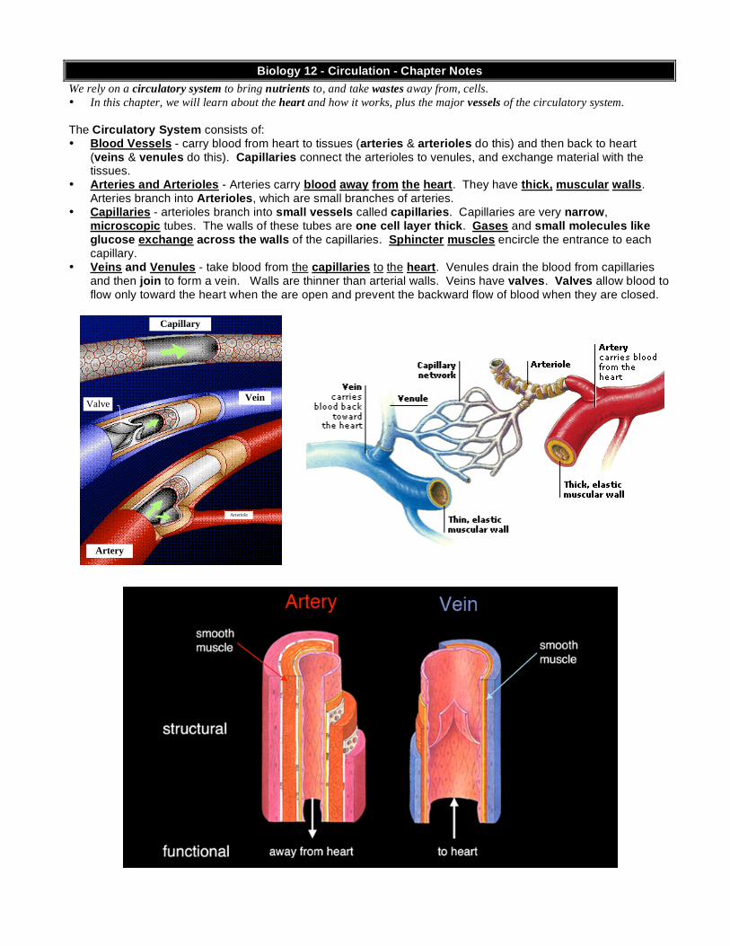
Capillary
Vein
Artery
Valve
Arteriole
Biology 12 - Circulation - Chapter Notes
We rely on a circulatory system to bring nutrients to, and take wastes away from, cells.
• In this chapter, we will learn about the heart and how it works, plus the major vessels of the circulatory system.
The Circulatory System consists of: • Blood Vessels - carry blood from heart to tissues (arteries & arterioles do this) and then back to heart
(veins & venules do this). Capillaries connect the arterioles to venules, and exchange material with the tissues.
• Arteries and Arterioles - Arteries carry blood away from the heart. They have thick, muscular walls. Arteries branch into Arterioles, which are small branches of arteries.
• Capillaries - arterioles branch into small vessels called capillaries. Capillaries are very narrow, microscopic tubes. The walls of these tubes are one cell layer thick. Gases and small molecules like glucose exchange across the walls of the capillaries. Sphincter muscles encircle the entrance to each capillary.
• Veins and Venules - take blood from the capillaries to the heart. Venules drain the blood from capillaries and then join to form a vein. Walls are thinner than arterial walls. Veins have valves. Valves allow blood to flow only toward the heart when the are open and prevent the backward flow of blood when they are closed.
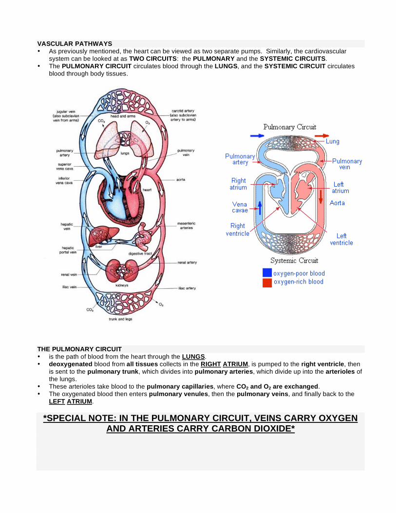
VASCULAR PATHWAYS • As previously mentioned, the heart can be viewed as two separate pumps. Similarly, the cardiovascular
system can be looked at as TWO CIRCUITS: the PULMONARY and the SYSTEMIC CIRCUITS. • The PULMONARY CIRCUIT circulates blood through the LUNGS, and the SYSTEMIC CIRCUIT circulates
blood through body tissues. THE PULMONARY CIRCUIT • is the path of blood from the heart through the LUNGS. • deoxygenated blood from all tissues collects in the RIGHT ATRIUM, is pumped to the right ventricle, then
is sent to the pulmonary trunk, which divides into pulmonary arteries, which divide up into the arterioles of the lungs.
• These arterioles take blood to the pulmonary capillaries, where CO2 and O2 are exchanged. • The oxygenated blood then enters pulmonary venules, then the pulmonary veins, and finally back to the
LEFT ATRIUM.
*SPECIAL NOTE: IN THE PULMONARY CIRCUIT, VEINS CARRY OXYGEN AND ARTERIES CARRY CARBON DIOXIDE*

THE SYSTEMIC CIRCUIT • The systemic circuit includes all blood vessels except those in the Pulmonary Circuit. It takes blood from the
LEFT VENTRICLE, through the tissues & organs of the body, and back to the RIGHT ATRIUM. • in the systemic system, veins carry deoxygenated blood, and arteries carry oxygenated blood. • The systemic circuit contains some blood vessels you should know: • AORTA: the largest artery. Branches of the aorta lead to all major body regions and organs. • SUPERIOR VENAE CAVA: large vein that collects blood from head, chest, and arms. • INFERIOR VENAE CAVA: large vein that collects blood from the lower body regions and organs. • HEPATIC PORTAL VEIN: connects the blood vessels of villi to the liver, carries nutrient rich blood to liver for
processing. • HEPATIC VEIN carries blood from liver to inferior venae cava. Summary of the Major Blood Vessels you Should know:
• SUBCLAVIAN ARTERY AND VEIN -= around clavicle • JUGULAR VEIN - blood from head
• CAROTID ARTERY - neck • MESENTERIC ARTERIES - connect to intestines
• ANTERIOR AND POSTERIOR VENAE CAVA - superior above, inferior below heart
• PULMONARY VEIN - carry oxygenated blood to left atrium
• HEPATIC VEIN - connects to inf. venae cava • HEPATIC PORTAL VEIN - connects intestine with liver
• RENAL ARTERY AND VEIN - connect kidneys and veins • ILIAC ARTERY AND VEIN - leads from aorta into legs
• CORONARY ARTERY AND VEIN • AORTA - largest artery, supplies all tissues
You should also be able to describe the flow of blood around the body through any major organ. For example: • e.g. path of blood to kidneys: Left ventricle aorta renal artery renal arterioles capillaries venules
renal vein venae cava right atrium

THE HEART : 3,000,000,000 beats in an 80 year lifetime! • The left and right side of the heart is divided by the SEPTUM. • On each side are two chambers. The smaller one, located on the top, is called the ATRIUM (plural =
“atria”). The larger one, on the bottom, is called the VENTRICLE. The left ventricle is considerably larger than the right ventricle because the right ventricle only pumps blood to the lungs and the left ventricle must pump to the rest of the body.
• There are VALVES between the atria and ventricles, collectively referred to as ATRIOVENTRICULAR VALVES. These valves control the flow of blood between the chambers, and prevent "backflow."
• Very strong, fibrous strings called the CHORDAE TENDINAE support the valves and prevent them from inverting. The chordae tendinae are firmly attached to muscular projections of the ventricular wall.
• Each ventricle also has a SEMILUNAR VALVE (called that because they look like half-moons) between it and its attached blood vessels. The blood flows through the semilunar vales on its way out of the heart. The right ventricle has a pulmonary semilunar valve and the left side has an aortic semilunar valve.
• The semilunar valves have no chordae tendinae. Why? semilunar valve
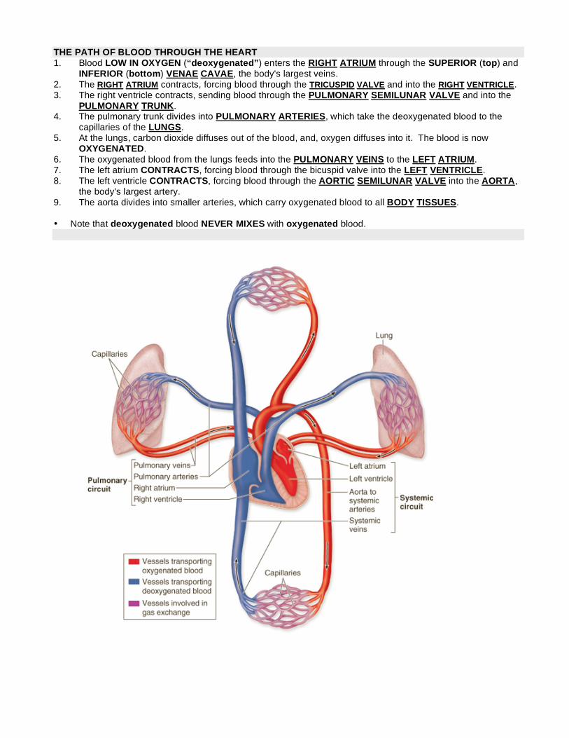
THE PATH OF BLOOD THROUGH THE HEART 1. Blood LOW IN OXYGEN (“deoxygenated”) enters the RIGHT ATRIUM through the SUPERIOR (top) and
INFERIOR (bottom) VENAE CAVAE, the body's largest veins. 2. The RIGHT ATRIUM contracts, forcing blood through the TRICUSPID VALVE and into the RIGHT VENTRICLE. 3. The right ventricle contracts, sending blood through the PULMONARY SEMILUNAR VALVE and into the
PULMONARY TRUNK. 4. The pulmonary trunk divides into PULMONARY ARTERIES, which take the deoxygenated blood to the
capillaries of the LUNGS. 5. At the lungs, carbon dioxide diffuses out of the blood, and, oxygen diffuses into it. The blood is now
OXYGENATED. 6. The oxygenated blood from the lungs feeds into the PULMONARY VEINS to the LEFT ATRIUM. 7. The left atrium CONTRACTS, forcing blood through the bicuspid valve into the LEFT VENTRICLE. 8. The left ventricle CONTRACTS, forcing blood through the AORTIC SEMILUNAR VALVE into the AORTA,
the body's largest artery. 9. The aorta divides into smaller arteries, which carry oxygenated blood to all BODY TISSUES. • Note that deoxygenated blood NEVER MIXES with oxygenated blood.

THE PATH OF BLOOD THROUGH THE HEART: Another look! THE HEARTBEAT • The heartbeat that you can hear (the "lub-DUB" sound) can be divided into TWO PHASES. • First the ATRIA CONTRACT (the "lub" part) while the ventricles are relaxed. • Then the VENTRICLES CONTRACT (the "-DUB" part) while the atria relax. • The actual sound you hear is caused b the vibrations of the heart when the VALVES CLOSE. "lub" = closing
of atrioventricular valves, "DUB" = closing of the semi-lunar valves. • If there is a problem with a valve closing, this can cause HEART MURMURS. • There are two terms that describe contraction and relaxation of heart muscle: SYSTOLE = CONTRACTION of heart muscle. (120 mmHg) DIASTOLE = RELAXATION of heart muscle. (80 mmHg)

WHAT CONTROLS THE HEARTBEAT? • The heart can beat without the brain telling it what to do! That is, the heartbeat is INTRINSIC. How is this
possible? • The heart has its own SPECIAL TISSUE, called NODAL TISSUE, which controls the heartbeat. • There are TWO nodal regions in the heart: 1. SA (sinoatrial) NODE (also called the PACEMAKER): located in the upper back wall of the right atrium.
The SA node INITIATES THE HEARTBEAT by sending out a signal automatically about every 0.85 seconds to make the ATRIA CONTRACT. The SA node is called the “PACEMAKER” because it keeps the beat regular. If it doesn't work, the heart will beat irregularly. This can be corrected by implanting an ARTIFICIAL PACEMAKER.
2. AV (atrioventricular) NODE: found in the base of the right atrium near the septum. The SA node sends its signal along fibers to the atria and also to the AV node. When the pulse sent out by the SA node reaches the AV node, the AV node relays the signal along special conducting fibers called PURKINJE FIBERS. These fibers take the message to the VENTRICLES, and cause them to contract. The contraction of the ventricles begins at the base of the heart and moves up like a wave.
• While the heart can keep a steady beat on its own, the how fast it goes (heart rate) is under NERVOUS
CONTROL. Specifically, there is a HEART-RATE CENTER in the brain (to be precise, in the MEDULLA OBLANGATA, which is an evolutionary ancient part of the brain right on top of the spinal cord).
• This center can speed up or slow down the heart rate. Various factors, such as stress, oxygen levels, and blood pressure determine how the medulla oblongata will affect heart rate.

CORONARY ARTERIES and VEINS • these are vitally important blood vessels that supply blood to the heart muscle itself (the heart does not use
the blood in its inner chambers. • arteries branch off the aorta just above the aortic semilunar valve, and lie on the outside of the heart. • coronary veins empty into the right atrium. if a coronary artery becomes plugged (e.g. with cholesterol), and blood is not supplied to part of the heart, a
heart attack occurs.
Angioplasty CORONARY BYPASS SURGERY: segments of leg veins are grafted between the aorta and coronary vessels, in order to bypass a blockage. Two to four such bypasses may be performed in a single operation.

Pulse, Blood Pressure • PULSE: the alternate expanding and recoiling of an arterial wall that can be felt in any artery that runs
near the surface of the body. Radial artery in wrist, carotid artery in neck are common places to check. Pulse rate indicates the rate of heartbeat.
• BLOOD PRESSURE: the pressure of the blood against the wall of a vessel, created by the pumping action of
the heart.
• HYPOTENSION: lower blood pressure than usual. • HYPERTENSION: higher blood pressure than normal. Measuring Blood Pressure • Measure blood pressure with an instrument called a SPHYGMOMANOMETER. • SYSTOLIC BLOOD PRESSURE: the highest arterial pressure reached during ejection of blood from the
heart. • DIASTOLIC BLOOD PRESSURE: lowest arterial pressure. Occurs while the ventricles are relaxing. • Normal resting blood pressure is 120 mm Hg over 80 mm Hg in brachial artery of arm (120/80). Of course,
blood pressure decreases with distance from left ventricle. It is higher in the arteries than in the arterioles, for example.

The Lymphatic System • The lymphatic system is another vascular system in your body. It has its own veins
and capillaries but is connected back with the cardiovascular system • Basically, the lymphatic system takes up excess tissue fluid from the tissues and
returns it to the cardiovasular system. • It is a one-way system that starts in the tissues and empties into the cardiovascular
system. • Lymph vessels consist of LYMPH CAPILLARIES and LYMPH VEINS (which have
valves). Note: there are no lymph “arteries” since there is no “pump” in this system. Once fluid enters the lymph vessels it is called LYMPH.
• Lymph is collected in vessels that join to right and left subclavian veins. (called the THORACIC DUCT) • Lacteal: blind ends of lymph vessels in villi of the small intestine. • LYMPH NODES: small oval or round structures that occur at places along the lymph vessels. They
produce and store white blood cells, and filter lymph of damaged cells and debris. • SPLEEN: located behind the stomach. Contains white blood cells and stores blood.

Differences between Fetal and Adult Systems • Heart develops in 3rd and 4th weeks in uterus. The fetus, first of all, can’t breathe air inside the womb, so
sending blood to the lungs won’t do much good. Likewise, the fetus must get all its nutrients from Mom, as well as let her take care of its wastes. Obviously, some serious plumbing problems must be solved.
• To solve these problems, the fetus has FOUR FEATURES not present in adults: 1. OVAL OPENING (foramen ovale): opening between the two atria, covered by a flap that acts like a valve.
Some of the blood bypasses the pulmonary circuit. • If the oval opening doesn’t close after birth, it can cause mixing of blood and “blue babies”. Correct with
open heart surgery. 2. ARTERIAL DUCT: (ductus arteriosus) connects pulmonary artery and aorta. Like the oval opening, the
arterial duct’s function is to bypass the pulmonary circuit. 3. UMBILICAL ARTERIES AND VEINS: vessels that travel to and from PLACENTA (a membrane shared by
the mother and baby across which gases, nutrients, and wastes are exchanged). The umbilical arteries are grafted to the iliac arteries.
4. VENOUS DUCT (ductus venosus): connects umbilical vein to the venae cava to bring the blood back to the baby’s heart. It attaches right at the babies liver, but bypasses most of the liver. This is why chemicals ingested by the mother can seriously affect the baby!

BIOLOGY 12 - CHAPTER 11 - BLOOD - CHAPTER NOTES • Blood is a liquid connective tissue. Blood functions in a) TRANSPORT (of gases, wastes, and nutrients) b)
CLOTTING (to seal injuries) c) INFECTION FIGHTING. Average person has about 5 to 6 liters of blood.
Blood is divided into TWO MAIN PARTS: Plasma and Formed Elements
A. PLASMA (the liquid portion of blood) - makes up about 55% of blood volume. Contains water and organic and inorganic substances (proteins, gases, salts, nutrients, wastes).
Looks like apple juice
Plasma Constituent Function
Water Maintains blood volume and transports molecules
Plasma Proteins a. Albumin b. Fibrinogen c. Globulins
All maintain blood osmotic pressure & pH Transport Clotting Fight Infection
Gases a. Oxygen b. CO2
Cellular Respiration End product of metabolism
Nutrients: Fats, glucose, amino acids, etc.
Food for cells
Salts Maintain blood osmotic pressure/pH, aid metabolism
Wastes End products of metabolism
Hormones, vitamins etc. Aid metabolism

B. FORMED ELEMENTS: the "solid part" of blood, consists of the following parts. a. RED BLOOD CELLS (ERYTHROCYTES) Contain Hemoglobin. Transport oxygen, formed in bone marrow. Over 95% of formed elements are erythrocytes. There are close to 30 trillion blood cells in an adult. In humans, red blood cells do have nuclei. The RBC shape is called biconcave b. WHITE BLOOD CELLS (LEUKOCYTES) Fight infection, formed in bone marrow and lymphoid tissue. c. PLATELETS (THROMBOCYTES) Function in blood clotting, formed from a burst Megakaryocyte.

Blood Proteins • Are required for the transport of many molecules. For example, cholesterol is a lipid that is insoluble in
plasma. It must be carried by proteins. • Blood proteins also contribute to osmotic pressure. Hemoglobin • O2 is carried by Hemoglobin. • Hemoglobin is always contained within red blood cells. Since hemoglobin is a red pigment, red blood cells
appear red. This colour can change based on what the hemoglobin is attached to.
Tissue-Blood Exchange • The main answer lies in battle of blood pressure versus osmotic pressure. The pressure of blood in blood
vessel would tend to push molecules out of the blood. Osmotic pressure is the opposing force trying to force molecules into the blood. Osmotic pressure is basically constant, but blood pressure varies considerable around a capillary bed. This causes some natural movement of molecules.
• At the arterial side of a capillary, blood pressure is higher than the osmotic pressure and therefore water, oxygen and glucose tend to leave the bloodstream.
• At the venous end of a capillary, the osmotic pressure is higher than the blood pressure and, therefore, water, ammonia, and carbon dioxide tend to enter the bloodstream.
BP vs Velocity vs X-sectional Area

BLOOD CLOTTING • After an injury, coagulation "or clotting" takes place to prevent
excessive blood loss. • This requires the action of 1) platelets 2) prothrombin, and 3)
fibrinogen. INFECTION FIGHTING: another major function of blood • The body's first line of defense against invading pathogens like
bacteria and viruses is the skin. • The second line of defense is the blood: specifically, white blood cells and antibodies. WHITE BLOOD CELLS • White blood cells multiply when the body is invaded and can manufacture large quantities antibodies. • White blood cells can also engulf foreign invaders. ANTIBODIES: very specific proteins that attach to invading pathogens • Antibodies combine with antigens in such a way that the antigens are inactivated. Each
antibody fits its antigen like a lock and key. • An individual is immune to an antigen if he/she has it. • The blood in the individual contains WBC that can remain in the system for years, ready to
produce antibodies if that antigen is detected. • Exposure to the antigen, either naturally or by way of a vaccine, can cause active immunity to develop. Ab-Ag interactions