Characterization ofRoot Surface and Endorhizosphere ...aem.asm.org/content/56/8/2462.full.pdf ·...
9
Vol. 56, No. 8 APPLIED AND ENVIRONMENTAL MICROBIOLOGY, Aug. 1990, p. 2462-2470 0099-2240/90/082462-09$02.00/0 Copyright © 1990, American Society for Microbiology Characterization of Root Surface and Endorhizosphere Pseudomonads in Relation to Their Colonization of Roots RON VAN PEER,'* HELMA L. M. PUNTE,1 LETTY A. DE WEGER,2 AND BOB SCHIPPERS' Willie Commelin Scholten Phytopathological Laboratory, Department of Plant Pathology, State University of Utrecht, Javalaan 20, 3742 CP Baarn,1 and Department of Plant Molecular Biology, Leiden University, 2311 VJ Leiden,2 The Netherlands Received 2 March 1990/Accepted 11 May 1990 An extensive colonization of the endorhizosphere by fluorescent pseudomonads was observed in tomato plants grown on artificial substrates. These studies reveal that a significantly higher percentage of pseudomo- nads obtained from the endorhizosphere (30%) reduced plant growth than those obtained from the root surface (4%). Lipopolysaccharide patterns, cell envelope protein patterns, and other biochemical characteristics indicated that Pseudomonas isolates obtained from the endorhizosphere are distinct from Pseudomonas isolates obtained from the root surface. Isolates from the endorhizosphere especially were able to recolonize the endorhizosphere of both sterile and nonsterile tomato roots. The ability of the endorhizosphere isolates to colonize the endorhizosphere significantly correlated with their agglutination by tomato root agglutinin but did not correlate with chemotaxis to seed exudates of tomato. No correlation between colonization of the endorhizosphere and agglutination by root agglutinin could be demonstrated for the root surface isolates. We propose that agglutination of specific Pseudomonas strains by root agglutinin is of importance in the initial phase of adherence of bacteria to the root surface. Previous studies demonstrated the presence of pseudo- monads both on the root surface and in the endorhizosphere of tomatoes (18). Pseudomonads were predominant in the endorhizosphere of tomatoes grown on artificial substrates such as rockwool or nutrient film cultures, compared with tomatoes grown in soil. Bacteria producing phytotoxic metabolites (particularly gaseous components) in the en- dorhizosphere of hydroponically grown plants are deleteri- ous to plant growth, because their metabolites are entrapped in the root tissue and do not easily diffuse, thereby exhibiting more dramatic effects on the plant physiology than metabo- lites produced on the root surface (B. Schippers, P. A. H. M. Bakker, A. W. Baker, and R. van Peer, Proc. Brighton Crop Prot. Conf., p. 611-614, 1988). The present study was initiated to elucidate the question of whether populations of pseudomonads inhabiting the endorhizosphere are different from those inhabiting the root surface. Chemotaxonomic characteristics such as lipopolysaccharide patterns and cell envelope protein patterns were used in addition to other biochemical characteristics. The observed differences in the colonization of the endorhizosphere between Pseudomonas spp. isolated from either the root surface or the endorhizo- sphere are related to specific properties, such as their interactions with the plant root agglutinins, adherence to the plant root, and chemotaxis. MATERIALS AND METHODS Samplings. Pseudomonads were isolated and selected from roots of glasshouse tomatoes (Lycopersicon esculen- tum Mill cv. Moneymaker or cv. Turbo) grown on rock- wool, on a nutrient film culture, or in a hydroponic system (18). A sample (0.3 g) of root pieces was shaken vigorously for 1 min in 5 ml of 0.1 M MgSO4 and dilution plated on modified King's medium B (KB) (10) to enumerate root * Corresponding author. surface-inhabiting pseudomonads. Root pieces were surface sterilized with H202 (10%; 15 s), rinsed three times with MgSO4 (0.1 M), ground with a mortar and pestle, and then homogenized and dilution plated to enumerate the en- dorhizosphere-inhabiting pseudomonads. Colonies representing different morphological types were randomly selected and purified. A total of 213 pseudomo- nads were collected in this way. Bacterial culture conditions. A loop of bacteria from a 24-h-old culture was suspended in 0.1 M MgSO4. A total of 500 [LI of this suspension was added to KB agar plates and incubated for 24 h at 27°C. These cultures were suspended in sterile distilled water (SDW) and were used for inoculation of plants (108 cells ml-'). For isolation of lipolysaccharides (LPS), a stationary-phase culture was diluted 100-fold in KB and cultivated for 24 h at 28°C. Screening of pseudomonads for effects on plant growth. For the screening of bacterial effects on plant growth, plastic containers (200 ml) were used. They were filled with a nutrient solution [0.6 g of Nutriflora-T (Windmill Holland B.V.) liter-' and 10 g of Ca(NO3)2 liter-']. Each container was covered with a petri dish lid with seven holes, each of which held a 1-ml pipette tip filled with sterilized vermicu- lite. Tomato seeds were sterilized by rinsing with 1% sodium hypochlorite for 1 min. After being rinsed with SDW, the seeds were germinated on sterile filter paper in a petri dish. One germinated seed was placed on the vermiculite in each pipette tip and covered with a thin layer of vermiculite. Seedlings were bacterized with 0.5 ml of a bacterial suspension (108 cells ml-1) from cultures grown for 24 h at 27°C. After 14 days of growth in a growth cabinet at 21°C, fresh weights of plants were recorded for four replicates of each treatment. Isolates with statistically significant effects on seedling growth and other randomly chosen isolates up to a total of either 50 root surface isolates or 50 endorhizo- sphere isolates were tested twice for their effects on growth of 5-week-old tomato plants grown in a hydroponic system 2462 on June 18, 2018 by guest http://aem.asm.org/ Downloaded from
Transcript of Characterization ofRoot Surface and Endorhizosphere ...aem.asm.org/content/56/8/2462.full.pdf ·...
Vol. 56, No. 8APPLIED AND ENVIRONMENTAL MICROBIOLOGY, Aug. 1990, p. 2462-24700099-2240/90/082462-09$02.00/0Copyright © 1990, American Society for Microbiology
Characterization of Root Surface and EndorhizospherePseudomonads in Relation to Their Colonization of RootsRON VAN PEER,'* HELMA L. M. PUNTE,1 LETTY A. DE WEGER,2 AND BOB SCHIPPERS'
Willie Commelin Scholten Phytopathological Laboratory, Department of Plant Pathology,State University of Utrecht, Javalaan 20, 3742 CP Baarn,1 and Department of Plant
Molecular Biology, Leiden University, 2311 VJ Leiden,2 The Netherlands
Received 2 March 1990/Accepted 11 May 1990
An extensive colonization of the endorhizosphere by fluorescent pseudomonads was observed in tomatoplants grown on artificial substrates. These studies reveal that a significantly higher percentage of pseudomo-nads obtained from the endorhizosphere (30%) reduced plant growth than those obtained from the root surface(4%). Lipopolysaccharide patterns, cell envelope protein patterns, and other biochemical characteristicsindicated that Pseudomonas isolates obtained from the endorhizosphere are distinct from Pseudomonas isolatesobtained from the root surface. Isolates from the endorhizosphere especially were able to recolonize theendorhizosphere of both sterile and nonsterile tomato roots. The ability of the endorhizosphere isolates tocolonize the endorhizosphere significantly correlated with their agglutination by tomato root agglutinin but didnot correlate with chemotaxis to seed exudates of tomato. No correlation between colonization of theendorhizosphere and agglutination by root agglutinin could be demonstrated for the root surface isolates. Wepropose that agglutination of specific Pseudomonas strains by root agglutinin is of importance in the initialphase of adherence of bacteria to the root surface.
Previous studies demonstrated the presence of pseudo-monads both on the root surface and in the endorhizosphereof tomatoes (18). Pseudomonads were predominant in theendorhizosphere of tomatoes grown on artificial substratessuch as rockwool or nutrient film cultures, compared withtomatoes grown in soil. Bacteria producing phytotoxicmetabolites (particularly gaseous components) in the en-dorhizosphere of hydroponically grown plants are deleteri-ous to plant growth, because their metabolites are entrappedin the root tissue and do not easily diffuse, thereby exhibitingmore dramatic effects on the plant physiology than metabo-lites produced on the root surface (B. Schippers, P. A. H. M.Bakker, A. W. Baker, and R. van Peer, Proc. Brighton CropProt. Conf., p. 611-614, 1988). The present study wasinitiated to elucidate the question of whether populations ofpseudomonads inhabiting the endorhizosphere are differentfrom those inhabiting the root surface. Chemotaxonomiccharacteristics such as lipopolysaccharide patterns and cellenvelope protein patterns were used in addition to otherbiochemical characteristics. The observed differences in thecolonization of the endorhizosphere between Pseudomonasspp. isolated from either the root surface or the endorhizo-sphere are related to specific properties, such as theirinteractions with the plant root agglutinins, adherence to theplant root, and chemotaxis.
MATERIALS AND METHODS
Samplings. Pseudomonads were isolated and selectedfrom roots of glasshouse tomatoes (Lycopersicon esculen-tum Mill cv. Moneymaker or cv. Turbo) grown on rock-wool, on a nutrient film culture, or in a hydroponic system(18). A sample (0.3 g) of root pieces was shaken vigorouslyfor 1 min in 5 ml of 0.1 M MgSO4 and dilution plated onmodified King's medium B (KB) (10) to enumerate root
* Corresponding author.
surface-inhabiting pseudomonads. Root pieces were surfacesterilized with H202 (10%; 15 s), rinsed three times withMgSO4 (0.1 M), ground with a mortar and pestle, and thenhomogenized and dilution plated to enumerate the en-dorhizosphere-inhabiting pseudomonads.
Colonies representing different morphological types wererandomly selected and purified. A total of 213 pseudomo-nads were collected in this way.
Bacterial culture conditions. A loop of bacteria from a24-h-old culture was suspended in 0.1 M MgSO4. A total of500 [LI of this suspension was added to KB agar plates andincubated for 24 h at 27°C. These cultures were suspended insterile distilled water (SDW) and were used for inoculationof plants (108 cells ml-'). For isolation of lipolysaccharides(LPS), a stationary-phase culture was diluted 100-fold in KBand cultivated for 24 h at 28°C.
Screening of pseudomonads for effects on plant growth. Forthe screening of bacterial effects on plant growth, plasticcontainers (200 ml) were used. They were filled with anutrient solution [0.6 g of Nutriflora-T (Windmill HollandB.V.) liter-' and 10 g of Ca(NO3)2 liter-']. Each containerwas covered with a petri dish lid with seven holes, each ofwhich held a 1-ml pipette tip filled with sterilized vermicu-lite. Tomato seeds were sterilized by rinsing with 1% sodiumhypochlorite for 1 min. After being rinsed with SDW, theseeds were germinated on sterile filter paper in a petri dish.One germinated seed was placed on the vermiculite in eachpipette tip and covered with a thin layer of vermiculite.
Seedlings were bacterized with 0.5 ml of a bacterialsuspension (108 cells ml-1) from cultures grown for 24 h at27°C. After 14 days of growth in a growth cabinet at 21°C,fresh weights of plants were recorded for four replicates ofeach treatment. Isolates with statistically significant effectson seedling growth and other randomly chosen isolates up toa total of either 50 root surface isolates or 50 endorhizo-sphere isolates were tested twice for their effects on growthof 5-week-old tomato plants grown in a hydroponic system
2462
on June 18, 2018 by guesthttp://aem
.asm.org/
Dow
nloaded from

ROOT SURFACE AND ENDORHIZOSPHERE PSEUDOMONADS 2463
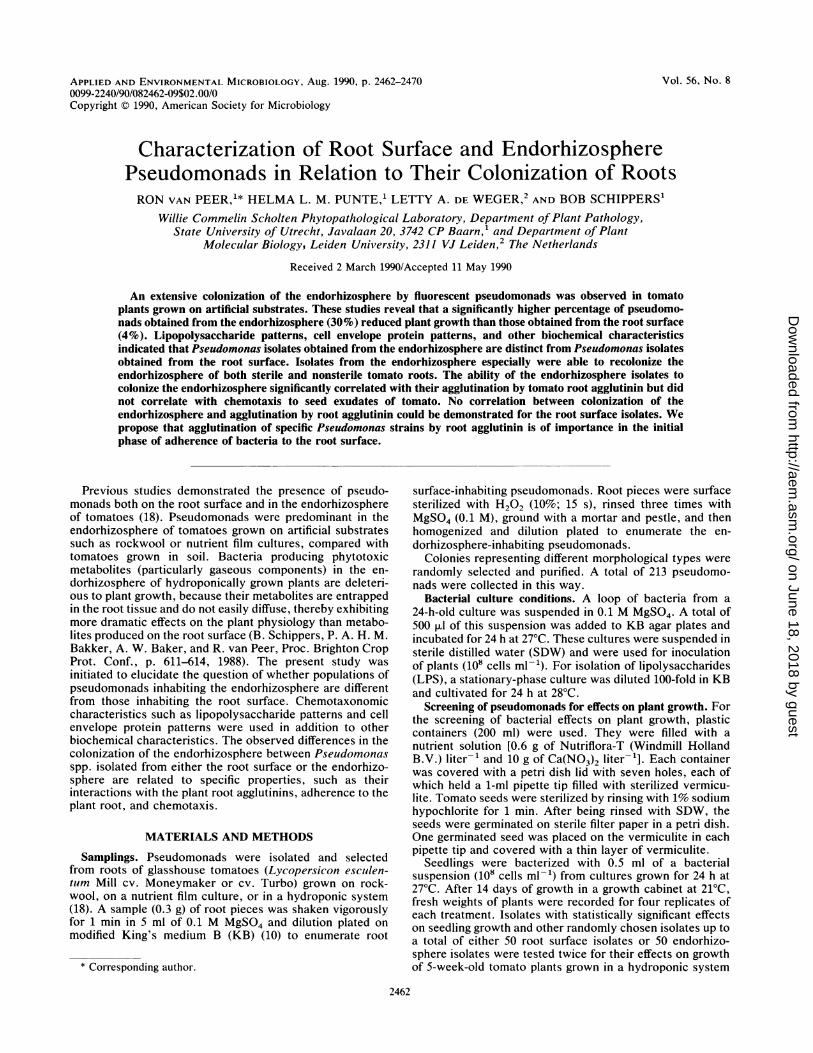
FIG. 1. Classes of agglutination of Pseudomonas cells by rootagglutinin of tomato plants. (A) Class 0; (B) class 1; (C) class 2; (D)class 3. See text for definition of classes.
(18). For each treatment, 10 plants were bacterized by theaddition of a bacterial suspension to the nutrient solution(final concentration, 106 cells ml-').
Antibiotic-resistant mutants. Spontaneous rifampin-resist-ant mutants of some isolates were obtained by streaking wildtypes of the isolates on KB agar plates supplemented with
% Tested isolates
0 6 10 15 20 25 30 35 40
150 mg of rifampin (KBrif) liter-1. Colonies that grew on
these plates were purified and cultured repeatedly on KBwith increasing concentrations of rifampin, up to 500 mg
liter-'. These mutants were compared with their wild typesregarding their fluorescence, growth rates, production ofC2H4 or HCN, and in vitro antagonism with some referencestrains (WCS417 Rifr, WCS358, and WCS374). None ofthese mutants differed from their parental strains.
Characterization of pseudomonads. (i) LPS and cell enve-
lope protein patterns. Cell envelopes were obtained bydifferential centrifugation after disruption of the cells byultrasonic treatment (12). Cell envelope samples containingapproximately 20 p,g of protein were solubilized in standardsample mixture and subjected to sodium dodecyl sulfate-polyacrylamide gel electrophoresis as described previously(12). Cells were stained with fast green. For the analysis ofLPS, the samples were first subjected to treatment withproteinase K (17), after which 10-fold dilutions were appliedto each lane. Gels were then stained with silver reagent (17).
(ii) Biochemical characterization. Representative isolatesfrom every group with reproducible stimulating, inhibiting,or neutral effects on plant growth were chosen for furtherexperimentation. A total of 26 isolates were identified bio-chemically by the methods described by Schaad (13) andStanier et al. (16). Results were scored positive or negative,and corresponding characteristics were expressed as clus-ters.Root colonization. Strains were tested for their potential to
colonize the root surface or endorhizosphere of stenrletomato roots. Roots of sterile seedlings were soaked for 5min in an aqueous suspension of bacterial strains (108 cellsml-') or a mixture of 100 isolates selected from either theroot surface or the endorhizosphere. These isolates were
mixed in a ratio of 1:1 (final concentration, 106 cells of each
% Tested isolates
5
4
3
2
% Deviation (fresh weight) of controls
-50-45-40-35-30-25-20-15-10 -5 0 6 10 15 20 25 30 35 40
% Deviation (fresh weight) of controls
FIG. 2. Effects of pseudomonads selected from the root surface (A) and from the endorhizosphere (B) on tomato seedlings. Controls were
not inoculated with pseudomonads.
V~~~~~B0
.0
10
0~~~~~I IUUEI
VOL. 56, 1990
1
on June 18, 2018 by guesthttp://aem
.asm.org/
Dow
nloaded from

2464 VAN PEER ET AL.
A
9B7k--e:
66k --
54k --
45k -1
24k -X
14k -1
B
97k--
66k-.*
55k-
45k-
24k-
14k -
2 3 4 5 6 7 8 9 10
FIG. 3. Cell envelope protein patterns of Pseudomonas isolates obtained from the root surface (A) and the endorhizosphere (B). Lanes:1, RS94; 2, RS194; 3, RS117; 4, RS112; 5, RS111; 6, RS8; 7, RS17; 8, RS12; 9, RS19; 10, RS3; 11, En4; 12, En413; 13, En25; 14, En27; 15,En23; 16, En415; 17, En7; 18, En401; 19, En3; 20, En89; 21, En59; 22, Enll4; 23, En416; 24, EnlO5. All isolates were run on the same gel.
isolate ml-1). Roots were then rinsed three times in SDWand incubated for different lengths of time in a moist petridish. The total root length of five seedlings was measured.Roots were macerated in 3 ml of 0.1 M MgSO4 with orwithout root surface disinfection (10% H202; 15 s) anddilution plated on KB. Contamination of roots was checkedby dilution plating of nonbacterized treatment samples.
Plates were incubated for 48 h at 27°C. Mean CFU centime-ter of root-' were determined by averaging log populationdensities of six replicates per treatment.
In addition, rifampin-resistant (Rif) mutants of isolatesRS48, RS17, En25, and En415 were tested for their root-colonizing capacity in a nonsterile nutrient film system (13).Tomato plants were bacterized with 10 ml of a bacterial
_r 'W two-, B
A
1 2 3 4 5 6 7 8 9 10 11 12 13 14 15 16 17 18 19 20 21 22 23 24
FIG. 4. Silver-strained LPS patterns of proteinase K-treated cell envelopes after gel electrophoresis of Pseudomonas isolates obtainedfrom the root surface (A) or from the endorhizosphore (B). Lane numbers correspond with those defined in the legend to Fig. 3. All isolateswere run on the same gel.
APPL. ENVIRON. MICROBIOL.
11 12 13 14 15 16 17 15 19 20 21 ZZ 23 Z4
on June 18, 2018 by guesthttp://aem
.asm.org/
Dow
nloaded from

ROOT SURFACE AND ENDORHIZOSPHERE PSEUDOMONADS 2465
suspension (108 CFU ml-1). Root pieces (0.3 g) were sam-
pled 2 weeks after bacterization to determine colonization ofthe rhizosphere by the isolates tested.Adherence to the root surface. Roots of sterile tomato
seedlings were immersed in bacterial suspensions (108 cellsml-1) for 30 min. Preliminary results demonstrated thatincubation for a longer period of time did not increase thenumber of adhering cells. Adherence of cells was quantitatedby the method described by Anderson et al. (3). Intact rootswere sequentially transferred under sterile conditionsthrough four washes in 10 ml of water and then homoge-nized. Cells present on the root surface after four washeswere considered to be firmly adhered. After each transfer,the tube was agitated for 60 s by a Vortex. The washed cellsand homogenates were diluted and plated (100 ,u) onto KBor KBrif. Each treatment was replicated six times, with tworoots per replicate.
Chemotaxis. In vitro chemotaxis towards seed exudates oftomato was assessed by using a modification of the methodof Adler (1). A total of 10 surface-sterilized tomato seedswere placed in 50 ml of SDW in 100-ml Erlenmeyer flasksand incubated on a rotary shaker (50 rpm) for 5 days at 21°C.The liquid containing the seed exudates was lyophilized andresuspended in 10 ml of SDW. Six capillary tubes (sterilizedin 70% ethanol) containing seed exudate or phosphate buffer(0.01 M; pH 7.0) were sealed at one end and placed in a
cuvette with an aqueous suspension of the individual isolates(108 CFU ml-1). The number of bacterial cells entering thecapillary tubes after 30 min was determined by plating theircontents onto KB. Chemotaxis was expressed as the ratioCFU entering the capillary tube with seed exudate/CFUentering the tube without seed exudate.
Agglutination. Tomato seeds were either grown on vermic-ulite and harvested after 21 days or grown on hydroponicculture and harvested after 42 days. Root agglutinin was
obtained by the method of Anderson (2). Roots were care-
fully removed from either the vermiculite or the nutrientsolution. Roots grown in vermiculite were swirled by handfor 15 min in 200 ml of SDW in a 1-liter beaker, whereas thesterile nutrient solution was used to obtain agglutinins fromthe roots grown under sterile conditions. Roots were thenallowed to drain for 10 min. The root wash was filtered(Whatman no. 1 filter paper) and centrifuged at 10,000 x g
for 20 min at 4°C. The supernatant was mixed with 1.5 g (dryweight) of prehydrated CM-Sephadex C-50 (Pharmacia FineChemicals) and gently stirred with a magnetic stirrer for 15min. The slurry was filtered and treated similarly with 1.5 g
(dry weight) of prehydrated DEAE-Sephadex A-50 (Pharma-cia Fine Chemicals). The filtrate was lyophilized and sus-
pended in 3 ml of SDW. Ethanol (9 ml; 96%) was added, thefiltrate was then held overnight at 4°C, and the precipitatewas collected by centrifugation. SDW was added to theprecipitate (1 ml g [fresh weight] of root-) and allowed tostand for 30 min at 4°C. Insoluble substances were removedby centrifugation at 10,000 x g for 15 min at 4°C. Rootagglutinins were also collected from 21-day-old sterile to-mato plants. Agglutination was examined microscopically.The activity was ranked from 0 to 3 on the following scale: 0,no agglutination; 1, weak agglutination of only two or threecells; 2, clear agglutination of several clusters of three ormore cells; and 3, complete agglutination of the majority ofcells (Fig. 1).
RESULTSEffects on plant growth. In the initial screening tests, 4% of
the 101 tested Pseudomonas isolates obtained from the root
TABLE 1. Clusters of Pseudomonas isolates that were obtainedfrom the root surface or the endorhizosphere based on LPS and
cell envelope protein patterns or biochemical characteristics
Clusters of isolates based on:Cluster no.
LPS and CEP' patterns Biochemical characteristics
1 RS94, RS194, En4, RS94, RS194, En3, En4O3,En4O3, En415 Enll4
2 RS8, RS111, RS112, RS8, RS111, RS112, RS117,RS117 RS48b
3 En401, En7 En401, En7, En9lb4 En23, En25, En27 En23, En25, En27 (En4)5 RS12, RS17 RS19, En4166 En89, En59Noncluster- RS3, RS19, EnlO5, RS3, RS12, RS17, En89,
ing iso- Enll4, En416, En3 EnlO5, En415, En59lates"
a CEP, Cell envelope protein.h LPS and cell envelope protein patterns were not determined.Each isolate demonstrated unique LPS and cell envelope protein patterns
or biochemical properties.
surface significantly reduced plant weight, whereas 7% dem-onstrated significant plant growth stimulation. In contrast,30% of the 112 Pseudomonas isolates obtained from theendorhizosphere reduced plant growth and only 2% stimu-lated plant growth (Fig. 2). In the second screening, only 2%of the root surface isolates and 18% of the endorhizosphereisolates showed reduced plant growth. For both populations,2% of the isolates stimulated plant growth.
Strain characterization. (i) LPS and cell envelope proteinpatterns. LPS and cell envelope protein patterns of rootsurface and endorhizosphere Pseudomonas isolates werevery diverse (Fig. 3 and 4). Of the 24 isolates, 6 demon-strated unique LPS and cell envelope protein patterns(Table 1). Six clusters consisting of two or more isolateswith identical or very similar patterns were distinguished(Table 1). In most cases, both LPS and cell envelope pro-ein patterns did not differ between individual isolates ofa cluster. Exceptions were isolate En3 (from the endo-hizosphere) of which the LPS pattern but not the cellenvelope protein pattern was clearly different from that ofisolates En89 and En59 and isolates RS12 and RS17 (fromthe root surface) which had the same LPS patterns as butdifferent cell envelope protein patterns from En89 and En59.On the basis of LPS and cell envelope protein patterns,isolates obtained from the root surface differed from thePseudomonas isolates obtained from the endorhizosphere,except for isolates RS94 and RS194; their patterns wereidentical to those of isolates En4, En4O3, and En415 (Table1).
(ii) Biochemical characterization. Biochemical characteris-tics of Pseudomonas isolates are summarized in Table 2. Ofthe 26 isolates, 7 exhibited unique characteristics that did notcorrespond with each other and that were not closely relatedto one of the clusters of other strains (Table 1). Five differentclusters of isolates with closely related biochemical charac-teristics could be distinguished (Table 1). Except for isolatesRS94 and RS194, the biochemical characteristics of whichwere identical to those of isolates EnlO5 and Enll4, identi-cal types were not found between isolates obtained from theroot surface and those obtained from the endorhizosphere.Isolates EnlO5 and RS12 also have very similar characteris-tics.Root colonization. Colonization of the total rhizosphere
(root surface and endorhizosphere) and of the endorhizo-
VOL. 56, 1990
on June 18, 2018 by guesthttp://aem
.asm.org/
Dow
nloaded from

2466 vAN PEER ETAL.APLENIN.Mcoo.
TABLE 2. Biochemical characterization of Pseudomonas isolates obtained from the root surface or the endorhizosphereResult or characteristic for isolate:
ParameterC,,C/~~ 00 00 00 00 00 A -P- D m m tTloo-00 0 o.C) CD))CD
Effects on plant growth' 0 0 0 0 + - 0 0 0 - 0 0 0 0 0 0 - +0 0
Fluorescence + + + + + + - - + + + + + + + + + + + + + + + + - +
+4 -4- + + ±- + + + +
Pectolytic activity
Arginine dihydrolase
Levan production
Production of poly-p3-hydroxybutyrate
Growth at:
40C
270C
370C
400C
±1 +
4-
+ + +
+.4+- + +- + +
-+ -4
Gelatine hydrolysis
Hypersensitive reaction
in tobacco by":
Cells
Supernatant
Denitrification
Soft rot in potato
Growth on various
C sources
Mannitol
Inositol
Trehalose
Sucrose
Cellobiose
Sorbitol
Calcium lactate
L-Arabinose
L-Rhamnose
L-Tartrate
Erythritol
+ + + + ±4 + + + + + + + + + + + + +
See Materials and Methods. Effects are distinguished by analysis of variance as inhibitory (-), promoting (+), or neutral (0).I Cells, Leaves injected with viable cells; Supernatant, leaves injected with the equivalent of cell-free culture supernatant.
sphere of sterile tomato roots by individually tested isolates
is presented in Table 3. After 2 h, significant differences in
root colonization were observed between many isolates.
However, after 6 h, only a few isolates significantly differed
in colonization of the total rhizosphere. In contrast, after 6 h
of incubation, many differences were still obvious with
respect to the colonization of the endorhizosphere (Table 3).The mean numbers of CFU centimeter of root-' obtained
from the endorhizosphere after inoculation with root surface
or endorhizosphere isolates were significantly different. The
same tendency was found when a mixture of 100 root surface
isolates or 100 endorhizosphere isolates was used (Fig. 5).This suggests that colonization of the endorhizospheremainly occurs within a specific group of pseudomonads.
Results from experiments performed under gnotobioticconditions cannot simply be applied to nonsterile conditions,
since properties such as antagonism and rhizosphere com-
petence, for example, are not measured in those experi-ments. Four isolates (RS48 Rif, RS17 Rif, En25 Rifr, and
En415 RifT) differing in their root colonization (Table 3) and
agglutination patterns (Table 4) were tested in a nonsterile
nutrient film system for their capacity to colonize the en-
dorhizosphere. All four isolates colonized the root surface,but only the isolates obtained from the endorhizospherecould be enumerated from both the root surface and the
endorhizosphere. In this system, the two isolates obtained
from the root surface could not be detected in the endorhizo-
sphere (Table 4).Adherence to the root surface. After the first wash, most of
the cells were removed from the root surface; thereafter,numbers in the washes slowly decreased with the wash steps
(Table 5). In particular, strain En7, and to some lesser extent
Oxidase
APPL. ENVIRON. MICROBIOL.
on June 18, 2018 by guesthttp://aem
.asm.org/
Dow
nloaded from

ROOT SURFACE AND ENDORHIZOSPHERE PSEUDOMONADS 2467
TABLE 3. Colonization of the rhizosphere of sterile tomato rootsby Pseudomonas isolates selected from the root surface
and from the endorhizosphere
Log CFU cm of root-' from:Pseudomonas Total rhizosphere Endorhizosphereisolates from:
0.5h 2h 6h 0.5h 2h 6h
Root surfaceRS94 5.5 6.2 6.3 3.3 4.9 4.2RS194 4.9 5.5 5.8 1.9 2.6 3.0RS117 4.3 5.8 5.9 1.5 3.7 3.6RS112 5.0 5.5 5.8 2.3 2.4 2.4RS111 5.4 5.7 5.8 2.3 3.0 3.2RS8 5.4 6.0 6.3 1.9 2.8 3.1RS17 4.8 5.6 5.3 1.1 1.3 1.5RS12 5.0 5.8 5.9 2.2 2.9 3.1RS19 5.2 5.4 5.7 1.8 3.0 3.1RS3 5.5 5.9 6.4 2.9 3.6 4.0RS48 4.1 5.5 6.0 2.4 2.8 2.9Mean 5.0 5.7 5.9 2.la 3 oa 3. 1"
EndorhizosphereEn4 4.7 5.8 6.0 3.0 4.4 5.0En403 5.0 5.4 5.6 2.2 3.1 3.3En23 4.4 4.9 5.7 2.9 3.6 3.9En25 5.3 5.9 5.9 2.8 3.5 3.5En27 5.2 6.0 5.8 2.6 3.4 3.2En415 4.9 5.8 6.3 3.0 3.9 4.9En7 5.8 6.0 6.0 5.0 4.2 4.6En401 4.7 5.6 6.1 4.0 4.9 5.2En3 5.3 5.8 5.9 2.8 3.3 3.4En89 5.1 5.5 5.7 3.0 3.6 3.8EnS9 4.8 5.4 5.8 3.6 4.1 4.3Enll4 4.4 5.8 6.4 2.8 3.6 4.1En416 5.4 6.3 6.5 2.8 3.3 3.6EnlO5 4.7 5.6 5.9 2.4 3.2 3.5En9l 5.2 5.5 6.4 2.2 3.4 3.8Mean 5.0 5.7 6.0 3.0a 3.7a 4 oa
a Differences in colonization (mean values) of the endorhizosphere betweenisolates obtained from the root surface and the endorhizosphere are signifi-cantly different (P - 0.05) after 0.5, 2, and 6 h of incubation (Wilcoxon test).
strain En415, adhered strongly to the root surface. Noprominent differences were observed between the isolatesfrom the root surface and those from the endorhizosphere.
Chemotaxis. Endorhizosphere pseudomonads exhibitedsignificantly higher capillary chemotaxis towards tomatoseed exudates than root surface pseudomonads (Table 6).
Agglutination. A total of 100 randomly chosen Pseudomo-nas isolates from both the root surface and the endorhizo-sphere were tested for their agglutinative responses to to-mato agglutinins present in root washes. A statisticallysignificantly higher percentage of endorhizosphere isolatesdemonstrated strong agglutinative responses (classes 2 and3) than root surface isolates (Table 7, Expt I). Similar resultswere found for experiments with a selected group of 26isolates (Table 7, Expt II). The agglutinative responses wereindependent of the age of the bacterial culture or the plantand demonstrated the same tendencies when root agglutininwas obtained from sterile roots (R. van Peer, unpublishedresults).
DISCUSSION
Compared with root surface isolates, a significantly higherpercentage of Pseudomonas isolates obtained from the en-dorhizosphere inhibited the growth of tomato seedlings (Fig.
2) and 5-week-old plants as well. These observations suggestthat endorhizosphere-inhabiting pseudomonads, comparedwith root surface-inhabiting pseudomonads, possibly exhibitdifferent characteristics that are responsible for their effectson plant growth. Growth inhibition by some isolates was stillmeasurable 2 months after bacterization (van Peer, unpub-lished results). When LPS patterns, cell envelope proteinpatterns, and biochemical characteristics were compared, itwas demonstrated that except for strains RS94 and RS194 noresemblances were found between isolates obtained from theroot surface and those obtained from the endorhizosphere(Table 1). Note that strains RS94 and RS194 are the onlyPseudomonas putida strains from the root surface. There-fore, we conclude that in general, the endorhizosphereisolates are distinct from root surface isolates. On the basisof similar LPS patterns, cell envelope protein patterns,and/or biochemical characteristics, 14 of 26 isolates could bearranged in six clusters of LPS-cell envelope protein pat-terns and five clusters based on biochemical differences(Table 1). Some isolates (e.g., RS12 and RS17) demonstratedidentical LPS and cell envelope protein patterns despitedifferences in biochemical characteristics. Consequently,care should be taken to use LPS and cell envelope proteinpatterns only for strain identification. On the basis of resem-blances of LPS, cell envelope protein patterns, and biochem-ical characteristics, it is tempting to conclude that isolatespresent in the same cluster represent different species orbiovars. Doing so, we distinguished at least P. putida andPseudomonas fluorescens (with three biovars), representedby two or more isolates. The 12 remaining isolates belong toother species or biovars, represented by single isolates(Table 8). This demonstrated once more that certain (domi-nant) species or biovars are present on the root surface whileothers inhabit the endorhizosphere.
In general, isolates obtained from the endorhizosphere(re)colonized the endorhizosphere in higher numbers thanisolates obtained from the root surface (Table 3). Endorhizo-sphere colonization occurs inter- and intracellularly, some-times as far as the endodermis (R. van Peer, W. L. M. Punte,and B. Schippers, unpublished data). Since no correlationwas demonstrated between endorhizosphere colonization ofthe individual isolates and inhibitory effects on plant growth,it is suggested that it is not the presence of pseudomonads assuch that is responsible for the growth reduction of plants.Presumably, growth reduction is mediated by the productionof certain metabolites. Metabolites have a more significanteffect on the physiology of the plant and thereby possibly onplant growth when they are produced and accumulated in theendorhizosphere than when they are produced on the rootsurface. Detailed electron microscopic studies demonstratedhistochemical differences between the root surface and theendorhizosphere (8, 9). These observations suggest thatcomponents that accumulate in the endorhizosphere aredifferent from those that accumulate on the root surface;these components may consequently favor the colonizationof certain pseudomonads in these areas. It is possible thatnot all isolates from the endorhizosphere are endorhizo-sphere colonizers. Some isolates may have establishedthemselves in the endorhizosphere because the presence ofwounds or dead cells in the root surface provided a substratefor these bacteria. They may also have entered the en-
dorhizosphere by aggregation with endorhizosphere coloniz-ers. Similarly, the root surface carries some endorhizo-sphere colonizers. However, this last group composes only a
minor proportion of the isolates obtained from the rootsurface (Fig. 5). Strains RS3, RS94, and RS117 may be
VOL. 56, 1990
on June 18, 2018 by guesthttp://aem
.asm.org/
Dow
nloaded from

2468 VAN PEER ET AL.
LOG CFU/CM ROOTl rhizosphere A
°- ROOT SURFACE Ps.
-&- ENDO Ps.
0 15' 30'
4.5
4
LOG CFU/CM ROOTendorhizosDhere B
3.5K
3
2.5 _
2
1.5
2 3 4 5 6
TIME (h)
I.. --.-r- - a
a/
a
bb E R
b
-8- ROOT SURFACE Ps.
-e- ENDO Ps.
15' 30' 2 3 4 5 6 7
TIME (h)FIG. 5. Recolonization of the total rhizosphere (root surface and endorhizosphere) (A) and the endorhizosphere only (B) of sterile tomato
roots by a mixture of 100 isolates obtained from either the root surface or the endorhizosphere. Ps, Pseudomonads.
designated endorhizosphere colonizers. However, strainsEn3, En25, En27, En9l, and En416 are probably rootsurface colonizers because their colonization of the en-dorhizosphere (expressed as a percentage of colonization ofthe total rhizosphere) is comparable to that of isolatesobtained from the root surface.The correlation coefficient for chemotactic response and
endorhizosphere colonization was low but was higher forisolates obtained from the endorhizosphere (0.4) than forisolates obtained from the root surface (0.1). This suggestsonly a minor role for chemotaxis as a factor in endorhizo-sphere colonization. Unlike Anderson et al. (3), we found nocorrelation between total rhizosphere colonization and ag-glutination with root agglutinin. It is possible that differencesin root surface colonization are masked because cells of astrain that strongly agglutinates do aggregate. When dilutionplated, these aggregates are represented only by a singlecolony and their numbers are underestimated. In contrast,
TABLE 4. Colonization of the rhizosphere of tomato plantsgrown in a nutrient film culture by rifampin-resistant mutants ofPseudomonas isolates RS48, RS17, En25, and En415 that wereobtained from either the root surface or the endorhizosphere
Strain (Rif) Agglutination CFU g (fresh wt) of root-' from:from: class' Root surface Endorhizosphere
Root surfaceRS48 0 5.1RS17 1 5.9
EndorhizosphereEn25 2 5.1 3.0En415 3 5.4 3.5
' For definition of classes, see text.
we demonstrated a significant correlation between aggluti-nation of isolates obtained from the endorhizosphere withroot agglutinin of sterile tomato roots and colonization of theendorhizosphere (r = 0.7). Such a correlation was not foundfor isolates obtained from the root surface. Adherence ofpseudomonads to the root surface was significantly corre-lated with their agglutinative response as well (Table 5).Agglutination of bacteria has been suggested to play a role inthe adherence and colonization of root surfaces (2, 3) butnever in the colonization of the endorhizosphere. Appar-ently, the ability of pseudomonads to agglutinate with rootagglutinin is beneficial for pseudomonads with the capacityto colonize the endorhizosphere. Strongly agglutinatingpseudomonads will firmly adhere to the root surface and arenot easily removed (Table 5). We suppose that agglutination
TABLE 5. Adherence and agglutination of several Pseudomonasstrains to sterile tomato roots after serial washings"
Pseudo- Log CFU No. of cells Agglutimonas on roots nationstrainwashi ~~~~~~~~(CFUclSbstrain Wash 1 Wash 2 Wash 3 Wash 4 cm-1) clas
RS48 7.5 (0.3) 5.4 (0.4) 3.9 (0.2) 2.4 (0.4) 3.6 (0.3) 0RS8 6.9 (0.5) 5.1 (0.2) 4.4 (0.3) 3.1 (0.4) 3.5 (0.3) 0RS17 7.5 (0.1) 4.1 (0.3) 3.0 (0.3) 1.9 (0.1) 3.9 (0.2) 1RS112 7.3 (0.3) 4.6 (0.2) 3.4 (0.2) 2.1 (0.3) 4.0 (0.1) 1RS3 7.6 (0.4) 5.1 (0.2) 3.8 (0.1) 2.7 (0.3) 4.0 (0.1) 3En3 7.1 (0.3) 5.0 (0.1) 3.3 (0.2) 2.4 (0.1) 3.9 (0.3) 1En25 7.5 (0.2) 4.8 (0.2) 3.6 (0.2) 1.6 (0.2) 3.9 (0.2) 2En9l 7.1 (0.1) 4.7 (0.3) 3.2 (0.1) 2.3 (0.1) 3.7 (0.2) 2En415 7.9 (0.2) 5.6 (0.1) 4.0 (0.4) 2.4 (0.2) 4.3 (0.3) 3En7 7.3 (0.4) 4.8 (0.3) 3.1 (0.1) 2.1 (0.2) 4.9 (0.4) 3
" For each volume, the standard error is given in parenthesis.b For definition of classes, see text.
6
5.5
5
4.5
3.5
3
APPL. ENVIRON. MICROBIOL.
1
I
on June 18, 2018 by guesthttp://aem
.asm.org/
Dow
nloaded from

ROOT SURFACE AND ENDORHIZOSPHERE PSEUDOMONADS 2469
TABLE 6. Chemotactic responses of Pseudomonas isolatesobtained from the root surface or the endorhizosphere
toward tomato seed exudate
Isolate Chemotacticresponse'
RS94 ........................................... 1.4RS194.................................. 1.1RS117 ....................................................... 1.1RS117...........................................1.1RS112 ............................................2.3RS111........................................... 1.3RS8 ........................................... 1.2RS17 ........................................... 1.3RS12 ........................................... 1.1RS19........................................... 1.8RS3 ........................................... 1.0RS48 ........................................... 1.3
Mean........................................... 1.4
En4 ........................................... 1.9En403 ........................................... 1.3En23 ........................................... 1.3En25 ........................................... 1.4En27 ........................................... 1.4En415............................................(17.8)En7 ............................................2.3En401............................................1.3En3 ...........................................2.2En89........................................... 1.8EnS9 ...........................................1.5Enll4......................2.0En416 ........................................... 2.4EnlO5 ........................................... 1.9En9l........................................... 2.3Mean ........................................... 1.8
a Expressed as the quotient of the log of CFU in capillary tubes containingseed exudate and the log of CFU in capillary tubes containing phosphatebuffer. Mean values for isolates obtained from the root surface and theendorhizosphere are significantly different (P s 0.05) by the Wilcoxon test.The value for isolate En415 is excluded.
of bacteria with root agglutinin is of significant importance inthe initial phase of adherence of the bacterium to the rootcell.The observation that four rifampin-resistant mutants
(RS48 Rifr, RS17 Rifr, En25 Rifr, and En415 Rifr) differed intheir abilities to colonize nonsterile tomato roots, whichcorrelated with agglutinability (Table 4), supports the hy-pothesis that agglutination indeed is important in coloniza-tion of the endorhizosphere. On the other hand, immobili-zation of bacteria through agglutination with plant agglutinin
TABLE 7. Percentage of Pseudomonas isolates from the rootsurface and from the endorhizosphere demonstrating agglutinative
responses with root agglutinin of tomatoes'
% of agglutinative isolates from:Aglutination Root surface EndorhizosphereclasSb_____________ ____________
Expt I Expt II Expt I Expt II
0 54 62 32 271 23 21 22 272 21 17 32 293 2 0 14 17
a Experiment I and experiment II were performed with 100 randomlychosen isolates and 26 selected isolates, respectively.
b For definition of classes, see text.
TABLE 8. Distinction of species and biovars of pseudomonadson the basis LPS and cell envelope protein patterns
and biochemical characteristicsa
Isolate(s) Species and biovar
RS94, RS194, En403 .................... P. putidaRS8, RS111, RS112, RS117 ...................P. fluorescens biovar IIRS12 .................... UnclassifiedRS17 .................... UnclassifiedRS19 .................... P. fluorsecens biovar IRS3.................... P. fluorescens biovar IVEn401, En7 .................... P. fluorescens biovar VEn23, En25, En27.................... P. fluorescens biovar IEn416 .................... P. fluorescens biovar IEnlO5 .................... UnclassifiedEnll4 .................... P. putidaEn415 .................... P. putidaEn3 .................... P. putidaEn4 .................... P. putidaEn89 .................... P. fluorescens biovar IIIEn59 .................... P. putida
" Isolates were obtained from the root surface or the endorhizosphere oftomato plants grown in hydroponic cultures.
has also been demonstrated (11, 14, 15). Notable was theobservation that differences in the colonization of the rhizo-sphere of nonsterile roots between strain En415 and strainEn25 were less pronounced than differences in the coloniza-tion of the rhizosphere of sterile roots (Tables 3 and 4).Further investigations are required to explain such a differ-ence.
LITERATURE CITED1. Adler, J. 1973. A method for measuring chemotaxis and use of
the method to determine optimum conditions for chemotaxis byEscherichia coli. J. Gen. Microbiol. 74:77-91.
2. Anderson, A. J. 1983. Isolation from root and shoot surfaces ofagglutinins that show specificity for saprophytic pseudomonads.Can. J. Bot. 61:3438-3443.
3. Anderson, A. J., P. Habibzadegah-Tari, and C. S. Tepper. 1988.Molecular studies on the role of a root surface agglutinin inadherence and colonization by Pseudomonas putida. Appl.Environ. Microbiol. 54:375-380.
4. Chao, W.-L., R.-K. Li, and W.-T. Chang. 1988. Effect of rootagglutinin on microbial activities in the rhizosphere. Appl.Environ. Microbiol. 54:1838-1841.
5. Darveau, R. P., and R. E. W. Hancock. 1983. Procedure forisolation of bacterial lipopolysaccharides from smooth andrough Pseudomonas aeruginosa and Salmonella typhimuriumstrains. J. Bacteriol. 155:831-838.
6. de Weger, L. A., B. Jann, K. Jann, and B. Lugtenberg. 1987.Lipolysaccharides of Pseudomonas spp. that stimulate plantgrowth: composition and use for strain identification. J. Bacte-riol. 169:1441-1446.
7. de Weger, L. A., M. C. M. van Loosdrecht, H. E. Klaassen, andB. Lugtenberg. 1989. Mutational changes in physicochemicalcell surface properties of plant-growth-stimulating Pseudomo-nas spp. do not influence the attachment properties of the cells.J. Bacteriol. 171:2756-2761.
8. Foster, R. C. 1981. The ultrastructure and histochemistry of therhizosphere. New Phytol. 89:263-273.
9. Foster, R. C. 1986. The ultrastructure of the rhizoplane andrhizosphere. Annu. Rev. Phytopathol. 24:211-234.
10. Geels, F. P., J. G. Lamers, 0. Hoekstra, and B. Schippers. 1986.Potato plant response to seed tuber bacterization in the field invarious rotations. Neth. J. Plant Pathol. 92:257-272.
11. Huang, J. S., and C. G. Van Dyke. 1978. Interaction of tobaccocallus tissue with Pseudomonas tabaci, P. pisi and P. fluo-rescens. Physiol. Plant Pathol. 13:65-72.
12. Lugtenberg, B. J., J. Meijers, R. Peters, P. van der Hoek, and L.
VOL. 56, 1990
on June 18, 2018 by guesthttp://aem
.asm.org/
Dow
nloaded from

APPL. ENVIRON. MICROBIOL.
van Alphen. 1975. Electrophoretic resolution of the "majorouter protein" of Escherichia coli K12 into four bands. FEBSLett. 58:254-258.
13. Schaad, N. W. (ed.). 1980. Laboratory guide for identification ofplant pathogenic bacteria. American Phytopathological Society,St. Paul, Minn.
14. Sequeira, L., and T. L. Graham. 1977. Agglutination of avirulentstrains of Pseudomonas solanacearum by potato lectin. Phys-iol. Plant Pathol. 11:43-54.
15. Sing, V. O., and M. N. Schroth. 1977. Bacterial-plant cellsurface interactions: active immobilization of saprophytic bac-
teria in plant leaves. Science 197:199-206.16. Stanier, R. Y., N. J. Palleroni, and M. Zucker. 1966. The aerobic
pseudomonads: a taxonomic study. J. Gen. Microbiol. 43:159-271.
17. Tsai, C. M., and C. E. Frash. 1982. A sensitive silver stain fordetecting lipolysaccharides in polyacrylamide gels. Anal. Bio-chem. 119:115-119.
18. van Peer, R., and B. Schippers. 1989. Plant growth responses tobacterization with selected Pseudomonas spp. strains and rhizo-sphere microbial development in hydroponic cultures. Can. J.Microbiol. 35:456-463.
2470 VAN PEER ET AL.
on June 18, 2018 by guesthttp://aem
.asm.org/
Dow
nloaded from