Chapter 6: Basic Instrumentation and Cystoscopy Peter Tran, D.O. Garden City Hospital 8/13/2008.
12
Chapter 6: Basic Instrumentation and Cystoscopy Peter Tran, D.O. Garden City Hospital 8/13/2008
-
Upload
camron-gibbs -
Category
Documents
-
view
218 -
download
0
Transcript of Chapter 6: Basic Instrumentation and Cystoscopy Peter Tran, D.O. Garden City Hospital 8/13/2008.

Chapter 6: Basic Instrumentation and
Cystoscopy
Peter Tran, D.O.Garden City Hospital
8/13/2008

Objectives
• Urethral Catherization
• Urethral Dilatation
• Cystourethroscopy
• Retrograde pyelography
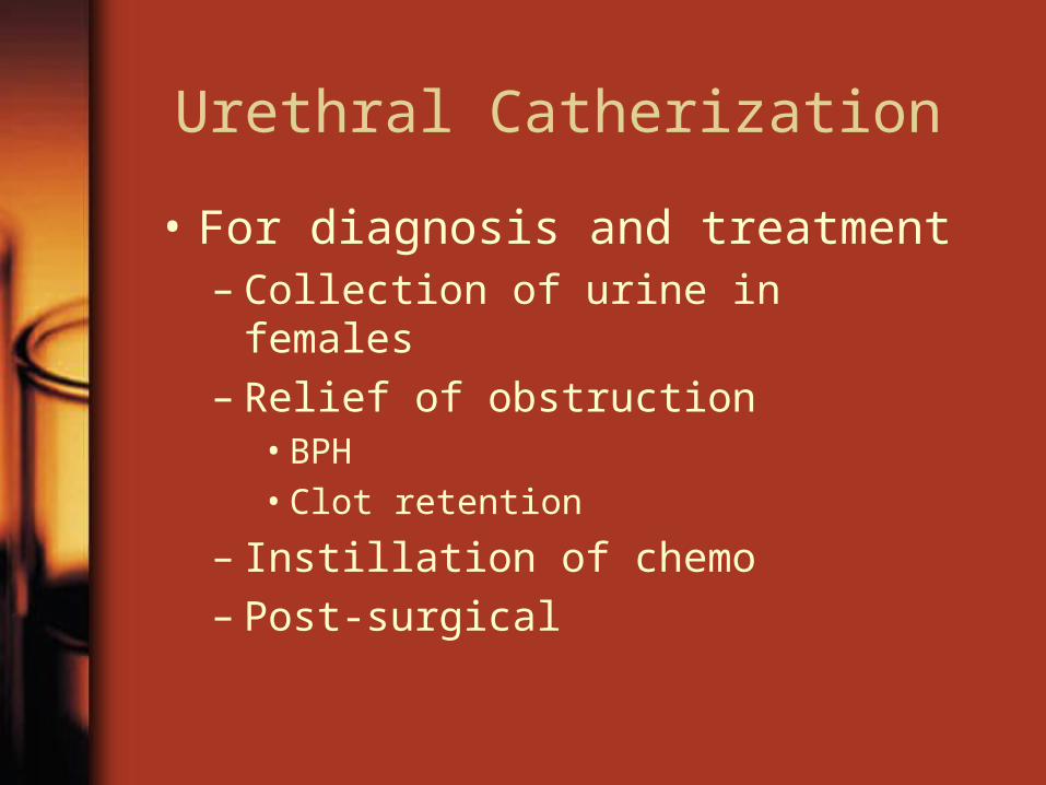
Urethral Catherization
• For diagnosis and treatment– Collection of urine in females– Relief of obstruction
• BPH• Clot retention
– Instillation of chemo– Post-surgical

Catheter Types and Size
• Size– 1 Fr = 0.33mm. Outside diameter not
luminal.
• Types– Straight– Coude– Malecot– Foley– 3-way

Downloaded from: Campbell-Walsh Urology (on 10 August 2008 03:12 AM)
© 2007 Elsevier
Catheter Types

Catheter Insertion
• Sterile Technique
• Use of Urojet
• Long-term use– Silicone better than latex or
polyurethane
• Size– 16-18 Fr for adults

Difficult Catherization
• Usually in males– BPH
• Use coude– Urethral Strictures– BNC
• May require the use of a glide-wire or filiform and followers
• In Females– Rare
• Obesity• Unable to locate urethral
meatus or difficult position– Use speculum

Difficult Catherization

Urethral Dilatation
• Typically in male– Urethral Stricture– BNC– Meatus for
transurethal surgery
• Sterile techniques– Filiforms and
followers– Urethral dilators– Amplatz dilators– balloon

Cystourethroscopy
• Diagnosis of lower urinary tract disease
• Sterile technique• Rigid cystoscope
– Better visualization– Larger working channel– Ease of orientation
• Flexible scope– More patient comfort– Pt. Stays supine– View from different
angles
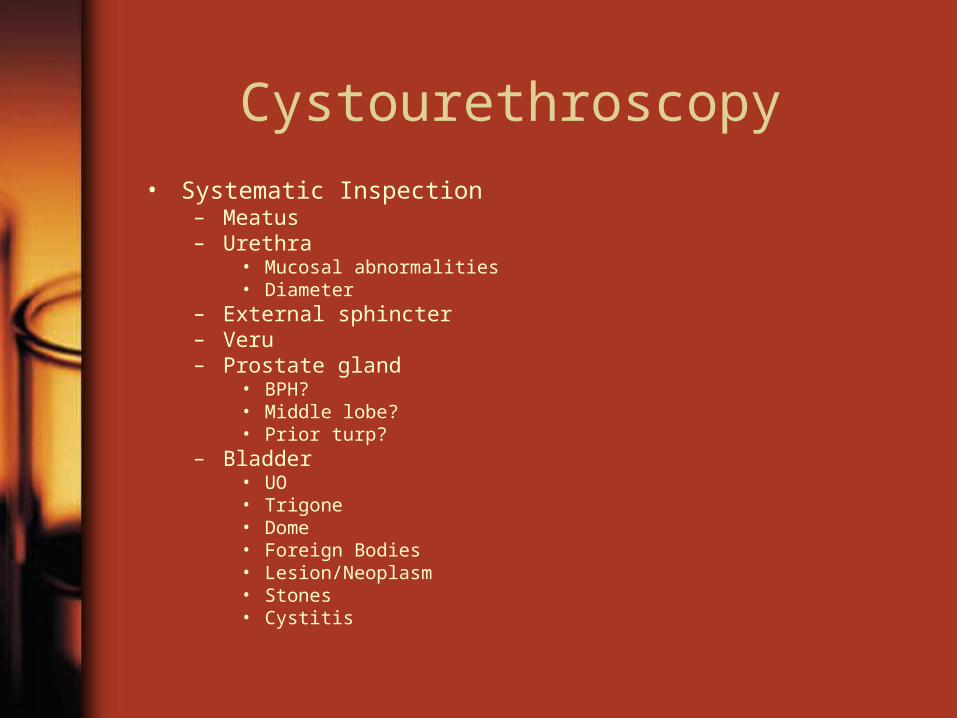
Cystourethroscopy• Systematic Inspection
– Meatus– Urethra
• Mucosal abnormalities• Diameter
– External sphincter– Veru– Prostate gland
• BPH?• Middle lobe?• Prior turp?
– Bladder• UO• Trigone• Dome• Foreign Bodies• Lesion/Neoplasm• Stones• Cystitis

Retrograde Pyelography
• Visualization of ureter and collecting system when other radiographic images are inadequate or when the patient is allergic to IV dye.– Hematuria– Filling defects– Positive cytology of ?
source– ? Obstruction
• Delayed films