Chapter 5 Fetal Central Nervous System · PDF fileChapter 5: Fetal Central Nervous System 72...
9
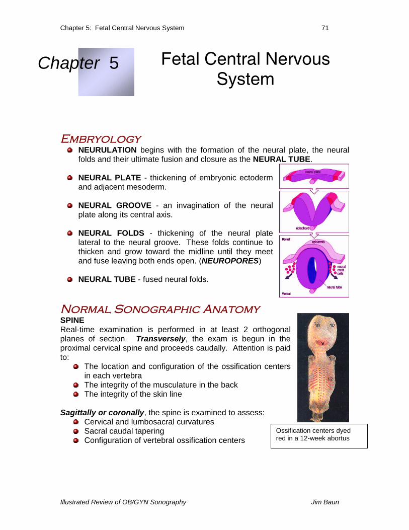
Chapter 5: Fetal Central Nervous System 71 Illustrated Review of OB/GYN Sonography Jim Baun Embryology NEURULATION begins with the formation of the neural plate, the neural folds and their ultimate fusion and closure as the NEURAL TUBE. NEURAL PLATE - thickening of embryonic ectoderm and adjacent mesoderm. NEURAL GROOVE - an invagination of the neural plate along its central axis. NEURAL FOLDS - thickening of the neural plate lateral to the neural groove. These folds continue to thicken and grow toward the midline until they meet and fuse leaving both ends open. (NEUROPORES) NEURAL TUBE - fused neural folds. Normal Sonographic Anatomy SPINE Real-time examination is performed in at least 2 orthogonal planes of section. Transversely, the exam is begun in the proximal cervical spine and proceeds caudally. Attention is paid to: The location and configuration of the ossification centers in each vertebra The integrity of the musculature in the back The integrity of the skin line Sagittally or coronally, the spine is examined to assess: Cervical and lumbosacral curvatures Sacral caudal tapering Configuration of vertebral ossification centers Chapter 5 Fetal Central Nervous System Ossification centers dyed red in a 12-week abortus
Transcript of Chapter 5 Fetal Central Nervous System · PDF fileChapter 5: Fetal Central Nervous System 72...
Chapter 5: Fetal Central Nervous System 71
Illustrated Review of OB/GYN Sonography Jim Baun
EmbryologyNEURULATION begins with the formation of the neural plate, the neuralfolds and their ultimate fusion and closure as the NEURAL TUBE.
NEURAL PLATE - thickening of embryonic ectodermand adjacent mesoderm.
NEURAL GROOVE - an invagination of the neuralplate along its central axis.
NEURAL FOLDS - thickening of the neural platelateral to the neural groove. These folds continue tothicken and grow toward the midline until they meetand fuse leaving both ends open. (NEUROPORES)
NEURAL TUBE - fused neural folds.
Normal Sonographic AnatomySPINEReal-time examination is performed in at least 2 orthogonalplanes of section. Transversely, the exam is begun in theproximal cervical spine and proceeds caudally. Attention is paidto:
The location and configuration of the ossification centersin each vertebraThe integrity of the musculature in the backThe integrity of the skin line
Sagittally or coronally, the spine is examined to assess:Cervical and lumbosacral curvaturesSacral caudal taperingConfiguration of vertebral ossification centers
Chapter 5 Fetal Central NervousSystem
Ossification centers dyedred in a 12-week abortus
Chapter 5: Fetal Central Nervous System 72
Illustrated Review of OB/GYN Sonography Jim Baun
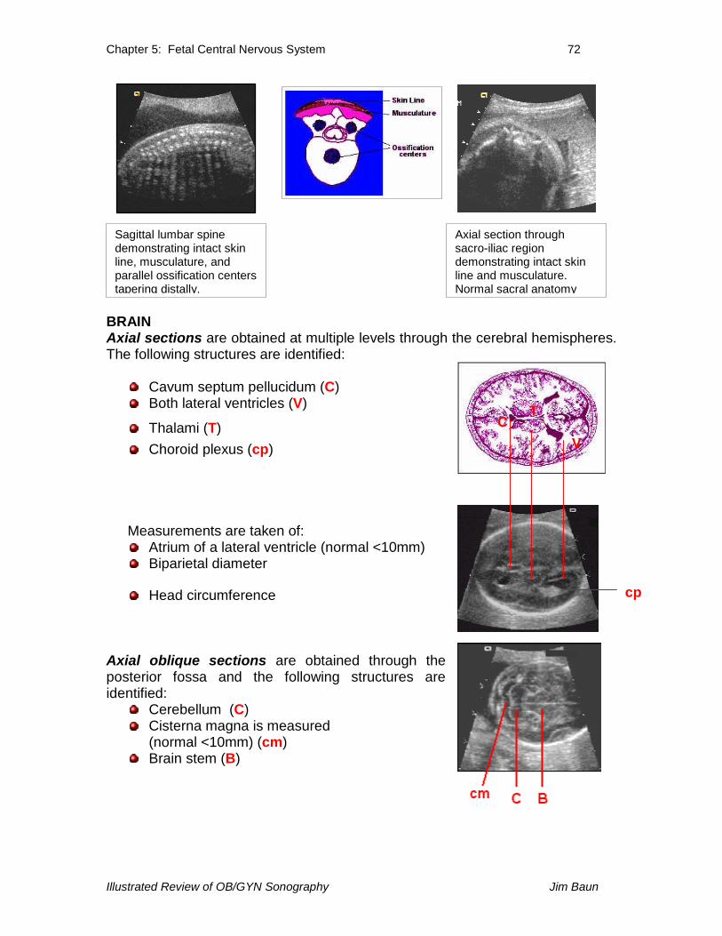
BRAINAxial sections are obtained at multiple levels through the cerebral hemispheres.The following structures are identified:
Cavum septum pellucidum (C)Both lateral ventricles (V)
Thalami (T)Choroid plexus (cp)
Measurements are taken of:Atrium of a lateral ventricle (normal <10mm)Biparietal diameter
Head circumference
Axial oblique sections are obtained through theposterior fossa and the following structures areidentified:
Cerebellum (C)Cisterna magna is measured(normal <10mm) (cm)Brain stem (B)
Sagittal lumbar spinedemonstrating intact skinline, musculature, andparallel ossification centerstapering distally.
TC
V
cp
Axial section throughsacro-iliac regiondemonstrating intact skinline and musculature.Normal sacral anatomy
Chapter 5: Fetal Central Nervous System 73
Illustrated Review of OB/GYN Sonography Jim Baun
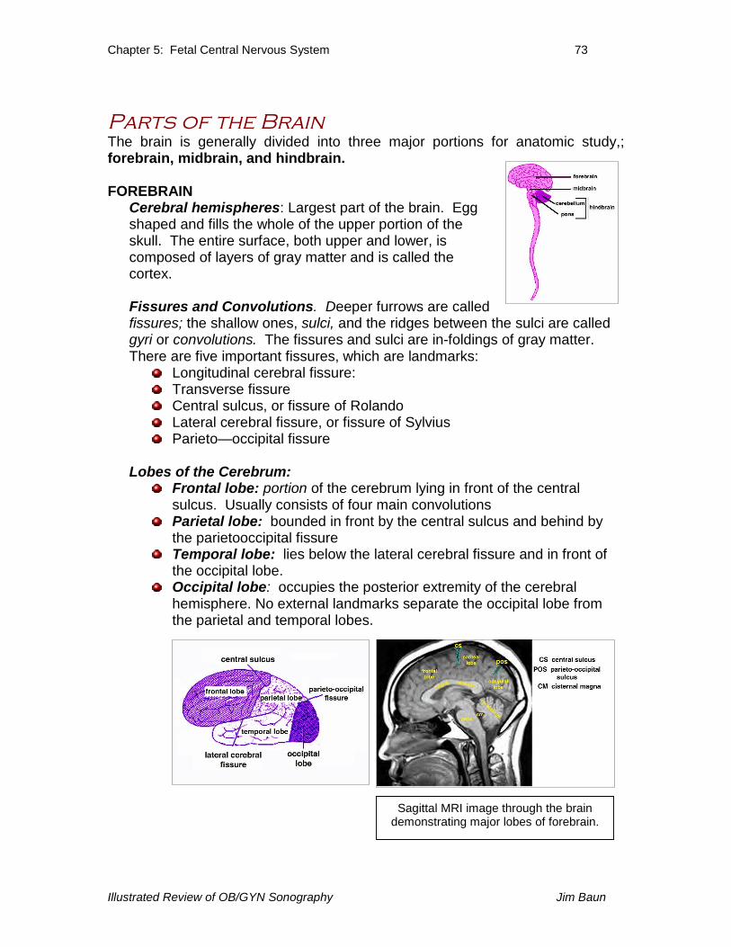
Parts of the BrainThe brain is generally divided into three major portions for anatomic study,;forebrain, midbrain, and hindbrain.
FOREBRAINCerebral hemispheres: Largest part of the brain. Eggshaped and fills the whole of the upper portion of theskull. The entire surface, both upper and lower, iscomposed of layers of gray matter and is called thecortex.
Fissures and Convolutions. Deeper furrows are calledfissures; the shallow ones, sulci, and the ridges between the sulci are calledgyri or convolutions. The fissures and sulci are in-foldings of gray matter.There are five important fissures, which are landmarks:
Longitudinal cerebral fissure:Transverse fissureCentral sulcus, or fissure of RolandoLateral cerebral fissure, or fissure of SylviusParieto—occipital fissure
Lobes of the Cerebrum:Frontal lobe: portion of the cerebrum lying in front of the centralsulcus. Usually consists of four main convolutionsParietal lobe: bounded in front by the central sulcus and behind bythe parietooccipital fissureTemporal lobe: lies below the lateral cerebral fissure and in front ofthe occipital lobe.Occipital lobe: occupies the posterior extremity of the cerebralhemisphere. No external landmarks separate the occipital lobe fromthe parietal and temporal lobes.
Sagittal MRI image through the braindemonstrating major lobes of forebrain.
Chapter 5: Fetal Central Nervous System 74
Illustrated Review of OB/GYN Sonography Jim Baun
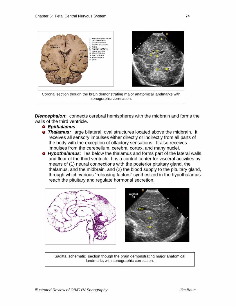
Diencephalon: connects cerebral hemispheres with the midbrain and forms thewalls of the third ventricle.
EpithalamusThalamus: large bilateral, oval structures located above the midbrain. Itreceives all sensory impulses either directly or indirectly from all parts ofthe body with the exception of olfactory sensations. It also receivesimpulses from the cerebellum, cerebral cortex, and many nuclei.Hypothalamus: lies below the thalamus and forms part of the lateral wallsand floor of the third ventricle. It is a control center for visceral activities bymeans of (1) neural connections with the posterior pituitary gland, thethalamus, and the midbrain, and (2) the blood supply to the pituitary gland,through which various “releasing factors” synthesized in thehypothalamusreach the pituitary and regulate hormonal secretion.
Coronal section though the brain demonstrating major anatomical landmarks withsonographic correlation.
Sagittal schematic section though the brain demonstrating major anatomicallandmarks with sonographic correlation.
Chapter 5: Fetal Central Nervous System 75
Illustrated Review of OB/GYN Sonography Jim Baun
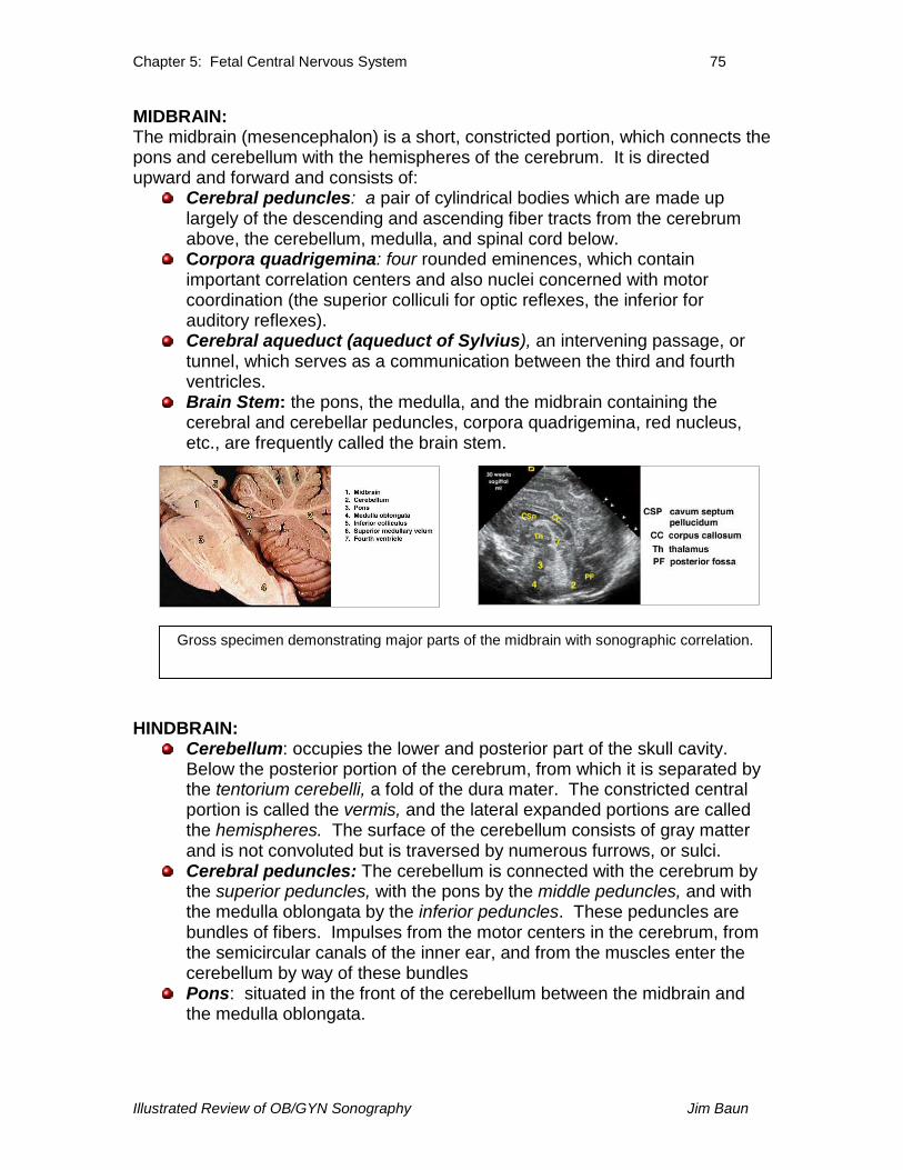
MIDBRAIN:The midbrain (mesencephalon) is a short, constricted portion, which connects thepons and cerebellum with the hemispheres of the cerebrum. It is directedupward and forward and consists of:
Cerebral peduncles: a pair of cylindrical bodies which are made uplargely of the descending and ascending fiber tracts from the cerebrumabove, the cerebellum, medulla, and spinal cord below.Corpora quadrigemina: four rounded eminences, which containimportant correlation centers and also nuclei concerned with motorcoordination (the superior colliculi for optic reflexes, the inferior forauditory reflexes).Cerebral aqueduct (aqueduct of Sylvius), an intervening passage, ortunnel, which serves as a communication between the third and fourthventricles.Brain Stem: the pons, the medulla, and the midbrain containing thecerebral and cerebellar peduncles, corpora quadrigemina, red nucleus,etc., are frequently called the brain stem.
HINDBRAIN:Cerebellum: occupies the lower and posterior part of the skull cavity.Below the posterior portion of the cerebrum, from which it is separated bythe tentorium cerebelli, a fold of the dura mater. The constricted centralportion is called the vermis, and the lateral expanded portions are calledthe hemispheres. The surface of the cerebellum consists of gray matterand is not convoluted but is traversed by numerous furrows, or sulci.Cerebral peduncles: The cerebellum is connected with the cerebrum bythe superior peduncles, with the pons by the middle peduncles, and withthe medulla oblongata by the inferior peduncles. These peduncles arebundles of fibers. Impulses from the motor centers in the cerebrum, fromthe semicircular canals of the inner ear, and from the muscles enter thecerebellum by way of these bundlesPons: situated in the front of the cerebellum between the midbrain andthe medulla oblongata.
Gross specimen demonstrating major parts of the midbrain with sonographic correlation.
Chapter 5: Fetal Central Nervous System 76
Illustrated Review of OB/GYN Sonography Jim Baun
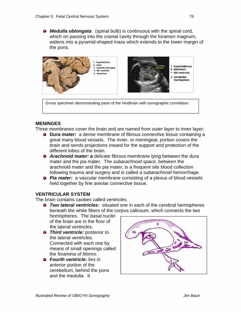
Medulla oblongata: (spinal bulb) is continuous with the spinal cord,which on passing into the cranial cavity through the foramen magnum,widens into a pyramid-shaped mass which extends to the lower margin ofthe pons.
MENINGESThree membranes cover the brain and are named from outer layer to inner layer:
Dura mater: a dense membrane of fibrous connective tissue containing agreat many blood vessels. The inner, or meningeal, portion covers thebrain and sends projections inward for the support and protection of thedifferent lobes of the brain.Arachnoid mater: a delicate fibrous membrane lying between the duramater and the pia mater. The subarachnoid space, between thearachnoid mater and the pia mater, is a frequent site blood collectionfollowing trauma and surgery and is called a subarachnoid hemorrhage.Pia mater: a vascular membrane consisting of a plexus of blood vesselsheld together by fine areolar connective tissue.
VENTRICULAR SYSTEMThe brain contains cavities called ventricles.
Two lateral ventricles: situated one in each of the cerebral hemispheresbeneath the white fibers of the corpus callosum, which connects the twohemispheres. The basal nucleiof the brain are in the floor ofthe lateral ventricles.Third ventricle: posterior tothe lateral ventricles.Connected with each one bymeans of small openings calledthe foramina of Monro.Fourth ventricle: lies inanterior portion of thecerebellum, behind the ponsand the medulla. It
Gross specimen demonstrating parts of the hindbrain with sonographic correlation.

Chapter 5: Fetal Central Nervous System 77
Illustrated Review of OB/GYN Sonography Jim Baun
communicates with the third ventricle via the slender canal called thecerebral aqueduct (aqueduct of Sylvius). In the roof of the fourth ventriclethere is an opening called the Foramen of Magendi. In the lateral wallsthere are, two openings called the foramina of Luschka. By means ofthese three openings, the ventricles communicate with the subarachnoidspace, and the cerebrospinal fluid can circulate from one to the other.
CEREBROSPINAL FLUID:The meningeal membranes and the spaces filled with fluid form a padenclosing the brain and cord on all sides. Cerebrospinal fluid is secreted anddiffused from the blood by the ependymal cells, which cover the choroidplexuses of the ventricles. The choroid plexuses are highly vascular folds orprocesses of the pia mater found in the ventricles.
Flow of CSF:Produced by choroid plexuses inlateral and 3rd ventriclesFills lateral ventricles firstFlows into 3rd via foramen ofMonroFlows into 4th via aqueduct ofSylvius
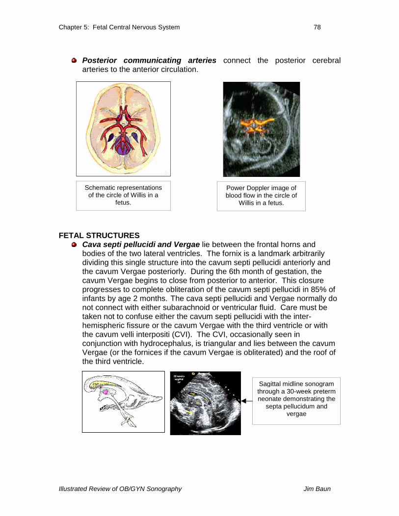
BLOOD SUPPLY.The internal carotid and vertebral arteries are the source of blood to the brain.Their intracranial branches anastomose at the base of the brain to form the circleof Willis from which arise the three primary sets of cerebral arteries:
Anterior cerebral arteries: arises from the internal carotid artery at theinner extremity of the Sylvian fissure. Passes anteriorly and medially tothe longitudinal fissure where it is connected to the artery of the oppositeside by the anterior communicating artery.Middle cerebral arteries: the largest branch of the internal carotid artery,which passes obliquely through the Sylvian fissure and, at the insula,divides into its terminal branches.Posterior cerebral arteries: branches of the basilar artery whoseterminal branches penetrate the cerebral cortex.The cerebellum receives its arterial perfusion from various branches of thevertebral and basilar arteries.
Chapter 5: Fetal Central Nervous System 78
Illustrated Review of OB/GYN Sonography Jim Baun
Posterior communicating arteries connect the posterior cerebralarteries to the anterior circulation.
FETAL STRUCTURESCava septi pellucidi and Vergae lie between the frontal horns andbodies of the two lateral ventricles. The fornix is a landmark arbitrarilydividing this single structure into the cavum septi pellucidi anteriorly andthe cavum Vergae posteriorly. During the 6th month of gestation, thecavum Vergae begins to close from posterior to anterior. This closureprogresses to complete obliteration of the cavum septi pellucidi in 85% ofinfants by age 2 months. The cava septi pellucidi and Vergae normally donot connect with either subarachnoid or ventricular fluid. Care must betaken not to confuse either the cavum septi pellucidi with the inter-hemispheric fissure or the cavum Vergae with the third ventricle or withthe cavum velli interpositi (CVI). The CVI, occasionally seen inconjunction with hydrocephalus, is triangular and lies between the cavumVergae (or the fornices if the cavum Vergae is obliterated) and the roof ofthe third ventricle.
Power Doppler image ofblood flow in the circle of
Willis in a fetus.
Sagittal midline sonogramthrough a 30-week pretermneonate demonstrating the
septa pellucidum andvergae
Schematic representationsof the circle of Willis in a
fetus.
Chapter 5: Fetal Central Nervous System 79
Illustrated Review of OB/GYN Sonography Jim Baun
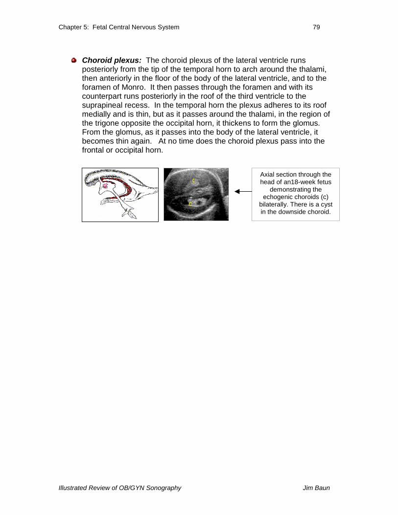
Choroid plexus: The choroid plexus of the lateral ventricle runsposteriorly from the tip of the temporal horn to arch around the thalami,then anteriorly in the floor of the body of the lateral ventricle, and to theforamen of Monro. It then passes through the foramen and with itscounterpart runs posteriorly in the roof of the third ventricle to thesuprapineal recess. In the temporal horn the plexus adheres to its roofmedially and is thin, but as it passes around the thalami, in the region ofthe trigone opposite the occipital horn, it thickens to form the glomus.From the glomus, as it passes into the body of the lateral ventricle, itbecomes thin again. At no time does the choroid plexus pass into thefrontal or occipital horn.
Axial section through thehead of an18-week fetus
demonstrating theechogenic choroids (c)
bilaterally. There is a cystin the downside choroid.
c
c