Chapter 5 5.0 EXPERIMENT 2 TITLE: FLUORESCENCE IMAGING...
52
172 Chapter 5 5.0 EXPERIMENT 2 TITLE: FLUORESCENCE IMAGING OF MOUSE OOCYTES 5.1 ABSTRACT This chapter has been is divided into four experimental parts. The main aims were to characterise maturation stages and factors affecting mouse oocyte competency by examining selected structural development using ICC staining. The factors included were maturation conditions, duration of incubation, oocyte retrieval times of the day and zona dissection (PZD). Structures detected were NM, MS, CG and actin filaments. Nuclear maturation was assessed through the activities of the nuclear materials and meiotic spindles arrangement and cytoplasmic maturation using cortical granules distribution. Factors involved in producing high yields of MII oocytes vary depending on strains and culture conditions. The production of MII oocytes of ICR mice were significantly higher in in vitro than in vivo condition. However, its CG appearance was low. This suggests activation of oocytes in in vitro environment that caused loss of CG through exocytosis. Low MS was also observed among F 1 mice during 6 hours of in vitro maturation as compared with 3 hours. Such reduction was not observed in C57BL/6J oocytes. Nevertheless, collecting oocytes at 1200 hours of the day improved MII oocytes by two-folds in both strains during both in vivo and in vitro conditions than at 0900 hours. This was also manifested by high correlation values between MII oocytes with structural appearances under study. The C57BL/6J oocytes were further shown to have higher structural formation under PZD treatment but not F 1 . In conclusions, the interplay of various factors including strains, culture conditions and post-hCG time of oocyte retrieval would determine maturation stages and structural distributions in the mouse oocytes.
Transcript of Chapter 5 5.0 EXPERIMENT 2 TITLE: FLUORESCENCE IMAGING...
172
Chapter 5
5.0 EXPERIMENT 2
TITLE: FLUORESCENCE IMAGING OF MOUSE OOCYTES
5.1 ABSTRACT
This chapter has been is divided into four experimental parts. The main aims were to
characterise maturation stages and factors affecting mouse oocyte competency by
examining selected structural development using ICC staining. The factors included were
maturation conditions, duration of incubation, oocyte retrieval times of the day and zona
dissection (PZD). Structures detected were NM, MS, CG and actin filaments. Nuclear
maturation was assessed through the activities of the nuclear materials and meiotic spindles
arrangement and cytoplasmic maturation using cortical granules distribution. Factors
involved in producing high yields of MII oocytes vary depending on strains and culture
conditions. The production of MII oocytes of ICR mice were significantly higher in in vitro
than in vivo condition. However, its CG appearance was low. This suggests activation of
oocytes in in vitro environment that caused loss of CG through exocytosis. Low MS was
also observed among F1 mice during 6 hours of in vitro maturation as compared with 3
hours. Such reduction was not observed in C57BL/6J oocytes. Nevertheless, collecting
oocytes at 1200 hours of the day improved MII oocytes by two-folds in both strains during
both in vivo and in vitro conditions than at 0900 hours. This was also manifested by high
correlation values between MII oocytes with structural appearances under study. The
C57BL/6J oocytes were further shown to have higher structural formation under PZD
treatment but not F1. In conclusions, the interplay of various factors including strains,
culture conditions and post-hCG time of oocyte retrieval would determine maturation
stages and structural distributions in the mouse oocytes.

173
Keywords: Fluorescence imaging, MI oocyte, MII oocyte, oocyte maturation, oocyte
competency.
5.2 INTRODUCTION
The morphology of mammalian oocytes including murine species have been extensively
reported in the literature as listed in Table 5.1. Assessment of good quality oocytes is
generally made based on its morphological appearances through microscopic observation
which could be enhanced by using various imaging devices and softwares (Vonesh et al.,
2006). The assessment has many significant implications especially for in vitro
applications of the oocytes such as IVF, nuclear transfer, embryonic stem cells production,
gene manipulation and oocyte manipulation. Good oocytes are categorised with a small
peri-vitelline space and a regular and distinct first polar body (Suppinyopong et al., 2000).
The above-mentioned criteria are usually associated with increased pregnancy (Ebner et al.,
1999). The morphological scoring of good oocytes for IVF application is mainly
determined by using microscopic imaging such as Differential Interference Contrast (DIC)
and Hoffman Modulation Contrast (HMC). However, oocyte intracellular structures could
only be determined by using staining methods. The conventional staining techniques
commonly used are Giemsa stain for DNA materials (Tateno and Kamiguchi, 2007) and
Coomasie stain for the cytoskeleton (Navarro-Garcia et al., 1999). Nowadays,
fluorescence staining is widely adopted by many laboratories to reveal the details of
internal architectures of mammalian oocytes (Schatten et al., 1985, 1993; Hewitson, 1995;
Abbot et al., 1998) for obtaining a better understanding not only on the complexity of its
structural development but also the important mechanisms associated with cellular
processes. Some other criteria reported for grading of oocytes included cytoplasmic
appearance and granular structures (Daniel, 1971), changes in the organisation of the

174
plasma membrane and microtubular morphology (Elder and Dale, 2000). These were
reported to play an important role in the acquisition of oocyte meiotic competence.
The fertilising capability of oocyte is determined mainly by their maturation stage.
Most mammalian eggs are meiotically arrested at dictyate stage of prophase I and resume
cell cycle progression to metaphase II (MII) only hours before ovulation. Hormonally
induced animals may take 10 to 13 hours for ovulation to occur (Daniel, 1971) and
subsequently mature. The rate at which an oocyte attains maturity varies and can be
affected by multitude of factors which include age, nutrients, strains, health, environmental
stress and oestrus stage. A mature oocyte is usually marked by the presence of polar body.
In contrast to the natural process, maturation stages of oocyte in vitro is influenced by
various factors such as the hormones used, the time at which hormones were injected,
retrieval time, the site from which oocytes were retrieved and also compositions of the
culture medium used. Oocytes directly retrieved from the follicles or even from the
oviducts may be of different maturation stages; germinal vesicles (GV), MI phase and MII
phase oocytes.
Two crucial processes of oocyte maturation include nuclear maturation and
cytoplasmic maturation. Grondahl (2008) has defined oocyte nuclear maturation as
modifications that take place during resumption of meiosis, producing a haploid
chromosome complement from the diploid state. He defined cytoplasmic maturation as the
unity of processes modifying the oocyte cytoplasm that are essential for fertilisation and
pre-implantation embryonic developmental competence. Nuclear maturation is essentially
required for fertilisation to take place. Nevertheless, cytoplasmic maturation is equally
important for cleavage and subsequent embryonic development in vitro and seems to be
independent of the nuclear division cycle (Elder and Dale, 2000). Cytoplasmic maturation
175
requires relocation of organelles and establishment of oocyte polarity, with an increase in
the number of mitochondria and ribosomes to support protein synthesis. The cytoplasmic
components of oocyte that are required to support early fertilisation include yolk granules,
pigment granules, mitochondria, cortical granules and a layer of membrane bound vesicles
located beneath the plasma membrane. Membrane bound vesicles of the cytoplasm include
crystalline bodies, fat droplets and glycogen granules. One peculiarity of the mouse
oocytes is the lack of centrioles (Brunet et al., 1999) and another is that oocytes which
failed to reach 60 µm would never developed in vitro (Epigg and Shcroeder, 1989). Also,
the frequency of premature metaphase I arrest decreased markedly as the age of the mice
and oocyte volume increased (Sorenson and Wassarman, 1976).
Many aspects of cellular and molecular developments of mouse oocytes need to be
studied and analysed during in vitro maturation (IVM) in contrast to its in vivo maturation
(IVO). There were factors reported to influence oocyte maturation including culture
conditions and strains (Ibanez et al., 2005). In vivo produced oocytes are generally of
superior quality with respect to production of healthy embryos and offspring. It is well
documented that nuclear maturation usually occurred among the in vitro matured oocytes
but complete cytoplasmic maturation is not really assured (Epigg et al., 1996). Liu et al.
(2005) also reported that tempos of nuclear maturation and cortical granules redistribution
were slower among in vitro than the in vivo matured oocytes.
Resumption of meiosis, also known as oocyte maturation is a crucial manifestation
of most mammalian egg development during which the oocyte would go through complex
changes; they grow and switch many genes on or off in preparation for the development.
These series of complex biological processes that the oocyte must go through in reaching
maturity states is also called meiotic maturation (Schultz et al., 1989). In mouse oocytes,

176
the first meiotic M-phase is very long; lasting from 6 to 11 hours, depending on the genetic
background (Brunet et al., 1999). The earliest morphological changes which occured
during maturation process is the breakdown of nuclear membrane or germinal vesicle
breakdown (GVBD), at which transcription of new RNA stops almost completely (Elder
and Dale, 2000). The progressive aggregation of numerous microtubule organising centres
(MTOC) forms the spindle poles, and a bipolar spindle is present during most of the first
meiotic M-phase (Brunet et al., 1999). This is followed by the formation of meiotic spindle
and the move of chromosomes to the periphery of the cell. The first meiotic division
separates homologous chromosomes. Also, cortical granules are redistributed as the cell
size increases to 100 µm in diameter. At this stage, one set of homologous chromosomes is
surrounded by a small amount of cytoplasm. This structure extrudes as the first polar body
and then arrested (Hogan et al., 1986). Polarisation is obvious at this stage of mouse
oocyte development, whereby a microvillus free area on the plasma membrane, adjacent to
the polar body overlying the meiotic spindle mark the animal pole of the oocyte. This stage
is also known as metaphase II stage oocyte, which is relatively quiet cell, with a pre-set
developmental programme.
In response to the preovulatory surge of LH, MII oocytes are expelled into the
infundibulum of the oviduct waiting to be fertilised by sperm. Eggs ovulated from the
follicles of mouse ovaries accumulated in the distal part of the oviducts called ampullae and
are surrounded by sticky mass cells called cumulus cell complex. These granulosa derived
cells communicate with an oocyte through gap junction. The arrest at MII stage of a
mature oocyte is maintained by maturation promoting factor (MPF) (Kono et al., 1995) and
the two components of the factor (MPF) identified are tyrosine kinases and cyclin B2,
which oscillate with meiotic cycles: increases during GVBD, remains high at MI,
177
disappears transiently at the time of first polar body extrusion, reappears at MII, and
remains elevated level until fertilisation. These components are stabilised by cytostatic
factor (CSF) that contained c-mos proto-oncogene product; protein that is responsible for
MPF activation. In mouse oocyte, this factor is only necessary for metaphase arrest at
meiosis II before fertilisation (Elder and Dale, 2000). Mos protein that is the activator of
MAP kinase (mitogen associated protein kinase) not only preventing the oocyte from
entering interphase between metaphase I and metaphase II but also maintaining
chromosomes condensation, reorganisation of microtubules, spindle formation at
metaphase I, stabilisation of the spindle at metaphase II, and therefore mediates as spindle
assembly checkpoint (Elder and Dale, 2000). In addition, Kubiak et al. (1993) suggested
that the mechanism maintaining the metaphase arrest in mouse oocyte involves the
equilibrium between cyclin synthesis and degradation, probably controlled by CSF and
which is also dependent upon the three dimensional organisation of the spindle. Thus,
progression beyond metaphase II requires destruction of MPF activity, which is stimulated
by calcium-mediated activation of calcium dependent protein kinase II (Kono et al., 1995).
It is noted generally, oocyte maturation to MII stage appears to be slower in in vitro follicle
culture as compared with in vivo maturation (Hu et al., 2001; Liu et al., 2005). Prolonging
culture of oocytes past 16 hours post-hCG may further increase the yield of oocyte in MII.
However, ageing of oocytes at MII may cause concomitantly a deterioration of the spindle
and displacement of chromosomes from the spindle equator.
Recent molecular study showed that among the many substances to influence
oocyte maturation is the endogenous signaling molecule; FF-MAS (4,4-dimethyl-5
cholest-8,14,24-trien-3-ol), an intermediate in the cholesterol biosynthetic pathway
present in cells (Byskov et al., 1995). This substance has triggered the resumption of
178
meiosis (nuclear maturation) of mouse oocyte in vitro (Grondahl et al., 1998). It is also
capable of initiating meiosis in isolated naked or cumulus cell-enclosed oocytes from
several species (Byskov et al., 1995). Another study showed that the formation of polar
body, asymmetrical organisation of meiotic spindles and arrangement of chromosmes at the
oocyte cortex in mice was associated with protein formis (Liu et al., 2003). Meanwhile, in
bovine, administration of cAMP in culture medium showed that granulosa cumulus oocyte
complexes (GCOCs) remained at prophase I with intact germinal vesicle but germinal
vesicle breakdown was observed among the cumulus oocyte complexes (COCs).
Nonetheless, both groups of oocytes were reported to resume meiotic division in hormone
and serum free medium (Aktas et al., 2003). In contrary, the recent finding reported that a
higher capability of cortical granules release was observed among oocyte matured in the
presence of serum than those matured without the serum (Ibanez et al., 2005). Also, Liu et
al. (2005) reaffirmed that the presence of serum in maturation media enhanced cortical
granules release after aging or activation of oocytes.
The cytoplasm of the mammalian oocytes consists of complex matrix of
cytoskeletal elements, including actin, tubulin and certain cytokeratins (Hogan et al., 1986).
The monomeric proteins that comprise microtubules are α and β tubulins. The γ tubulin, a
third tubulin protein is not present in microtubules, but is thought to nucleate the initial
polymerisation of α and β tubulins. Tubulin filaments are mostly found in the cytoskeleton
and meiotic spindles of an oocyte. Gobarsky et al. (1990) injected biotinylated tubulin into
living oocyte that was subsequently incorporated into the meiotic spindle and found that
this component of the microtubules within the arrested metaphase spindle of the mouse
oocyte undergo rapid cycles of assembly and disassembly. Microtubules of the telophase
midbody are more stable. These spindle shaped microtubules that partition chromosomes

179
are classified as short lived structures. They appear after interphase and disappear after
mitosis is completed. Although microtubules in a cell may seem to be arranged in a
somewhat random pattern, in fact, they are usually in a hub and spoke formation with the
hub near the centre of the cell. This hub is called the centrosome and is the primary
microtubules organising center (MTOC) of the cell. Polymerisation of microtubules is
temperature dependent. At low temperatures, microtubules tend to depolymerise, whereas
at higher temperatures, they spontaneously polymerise. Above a critical concentration
polymerisation is favoured and vice versa. During polymerisation, microtubules radiate
outward with the +ve ends moving away from the centrosomes.
The role of cytoskeletal microtubules and microfilaments in the process of egg
maturation and fertilisation in various kinds of mammalian oocytes has been reported in the
last decade. In Xenopus egg, interactions between individual cytoskeletal components and
internal membranes are important for organisation of the cortex and cortical actin is
required for anchoring and rotation of the meiotic spindle. It has also been suggested that
the first contact of mammalian sperm with an oocyte involves an early reorganisation of the
oocyte cytoskeleton. Clusters of mouse oocyte chromosomes are redistributed around the
cortex in a microfilament dependent process (Veselska and Janish, 2001). The system has
been reported to coordinate the migration of maternal and paternal pronuclei when sperm
fertilised an oocyte. Microtubules forming within the mouse egg during fertilisation are
required for the movements leading to the union of the sperm and egg nuclei. In an
unfertilised oocyte, microtubules are predominantly found in the arrested meiotic spindles.
At the time for sperm incorporation, a dozen cytoplasmic asters assemble, often associated
with the pronuclei formation (Schatten et al., 1985). They also reported that meiotic
spindle of unfertilised oocyte is anastral, barrel shaped, and attached to the oocyte cortex
180
and probably interact with the perinuclear actin found in pronucleate eggs. It has been
postulated that the activity of MPF: histone H1 phosphorylation not only contribute to
chromosome condensation and nucleosome packing alteration but also rearrangement of
microfilament, reorganisation of intermediate filament network, and nuclear disassembly.
Hu et al. (2001) also reported that spindle morphology and chromosome alignment can be
used as one indicator of the oocyte’s capacity to form chromosomally balanced embryo. In
mice, mirotubules in the pronucleate stage zygote are organised as multiple cytoasters
instead of the expected single sperm aster. Furthermore, centrosomal material is detected
in unfertilised oocyte but not in the sperm (Navara et al., 1994; Schatten, 1994; Schatten et
al., 1986). Schatten et al. (1991) had conducted studies involving parthenogenesis,
polyspermy and recovery from microtubule inhibitors to support the hypothesis that the
mouse centrosome is of maternal origin. The evolutionary shift from paternal to maternal
centrosomes in mice is probably due to their short generation time in which the females are
sexually mature around 1.5 months versus the 1.5 decade norm of humans (Schatten, 1994).
It has been proven that in murine oocyte development, microfilaments are crucial for
cortical meiotic spindle positioning and maintenance, formation of the first and second
polar bodies, incorporation of cone formation, pronuclear apposition, and cytokinesis.
Nevertheless, actin assembly is not required for sperm head penetration in the mouse
(Simerly et al., 1998). In addition, the earliest developmental changes in actin organisation
in the egg are seen at fertilisation. In the ovulated oocytes the plasma membrane above the
meiotic spindle is devoid of concanavalin A (Con A) binding sites and microvilli, and is
underlaid by actin rich subcortical layer. Fertilisation results in the formation of a second
Con A free zone, the fertilisation zone, which is around the site of sperm entry. The plasma
membrane of this region is also underlaid by an actin rich layer. As the pronuclei move

181
toward the centre of the egg, the distribution of actin filaments become more uniform and
the Con A free regions disappear. The microfilaments of human oocytes formed a very fine
meshwork localised particularly in the cortical region. Individual fibres either extended
into remnants of the zona pellucida or, occasionally, formed cell surface protrusion called
microspikes or microvilli. The bundles of microfilaments in a mouse oocyte were thicker
as compared with the actin filaments in human oocyte (Veselska and Janish, 2001). Actin
structures are also involved in the process of meiotic division I and extrusion of first polar
body. It has also been confirmed that the migration of cortical granules is driven by
microfilaments and not by microtubules. Anchorage of cortical granules to the cortex is
independent of these cytoskeletal structures. Neither microtubules nor microfilaments are
involved in exocytosis of cortical granules (Veselska and Janish, 2001). It has been
reported that at 2 hours, fertilised MII mouse eggs underwent a mean CG loss of 69% and
the entire cortex occupied by CG but not in the fertilised GVBD and the prometaphase I.
The mechanism of propagating a normal wave of CG loss from the site of sperm entry
develops between MI and MII oocyte (Ducibella and Buetow, 1994).
The ICC technique utilised in this study had revealed some of the structural
abnormalities that may have contributed to the failure of meiotic completion and the search
for superior eggs suitable for cultivation continues. Some of the past studies on the
morphology of mammalian oocytes are shown in Chapter 4 (Table 4.1) and described in
chronological order in Table 5.1. In brief, Downs et al. (1986) revealed that in vitro
maturation of mouse oocytes can be inhibited simultaneously by adding hypoxanthine and
adenosine into the incubation medium. Hypoxanthine also blocked GVBD, postponed
cortical granules migration and prevented cortical granules free domain formation (Liu et
al., 2005). Meanwhile, the metaphase plate of MI stage in bovine oocyte occurred by 6.6

182
hours of culture following GVBD (De Loos et al., 1994). Another study conducted by
Sirad and Blondin (1996), reported that different developmental potential was observed
between IVO and IVM bovine oocytes. However, the groups displayed similar rates of
nuclear maturation, fertilisation and cleavage formation. Retardation of spontaneous cell
cycle progression and meiotic competence in mouse oocytes cultured in vitro was detected
by using DAPI to stain the nuclear materials and LCA-biotin texas red to detect blockage of
cortical granules exocytosis (Abbot et al., 1998). A study on porcine oocytes showed that
cooling can cause irreversible damage of both the chromosome and meiotic spindle (Leader
et al., 2002).
Mouse oocyte has been claimed to be the excellent model for exploring
microtubular fibres such as myosin structure and function (Lee et al., 2000). It has also
been reported that immature germinal stage oocyte matures spontaneously in vitro and can
be arrested at specific cell cycle during first meiosis. This permits observations on dynamic
motility events such as peripheral spindle migration, cortical spindle anchoring, and cell
surface modification (Lee et al., 2000). Ovulated oocyte is arrested at metaphase of the
second meiosis and can be artificially activated or fertilised in vitro, permitting detailed
investigations on spindle rotation and cytokinesis during second polar body formation and
the event during sperm incorporation, pronuclear formation, and migration, as well as
mitosis, can be investigated.
5.3 OBJECTIVES
The aims of this study were, a) to reveal the general architectures of the in vivo and in vitro
matured oocytes in different strains of mice by using fluorescence imaging, b) to study the
effects of retrieval hours post-hCG injection on oocyte structures and c) to evaluate the
effect of micromanipulation such as partial zona dissection on the oocyte structures.

183
Year Author Species Description
1986
1994
1996
1998
2002
2003
2003
2005
2008
Downs et al.
De Loos et al.
Sirard and
Blondin
Abbott et al.
Leader et al.
Liu et al.
Aktas et al.
Ibanez et al.
Grondahl
Mouse
Cattle
Cattle
Mouse
Pig
Mouse
Cattle
Mouse
Horse,
pig
The simultaneous addition of hypoxanthine and
adenosine into the incubation medium inhibits in
vitro maturation of mouse oocyte
GVBD occurs within hours after removal from the
follicle or the ovulatory LH signal. By 6.6 hours of
culture, approximately 50% of the oocyte
undergone GVBD. Then, the chromosomes
condensed further. The kinetochores appear and
the microtubules pull the chromosomes and they
form the metaphasic plate of MI.
Oocyte matured in vitro or in vivo have similar
rates of nuclear maturation, fertilization and
cleavage, but clearly differ in their developmental
potential
In vitro culture retards spontaneous cell cycle
progression and the meiotic status was determined
by DNA staining using DAPI. Cortical granule
exocytosis was disrupted too and this was detected
by using Lens culinaris –biotin-Texas red
Cooling caused non-reversible damage of both
chromosomes and meiotic spindle.
Formis (FH proteins) is associated with the
formation of polar body and also organisation of
asymmetry spindle and arrangement of
chromosomes at the oocyte cortex.
Regulation of meiotic state in granulose cumulus
complex oocyte (GCOC) and cumulus oocyte
complex (COC) with cAMP showed GCOC
remained at prophase with intact germinal vesicle
(GV) but COC underwent germinal breakdown
(GVBD) but in hormone and serum free medium
they resume meiosis.
Strains and culture condition affect nuclear and
cytoplasmic maturations in mouse oocyte.
Addition of FF-MAS to the maturation medium
improves the cytoplasmic maturation and yields
higher quality pre-embryos.
Table 5.1: Timeline on some of the significance findings on cellular architectures of
mammalian oocytes
184
5.4 SIGNIFICANCE OF THE STUDY
Determining oocyte morphology using fluorescence staining could reveal intracellular
architectural dynamics which are directly or indirectly related to oocyte developmental
competence. Some of the structures such as cytoskeleton are generally not clearly revealed
through conventional staining that consequently lead to uncertainties on specific
morphological development at different maturation stages of oocytes. Determining oocyte
developmental competence between strains and culture conditions enables ones to identify
suitable criteria to be included for cultivating high quality oocytes, consequently improving
fertilisation rate and subsequent embryonic development in vivo and in vitro. Also, the
study may reveal more information on architectural development of in vivo and in vitro
matured oocytes and variation that occur between strains. In other words, by comparing
oocyte architectural topography, susceptibility of different strains of mouse oocytes
towards in vivo and in vitro environment especially during micromanipulation could be
assessed and compared. Thus, their further survival and developmental competence can be
predicted.
5.5 MATERIALS
The materials required in this study were as described in Sections 3.3, 3.4, 3.5 and 3.6. In
brief, female mice from the following strains which were ICR, C57BL/6J and F1 hybrids of
CBAC57BL/CJ were used and sacrificed at the age between 8-10 weeks old.
Superovulation through PMSG and hCG admininstrations was described in Section 3.3.3.
The TYH medium and hyaluronidase acid (HA, 0.1%) were used to prepare naked oocytes.
Zona dissolution was conducted using acid Tyrode’s solution (pH 2) and zona dissection
method followed Nakagata (2000) with modifications. Washing and rinsing of the oocytes

185
were conducted in sodium hepes-containing medium (HWM). The fluorescent dyes were
as listed in Section 3.6.3.3.1.
5.6 PROCEDURES
The experiments were divided into four parts. The procedures for each part were described
in the following subsections.
5.6.1 Fluorescence Imaging of Mouse Oocyte In Vivo and In Vitro
Female mice of three different strains, which were ICR, C57/6J, and F1 (CBAxC57/6J),
were superovulated with PMSG and hCG, 5 IU of each, 48 hours apart. They were
sacrificed 13 hours later and the oocytes were processed for fluorescence staining and
imaging. Prepared oocytes were eventually stained for detecting its microtubules, cortical
granules and the nuclear materials. Some of the oocytes were further incubated in TYH
medium inside the CO2 incubator for 3 to 5 hours before they were finally prepared for
fluorescence staining. The oocyte preparation and staining procedures were as described in
Sections 3.4.5 and 3.6.3.6, respectively.
5.6.2 Effect of Post-hCG Retrieval Time on Ooocyte Morphology in Different
Culture Conditions
Following hCG injection, superovulated female mice of F1 hybrids were sacrificed next day
at two different times, which were 900 to 1100 hours and 1200 to1400 hours. The in vivo
group was immediately stained, while the in vitro group was matured further in in vitro
condition for another two to three hours. The morphology of the oocyte was compared by
studying the nuclear materials, the meiotic spindle and the cortical granules.
186
5.6.3 Effect of In Vitro Incubation Hours on Mouse Oocytes
5.6.4 Effect of Zona Dissection on Mouse Oocytes
Oocytes retrieved from female mice were exposed to in vitro culture for three hours
following zona dissolution and zona dissection. The procedure for zona dissection
followed Nakagata (2000) as briefly described in Section 3.5.5.3. The oocytes were then
prepared for fluorescence staining (Section 3.6.3.6.5) and observed under the fluorescent
microscope.
5.7 MICROSCOPIC OBSERVATION
All stained oocytes were examined under the Nikon Ohptiphot UFX fluorescent
microscope and the Carl Zeiss inverted fluorescent microscope. Most of the images were
captured at 400x magnification using Nikon camera loaded with Kodak film which were
then processed and edited using Photoshop editor for colour tones and sharpness. Some
observations were digitally recorded using cooled CCD camera linked to Pro-plus image
editing software.
5.8 EXPERIMENTAL DESIGN
The summary of experimental design is shown in Table 5.2. In the first part, three criteria
of oocyte morphology namely MS, MTOC and CG were compared among three different
strains of mice in two culture conditions. The second part included F1 females only, due to
its availability when the experiment was conducted. In this part, oocytes were retrieved at
two different hours and their morphological differences were compared. Further analysis
was conducted to study the effects of incubation hours on IVM oocytes. Two strains which
were F1 and C57BL/6J were included and oocytes were incubated at two incubation hours.
187
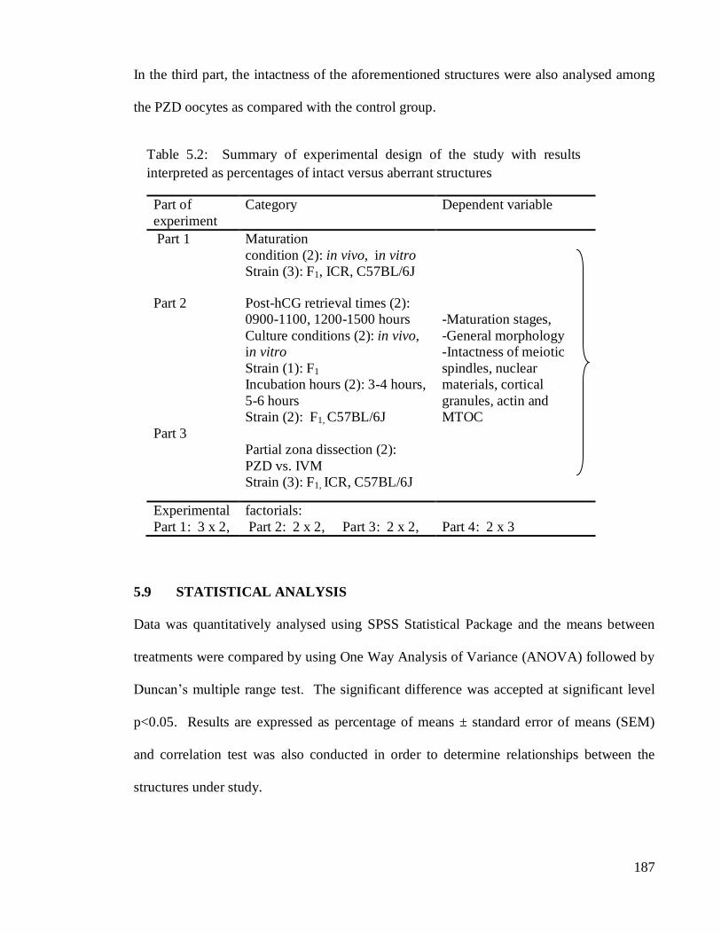
In the third part, the intactness of the aforementioned structures were also analysed among
the PZD oocytes as compared with the control group.
Part of
experiment
Category Dependent variable
Part 1 Maturation
condition (2): in vivo, in vitro
Strain (3): F1, ICR, C57BL/6J
Part 2
Post-hCG retrieval times (2):
0900-1100, 1200-1500 hours
Culture conditions (2): in vivo,
in vitro
Strain (1): F1
-Maturation stages,
-General morphology
-Intactness of meiotic
spindles, nuclear
Part 3
Incubation hours (2): 3-4 hours,
5-6 hours
Strain (2): F1, C57BL/6J
Partial zona dissection (2):
PZD vs. IVM
Strain (3): F1, ICR, C57BL/6J
materials, cortical
granules, actin and
MTOC
Experimental
Part 1: 3 x 2,
factorials:
Part 2: 2 x 2, Part 3: 2 x 2,
Part 4: 2 x 3
5.9 STATISTICAL ANALYSIS
Data was quantitatively analysed using SPSS Statistical Package and the means between
treatments were compared by using One Way Analysis of Variance (ANOVA) followed by
Duncan’s multiple range test. The significant difference was accepted at significant level
p<0.05. Results are expressed as percentage of means ± standard error of means (SEM)
and correlation test was also conducted in order to determine relationships between the
structures under study.
Table 5.2: Summary of experimental design of the study with results
interpreted as percentages of intact versus aberrant structures

188
5.10 RESULTS
The results of each experiment are grouped into several sections as follows:
5.10.1 Fluorescence images of in vivo and in vitro oocytes in different strains of mouse.
5.10.2 Effects of post-hCG retrieval times on oocytes maturation and morphology.
5.10.3 Effects of in vitro maturation durations on oocyte structures.
5.10.4 Effects of partial zona dissection on oocyte structures.
5.10.1 Fluorescence Images of In Vivo and In Vitro Oocytes in Different Strains of
Mice
The results are discussed and divided into various sections as the following order.
5.10.1.1 General morphology
The fluorescently stained in vivo as well as in vitro treated mouse oocytes revealed
comparable morphology and polarity for the architectures and distributions of the meiotic
spindles, cortical granules and the nuclear materials between strains. Fluorescence staining
clearly distinguished the animal from the vegetal pole (Figure 5.1), which was usually
inconspicuously observed under the conventional bright field microscope, unless the
vesicular structures were found, aggregated at the vegetal pole as shown in Figure 5.2. In
Figure 5.1, eccentric location of cortical granules (Figure 5.1b, red), meiotic spindle (Figure
5.1c, yellowish green) and the nuclear materials (Figure 5.1d, blue) were clearly shown as
compared with the oocyte’s corresponding image obtained from the bright field image
(Figure 5.1a). Little information could be extracted from the bright field image. However,
in certain circumstances, the nuclear materials and the meiotic spindles that were fixed in
formaldehyde solution could be traced under the bright field microscope. The structures
then were reaffirmed and clearly visualised under the fluorescent light (Figures 5.3).

189
Figure 5.2: Bright-field image.
Arrows showing granular
vesicles at the vegetal pole of
the oocyte. (400x)
Figure 5.1: Comparative images between the bright-field and fluorescently
stained oocytes. (a) Bright-field image of MI oocyte, (b) the corresponding
oocyte stained with TRITC-LCA, (c) meiotic spindles, (d) composite image of
bright-field and nuclear materials stained with Hoechst. ap-animal pole, ms-
meiotic spindle, nm-nuclear materials, pm-plasma membrane. (400x)
Debris
nm
Artifact
ms
ap
pm
d c b a

190
Shape of oocytes was not strain-specific too. Images showed that most of the
oocytes were of round shape, while others appeared oval, potato shape, egg-shape and
irregular round shape. One of the contributing factors for such variations could be due to
how they were placed and positioned onto the coverslips or the glass slides. The shape of
an oocyte could also be used as the indicative criterion for determining the oocyte poles.
Although, it might not be always applicable to all oocytes, nevertheless, for most of the
Figure 5.3: Top: Bright-field images of MI oocyte. (a1), (b1)
arrows showing the nuclear materials, (c1) arrow showing meiotic
spindles. Bottom: Fluorescence images of the corresponding
structures. (a2), (b2) arrows showing the nuclear materials that were
stained with Hoechst, (c2) the meiotic spindle (arrow) that was
stained with FITC. ms-meiotic spindle, nm-nuclear materials.
(400x)
a1
a2
b1
b2 c2
c1
nm
nm
nm
nm
ms
ms
191
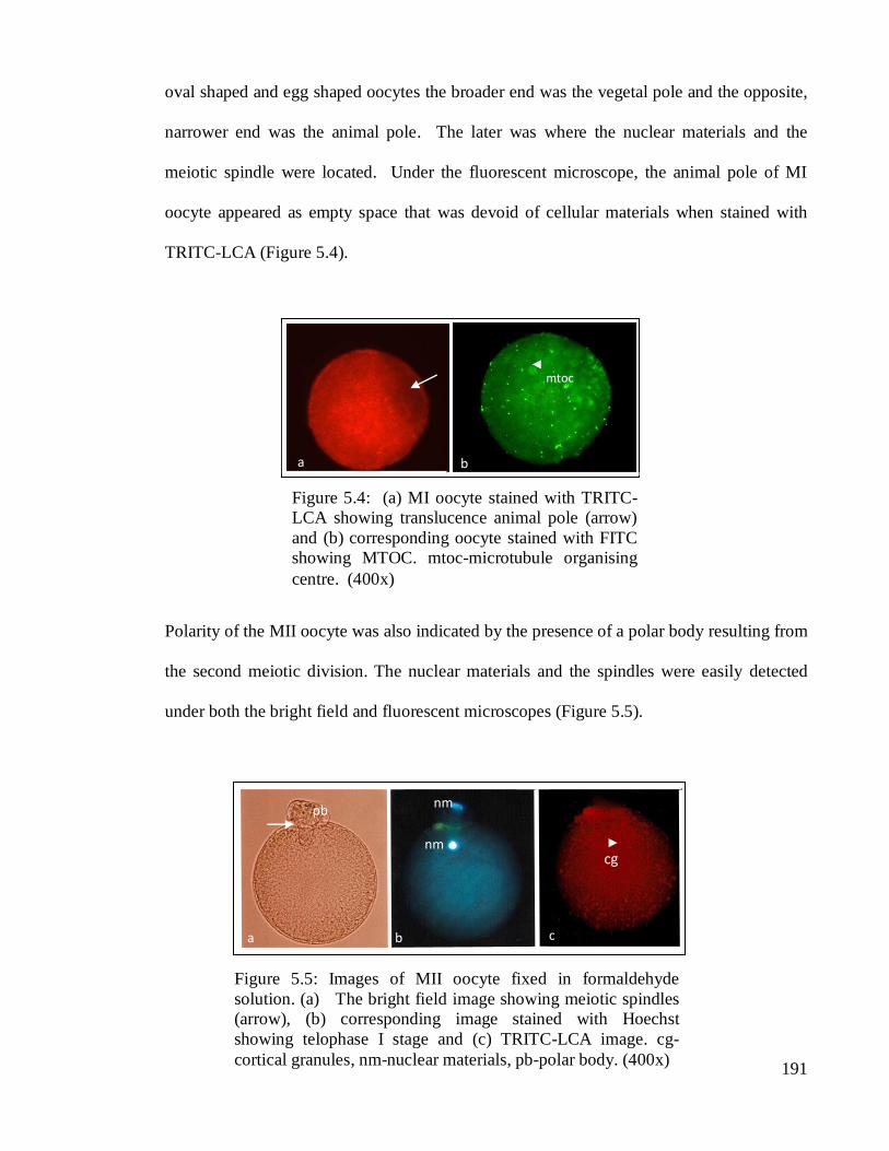
oval shaped and egg shaped oocytes the broader end was the vegetal pole and the opposite,
narrower end was the animal pole. The later was where the nuclear materials and the
meiotic spindle were located. Under the fluorescent microscope, the animal pole of MI
oocyte appeared as empty space that was devoid of cellular materials when stained with
TRITC-LCA (Figure 5.4).
Polarity of the MII oocyte was also indicated by the presence of a polar body resulting from
the second meiotic division. The nuclear materials and the spindles were easily detected
under both the bright field and fluorescent microscopes (Figure 5.5).
Figure 5.4: (a) MI oocyte stained with TRITC-
LCA showing translucence animal pole (arrow)
and (b) corresponding oocyte stained with FITC
showing MTOC. mtoc-microtubule organising
centre. (400x)
a b
mtoc
Figure 5.5: Images of MII oocyte fixed in formaldehyde
solution. (a) The bright field image showing meiotic spindles
(arrow), (b) corresponding image stained with Hoechst
showing telophase I stage and (c) TRITC-LCA image. cg-
cortical granules, nm-nuclear materials, pb-polar body. (400x)
a b c
pb nm
nm cg
192
In general, most of the oocytes displayed their nuclear materials and the meiotic
spindles at the periphery of the animal pole while cortical granules were abundance either
at the centre or at the bottom hemisphere towards the vegetal pole. The spindles of the MI
and MII oocytes were arranged and positioned differently. In MI oocytes the meiotic
spindles extended in tangential position but radial or vertical arrangement for the MII
oocytes. The meiotic spindles among the MI oocytes might be less indicative of the
maturation stage because its position is also determined by the angle at which an oocyte
was attached onto the coverslips. However, staining had revealed various stages of nuclear
and cytoplasmic maturation among the in vivo and in vitro oocytes. In Figures 5.6, 5.7, 5.8
and 5.9 archives of fluorescence images of the MI and MII oocytes obtained from the
different strains of mice were shown. Figure 5.6 shows various arrangements and angles of
the meiotic spindles in MI oocytes, revealing the metaphase I stage. A few asters were
also seen too in Figure 5.6a. In Figure 5.7, the different views of the nuclear materials were
displayed representing metaphase I and metaphase II stages, respectively, among the
oocytes. For example, 2n set of chromosomes arranged in circular pattern could be
observed during the metaphase II stage (Figures 5.7c, 5.7d).

193
Figure 5.6: Meiotic spindles (MS) of the MI oocytes. (a) Zona-intact oocyte of C57BL/6J, (b)
and (c) showing the different shapes and sizes of MS in ICR oocytes and (d) zonaless oocyte of
C57BL/6J. ms-meiotic spindle, mtoc-microtubule organising centre. (400x)
b a
ms ms
c d
mtoc
Figure 5.7: Nuclear maturation stages: (a) Metaphase I of F1 hybrid, (b) MII of C57BL/6J
oocyte, (c) MII of F1oocyte and (d) MII of ICR oocytes. Inset of Figure 5.7b shows
nuclear materials of MII stage at 1000x magnification.
MII
c a d
MI MII MII
b
Figure 5.8: Cortical granules (arrows) in MI (a and b) and MII (c) oocytes. (d) Composite of
three fluorescent dyes (blue for NM, green for MS and red for CG). cg-cortical granules, pb-
polar body, nm-nuclear materials. (400x)
b a c d
cg cg
pb cg nm
194
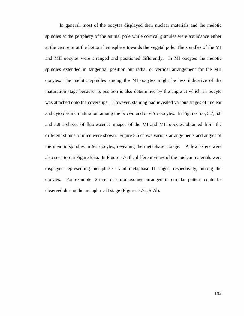
Figure 5.8 reveals the distributions of cortical granules in MI and MII oocytes, respectively.
Healthy unfertilised oocytes have CG centrally distributed or aggregated at the vegetal pole
meanwhile uneven distribution was observed among the unhealthy ones as shown in Figure
5.8b. As previously reported (Schatten et al., 1992), a unique characteristic of murine
oocytes was the presence of microtubule organising centre (MTOC) among the unfertilised
oocytes. It was observed as small dense round structures with green fluorescence as
exhibited in Figures 5.9a, c. The MTOC numbers varied between oocytes and treatments
and were found to be distributed at all locations regardless of the poles.
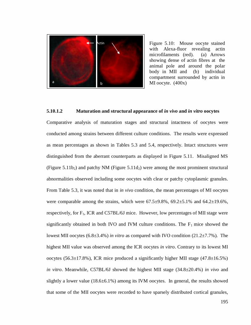
Compartmentalisation could be observed in some oocytes as shown in Figure 5.9a. This
could be related with organisation of the microfilaments such as actin as shown in Figure
5.10. In Figure 5.10a, actin appeared as a thin layer surrounding an oocyte and it was
thicker toward the animal pole especially around the polar body of MII oocyte. Also, actin
distribution was observed to be related to smaller compartments of MI oocyte as shown by
arrows in Figure 5.10b.
MTOC
Figure 5.9: Microtubules organising centres (MTOC) in MI oocyte . (a) F1
oocyte with arrows showing tiny compartments at the animal pole, (b)
C57BL/6J oocyte. Arrow showing the meiotic spindles, (c) compacted nuclear
materials (arrow) of oocyte, (d) arrow showing villi of oocyte. (400x)
b c d a
villi
195
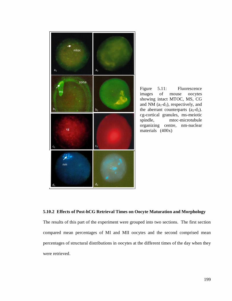
5.10.1.2 Maturation and structural appearance of in vivo and in vitro oocytes
Comparative analysis of maturation stages and structural intactness of oocytes were
conducted among strains between different culture conditions. The results were expressed
as mean percentages as shown in Tables 5.3 and 5.4, respectively. Intact structures were
distinguished from the aberrant counterparts as displayed in Figure 5.11. Misaligned MS
(Figure 5.11b2) and patchy NM (Figure 5.11d2) were among the most prominent structural
abnormalities observed including some oocytes with clear or patchy cytoplasmic granules.
From Table 5.3, it was noted that in in vivo condition, the mean percentages of MI oocytes
were comparable among the strains, which were 67.5±9.8%, 69.2±5.1% and 64.2±19.6%,
respectively, for F1, ICR and C57BL/6J mice. However, low percentages of MII stage were
significantly obtained in both IVO and IVM culture conditions. The F1 mice showed the
lowest MII oocytes (6.8±3.4%) in vitro as compared with IVO condition (21.2±7.7%). The
highest MII value was observed among the ICR oocytes in vitro. Contrary to its lowest MI
oocytes (56.3±17.8%), ICR mice produced a significantly higher MII stage (47.8±16.5%)
in vitro. Meanwhile, C57BL/6J showed the highest MII stage (34.8±20.4%) in vivo and
slightly a lower value (18.6±6.1%) among its IVM oocytes. In general, the results showed
that some of the MII oocytes were recorded to have sparsely distributed cortical granules,
Figure 5.10: Mouse oocyte stained
with Alexa-fluor revealing actin
microfilaments (red). (a) Arrows
showing dense of actin fibres at the
animal pole and around the polar
body in MII and (b) individual
compartment surrounded by actin in
MI oocyte. (400x) a b
Actin

196
aggregation of granular materials at the oocyte circumference, circular arrangement of
nuclear materials, darker image at the animal pole which contained mass of condensed
nuclear materials and radial arrangement of meiotic spindles (Figure 5 (a-d)).
The mean percentages of structural occurrences among the cohorts of IVO and IVM
matured oocytes between strains were tabulated in Table 5.4. The NM and CG were found
to be consistently higher among the IVO and IVM matured oocytes. The highest value for
the respective structures was 93.53.1% in IVM of C57BL/6J and 98.31.7%
in IVO of F1
mice. The highest percentages of MS for both IVM and IVO oocytes were detected in
C57BL/6J oocytes with values of 78.515.7% and 65.716.5%, respectively. These were
significantly higher (p<0.05) than the MS percentage in IVO oocyte of F1; the lowest with
23.87.9%. However, higher percentage of MS was obtained among its IVM counterpart
Strain Maturation condition
IVO (%)
(Mean±SEM)
IVM (%)
(Mean±SEM)
MI MII MI MII
F1 67.5± 9.8a,y
21.2±7.7a,x
77.4±10.8a,y
6.8±3.4a,x
ICR 69.2± 5.1a,y
27.3±5.5a,x
56.3±17.8a, x, y
47.8±16.5b, x, y
C57BL/6J 64.2± 19.6a, y, z
34.8± 20.4a, x, y
78.8± 8.6a, z
18.6± 6.1a, x
Table 5.3: Mean percentages of MI and MII oocytes in different maturation
conditions
a, b means within a column in a group differ significantly at p<0.05.
x, y, z means within a row in a group differ significantly at p<0.05.

197
with 40.68.4%. In addition to MS, the IVM oocytes of C57BL/6J also showed highest
percent of NM with value of 93.53.9%. This, as well as the percentage of NM in its IVO
oocytes were significantly higher (p<0.05) than that obtained from IVO of ICR oocytes
which was 67.311.1%. It was revealed that approximately only 7.5% of MI oocytes of the
IVO groups showed nuclear and cytoplasmic degradations. The F1 oocytes displayed high
anomalies of nuclear materials whereas patchy cortical granules were found to be higher in
ICR mice. Higher values of degraded nuclear materials were observed among the MI
oocytes cultured in vitro with approximately 10.3% and 15.7%, respectively, for ICR and
C57BL/6J. Approximately 5% patchy cytoplasmic granules were observed among ICR and
F1 oocytes, respectively. The MII oocytes showed that there were approximately 7% patchy
nuclear materials and 10% patchy cytoplasmic materials detected among the IVM groups as
compared with smaller percentages found among the IVO groups.
Contrary to MS and NM, MTOC structure was comparatively higher among the
IVO than the IVM oocytes in all the strains, especially for F1 oocytes, which differed
significantly (p<0.05) between the maturation groups. The percentages for its IVO and
IVM matured oocytes were 54.810.9% and 21.37.3%, respectively. Mouse oocytes
displayed maternally derived MTOC. It was observed that MTOC numbers ranged
between 1 to 33 and 1 to 28, in IVO and IVM oocytes, respectively. However, a majority
of the oocytes across the strains produced either 1 or 2 counts of MTOC only and
approximately 5% or less showed more than 3 or bigger amount of MTOC per oocyte.
Large amount of IVO and IVM produced metaphase I oocytes lacked MTOC. Nonetheless,
among those with intact MTOC, the F1 strain displayed varying amount of the structure
ranging from 0-33 in vivo and 0-18 in vitro. The ICR and C57BL/6J each displayed
between 0-19 and 0-23 MTOC, respectively, during in vivo condition and a range between

198
0-28 and 0-15, respectively, during in vitro condition. In comparison to MI, the amount of
MII oocyte with MTOC structure was higher for both culture conditions and most of them
displayed either 1 or 2 MTOC only. In addition to MTOC, the highest percentage of CG
appearance was also noted among the IVO oocytes of F1 with value of 98.3 1.7%.
Nevertheless, no significant difference was observed among the strains as well as between
the maturation groups.
High correlation was determined between selected structures of oocytes. The CG
was highly correlated with MS, MTOC and NM in IVO oocytes of F1 mice, but weak
correlations between structures of IVM oocytes. Low correlation values also were found
among the structures of IVO oocytes in ICR mice. However, the MTOC structure in its
IVM counterparts was highly correlated with NM and MS. On the other hand, the CG of
C57BL/6J oocytes was found to correlate strongly with all the structures being studied in
both maturation groups. Also, a high correlation was observed between the MS and NM in
the IVM oocytes of C57BL/6J.
Treatment
Oocyte
structures
IVO (%)
(Mean SEM)
IVM (%)
(Mean SEM)
F1 ICR C57BL/6J F1 ICR C57BL/6J
MS 23.87.9a
(n=54)
40.214.0 a,b (n=57)
65.716.5b
(n=41)
40.68.4a,b
(n=95)
42.719.3 a,b
(n=44)
78.5 15.7b
(n=48)
MTOC 54.8 10.9a
(n=54)
51.65.0a,b
(n=57)
40.38.8ª,b
(n=41)
21.37.3b
(n=95)
45.214.9ª,b
(n=44)
30.3 10.7ª,b
(n=48)
NM 89.93.3ª,b
(n=83)
67.311.1a
(n=77)
91.3 5.4b
(n=53)
79.14.0 a,b
(n=107)
81.914.3ª,b
(n=45)
93.5 3.9b
(n=72)
CG 98.31.7a
(n=29)
94.23.9 a
(n=46)
80.610.0a
(n=15)
83.65.6a
(n=57)
73.013.1a
(n=27)
80.45.4a
(n=11)
Table 5.4: Mean percentages of structural occurrences of IVO and IVM oocytes in the different
strains of mice
199
5.10.2 Effects of Post-hCG Retrieval Times on Oocyte Maturation and Morphology
The results of this part of the experiment were grouped into two sections. The first section
compared mean percentages of MI and MII oocytes and the second comprised mean
percentages of structural distributions in oocytes at the different times of the day when they
were retrieved.
Superscripts a,b in a row differ significantly at P<0.05
a,b means in a row in a group differ significantly at P<0.05
Figure 5.11: Fluorescence
images of mouse oocytes
showing intact MTOC, MS, CG
and NM (a1-d1), respectively, and
the aberrant counterparts (a2-d2).
cg-cortical granules, ms-meiotic
spindle, mtoc-microtubule
organizing centre, nm-nuclear
materials (400x)
MTOC
d2 d1
a2
b1 b2
c1 c2
ms
cg
nm
zona
a1
mtoc
200
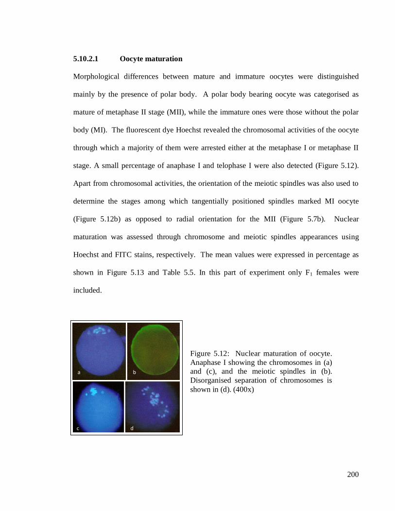
5.10.2.1 Oocyte maturation
Morphological differences between mature and immature oocytes were distinguished
mainly by the presence of polar body. A polar body bearing oocyte was categorised as
mature of metaphase II stage (MII), while the immature ones were those without the polar
body (MI). The fluorescent dye Hoechst revealed the chromosomal activities of the oocyte
through which a majority of them were arrested either at the metaphase I or metaphase II
stage. A small percentage of anaphase I and telophase I were also detected (Figure 5.12).
Apart from chromosomal activities, the orientation of the meiotic spindles was also used to
determine the stages among which tangentially positioned spindles marked MI oocyte
(Figure 5.12b) as opposed to radial orientation for the MII (Figure 5.7b). Nuclear
maturation was assessed through chromosome and meiotic spindles appearances using
Hoechst and FITC stains, respectively. The mean values were expressed in percentage as
shown in Figure 5.13 and Table 5.5. In this part of experiment only F1 females were
included.
Supercripts a,b
in a row differ significantly at P<0.05
Figure 5.12: Nuclear maturation of oocyte.
Anaphase I showing the chromosomes in (a)
and (c), and the meiotic spindles in (b).
Disorganised separation of chromosomes is
shown in (d). (400x)
a b
c d

201
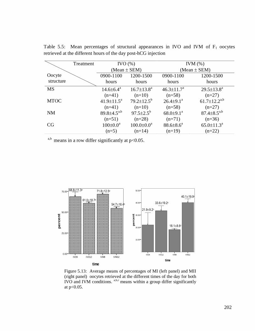
The means percentages of the MI and MII oocytes varied between the retrieval
times post-hCG injection in both groups. The IVM group showed the highest frequency of
MI oocytes at 0900-1100 hours and the lowest at 1200-1500 hours of the day.
Correspondingly, its low and high values of the MII stage were also detected during the
respective hours. The percentages of MII oocytes for both IVO and IVM groups were
higher during 1200-1500 hours, which were 33.6±19.2% and 40.1±19.9%, respectively. In
addition, high correlation values were obtained among the structures of IVM oocytes
retrieved at the later hours (Appendix 3.5). The percentages of MII oocytes were not
significantly different between the different retrieval hours of the day in both conditions.
However, a significantly low MI (54.718.4%) was found at 1200-1500 hours in vitro as
compared to early hours of oocyte retrieval (71.812.5%).
5.10.2.2 Structural appearance of IVO and IVM oocytes retrieved at the different
times of the day post hCG injection
The percentages of MS, MTOC, NM and CG appearances in both IVO and IVM oocytes
are summarised in Table 5.5. Significant variations (p<0.05) were detected in only MTOC
and NM among the treatment groups. The values of MTOC (79.212.5%) and NM
(97.52.5%) were significantly higher among IVO oocytes retrieved at 1200-1500 hours of
the day. Comparatively, most of the structures were higher among IVO than IVM oocytes,
except for the MS. This was especially so at 1200-1500 hours of retrieval time. The MS
was highest at 0900-1100 hours of retrieval time (46.311.7%) in in vitro condition. The
CG values were consistently higher at both retrieval times especially among the IVO
oocytes and markedly reduced at 1200-1500 hours in vitro.
a,b means differ significantly at P<0.05 between the MI stage oocyte.
n=number of oocyte
(*) significant at p<0.05
(**) Significant at p<0.01
202
Error Bars show Mean +/- 1.0 SE
Bars show Means
IVO9 IVO12 IVM9 IVM12
time
0.00
25.00
50.00
75.00
pe
rce
nt
]
]
]
]
Error Bars show Mean +/- 1.0 SE
Bars show Means
IVO9 IVO12 IVM9 IVM12
time
10.00
20.00
30.00
40.00
50.00
pe
rce
nt
V
V
V
V
68.811.3a
61.018.7a
71.812.5a
54.718.4b
21.99.2c
33.619.2c
18.18.6c
40.119.9c
Treatment
Oocyte
structure
IVO (%)
(Mean SEM)
IVM (%)
(Mean SEM)
0900-1100
hours
1200-1500
hours
0900-1100
hours
1200-1500
hours
MS 14.66.4a
(n=41)
16.713.8a
(n=10)
46.311.7a
(n=58)
29.513.8a
(n=27)
MTOC 41.911.5a
(n=41)
79.212.5b
(n=10)
26.49.1a
(n=58)
61.712.2a,b
(n=27)
NM 89.84.5a,b
(n=51)
97.52.5b
(n=28)
68.09.1a
(n=71)
87.48.5a,b
(n=36)
CG 1000.0a
(n=5)
100.00.0a
(n=14)
88.68.6a
(n=19)
65.011.3a
(n=22)
Table 5.5: Mean percentages of structural appearances in IVO and IVM of F1 oocytes
retrieved at the different hours of the day post-hCG injection
a,b means in a row differ significantly at p<0.05.
n=number of oocyte
Figure 5.13: Average means of percentages of MI (left panel) and MII
(right panel) oocytes retrieved at the different times of the day for both IVO and IVM conditions.
a,b,c means within a group differ significantly
at p<0.05.
203
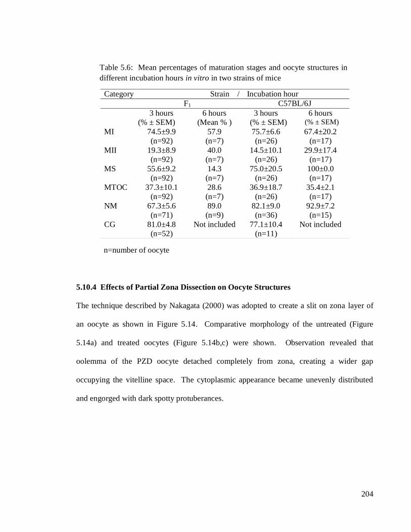
5.10.3 Effect of Incubation Hours on Maturation Stages and Structural Appearance of
IVM Oocyte.
In this experiment, oocytes from two strains of mice which were the F1 and C57BL/6J were
retrieved 13 hours post-hCG and cultured in vitro. The durations of incubation were 3 hours
and 6 hours, respectively, for each group. The results entailing maturation stages and
structural appearance of the oocytes are presented in Table 5.6. For 3 hours incubation, the
percentages of MI and MII oocytes of C57BL/CJ were negatively correlated (p<0.05) as
well as between the MII stage with the MS. The correlation values were r=-0.954 and r=-
0.992, respectively. Its MS however, was positively correlated with MI stage (r=0.962).
Among the F1 oocytes, a negative correlation (r=-0.9720) was also determined between the
MI and MII stages during 3 hours incubation in vitro. Meanwhile, among MII stage,
positive correlations were found between MTOC and MS with CG, with r=0.634 and
r=0.678, respectively. No correlation values were computed for 6 hours incubation due to
small sample sizes in both strains.
The mean percentages of MII oocytes were found to be higher during 6 hours
incubation in both strains. The values amounted to 40% for the F1 and 30% for C57BL/6J
oocytes. The MS and NM appearances were consistently high during both incubation
hours with C57BL/6J oocytes outnumbered the MS of F1‘s when almost all of the oocytes
(100%) showed intact MS. It was 56% and 14%, respectively, of MS for 3 hours and 6
hours incubation for the later strain. The NM percentage was also higher during 6 hours
incubation for both F1 and C5BL/6J oocytes. The highest value was 93% of C57BL/6J
oocytes. The MTOC of both strains were low in which less than 50% of the oocytes
displayed the structure during both hours of incubation period.
204
5.10.4 Effects of Partial Zona Dissection on Oocyte Structures
The technique described by Nakagata (2000) was adopted to create a slit on zona layer of
an oocyte as shown in Figure 5.14. Comparative morphology of the untreated (Figure
5.14a) and treated oocytes (Figure 5.14b,c) were shown. Observation revealed that
oolemma of the PZD oocyte detached completely from zona, creating a wider gap
occupying the vitelline space. The cytoplasmic appearance became unevenly distributed
and engorged with dark spotty protuberances.
Category Strain / Incubation hour
F1 C57BL/6J
3 hours
(% ± SEM)
6 hours
(Mean % )
3 hours
(% ± SEM)
6 hours (% ± SEM)
MI 74.5±9.9
(n=92)
57.9
(n=7)
75.7±6.6
(n=26)
67.4±20.2
(n=17)
MII 19.3±8.9
(n=92)
40.0
(n=7)
14.5±10.1
(n=26)
29.9±17.4
(n=17)
MS 55.6±9.2
(n=92)
14.3
(n=7)
75.0±20.5
(n=26)
100±0.0
(n=17)
MTOC 37.3±10.1
(n=92)
28.6
(n=7)
36.9±18.7
(n=26)
35.4±2.1
(n=17)
NM 67.3±5.6
(n=71)
89.0
(n=9)
82.1±9.0
(n=36)
92.9±7.2
(n=15)
CG 81.0±4.8
(n=52)
Not included 77.1±10.4
(n=11)
Not included
Table 5.6: Mean percentages of maturation stages and oocyte structures in
different incubation hours in vitro in two strains of mice
n=number of oocyte

205
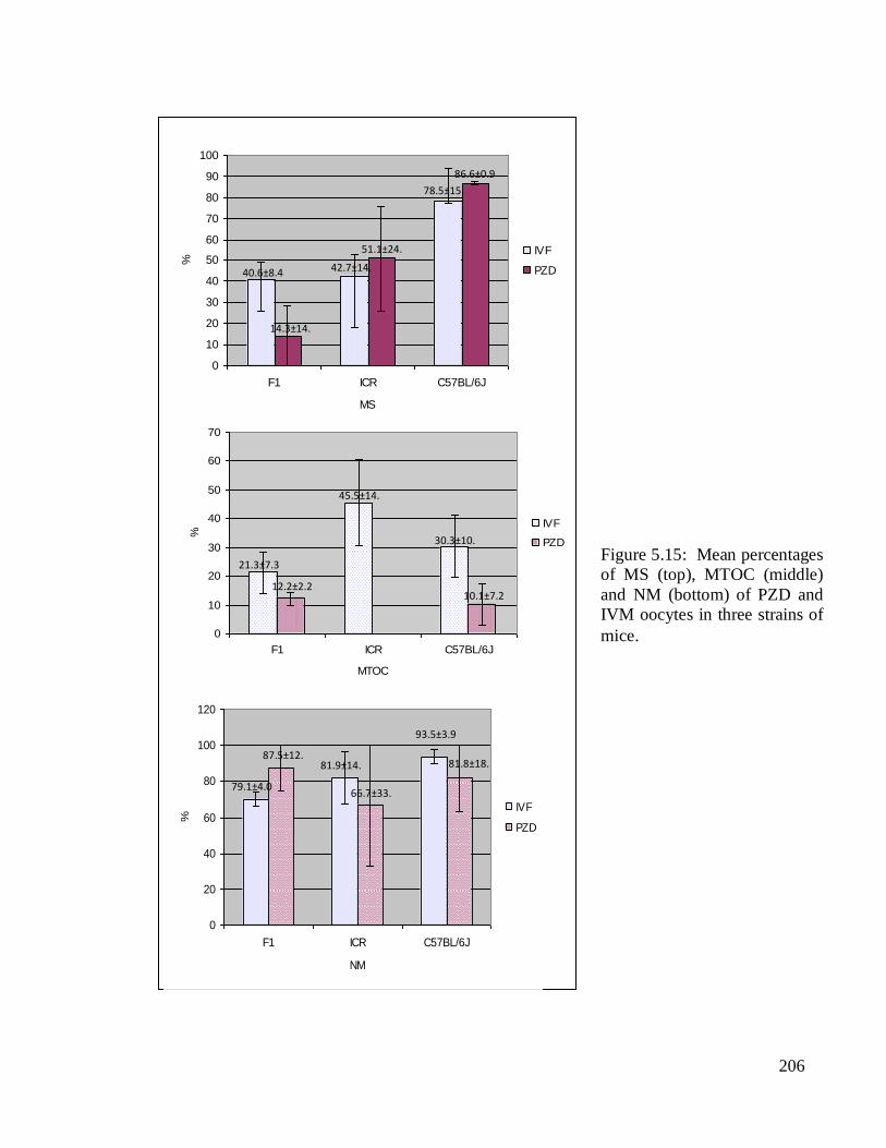
Fluorescence staining of the MS, MTOC and NM revealed insignificant structural
appearances between the treated and untreated oocytes in the three strains of mice. The
mean percentages of each category were represented by the bar charts as displayed in
Figure 5.15. Higher percentages of MS were comparatively observed in both of PZD
oocytes of ICR (51.1±24.8%) and C57BL/6J (86.6±0.9%) than their IVM counterparts.
The lowest MS value (14.3±14.3%) was detected in PZD oocytes of F1. The NM in both of
PZD and IVM oocytes were consistently high with the highest value detected in IVM
oocytes of C57BL/6J and the lowest was among PZD oocytes of ICR. The respective
values were 93.5±3.9% and 66.7±33.3%. Unlike the NM, the percentages of MTOC were
comparatively lower in PZD than IVM oocytes. This was especially so in ICR mice when
none of its oocytes showed MTOC structure. Low MTOC was similarly observed among
PZD oocytes of both C57BL/6J and F1, with values of 10.1±7.2% and 12.2±2.2%,
respectively.
Figure 5.14. Bright-field images of a normal MII oocyte (a) and PZD oocytes
(b) and (c). Arrows showing the slits made on the zona layer. pb-polar body, vs-
peri-vitelline space. (400x)
vs
pb
a b c
206
Figure 5.15: Mean percentages
of MS (top), MTOC (middle)
and NM (bottom) of PZD and
IVM oocytes in three strains of
mice.
0
10
20
30
40
50
60
70
80
90
100
F1 ICR C57BL/6J
MS
%
IVF
PZD40.6±8.4
14.3±14.
3
42.7±14.
3
51.1±24.
8
78.5±15.
1
86.6±0.9
0
10
20
30
40
50
60
70
F1 ICR C57BL/6J
MTOC
%
IVF
PZD
21.3±7.3
12.2±2.2
45.5±14.
9
30.3±10.
7
10.1±7.2
0
20
40
60
80
100
120
F1 ICR C57BL/6J
NM
%
IVF
PZD
79.1±4.0
81.9±14.
3
81.8±18.
2
87.5±12.
5
66.7±33.
3
93.5±3.9
207
5.11 DISCUSSION
The fate of mammalian oocytes relies greatly on its molecular machinery functions and
interactions, which is manifested into structural appearance and functions. However, the
developmental competency of oocytes varies due to external influences and manipulative
environment. In this experiment, oocyte maturation stages and morphological appearance
of selected structures namely MS, MTOC, NM and CG using ICC technique have been
extensively described and compared between several treatments and strains of mice.
Although the works depended mostly on conventional apparatus and instrumentations,
nevertheless, the objectives of the experiment had been successfully accomplished
revealing enormous findings on microscopic anatomy of the mouse oocytes. Using
fluorescence staining technique, observation on temporal and spatial distribution of selected
structures of oocytes had been successfully compared and elucidated. Comprehensive
information from both the brightfield and the fluorescence images of the oocytes were
collectively gathered to advance our understanding on structural organisation and factors
affecting mammalian oocyte maturation. The objectives of the experiments; a) to compare
structural appearance between MI and MII oocytes, b) to analyse structural development of
oocyte in different maturation conditions, c) to identify structural differences of oocytes
retrieved at different hours of the day and d) to determine the effect of mechanical stress
such as partial zona dissection on oocyte structures.
In natural condition, mammalian oocytes which are quiescence at prophase I,
resume its meiotic division to metaphase II at ovulation and eventually completing meiotic
II only at time when it is triggered by sperm at fertilisation. A mature ovulated oocyte can
be morphologically distinguished from immature ones by the presence of polar body. In
laboratory environment, oocytes are produced in prodigious amount by the ovaries which
208
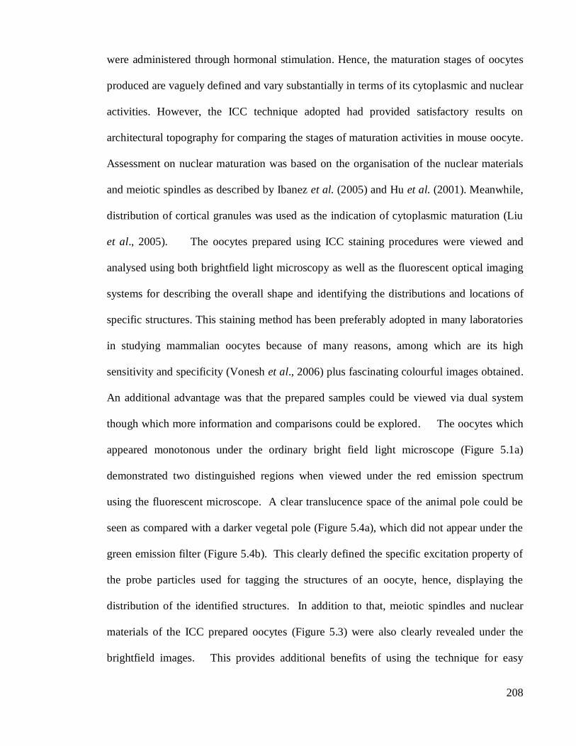
were administered through hormonal stimulation. Hence, the maturation stages of oocytes
produced are vaguely defined and vary substantially in terms of its cytoplasmic and nuclear
activities. However, the ICC technique adopted had provided satisfactory results on
architectural topography for comparing the stages of maturation activities in mouse oocyte.
Assessment on nuclear maturation was based on the organisation of the nuclear materials
and meiotic spindles as described by Ibanez et al. (2005) and Hu et al. (2001). Meanwhile,
distribution of cortical granules was used as the indication of cytoplasmic maturation (Liu
et al., 2005). The oocytes prepared using ICC staining procedures were viewed and
analysed using both brightfield light microscopy as well as the fluorescent optical imaging
systems for describing the overall shape and identifying the distributions and locations of
specific structures. This staining method has been preferably adopted in many laboratories
in studying mammalian oocytes because of many reasons, among which are its high
sensitivity and specificity (Vonesh et al., 2006) plus fascinating colourful images obtained.
An additional advantage was that the prepared samples could be viewed via dual system
though which more information and comparisons could be explored. The oocytes which
appeared monotonous under the ordinary bright field light microscope (Figure 5.1a)
demonstrated two distinguished regions when viewed under the red emission spectrum
using the fluorescent microscope. A clear translucence space of the animal pole could be
seen as compared with a darker vegetal pole (Figure 5.4a), which did not appear under the
green emission filter (Figure 5.4b). This clearly defined the specific excitation property of
the probe particles used for tagging the structures of an oocyte, hence, displaying the
distribution of the identified structures. In addition to that, meiotic spindles and nuclear
materials of the ICC prepared oocytes (Figure 5.3) were also clearly revealed under the
brightfield images. This provides additional benefits of using the technique for easy

209
searching and locating the structures and most importantly to minimise bleaching effect due
to long exposure of samples to the UV light. A smaller end of the animal pole where the
meiotic spindles and nuclear materials were eccentrically assembled was void of granular
materials in contrast to a broader vegetal pole with numerous aggregations of vesicular
structures as seen in Figure 5.2. Segregation of oocyte structures that are associated with
anterior-posterior positioning of an oocyte are determined by both genes and signals. Both
of these elements have been successfully identified (Martin, et al., 2003). In relation to that,
further studies could be conducted that include advanced search on signaling
communication (Schubach, 2008) for regulating structural distribution associated with
oocyte polarity. Variation in signaling pathways of oocyte amongst the strains and culture
conditions are yet to be compared and resolved.
The general morphology and maturation stages of mouse oocytes have been
extensively described (Schatten et al., 1992; Segers et al., 2008). These included the
patterns of spindle appearance and nuclear maturation (Hu et al., 2001; Ibanez et al., 2005)
and cortical granules distribution (Liu et al., 2005). Morphological observation revealed
variation of shapes among the oocytes, most of which were round. Others have oval shape,
potato shape and egg shape. It was difficult to correlate shape of oocytes with strains of
mice. The images observed were also affected by the way oocytes were positioned onto
coverslips. The use of a chambered glass slide is recommended in future microscopic
analyses to avoid cell damage and cell loss. The spacer of the slide would ease the
mounting process underneath the coverslip and may allow ones to observe intact live cells.
Observation also revealed compartmentalised segments in some oocytes. This was
especially so among the Alexa-flour®568 stained oocytes, which defined actin distribution.
The similar segments were inconspicuously shown in oocyte stained with FITC-conjugated

210
anti-tubulin (Figure 5.9). The result not only characterised the distribution of actin but also
its relationships with other structures of an oocyte. This unexceptional appearance of actin
in MI oocyte might be correlated with its important role in aggregation of MTOC and
movement of spindle to oocyte periphery (Calarco, 2005). In MII oocytes, actin
concentrated mostly at the animal pole surrounding the polar body, revealing its role in
cytokinesis. Actin has also been reported to be inseparable with cortical granules
exocytosis (Wessels et al., 2002; Schietrome et al., 2007) and migration of nuclear
materials to the periphery of the mammalian oocytes (Sun and Schatten, 2006). Actin
distribution was briefly highlighted in this section because of several limitations among
which was insufficient number of fluorescent filters to perform multiple staining. Due to
that, ICC staining for actin was separately conducted on the different batches of oocytes.
Consequently, it involved extra samples, time and energy inputs.
In this experiment, fluorescence images clearly distinguished immature from mature
oocytes. This was achieved by assessing both its nuclear and cytoplasmic maturations. A
prominent morphological comparison that distinguishes MI from MII oocytes is the
appearance of a polar body in the later stage. Meanwhile, cellular responses to mark
cytoplasmic maturation are relocation of organelles and exocytosis of the cortical granules
(Liu et al., 2005). Although it is basically known that only a small percentage of
mammalian oocytes would attain maturity, nevertheless, some variations were observed
among the strains as well as between the culture conditions. Overall results showed less
than 50% of oocytes attained MII stage in most strains and treatment groups in both in vivo
and in vitro conditions. In general, in vitro culture had profoundly increased the number of
mature oocytes. This was proven in ICR oocytes. In other strains such as the F1 and
C57BL/6 mice, longer exposure to in vitro culture had improved MII oocyte collection.
211
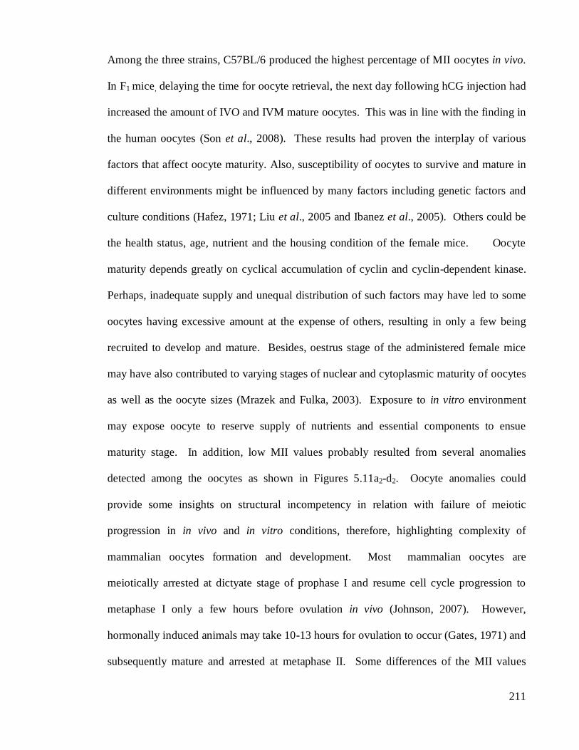
Among the three strains, C57BL/6 produced the highest percentage of MII oocytes in vivo.
In F1 mice, delaying the time for oocyte retrieval, the next day following hCG injection had
increased the amount of IVO and IVM mature oocytes. This was in line with the finding in
the human oocytes (Son et al., 2008). These results had proven the interplay of various
factors that affect oocyte maturity. Also, susceptibility of oocytes to survive and mature in
different environments might be influenced by many factors including genetic factors and
culture conditions (Hafez, 1971; Liu et al., 2005 and Ibanez et al., 2005). Others could be
the health status, age, nutrient and the housing condition of the female mice. Oocyte
maturity depends greatly on cyclical accumulation of cyclin and cyclin-dependent kinase.
Perhaps, inadequate supply and unequal distribution of such factors may have led to some
oocytes having excessive amount at the expense of others, resulting in only a few being
recruited to develop and mature. Besides, oestrus stage of the administered female mice
may have also contributed to varying stages of nuclear and cytoplasmic maturity of oocytes
as well as the oocyte sizes (Mrazek and Fulka, 2003). Exposure to in vitro environment
may expose oocyte to reserve supply of nutrients and essential components to ensue
maturity stage. In addition, low MII values probably resulted from several anomalies
detected among the oocytes as shown in Figures 5.11a2-d2. Oocyte anomalies could
provide some insights on structural incompetency in relation with failure of meiotic
progression in in vivo and in vitro conditions, therefore, highlighting complexity of
mammalian oocytes formation and development. Most mammalian oocytes are
meiotically arrested at dictyate stage of prophase I and resume cell cycle progression to
metaphase I only a few hours before ovulation in vivo (Johnson, 2007). However,
hormonally induced animals may take 10-13 hours for ovulation to occur (Gates, 1971) and
subsequently mature and arrested at metaphase II. Some differences of the MII values
212
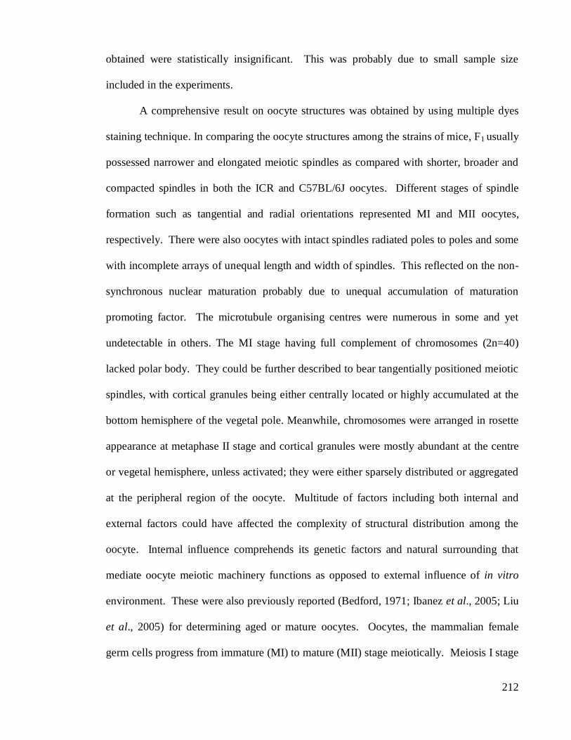
obtained were statistically insignificant. This was probably due to small sample size
included in the experiments.
A comprehensive result on oocyte structures was obtained by using multiple dyes
staining technique. In comparing the oocyte structures among the strains of mice, F1 usually
possessed narrower and elongated meiotic spindles as compared with shorter, broader and
compacted spindles in both the ICR and C57BL/6J oocytes. Different stages of spindle
formation such as tangential and radial orientations represented MI and MII oocytes,
respectively. There were also oocytes with intact spindles radiated poles to poles and some
with incomplete arrays of unequal length and width of spindles. This reflected on the non-
synchronous nuclear maturation probably due to unequal accumulation of maturation
promoting factor. The microtubule organising centres were numerous in some and yet
undetectable in others. The MI stage having full complement of chromosomes (2n=40)
lacked polar body. They could be further described to bear tangentially positioned meiotic
spindles, with cortical granules being either centrally located or highly accumulated at the
bottom hemisphere of the vegetal pole. Meanwhile, chromosomes were arranged in rosette
appearance at metaphase II stage and cortical granules were mostly abundant at the centre
or vegetal hemisphere, unless activated; they were either sparsely distributed or aggregated
at the peripheral region of the oocyte. Multitude of factors including both internal and
external factors could have affected the complexity of structural distribution among the
oocyte. Internal influence comprehends its genetic factors and natural surrounding that
mediate oocyte meiotic machinery functions as opposed to external influence of in vitro
environment. These were also previously reported (Bedford, 1971; Ibanez et al., 2005; Liu
et al., 2005) for determining aged or mature oocytes. Oocytes, the mammalian female
germ cells progress from immature (MI) to mature (MII) stage meiotically. Meiosis I stage
213
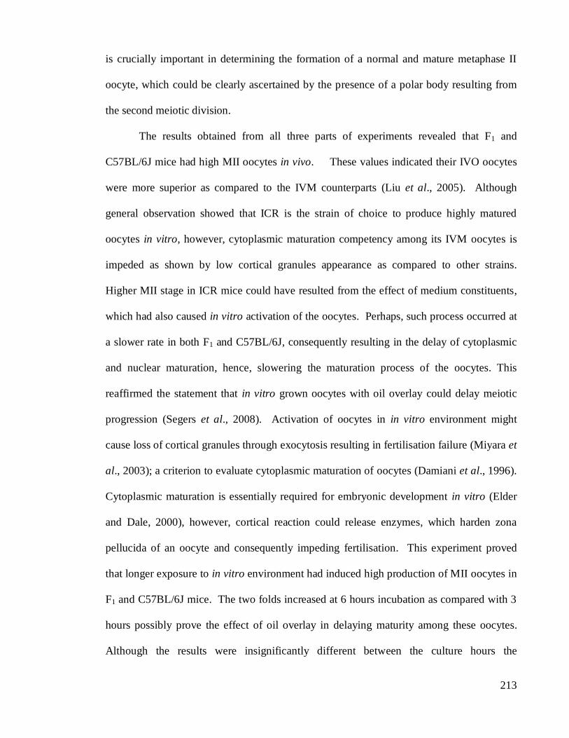
is crucially important in determining the formation of a normal and mature metaphase II
oocyte, which could be clearly ascertained by the presence of a polar body resulting from
the second meiotic division.
The results obtained from all three parts of experiments revealed that F1 and
C57BL/6J mice had high MII oocytes in vivo. These values indicated their IVO oocytes
were more superior as compared to the IVM counterparts (Liu et al., 2005). Although
general observation showed that ICR is the strain of choice to produce highly matured
oocytes in vitro, however, cytoplasmic maturation competency among its IVM oocytes is
impeded as shown by low cortical granules appearance as compared to other strains.
Higher MII stage in ICR mice could have resulted from the effect of medium constituents,
which had also caused in vitro activation of the oocytes. Perhaps, such process occurred at
a slower rate in both F1 and C57BL/6J, consequently resulting in the delay of cytoplasmic
and nuclear maturation, hence, slowering the maturation process of the oocytes. This
reaffirmed the statement that in vitro grown oocytes with oil overlay could delay meiotic
progression (Segers et al., 2008). Activation of oocytes in in vitro environment might
cause loss of cortical granules through exocytosis resulting in fertilisation failure (Miyara et
al., 2003); a criterion to evaluate cytoplasmic maturation of oocytes (Damiani et al., 1996).
Cytoplasmic maturation is essentially required for embryonic development in vitro (Elder
and Dale, 2000), however, cortical reaction could release enzymes, which harden zona
pellucida of an oocyte and consequently impeding fertilisation. This experiment proved
that longer exposure to in vitro environment had induced high production of MII oocytes in
F1 and C57BL/6J mice. The two folds increased at 6 hours incubation as compared with 3
hours possibly prove the effect of oil overlay in delaying maturity among these oocytes.
Although the results were insignificantly different between the culture hours the
214
result may provide some significant impacts on IVM or IVF practices and eventually its
end results. Nowadays, many IVF procotols especially in mice commonly required 3 to 4
hours of in vitro insemination (Hogan, 1986; Jackson Laboratory, 2008; Elder and Dale,
2000) prior to in vitro culture. Accordingly, optimal insemination and maturation hours in
vitro should be more specific among the strains due to their varied responses. This is in
lined with many reports on the influence of genetic background (Polanski, 1986; Ibanez et
al., 2005) and constituents of the culture medium (Liu et al., 2005) on oocyte maturation
and IVF. Moreover, in vitro maturation is known to alter the meiotic time course as
reported by Segers et al. (2008). They found that oil overlay for in vitro prepared oocytes
caused a differential hormonal exposure among in vitro grown mouse follicles by a
differential segregation into the oil overlay. However, this did not impair the competence
of cultured oocytes to proceed to MII, but delayed meiosis I progression. In ICR,
relocation of cortical granules could have been related to in vitro exposure which induced
oocyte meiotic progression from metaphase II to anaphase II and subsequently telophase II.
Spontaneous activation of some mammalian oocytes has been reported once they were
aspirated from mature follicles (Grondahl, 2008). Overall, oocyte maturation is a process
that could arrest and resume in response to different stimuli (Persson et al., 2005).
Developmental competency of IVO and IVM oocytes depends upon synchronous
maturation of both nuclear and cytoplasmic materials which are manifested by
reorganisation of oocyte structures including chromosomal and meiotic spindles assembly
and redistribution of vesicular structures such as cortical granules. A significantly high
prevalence of meiotic spindles in in vitro condition was found among all strains of mice as
compared with the IVO groups, especially in C57BL/6J oocytes. This was consistent with
the earlier report (Ibanez et al., 2005) that the environment in which the oocyte undergoes

215
maturation is a critical factor in establishing and maintaining microtubule patterning in
mouse oocytes. Moreover, high meiotic spindles occurrence in C57BL/6J oocytes was also
observed during 6 hours of incubation periods as compared with the F1 oocytes. Although
genetic influence on superiority of strains to survive in in vitro condition has been clearly
defined, however, factors that influenced C57BL/6J oocytes to produce a consistently high
occurrence of the entire structures understudy in both IVO and IVM oocytes need further
investigation. The IVM oocytes of F1 showed low MII count, however, it was noted that
its structural development was comparable with those of C57BL/6J except lower
percentages of MS and NM. In addition, delaying the time of oocyte retrieval not only
improved its survival in vitro but also consistently produced higher appearances of
structural development especially MTOC and NM in both of the IVO and IVM oocytes.
This was also proven by low correlation values obtained between oocyte maturity and
structural appearance at the earlier hours of retrieval time than the later hours. This showed
that structural development in F1 oocytes was time dependence. Interestingly, it was
reported by Son et al. (2008) that extending the period of hCG priming time from 35 to 38
hours for immature oocyte retrieval promotes oocyte maturation in vivo and increases the
IVM rate of immature oocytes. Therefore, oocyte retrieval after 38 h of hCG priming
may
improve subsequent pregnancy outcome, in cycles programmed
for IVM treatment.
Although this indicate the optimal time of oocyte collection by referring to its nuclear
maturation, however, the reduction in CG and MS appearance at 1200-1500 hours among
the IVM group in F1 oocyte, might hamper its fertilising competency. In addition to that,
the CG was also vaguely correlated with other structures among the IVM oocytes as
compared with the IVO counterparts. Since in vitro environment generally improved
oocyte maturity, therefore oocyte collection time of the day for IVM and IVF applications

216
in mice prior to midday is highly encouraged to avoid spontaneous cytoplasmic aging.
Oocyte collected later than the time may experience structural incompetency during
prolonged in vitro exposure that consequently may affect IVF results.
Recent technology has developed various methods and devices to circumvent
infertility. Micromanipulation technique usually requires oocyte to be handled with
mechanical instruments such as micromanipulation needles. However, little information is
available about its effects on oocyte structural developmental competency. In this
experiment, PZD method has been utilised to impose mechanical stress through zona
dissection and eventually exposed the oocyte contents to in vitro environment. There were
insignificant variations with respect to MS, MTOC and NM appearances between the IVM
and PZD oocytes. However, MS appearance in F1 was lower as compared with ICR and
C57BL/6J oocytes. Low MTOC and NM percentages were also observed in all the strains.
Hence, F1 oocytes could be more liable to experience structural impairment as compared
with ICR and C57BL/6J. Strain superiority for manipulative application such as PZD was
again shown by C57BL/6J oocytes based on its MS value and comparable results of MTOC
and NM with the IVM group. Thus, strain selection is crucial for manipulation purpose in
mouse oocytes. For example, many laboratories favour C57BL/6J as the experimental
strain in IVF and transgenic research. In this part of the experiment, CG appearance was
not detected due to various constraints especially sample size and technical problem.
Analyses on CG appearance would be advantageous to study oocyte cytoplasmic
maturation or aging among PZD oocytes. This may be paralleled with the results proposed
by Sigurdson et al. (1992) that mechanical stress can initiate Ca wave and ATP release
(Grygorzyk and Hanrahan, 1997; Yamamoto et al., 2000), which simulate the influence of
sperm penetration at fertilisation process. The disadvantages of using PZD method were; a)

217
it may cause severe damage to oocytes with rigorous cut and b) lysis of cytosol due to
osmotic influx of medium.
5.12 CONCLUSIONS
In conclusion, the experiments conducted provided archives of both brightfield and
fluorescence images of the MI and MII oocytes. Comparative maturation stages were
morphologically distinguished by the presence of polar body through bright-field
observation. Meanwhile nuclear and cytoplasmic maturations were determined through
ICC staining of the respective structures namely NM, MS and CG. Multiple staining of
ICC technique produces more informative and comprehensive morphological appearances
of the mouse oocytes. However, more structural analysis could be performed such as actin
distribution, if adequate supply of fluorescent filters is to be installed in the existing system.
Overall, the percentages of MII oocyte among the strains and treatment groups were low.
However, variation in the results revealed the effects of the following factors in MII yields.
Among all, C57BL/6J mice displayed strain superiority in both in vivo and in vitro
environments. This was proven by its consistently high outputs of MII and structural
appearances in both environments. The F1 oocytes have proven to increase the production
of MII stage in both IVO and IVM oocytes if retrieved at the later hours of the day
following hCG injection. This explains the delay in maturation stages. Extending the time
between hCG priming and oocyte collection has been associated with oocyte undergoing
complete cellular and maturation processes, hence, improved survival and competency (Son
et al., 2008). This was also emphasised by high correlation values among the structures
obtained among the MII oocytes collected at a later time of the day. Meanwhile, in vitro
environment has generally produced higher MII oocytes especially the ICR strain. Longer
exposure to in vitro culture had also influenced MII production in both C57BL/6J and F1
218
oocytes. Nevertheless, longer in vitro exposure had also resulted in cytoplasmic aging in
the F1 and ICR oocytes due to reduction of MS and CG appearance. Thus, the optimal
hours of oocyte collection is recommended between 13-15 hours post hCG injection, to
compromise the effect of in vitro activation and nuclear maturation. Structural anomalies
detected among the oocytes, which have probably contributed to low count of MII were
fragmented NM, disarrayed MS and patchy cytoplasm and CG. The effect of PZD
dissection showed insignificant effect among the oocytes, especially C57BL/6J mice.
Nevertheless, low appearance of MS and CG in PZD oocytes of F1 mice has shown its
lower competency for such treatment. The study had successfully identified some
contributing factors, with regards to oocyte survival and competency in in vivo and in vitro
environments that corresponded significantly with its structural developments. These
findings could be useful as reference for future research in mammalian reproductive
technology. Nevertheless, the application of advanced technique such as confocal imaging
is recommended and could possibly provide more insights on oocyte structural
development. Also, the use of chambered-slide is also proposed to ease staining steps when
dealing with round cells such as mammalian oocytes.
REFERENCES
Abbott, A.L., Z. Xu, G. S. Kopf, T. Duibella and R.M. Schultz. 1998. In vitro culture
retards spontaneous activation of cell cycle progression and cortical granule
exocytosis that normally occur in in vivo unfertilised mouse eggs. Biology of
Reproduction. 59(6):1515-1521.
Aktas, H., M.L. Lorainne and N.L. First. 2003. Meiotic state of bovine oocyte is regulated
by interactions between cAMP, cumulus, and granulosa. Molecular Reproduction
and Development. 65(3):336-343.
Bedford, J.M. 1971. Techniques and criteria used in the study of fertilisation. In: Methods
in Mammalian Embryology. Freeman International Edition. 37-63.
219
Brunet, S., A.S. Maria, P. Guillaud, D. Dujardin, J.Z. Kubiak and B. Maro. 1999.
Kinetochore fibres are not involved in the formation of the first meiotic spindle in
mouse oocyte, but control the exit from the first meiotic M phase. Journal of Cell
Biology. 146:1-11.
Byskov, A.G., C.Y. Anderson and L. Nordholm. 1995. Chemical structure of sterols that
activate oocyte meiosis. Nature. 374:559-562.
Calarco, P.G. 2005. The role of microfilaments in early meiotic maturation of mouse
oocyte. Microscopy and Microanalysis. 11:146-153.
Damiani, P., R.A. Fissore, J.B. Cibelli, C.R. Long, J.J. Balise, J.M. Robl and R.T. Duby.
1996. Evaluation of developmental competence, nuclear and ooplasmic maturation
of calf oocyte. Molecular Reproduction and Development. 45:521-534.
Daniel, J.C. 1971. Methods in Mammalian Embryology. Freeman International Edition.
De Loos, F.A.M., E. Zeinstra and M.M. Bevers. 1994. Follicular wall maintains meiotic
arrest in bovine oocyte cultured in vitro. Gamete Biology. (Abstract).
Downs, S.M., A.C. Schroeder and J.J. Eppig. 1986. Serum maintains the fertilising ability
of mouse oocyte matured in vitro by preventing the hardening of the zona pellucida.
Gamete Research. 15:115-122.
Ducibella, T. and J. Buetow. 1994. Competence to undergo normal, fertilisation-induced
cortical activation develops after metaphase I of cAMP levels and cAMP
phosphodiesterase activity. Developmental Biology. 165:95-104.
Ebner, T., M. Moser, C. Yamaan, O. Feichtinger, J. Harti and G. Tews. 1999. Elective
transfer of embryos selected on the basis of the first polar morphology is associated
with increased rates of implantation and pregnancy. Fertility and Sterility. 72:599-
603.
Elder, K. and B. Dale. 2000. In Vitro Fertilisation. Second Edition. Cambridge
University. 2-27.
Epigg, J.J. and A.C. Schroeder. 1989. Capacity of mouse oocyte from preantral follicles to
undergo embryogenesis and development to live young after growth, maturation
and fertilisation in vitro. Biology of Reproduction. 41(2):268-276.
Epigg, J.J., M. O’Brien and K. Wigglesworth. 1996. Mammalian oocyte growth and
development in vitro. Molecular Reproduction and Development. 44:260-273.
Gates, A.H. 1971. Maximal yield and developmental uniformity of eggs. In: Methods in
Mammalian Embryology. Freeman International Edition. 64-75.
220
Gobarsky, G.J., C. Simerly, G. Schatten and G.G. Borisy. 1990. Microtubule in the
metaphase-arrested mouse oocyte turn over rapidly. Proceedings of the National
Academy of Sciences USA. 87(16):6049-6053.
Grondahl, C., J.L. Otteson and M. Lessl. 1998. Meiosis-activating sterol promotes
resumption of meiosis in mouse oocyte cultured in vitro in contrast to related
oxysterols. Biology of Reproduction. 58:1297-1302.
Grondahl, C. 2008. Oocyte maturation. Danish Medical Bulletin. 55(1):1-16.
Grygorzyk, R. and J.W. Hanrahan. 1997. CFTR-independent ATP release from epithelial
cells triggered by mechanical stimuli. American Journal of Physiology and Cell
Physiology. 272: 1058-1066.
Hafez, E.S.E. 1971. Egg storage. In: Methods in Mammalian Embryology. Freeman
International Edition. 117-132.
Hewitson, L.C, C.R. Simerly, M.W. Tengowski, P. Sutovsky, C.S. Navara, A.J. Haavisto
and G. Schatten. 1996. Microtubule and chromatin configurations during rhesus
intracytoplasmic sperm injection: successes and failures. Biology of Reproduction.
55: 271-280.
Hogan, B., F. Constantini and E. Lacy. 1986. Manipulating the Mouse Embryo: A
Laboratory Manual. Cold Spring Harbor Laboratory, Cold Spring Harbor, NY,
USA.
Hu, Y., I. Betzendahl, R. Cortvrind, J. Smitz and U. Eichenlaub-Ritter. 2001. Effects of
low O2 and ageing on spindles and chromosomes in mouse oocyte from preantral
follicle culture. Human Reproduction. 16(4):737-748.
Ibanez, E., A. Sanfins, C.M.H. Combelles, E.W. Overstrom and D.F. Albertini. 2005.
Genetic strain variations in the metaphase-II phenotype of mouse oocyte matured in
vivo or in vitro. Reproduction. 130:845-855.
Jackson Laboratory. 2008. IVF protocol. Cryo.jax.org/ivf.html. Retrieved on June 23,
2008.
Johnson, H.M. 2007. Essential Reproduction. Sixth Edition. Blackwell Publishing. 185-
186.
Kono, T., J. Carroll, K. Swann and D.G. Whittingham. 1995. Nuclei from fertilised mouse
embryos have calcium-releasing activity. Development. 12:1123-1128.
Kubiak, J.Z., M. Weber, H. de Pennant, N.J. Winston and B. Maro. 1993. The metaphase
II arrest in mouse oocyte is controlled through microtubule-dependent destruction of
cyclin B in the presence of CSF. EMBO Journal 12:3773-3778.

221
Leader, B., H. Lim, M.J. Carabatsos, A. Harrington, J. Ecsedy, D. Pellman, R. Mass and P.
Leder. 2002. Formin-2, polyploidy, hypofertility and positioning of the meiotic
spindle in mouse oocyte. Biology of Reproduction. 61:1-7.
Lee, J. T. Miyano and R.M. Moor. 2000. Spindle formation and dynamics of γ-tubulin and
nuclear mitotic apparatus protein distribution during meiosis in pig and mouse
oocyte. Biology of Reproduction. 62:1184-1192.
Liu, J., A. Rybouchkin, J. Van der Eist and M. Dhont. 2003. Fertilisation of mouse oocyte
from in vitro matured preantral follicles using classical in vitro fertilisation or
intracytoplasmic sperm injection. Biology of Reproduction. 67(2):575-579.
Liu, X.Y., S.F. Mal, D.Q. Miao, D.J. Liu, S. Bao and J.H. Tan. 2005. Cortical granules
behave differently in mouse oocyte martyred under different conditions. Human
Reproduction. 20(12):3402-3413.
Martin, S.G., V. Leclerc, K. Smith-Litiere and D. St. Johnson. 2003. The identification of
novel genes required for Drosophila anteroposterior axis formation in germline
clone screen using GFP-Staufen. Development. 130:4201-4215.
Miyara, F., F.X. Aubriot, A. Glissant, C. Nathan, S. Douard, A. Stanovici, F. Herve, M.
Dumont-hassan, A.LeMeur, P. Cohen-Bacrie and P. Debey. 2003. Multiparameter
analysis of human oocyte at metaphase II stage after IVF failure in non-male
infertility. Human Reproduction. 18:1494-1503.
Mrazek, M. and J. F. Jr. Navara. 1994. Failure of oocyte maturation. Possible mechanisms
for oocyte maturation arrest. Human Reproduction. 18(1):2249-2252.
Nakagata, N. 2000. A Review. Mouse spermatozoa cryopreservation. Journal of
Mammalian Ova Research. 17:1-8.
Navara, C.S., N.L. First and G. Schatten. 1994. Microtubules organisation in the cow
during fertilisation, polyspermy, parthenogenesis and nuclear transfer. The role of
the sperm aster. Developmental Biology. 162:29-40.
Navarro-Garcia, F., C. Sears, C. Eslava, A. Cravioto and J.P. Nataro. 1999. Cytoskeletal
effects induced by pet, the serine protease enterotoxin of enteroaggregative
Escherichia coli . Infection and Immunity. 67(5):2184-2192.
Navarro, P.A., L. Liu, J.R. Trimachi and A.R. Ferriani. 2005. Non-invasive imaging of
spindle dynamics during mammalian oocyte activation. Fertility and Sterility.
83(1):1197-1205.
Persson, J.L., Q. Zhang, X.Y. Wang, S.E. Ravnik, S. Muhirad and D.J. Wolgemuth. 2005.
Distinct roles for the mammalian A-type cyclins during oogenesis. Reproduction.
130: 411-422.

222
Polanski, Z. 1986. In-vivo and in-vitro maturation rates of oocyte from two strains of mice.
Journal of Reproduction and Fertility. 78:103-109.
Schatten, G., C. Simerly and H. Schatten. 1985. Microtubule configurations during
fertilisation, mitosis, and early development in the mouse and the requirement for
egg microtubule-mediated motility during mammalian fertilisation. Proceedings of
the National Academy of Sciences USA. 82:4152–4156.
Schatten, H. and G. Schatten. 1986. Motility and centrosomal organisation during sea
urchin and mouse fertilisation. Cell Motility and Cytoskeleton. 6:163-175.
Schatten, G., C. Simerly and H. Schatten. 1991. Maternal inheritance of centrosomes in
mammals? Studies on parthenogenesis and polyspermy in mice. Proceedings of the
National Academy of Sciences USA. 88:6785-6789.
Schatten, H., M. Walter, H. Biessmann and G. Schatten. 1992. Activation of maternal
centrosomes in unfertilised sea urchin eggs. Cell Motility and Cytoskeleton. 23:61-
70.
Schatten, G.1994. The centrosome and its mode of inheritance: The reduction of the
centrosome during gametogenesis and its restoration during fertilisation.
Developmental Biology. 165:299-335.
Schietrome, C. H.Y. Yu, M.C. Wagner, J.A. Umbach, W.M. Bemen and C.B. Gundersen.
2007. A role for myosin in cortical granule exocytosis in Xenopus oocyte. Journal
of Biological Chemistry. 282:29504-29513.
Schultz, R.M., S. Kurasawa, Y. Endo and G.S. Kopf. 1989. Molecular events pre- and
post-fertilisation of mouse eggs. Oocyte maturation, egg activation and polyspermy
block. In: Medically Assisted Conception: Agenda for Research. National
Academy Press. Washington DC.
Schubach, T. 2008. Establishment of asymmetries during Drosophila oogenesis. Research
Abstract. Howard Hughes Medical Institute.
Segers, I., T. Adriaenssens, W. Coucke, R. Cortvrindt and J. Smitz. 2008. Timing of
nuclear maturation and postovulatory aging in oocyte of in vitro grown mouse
follicles with or without oil overlay. Biology of Reproduction. 78:839-868.
Simerly, C. and G. Schatten. 1993. Techniques for localisation of specific molecules in
oocyte and embryos. Methods in Enzymology. 225:516-553.
Sigurdson, W., A. Ruknudin and F. Sachs. 1992. Calcium imaging of mechanically
induced fluxes in tissue-cultured chick heart. Role of stretch activated ion channels.
American Journal of Physiology: Heart Circulation Physiology. 262:1110-1115.
Sirad, M.A. and P. Blondin. 1996. Oocyte maturation and IVF in cattle. Animal
Reproduction Science, Elsevier. 42:417-426.

223
Son, W.Y., J.T. Chung, R.C. Chian, B. Herrero, E. Demirtas, S. Elizur, Y. Gidoni, C.
Sylvestre, N. Dean and S.L. Tan. 2008. A 38 h interval between hCG priming and
oocyte retrieval increases in vivo and in vitro oocyte maturation rate in programmed
IVM cycles. Human Reproduction. 23(9):2010-2016.
Sun, Q.Y. and H. Schatten. 2006. Regulation of dynamic events by microfilaments during
oocyte maturation and fertilisation. Reproduction. 131:93-205.
Sorenson, R.A. and P.M. Wassarman. 1976. Relationship between growth and meiotic
maturation of the mouse oocyte. Developmental Biology. 50(2):(Abstract).
Suppinyopong, S., R. Choavaratana and C. Karavakul. 2000. Correlation of oocyte
morphology with fertilisation rate and embryo quality after intracytoplasmic sperm
injection. Journal of Medical Association, Thai. 83:627-632.
Tateno, H., and Y. Kamiguchi. 2007. Evaluation of chromosomal risk following
intracytoplasmic sperm injection in the mouse. Biology of Reproduction. Retrieved
on April 4, 2007 as DOI:10.1095/biolreprod.106.057778.
Veselska, R. and R. Janish. 2001. Cortical actin cytoskeleton in human oocyte: a
comparison with mouse oocyte. Scripta Medica. 74(4):265-274.
Vonesh, C., F. Aguet, J.L. Vonesh and M. Unser. 2006. The colored revolution of
bioimaging. IEEE Signal Processing Magazine. 3(23):20-31.
Wessel, G.M., S.D. Corner and L. Berg. 2002. Cortical granule translocation is
microfilament mediated and linked to meiotic maturation in the sea urchin oocyte.
Development. 129:4315-4325.
Yamamoto, K., R. Korenaga, A. Kamiya and J. Ando. 2000. Fluid shear stress activates Ca
influx into human endothelial cells via P2X4purinoceptors. Circulation Research.
87: 385-391.