
Chapter 25 - Napa Valley College 218/Martini PPT... · An Overview of the Digestive System •The...
158
Lecture Presentation by Steven Bassett Southeast Community College Chapter 25 The Digestive System © 2015 Pearson Education, Inc.
Transcript of Chapter 25 - Napa Valley College 218/Martini PPT... · An Overview of the Digestive System •The...
Lecture Presentation by
Steven Bassett
Southeast Community College
Chapter 25
The Digestive
System
© 2015 Pearson Education, Inc.

Introduction
• The digestive system consists of:
• The digestive tract
• Accessory organs of digestion
• Digestive tract
• Mouth
• Pharynx
• Esophagus
• Stomach
• Small intestine
• Large intestine
© 2015 Pearson Education, Inc.

Introduction
• Accessory Organs of the Digestive Tract
• Teeth
• Tongue
• Salivary glands
• Pancreas
• Liver
• Gallbladder
© 2015 Pearson Education, Inc.

Figure 25.1 Components of the Digestive System
© 2015 Pearson Education, Inc.
Major Subdivisions of
the Digestive Tract
Oral Cavity
Mechanical processing, moistening,
mixing with salivary secretions
Pharynx
Muscular propulsion of materials into
the esophagus
Mouth
Esophagus
Transport of materials to the stomach
Stomach
Chemical breakdown of materials via acid
and enzymes; mechanical processing
through muscular contractions
Small Intestine
Enzymatic digestion and absorption of
water, organic substrates, vitamins, and ions
Large Intestine
Enzymatic digestion and absorption of
water, organic substrates, vitamins, and ions
Anus
Accessory Organs of
the Digestive System
Salivary Glands
Secretion of lubricating fluid
containing enzymes that
break down carbohydrates
Liver
Secretion of bile (important
for lipid digestion), storage
of nutrients, many other
vital functions
Gallbladder
Storage and concentration
of bile
Pancreas
Exocrine cells secrete
buffers and digestive
enzymes; endocrine cells
secrete hormones

Introduction
• Functions of the Digestive System
• Ingestion
• Mechanical processing
• Digestion
• Secretion
• Absorption
• Excretion
• Compaction
© 2015 Pearson Education, Inc.

Introduction
• Functions of the Digestive System (details)
• Ingestion
• Bringing food and liquids into the mouth
• Mechanical processing
• Chewing and swallowing food
• Digestion
• Chemical breakdown of food into nutrient form
• Secretion
• Secretion of products by the lining of the digestive
tract
• Secretion of products by the accessory organs of
digestion© 2015 Pearson Education, Inc.

Introduction
• Functions of the Digestive System (continued)
• Absorption
• The movement of nutrients from the small intestine
to the bloodstream
• Excretion
• The removal of waste products from the digestive
tract
• Compaction
• Progressive dehydration of organic wastes
© 2015 Pearson Education, Inc.

An Overview of the Digestive System
• Histological Organization of the Digestive Tract
• There are four major layers of the digestive tract
• The mucosa
• The submucosa
• The muscularis externa
• The serosa
© 2015 Pearson Education, Inc.

Figure 25.2a Histological Structure of the Digestive Tract
© 2015 Pearson Education, Inc.
Three-dimensional view of the histological
organization of the general digestive tube
a
Mesenteric artery and vein
Mesentery Plica
Mucosa
Submucosa
Muscularis
externa
Serosa
(visceral
peritoneum)

An Overview of the Digestive System
• The Mucosa
• The inner lining of the digestive tract
• This is a mucous membrane
• Cells of the mucosa are either stratified or simple
• Oral cavity and esophagus are lined with
nonkeratinized stratified squamous cells (resist
stress and abrasion)
• Stomach, small intestine, and large intestine are
lined with simple columnar cells (for secretion and
absorption)
© 2015 Pearson Education, Inc.

An Overview of the Digestive System
• The Mucosa
• The mucosa of the small intestine makes up folds
called plicae
• Plicae increase the surface area for increased
absorption
• Lamina propria
• Contains blood vessels / nerves / smooth muscle /
lymphatic vessels
© 2015 Pearson Education, Inc.

Figure 25.2ab Histological Structure of the Digestive Tract
© 2015 Pearson Education, Inc.
Three-dimensional view of the histological
organization of the general digestive tube
a
Mesenteric artery and vein
Mesentery Plica
Mucosa
Submucosa
Muscularis
externa
Serosa
(visceral
peritoneum)
An enlarged section of the digestive
tube showing the structure of a plica
Plica
Mucosal
epithelium
Lamina
propria
Mucosa
Villi
Mucosal glands
Submucosal gland
Muscularis
mucosae
Lymphatic vessel
Artery and vein
Submucosal
plexus
Circular muscle
layer
Myenteric plexus
Longitudinal
muscle layer
b

An Overview of the Digestive System
• The Submucosa
• Surrounds the muscularis mucosae
• Large blood vessels and lymphatics are in this
layer
• Submucosal plexus innervates the mucosa
• Consists of sensory neurons
• Consists of parasympathetic ganglia
• Consists of sympathetic postganglionic fibers
© 2015 Pearson Education, Inc.

Figure 25.2ab Histological Structure of the Digestive Tract
© 2015 Pearson Education, Inc.
Three-dimensional view of the histological
organization of the general digestive tube
a
Mesenteric artery and vein
Mesentery Plica
Mucosa
Submucosa
Muscularis
externa
Serosa
(visceral
peritoneum)
An enlarged section of the digestive
tube showing the structure of a plica
Plica
Mucosal
epithelium
Lamina
propria
Mucosa
Villi
Mucosal glands
Submucosal gland
Muscularis
mucosae
Lymphatic vessel
Artery and vein
Submucosal
plexus
Circular muscle
layer
Myenteric plexus
Longitudinal
muscle layer
b

An Overview of the Digestive System
• The Muscularis Externa
• Surrounds the submucosa
• Dominated by smooth muscle fibers
• Forms sphincters or valves
• Innervated by myenteric plexus
• This is a network of parasympathetic ganglia and
sympathetic postganglionic fibers
© 2015 Pearson Education, Inc.

Figure 25.2ab Histological Structure of the Digestive Tract
© 2015 Pearson Education, Inc.
Three-dimensional view of the histological
organization of the general digestive tube
a
Mesenteric artery and vein
Mesentery Plica
Mucosa
Submucosa
Muscularis
externa
Serosa
(visceral
peritoneum)
An enlarged section of the digestive
tube showing the structure of a plica
Plica
Mucosal
epithelium
Lamina
propria
Mucosa
Villi
Mucosal glands
Submucosal gland
Muscularis
mucosae
Lymphatic vessel
Artery and vein
Submucosal
plexus
Circular muscle
layer
Myenteric plexus
Longitudinal
muscle layer
b

An Overview of the Digestive System
• The Serosa
• Covers the muscularis externa
• Outermost layer of the digestive system
© 2015 Pearson Education, Inc.

Figure 25.2ab Histological Structure of the Digestive Tract
© 2015 Pearson Education, Inc.
Three-dimensional view of the histological
organization of the general digestive tube
a
Mesenteric artery and vein
Mesentery Plica
Mucosa
Submucosa
Muscularis
externa
Serosa
(visceral
peritoneum)
An enlarged section of the digestive
tube showing the structure of a plica
Plica
Mucosal
epithelium
Lamina
propria
Mucosa
Villi
Mucosal glands
Submucosal gland
Muscularis
mucosae
Lymphatic vessel
Artery and vein
Submucosal
plexus
Circular muscle
layer
Myenteric plexus
Longitudinal
muscle layer
b

An Overview of the Digestive System
• Muscularis Layers and the Movement of
Digestive Materials
• The digestive tract consists of smooth muscle
• Muscularis mucosa and muscularis externa have
pacemaker cells
• Produce two types of muscle contractions
• Peristalsis
• Segmentation
© 2015 Pearson Education, Inc.
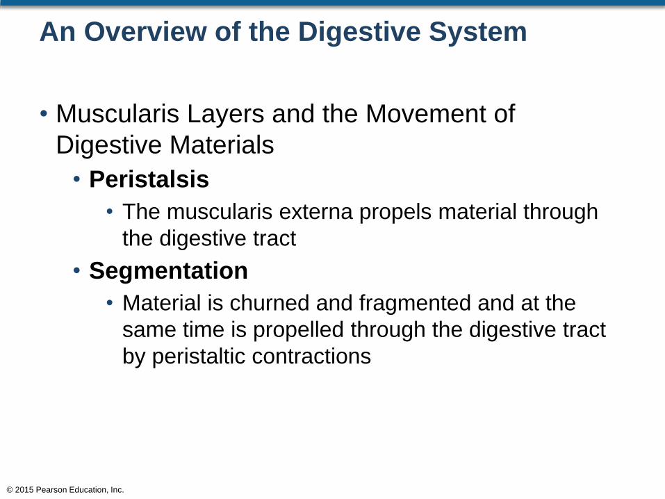
An Overview of the Digestive System
• Muscularis Layers and the Movement of
Digestive Materials
• Peristalsis
• The muscularis externa propels material through
the digestive tract
• Segmentation
• Material is churned and fragmented and at the
same time is propelled through the digestive tract
by peristaltic contractions
© 2015 Pearson Education, Inc.
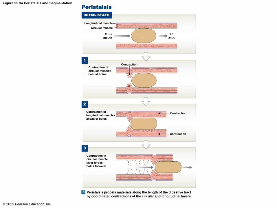
Figure 25.3a Peristalsis and Segmentation
© 2015 Pearson Education, Inc.
1
Longitudinal muscle
Peristalsis
INITIAL STATE
Circular muscle
From
mouth
To
anus
Contraction of
circular muscles
behind bolus
Contraction
2
Contraction
Contraction
Contraction of
longitudinal muscles
ahead of bolus
Contraction in
circular muscle
layer forces
bolus forward
3
a Peristalsis propels materials along the length of the digestive tract
by coordinated contractions of the circular and longitudinal layers.

Figure 25.3b Peristalsis and Segmentation
© 2015 Pearson Education, Inc.
b Segmentation movements primarily involve the circular muscle
layers. These activities churn and mix the contents of the digestive
tract, but do not produce net movement in a particular direction.
4
3
2
1
Segmentation

An Overview of the Digestive System
• The Peritoneum
• The serosa (visceral peritoneum) is continuous
with the parietal peritoneum
• The abdominal organs lie within the peritoneal
cavity or the abdominal cavity
• Intraperitoneal organs
• Retroperitoneal organs
• Secondarily retroperitoneal organs
© 2015 Pearson Education, Inc.
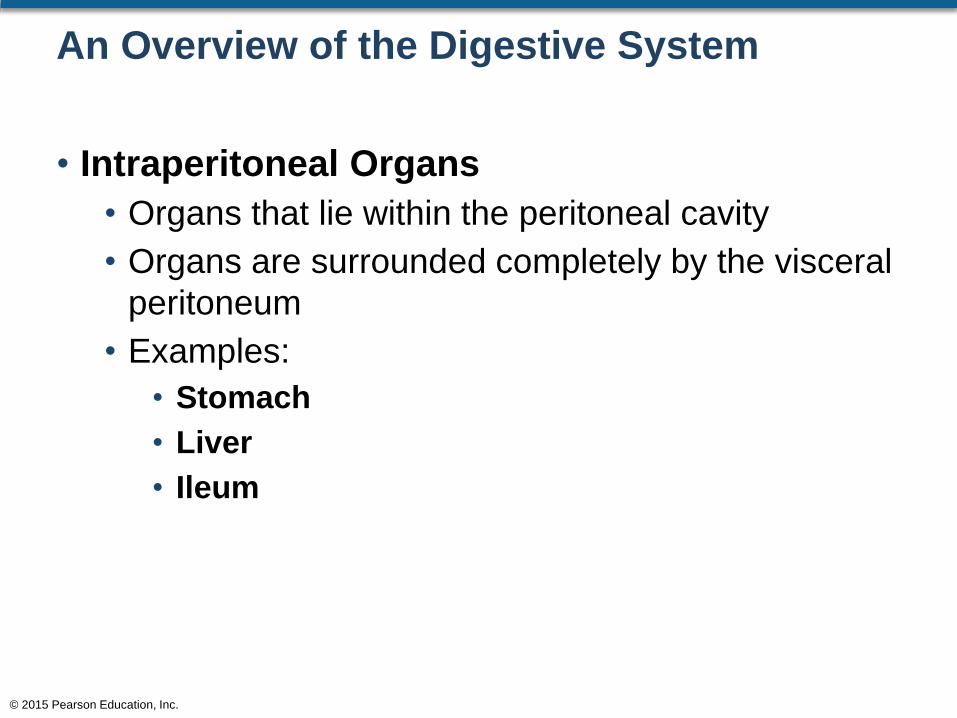
An Overview of the Digestive System
• Intraperitoneal Organs
• Organs that lie within the peritoneal cavity
• Organs are surrounded completely by the visceral
peritoneum
• Examples:
• Stomach
• Liver
• Ileum
© 2015 Pearson Education, Inc.

An Overview of the Digestive System
• Retroperitoneal Organs
• Organs are covered by the visceral peritoneum on
their anterior surface
• These organs lie outside the visceral peritoneum
• Examples:
• Kidneys
• Ureters
• Abdominal aorta
© 2015 Pearson Education, Inc.

An Overview of the Digestive System
• Secondarily Retroperitoneal Organs
• These organs form as intraperitoneal but soon
become retroperitoneal
• The change occurs during embryonic development
as the associated visceral peritoneum fuses with
the opposing parietal peritoneum
• Examples are:
• Pancreas
• Duodenum
© 2015 Pearson Education, Inc.

An Overview of the Digestive System
• Mesenteries
• These are fused double sheets of peritoneal
membrane
• Function:
• Stabilize the position of organs
• Stabilize the position of blood vessels
• Provide the attachment of blood vessels going to
and from the small intestine
© 2015 Pearson Education, Inc.

An Overview of the Digestive System
• Mesenteries (continued)
• All but the duodenum is suspended in a sheet of
mesentery called the mesentery proper
• Mesocolon
• Mesentery attached to the large intestine
• Transverse mesocolon
• Mesentery attached to the transverse colon
• Sigmoid mesocolon
• Mesentery attached to the sigmoid colon
© 2015 Pearson Education, Inc.

An Overview of the Digestive System
• Mesenteries (continued)
• Fusion Fascia
• The ascending colon, descending colon, and
rectum are attached to the posterior abdominal wall
via this fused mesentery
• Lesser Omentum
• This mesentery lies between the stomach and the
liver
• Greater Omentum
• This mesentery extends from the stomach and
covers the rest of the abdominal organs on the
anterior surface
© 2015 Pearson Education, Inc.

Figure 25.4d Mesenteries
© 2015 Pearson Education, Inc.
The organization of mesenteries in the adult.
This is a simplified view; the length of the
small intestine has been greatly reduced.
d
Lesser
omentum
Transverse
colon
Ascending
colon
Mesentery
proper
(mesenterial
sheet)
Greater
omentum (cut)
Transverse
mesocolon
Fusion fascia of
ascending and
descending
colons fuses
to dorsal
peritoneum
Descending
colon
Small
intestine
Sigmoid
colon

Figure 25.4b Mesenteries
© 2015 Pearson Education, Inc.
Mesenteries of the abdominopelvic cavity,
as seen in a diagrammatic sagittal section.
b
Falciform
ligament
Pancreas
Duodenum
Mesentery
proper
Sigmoid
mesocolon
Rectum
Urinary bladder
Liver
Diaphragm
Lesser
omentum
Stomach
Transverse
mesocolon
Transverse
colon
Greater
omentum
Parietal
peritoneum
Small
intestine
Uterus

The Oral Cavity
• Structures within the Oral Cavity
• Tongue
• Uvula
• Palatoglossal arches
• Salivary glands
• Teeth
© 2015 Pearson Education, Inc.

The Oral Cavity
• Anatomy of the Oral Cavity
• Lined by oral mucosa
• Consists of nonkeratinized stratified squamous
cells
• The oral mucosa is continuous with:
• Lining of the cheeks
• Lining of the lips
• Lining of the gums
© 2015 Pearson Education, Inc.
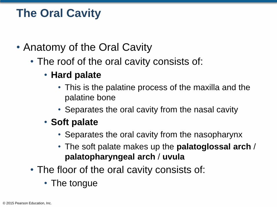
The Oral Cavity
• Anatomy of the Oral Cavity
• The roof of the oral cavity consists of:
• Hard palate
• This is the palatine process of the maxilla and the
palatine bone
• Separates the oral cavity from the nasal cavity
• Soft palate
• Separates the oral cavity from the nasopharynx
• The soft palate makes up the palatoglossal arch /
palatopharyngeal arch / uvula
• The floor of the oral cavity consists of:
• The tongue
© 2015 Pearson Education, Inc.

The Oral Cavity
• Anatomy of the Oral Cavity
• The oral cavity also houses the palatine tonsils
• These lie between the palatoglossal and
palatopharyngeal arches
• They are lateral to the uvula
© 2015 Pearson Education, Inc.

Figure 25.5b The Oral Cavity
© 2015 Pearson Education, Inc.
An anterior view of the oral cavity
as seen through the open mouth
Frenulum
of upper lip
Fauces
Pharyngeal
Arches
Palatoglossal
arch
Palatopharyngeal
arch
Palatine
tonsil
Lingual
frenulum
Gingiva
Vestibule
Frenulum
of lower lip
Tongue
Hard palate
Soft palate
Uvula
Openings of
submandibular
ducts
b

Figure 25.5a The Oral Cavity
© 2015 Pearson Education, Inc.
Hard
palate
a The oral cavity as seen in sagittal section
Soft
palate
Nasal cavity
Palatoglossal
arch
Opening of
parotid duct
Upper lip
Cheek
Dorsum of
tongue
Lower lip
Gingiva
Vestibule
Body of
tongue
Root of
tongue
Hyoid bone
Laryngopharynx
Epiglottis
Lingual tonsil
Oropharynx
Palatopharyngeal
arch
Fauces
Palatine tonsil
Uvula
Nasopharynx
Entrance to
auditory tube
Pharyngeal
tonsil

The Oral Cavity
• The Tongue
• Has numerous functions
• Manipulation of food
• Sensory analysis
• Secretion of enzymes to aid in fat digestion
• Movement for the formulation of words
© 2015 Pearson Education, Inc.

The Oral Cavity
• Tongue (continued)
• Can be divided into different areas
• Body
• Anterior portion of the tongue
• Root
• Posterior portion of the tongue
• Dorsum
• Superior portion of the tongue
• Contains the papillae
• Papillae contain the taste buds
© 2015 Pearson Education, Inc.

Figure 25.5a The Oral Cavity
© 2015 Pearson Education, Inc.
Hard
palate
a The oral cavity as seen in sagittal section
Soft
palate
Nasal cavity
Palatoglossal
arch
Opening of
parotid duct
Upper lip
Cheek
Dorsum of
tongue
Lower lip
Gingiva
Vestibule
Body of
tongue
Root of
tongue
Hyoid bone
Laryngopharynx
Epiglottis
Lingual tonsil
Oropharynx
Palatopharyngeal
arch
Fauces
Palatine tonsil
Uvula
Nasopharynx
Entrance to
auditory tube
Pharyngeal
tonsil
The Oral Cavity
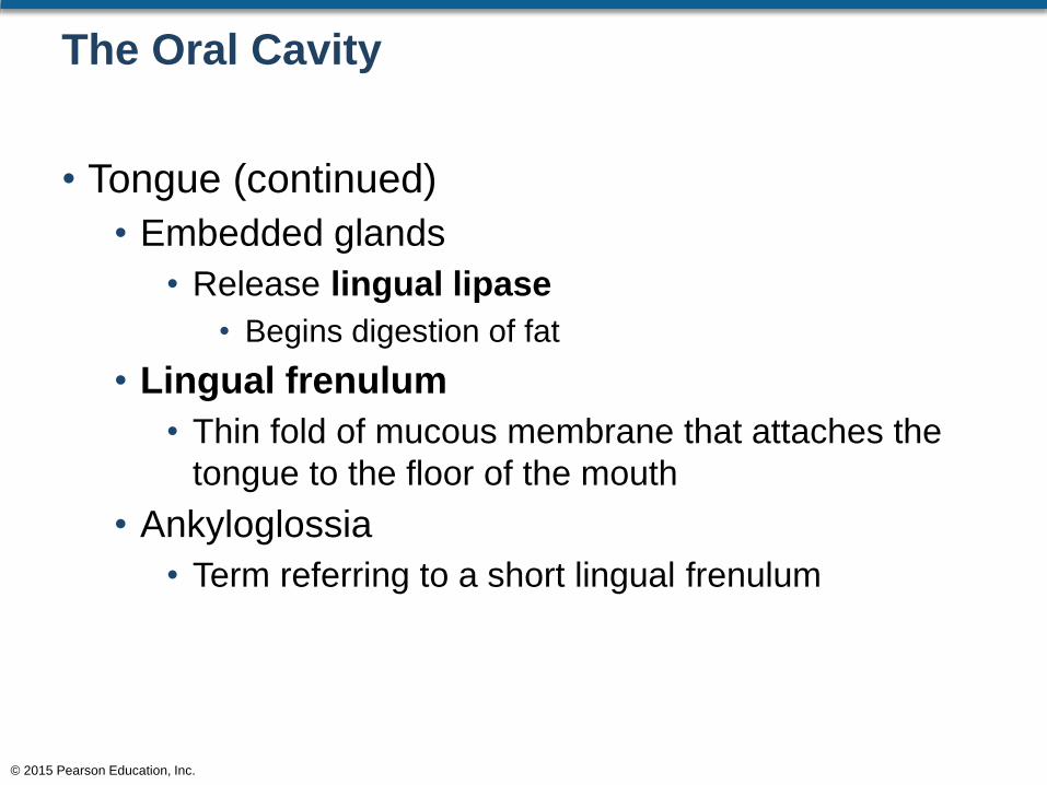
• Tongue (continued)
• Embedded glands
• Release lingual lipase
• Begins digestion of fat
• Lingual frenulum
• Thin fold of mucous membrane that attaches the
tongue to the floor of the mouth
• Ankyloglossia
• Term referring to a short lingual frenulum
© 2015 Pearson Education, Inc.

Figure 25.5b The Oral Cavity
© 2015 Pearson Education, Inc.
An anterior view of the oral cavity
as seen through the open mouth
Frenulum
of upper lip
Fauces
Pharyngeal
Arches
Palatoglossal
arch
Palatopharyngeal
arch
Palatine
tonsil
Lingual
frenulum
Gingiva
Vestibule
Frenulum
of lower lip
Tongue
Hard palate
Soft palate
Uvula
Openings of
submandibular
ducts
b

The Oral Cavity
• Tongue (continued)
• Consists of two muscle groups
• Intrinsic tongue muscles
• Alter the shape of the tongue
• Extrinsic tongue muscles
• Gross movements of the tongue
• Examples:
• Hyoglossus / Styloglossus / Genioglossus /
Palatoglossus
• Both sets of muscles are controlled by N XII
© 2015 Pearson Education, Inc.

Figure 10.7 Muscles of the Tongue
© 2015 Pearson Education, Inc.
Styloid process
Styloglossus
Genioglossus
Hyoglossus
Hyoid bone
Palatoglossus (cut)
Mandible
(cut)
Muscles of the Tongue

The Oral Cavity
• Salivary Glands
• There are three pairs of salivary glands
• Parotid
• Sublingual
• Submandibular
• All three glands produce salivary amylase
• Partially digests carbohydrates
© 2015 Pearson Education, Inc.

The Oral Cavity
• Salivary Glands
• Parotid salivary glands
• The largest of the three salivary glands
• Located on the lateral side of the face in the area of
the ramus of the mandible
• Enzyme drains to the mouth cavity via the parotid
duct
• Parotid duct lies on the masseter muscle
© 2015 Pearson Education, Inc.

Figure 25.6a The Salivary Glands
© 2015 Pearson Education, Inc.
a Lateral view showing the relative positions of the salivary glands and
ducts on the left side of the head. Much of the left half of the body
and the left ramus of the mandible have been removed. For the positions of the ducts inside the oral cavity, see Figure 25.5.
Parotid salivary
gland
Sublingual
salivary gland
Submandibular
salivary gland
Salivary Glands
Parotid duct
Openings of
sublingual ducts
Lingual frenulum
Opening of left
submandibular
duct
Submandibular duct

The Oral Cavity
• Salivary Glands
• Sublingual salivary glands
• Covered by the mucous membrane of the floor of
the mouth
• Consists of numerous sublingual ducts that open
along either side of the lingual frenulum
• Submandibular salivary glands
• Located on the floor of the mouth, deep into the
mandible, inferior to the mylohyoid line
• Submandibular ducts open posterior to the
mandibular teeth
© 2015 Pearson Education, Inc.
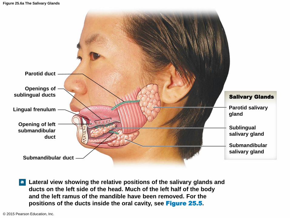
Figure 25.6a The Salivary Glands
© 2015 Pearson Education, Inc.
a Lateral view showing the relative positions of the salivary glands and
ducts on the left side of the head. Much of the left half of the body
and the left ramus of the mandible have been removed. For the positions of the ducts inside the oral cavity, see Figure 25.5.
Parotid salivary
gland
Sublingual
salivary gland
Submandibular
salivary gland
Salivary Glands
Parotid duct
Openings of
sublingual ducts
Lingual frenulum
Opening of left
submandibular
duct
Submandibular duct
The Oral Cavity
• Regulation of the Salivary Glands
• Secretions are controlled by the autonomic
nervous system
• Parasympathetic
• Accelerates salivary secretions
• Sympathetic
• Reduces salivary secretions
© 2015 Pearson Education, Inc.

The Oral Cavity
• The Teeth
• Designed for mastication
• Anatomy of teeth
• Crown
• Neck
• Root
• Dentine
• Pulp cavity
• Root canal
• Apical foramen
• Periodontal ligament
© 2015 Pearson Education, Inc.

The Oral Cavity
• Teeth Anatomy
• Crown
• Covered by enamel
• Consists of dentine
• Consists of pulp (highly vascularized)
• Neck
• Area of gingiva
• Root
• Consists of root canal
• Consists of artery, vein, and nerve
© 2015 Pearson Education, Inc.

The Oral Cavity
• Teeth Anatomy
• Dentine
• Mineralized matrix
• Different than bone; it does not contain cells
• Pulp cavity
• Spongy area and highly vascularized
• Root canal
• Arteries and veins and nerves pass through the
root canal to the pulp cavity area
© 2015 Pearson Education, Inc.

The Oral Cavity
• Teeth Anatomy
• Apical foramen
• An opening at the distal end of the root canal
• Periodontal ligament
• Anchors the root of the tooth to the alveolar sockets
• The articulation at this point is called gomphosis
© 2015 Pearson Education, Inc.

Figure 25.7a Teeth
© 2015 Pearson Education, Inc.
Diagrammatic section through
a typical adult tooth.
a
Crown
Neck
Root
Enamel
Dentine
Pulp cavity
Gingiva
Gingival sulcus
Cement
Periodontal ligament
Root canal
Bone of alveolus
Apical foramen
Dental vessels
and nerve

The Oral Cavity
• Types of Teeth
• Designed for mastication
• Four incisors per jaw
• Two cuspids per jaw
• Four bicuspids per jaw
• Four to six molars per jaw
© 2015 Pearson Education, Inc.

Figure 25.7c Teeth
© 2015 Pearson Education, Inc.
The normal orientation of adult teeth. The normal range of
ages at eruption for each tooth is shown in parentheses.
c
Maxillary
dental
arcade
Hard palate
Mandibular
dental
arcade
Central incisors (6–7 yr)
Lateral incisor (7–8 yr)
Cuspid (9–10 yr)
1st Premolar
(10–12 yr)
2nd Premolar
(11–12 yr)
1st Molar
(6–7 yr)
2nd Molar
(11–13 yr)
3rd Molar
(17–21 yr)
3rd Molar
(17–21 yr)
2nd Molar
(12–13 yr)
1st Molar
(6–7 yr)
2nd Premolar
(10–12 yr)
1st Premolar
(10–11 yr)
Cuspid
(11–12 yr)
Lateral incisor
(8–9 yr)
Central incisors (7–8 yr)

Figure 25.7b Teeth
© 2015 Pearson Education, Inc.
Upper jaw
Lower jaw
The adult teeth.b
Incisors MolarsCuspids
(canines)
Bicuspids
(premolars)

The Oral Cavity
• Dental Succession
• During development, two sets of teeth form
• Deciduous teeth
• Usually 20 deciduous teeth
• Permanent teeth
• Usually 32 permanent teeth
© 2015 Pearson Education, Inc.

Figure 25.7d Teeth
© 2015 Pearson Education, Inc.
The deciduous teeth with the age
at eruption given in months.
Central incisors (7.5 mo)
Lateral incisor
(9 mo)
Cuspid (18 mo)
Deciduous 1st
molar (14 mo)
Deciduous 2nd
molar (24 mo)
Deciduous 2nd
molar (20 mo)
Deciduous 1st
molar (12 mo)
Cuspid (16 mo)
Lateral incisor
(7 mo)
Central incisors (6 mo)
d

Figure 25.7c Teeth
© 2015 Pearson Education, Inc.
The normal orientation of adult teeth. The normal range of
ages at eruption for each tooth is shown in parentheses.
c
Maxillary
dental
arcade
Hard palate
Mandibular
dental
arcade
Central incisors (6–7 yr)
Lateral incisor (7–8 yr)
Cuspid (9–10 yr)
1st Premolar
(10–12 yr)
2nd Premolar
(11–12 yr)
1st Molar
(6–7 yr)
2nd Molar
(11–13 yr)
3rd Molar
(17–21 yr)
3rd Molar
(17–21 yr)
2nd Molar
(12–13 yr)
1st Molar
(6–7 yr)
2nd Premolar
(10–12 yr)
1st Premolar
(10–11 yr)
Cuspid
(11–12 yr)
Lateral incisor
(8–9 yr)
Central incisors (7–8 yr)
The Oral Cavity
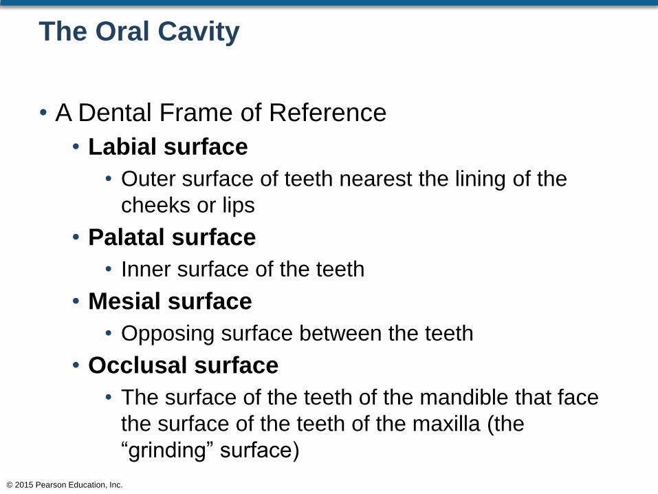
• A Dental Frame of Reference
• Labial surface
• Outer surface of teeth nearest the lining of the
cheeks or lips
• Palatal surface
• Inner surface of the teeth
• Mesial surface
• Opposing surface between the teeth
• Occlusal surface
• The surface of the teeth of the mandible that face
the surface of the teeth of the maxilla (the
“grinding” surface)
© 2015 Pearson Education, Inc.

The Pharynx
• The Pharynx
• Serves as a common passageway for food, liquid,
and air
• Pharyngeal muscles involved in swallowing:
• Pharyngeal constrictors
• Palatopharyngeus
• Stylopharyngeus
• Palatal
© 2015 Pearson Education, Inc.

The Pharynx
• The Pharynx
• Pharyngeal constrictors
• Push the bolus toward the esophagus
• Palatopharyngeus
• Elevates the larynx
• Stylopharyngeus
• Elevates the larynx
• Palatal muscles
• Raise the soft palate
© 2015 Pearson Education, Inc.

Figure 10.8a Muscles of the Pharynx
© 2015 Pearson Education, Inc.
Palatal Muscles
Tensor veli
palatini
Levator veli
palatini
Laryngeal Elevators
Stylopharyngeus
Palatopharyngeus
Pharyngeal Constrictors
Superior pharyngeal constrictor
Middle pharyngeal constrictor
Inferior pharyngeal constrictor
Esophagus
Lateral viewa

The Pharynx
• The Swallowing Process
• Process of swallowing is called deglutition
• There are three phases
• Buccal phase
• The tongue pushes the food to the oropharynx area
• Pharyngeal phase
• The epiglottis closes over the glottis and swallowing
begins
• Esophageal phase
• Upper esophageal sphincter opens and the bolus
begins moving down the esophagus
© 2015 Pearson Education, Inc.

Figure 25.8 The Swallowing Process
© 2015 Pearson Education, Inc.
1Buccal Phase
Hard palate
Tongue
Epiglottis
Larynx
Soft palate
Bolus
Esophagus
2Pharyngeal Phase
3Esophageal Phase
Esophagus
Diaphragm
Stomach
Thoracic
cavity
Peristalsis

The Esophagus
• This is a hollow muscular tube that extends from
the pharynx region to the stomach
• It is 25 cm long and 2 cm in diameter
• Located posterior to the trachea
• Enters the peritoneal cavity by passing through
the esophageal hiatus of the diaphragm
• Innervated by the vagus nerve from the
esophageal plexus
• Contains upper and lower esophageal sphincters
© 2015 Pearson Education, Inc.

Figure 25.1 Components of the Digestive System
© 2015 Pearson Education, Inc.
Major Subdivisions of
the Digestive Tract
Oral Cavity
Mechanical processing, moistening,
mixing with salivary secretions
Pharynx
Muscular propulsion of materials into
the esophagus
Mouth
Esophagus
Transport of materials to the stomach
Stomach
Chemical breakdown of materials via acid
and enzymes; mechanical processing
through muscular contractions
Small Intestine
Enzymatic digestion and absorption of
water, organic substrates, vitamins, and ions
Large Intestine
Enzymatic digestion and absorption of
water, organic substrates, vitamins, and ions
Anus
Accessory Organs of
the Digestive System
Salivary Glands
Secretion of lubricating fluid
containing enzymes that
break down carbohydrates
Liver
Secretion of bile (important
for lipid digestion), storage
of nutrients, many other
vital functions
Gallbladder
Storage and concentration
of bile
Pancreas
Exocrine cells secrete
buffers and digestive
enzymes; endocrine cells
secrete hormones

The Esophagus
• Histology of the Esophageal Wall
• The esophageal wall is made of:
• Mucosa lining
• Submucosa
• Smooth muscle layer (muscularis mucosae)
• Muscularis externa
• The esophagus does not have a serosa layer
© 2015 Pearson Education, Inc.

Figure 25.9 Histology of the Esophagus
© 2015 Pearson Education, Inc.
a Low-power view of a section through
the esophagus
Muscularis
mucosae
LM x 5
Mucosa
Submucosa
Muscularis
externa
Adventitia
The esophagus The esophageal mucosa
b The esophageal mucosa
LM x 300
Stratified
squamous
epithelium
Lamina
propria
Muscularis
mucosae
The Stomach
• The stomach performs three major functions:
• Bulk storage of ingested food
• Mechanical breakdown of ingested food
• Chemical digestion of ingested food
• The end result is the production of chyme
© 2015 Pearson Education, Inc.

The Stomach
• Anatomy of the Stomach
• The stomach is intraperitoneal and is located:
• In the left hypochondriac, epigastric, and a portion
of the umbilical and left lumbar regions
• The stomach consists of:
• Lesser curvature
• Greater curvature
• Cardia
• Fundus
• Body
• Pylorus
© 2015 Pearson Education, Inc.

Figure 25.1 Components of the Digestive System
© 2015 Pearson Education, Inc.
Major Subdivisions of
the Digestive Tract
Oral Cavity
Mechanical processing, moistening,
mixing with salivary secretions
Pharynx
Muscular propulsion of materials into
the esophagus
Mouth
Esophagus
Transport of materials to the stomach
Stomach
Chemical breakdown of materials via acid
and enzymes; mechanical processing
through muscular contractions
Small Intestine
Enzymatic digestion and absorption of
water, organic substrates, vitamins, and ions
Large Intestine
Enzymatic digestion and absorption of
water, organic substrates, vitamins, and ions
Anus
Accessory Organs of
the Digestive System
Salivary Glands
Secretion of lubricating fluid
containing enzymes that
break down carbohydrates
Liver
Secretion of bile (important
for lipid digestion), storage
of nutrients, many other
vital functions
Gallbladder
Storage and concentration
of bile
Pancreas
Exocrine cells secrete
buffers and digestive
enzymes; endocrine cells
secrete hormones
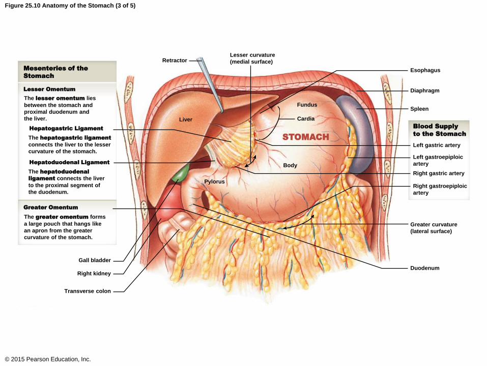
Figure 25.10 Anatomy of the Stomach (3 of 5)
© 2015 Pearson Education, Inc.
Mesenteries of the
Stomach
Lesser Omentum
The lesser omentum lies
between the stomach and
proximal duodenum and
the liver.
Hepatogastric Ligament
The hepatogastric ligament
connects the liver to the lesser
curvature of the stomach.
Hepatoduodenal Ligament
The hepatoduodenal
ligament connects the liver
to the proximal segment of
the duodenum.
Greater Omentum
The greater omentum forms
a large pouch that hangs like
an apron from the greater
curvature of the stomach.
Gall bladder
Right kidney
Transverse colon
Pylorus
Liver
Body
Fundus
Cardia
STOMACH
RetractorLesser curvature
(medial surface)
Esophagus
Diaphragm
Spleen
Left gastric artery
Left gastroepiploic
artery
Right gastric artery
Right gastroepiploic
artery
Blood Supply
to the Stomach
Greater curvature
(lateral surface)
Duodenum

The Stomach
• Anatomy of the Stomach (continued)
• Gastric rugae
• Relaxed stomach: mucosa forms numerous
muscular ridges
• Rugae permits expansion of the stomach
• A stretched stomach exhibits less prominent rugae
• Smooth muscle layers
• Circular muscles
• Longitudinal muscles
• Oblique muscles
© 2015 Pearson Education, Inc.

Figure 25.10 Anatomy of the Stomach (3 of 5)
© 2015 Pearson Education, Inc.
Mesenteries of the
Stomach
Lesser Omentum
The lesser omentum lies
between the stomach and
proximal duodenum and
the liver.
Hepatogastric Ligament
The hepatogastric ligament
connects the liver to the lesser
curvature of the stomach.
Hepatoduodenal Ligament
The hepatoduodenal
ligament connects the liver
to the proximal segment of
the duodenum.
Greater Omentum
The greater omentum forms
a large pouch that hangs like
an apron from the greater
curvature of the stomach.
Gall bladder
Right kidney
Transverse colon
Pylorus
Liver
Body
Fundus
Cardia
STOMACH
RetractorLesser curvature
(medial surface)
Esophagus
Diaphragm
Spleen
Left gastric artery
Left gastroepiploic
artery
Right gastric artery
Right gastroepiploic
artery
Blood Supply
to the Stomach
Greater curvature
(lateral surface)
Duodenum

Figure 25.10 Anatomy of the Stomach (5 of 5)
© 2015 Pearson Education, Inc.
Lesser curvature
(medial surface)
Pyloric sphincter
Duodenum
Pyloric canal
Pyloric antrum
Longitudinal
muscle layer
Circular
muscle layer
Oblique
muscle layer
(overlying mucosa)
Musculature of
the Stomach
Anterior
surface
Rugae
Esophagus
Gastroesophageal junction Regions of the Stomach
Fundus
Cardia
Body
Pyloris
The fundus is the region of the
stomach superior to the junction
between the stomach and the
esophagus (the gastroesophageal
junction).
The cardia is the superior, medial
portion of the stomach within 3 cm of
the gastroesophageal junction.
The body, the largest region of the
stomach, is the area between the
fundus and the pylorus.
The pylorus extends to the entrance
to the duodenum. It is divided into the
pyloric antrum and the pyloric
canal. A muscular pyloric sphincter
regulates the passage of materials into
the duodenum.
Greater curvature (lateral surface)
Right gastroepiploic vessels

The Stomach
• Mesenteries of the Stomach
• The mesenteries associated with the stomach are
called the greater and lesser omentum
• Greater omentum
• Extends from the greater curvature of the stomach
and drapes across the surface of the small
intestine
• Lesser omentum
• Extending from the lesser curvature of the stomach
to the liver is the hepatogastric ligament
• Extending from the pylorus/duodenum region to the
liver is the hepatoduodenal ligament
© 2015 Pearson Education, Inc.

Figure 25.10 Anatomy of the Stomach (3 of 5)
© 2015 Pearson Education, Inc.
Mesenteries of the
Stomach
Lesser Omentum
The lesser omentum lies
between the stomach and
proximal duodenum and
the liver.
Hepatogastric Ligament
The hepatogastric ligament
connects the liver to the lesser
curvature of the stomach.
Hepatoduodenal Ligament
The hepatoduodenal
ligament connects the liver
to the proximal segment of
the duodenum.
Greater Omentum
The greater omentum forms
a large pouch that hangs like
an apron from the greater
curvature of the stomach.
Gall bladder
Right kidney
Transverse colon
Pylorus
Liver
Body
Fundus
Cardia
STOMACH
RetractorLesser curvature
(medial surface)
Esophagus
Diaphragm
Spleen
Left gastric artery
Left gastroepiploic
artery
Right gastric artery
Right gastroepiploic
artery
Blood Supply
to the Stomach
Greater curvature
(lateral surface)
Duodenum

The Stomach
• Blood Supply to the Stomach
• There are three branches from the celiac trunk
that supply the stomach
• Left gastric artery
• Supplies blood to the lesser curvature and cardia
• Splenic artery
• Supplies blood to the fundus
• Branches to form the left gastroepiploic artery, which
supplies the greater curvature
• Common hepatic artery
• Branches to form the right gastric, right
gastroepiploic, and gastroduodenal artery to supply
the greater and lesser curvatures© 2015 Pearson Education, Inc.

Figure 22.15a Arteries of the Abdomen
© 2015 Pearson Education, Inc.
LiverCeliac trunk
Right gastric
Ileocolic
Left common iliac
Rectal
Sigmoid colon
Rectum
Right external iliac
Inferior
pancreaticoduodenal
Inferior vena cava
Left gastric
Right gastroepiploic Left gastroepiploic
PancreaticPancreas
Sigmoid
Small intestine
Ascending colon
Superior mesenteric
Inferior mesenteric
Stomach
Superior
pancreaticoduodenal
ABDOMINAL AORTA
Duodenal
Right internal iliac
Major arteries supplying the abdominal viscera
THORACIC AORTA
Splenic
Spleen
Left colic
Common hepatic
Hepatic artery proper
Cystic
Gastroduodenal
Middle colic (cut)
Right colic
Intestinal
a

The Stomach
• Histology of the Stomach
• Lined with simple columnar epithelium
• Structures within the lining of the stomach
• Gastric pits
• Gastric secretory cells
• Mucous neck cells
• Parietal cells
• Chief cells
• Enteroendocrine cells
© 2015 Pearson Education, Inc.

The Stomach
• Histology of the Stomach
• Gastric pits
• Produce cells to continuously replace lost stomach
cells
• Mucous surface cells
• Produce copious amounts of mucus to protect the
lining of the stomach
• Mucous neck cells
• Produce mucus to lubricate the food entering the
stomach
© 2015 Pearson Education, Inc.

The Stomach
• Histology of the Stomach
• Parietal cells
• Secrete intrinsic factor and hydrochloric acid
• Intrinsic factor
• Facilitates the absorption of vitamin B12 from the
small intestine into the bloodstream, which is used
during erythropoiesis
• Hydrochloric acid
• Kills microorganisms and activates pepsinogen
© 2015 Pearson Education, Inc.

The Stomach
• Histology of the Stomach
• Chief cells
• Secrete pepsinogen, which is converted to pepsin
via the action of hydrochloric acid
• Enteroendocrine cells
• These are cells of the stomach that produce
hormones. The G cells produce the hormone
gastrin. Gastrin causes the parietal and chief cells
to release their products
© 2015 Pearson Education, Inc.
Figure 25.12ab Histology of the Stomach Wall
© 2015 Pearson Education, Inc.
Diagrammatic view of the stomach and mucosa.a
Esophagus
Colorized SEM of the gastric mucosa.
Diaphragm
Cardia
Greater omentum
Greater curvature
FundusBody
Lesser curvature
Lesser omentum
Pylorus
Rugae
Gastric mucosa SEM x 35
Mucous
epithelial
cells
Entrances to
gastric pits
b

Figure 25.12cd Histology of the Stomach Wall
© 2015 Pearson Education, Inc.
c
Layers of the
Stomach Wall
Mucosa
Gastric pit (opening
to gastric gland)
Mucous epithelium
Muscularis mucosae
Submucosa
Muscularis externa
Oblique muscle
Circular muscle
Longitudinal muscle
Serosa
Myenteric
plexus
Artery
and
vein
Diagrammatic view of the organization of the stomach wall. This corresponds
to a sectional view through the area indicated by the box in part (b).
Lamina propria
Lymphatic
vessel
Gastric
pit
Gastric
gland
Diagrammatic view of a gastric gland and
micrograph of the gastric mucosa.
d
LM x 200
Luminal
surface
Lamina
propria
Mucous
neck cells
Cells of
Gastric
Glands
Parietal
cells
G cell
Chief
cells
Smooth
muscle cell
Muscularis
mucosae

The Stomach
• Regulation of the Stomach
• The production of stomach acid and enzymes is
controlled by the CNS
• CNS regulation involves:
• Vagus nerve (parasympathetic innervation)
• Triggered by the sight and thought of food
• Celiac plexus (sympathetic innervation)
© 2015 Pearson Education, Inc.

The Stomach
• Regulation of the Stomach
• Food enters the stomach and the stomach
stretches
• Stretching causes the G cells to release gastrin
• Gastrin causes the parietal and chief cells to
release their products
© 2015 Pearson Education, Inc.

The Small Intestine
• Features of the Small Intestine
• Approximately 20 feet in length / 1.5–2.5 inches in
diameter
• Consists of:
• Duodenum
• 10 inches long; receives digestive enzymes from the
pancreas, bile from the liver and gallbladder
• Jejunum
• 8 feet long; most of the digestion and absorption
occurs in the jejunum
• Ileum
• 12 feet long
© 2015 Pearson Education, Inc.

Figure 25.1 Components of the Digestive System
© 2015 Pearson Education, Inc.
Major Subdivisions of
the Digestive Tract
Oral Cavity
Mechanical processing, moistening,
mixing with salivary secretions
Pharynx
Muscular propulsion of materials into
the esophagus
Mouth
Esophagus
Transport of materials to the stomach
Stomach
Chemical breakdown of materials via acid
and enzymes; mechanical processing
through muscular contractions
Small Intestine
Enzymatic digestion and absorption of
water, organic substrates, vitamins, and ions
Large Intestine
Enzymatic digestion and absorption of
water, organic substrates, vitamins, and ions
Anus
Accessory Organs of
the Digestive System
Salivary Glands
Secretion of lubricating fluid
containing enzymes that
break down carbohydrates
Liver
Secretion of bile (important
for lipid digestion), storage
of nutrients, many other
vital functions
Gallbladder
Storage and concentration
of bile
Pancreas
Exocrine cells secrete
buffers and digestive
enzymes; endocrine cells
secrete hormones

Figure 25.13 Regions of the Small Intestine
© 2015 Pearson Education, Inc.
Transverse
colon
Regions of the
Small Intestine
Duodenum
Jejunum
Ileum
Ascending
colon
Cecum
Descending
colon
Sigmoid
colon
Rectum

The Small Intestine
• Support of the Small Intestine
• Jejunum and ileum are supported by the
mesentery proper
• Duodenum is not associated with any mesentery
• Blood supply
• Branches of the superior mesenteric artery and
intestinal arteries
• Nerve supply
• Parasympathetic innervation via the vagus nerve
• Sympathetic innervation via the superior
mesenteric ganglion
© 2015 Pearson Education, Inc.
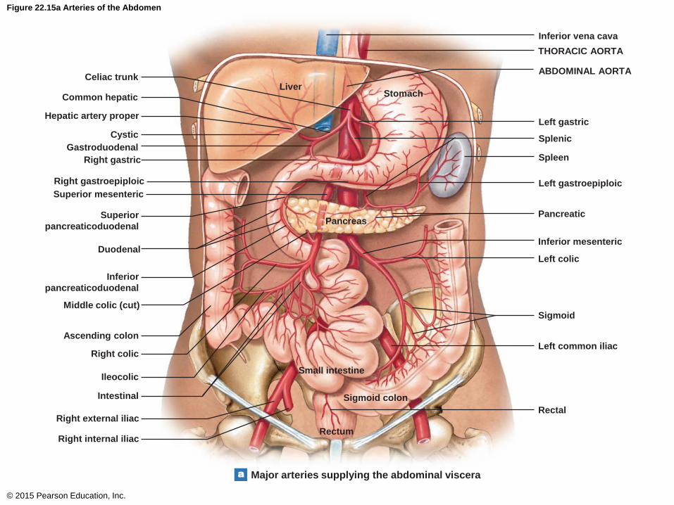
Figure 22.15a Arteries of the Abdomen
© 2015 Pearson Education, Inc.
LiverCeliac trunk
Right gastric
Ileocolic
Left common iliac
Rectal
Sigmoid colon
Rectum
Right external iliac
Inferior
pancreaticoduodenal
Inferior vena cava
Left gastric
Right gastroepiploic Left gastroepiploic
PancreaticPancreas
Sigmoid
Small intestine
Ascending colon
Superior mesenteric
Inferior mesenteric
Stomach
Superior
pancreaticoduodenal
ABDOMINAL AORTA
Duodenal
Right internal iliac
Major arteries supplying the abdominal viscera
THORACIC AORTA
Splenic
Spleen
Left colic
Common hepatic
Hepatic artery proper
Cystic
Gastroduodenal
Middle colic (cut)
Right colic
Intestinal
a

The Small Intestine
• Histology of the Small Intestine
• The lining contains:
• Plicae
• Each plica consists of numerous microvilli (villi)
• Within each villus are capillaries
• Villi will absorb the digested nutrients from the
lumen of the small intestine into the capillaries
© 2015 Pearson Education, Inc.

Figure 25.14a-c Histology of the Intestinal Wall
© 2015 Pearson Education, Inc.
Characteristic features
of the intestinal lining
a
The organization
of villi and the
intestinal crypts
b
Villi
Plica circularis
Villi Intestinal
crypt
Lymphoid
noduleLacteal
Muscularis
mucosae
Layers of the
Small Intestine
Mucosa
Submucosa
Muscularis
externa
Serosa
Submucosal
artery and vein
Lymphatic
vessel
Submucosal
plexus
Circular layer
of smooth
muscle
Myenteric plexus
Diagrammatic view
of a single villus
showing the capillary
and lymphatic supply
c
Lymphatic
vesselVenuleArteriole
Goblet cell
Columnar
epithelial
cell
Lacteal
Nerve
Capillary
network
Lamina
propria
Longitudinal layer
of smooth muscle

The Small Intestine
• Histology of the Small Intestine
• Intestinal crypts
• Appear at the base of the villi
• New epithelial cells are formed in this area
• Contain enteroendocrine cells
• These cells produce intestinal hormones, including
cholecystokinin and secretin
• These cells produce enzymes with antibacterial
activity
© 2015 Pearson Education, Inc.

Figure 25.14b Histology of the Intestinal Wall
© 2015 Pearson Education, Inc.
The organization
of villi and the
intestinal crypts
b
Villi Intestinal
crypt
Lymphoid
noduleLacteal
Muscularis
mucosae
Layers of the
Small Intestine
Mucosa
Submucosa
Muscularis
externa
Serosa
Submucosal
artery and vein
Lymphatic
vessel
Submucosal
plexus
Circular layer
of smooth
muscle
Myenteric plexus
Longitudinal layer
of smooth muscle

The Small Intestine
• Histology of the Small Intestine
• Each villus also contains a lacteal
• Lacteals absorb material that cannot be absorbed
by the capillaries
• Examples would be large lipid-protein complexes
© 2015 Pearson Education, Inc.

Figure 25.14c Histology of the Intestinal Wall
© 2015 Pearson Education, Inc.
Diagrammatic view
of a single villus
showing the capillary
and lymphatic supply
c
Lymphatic
vesselVenuleArteriole
Goblet cell
Columnar
epithelial
cell
Lacteal
Nerve
Capillary
network
Lamina
propria

The Small Intestine
• Regional Specialization
• The Duodenum
• Contains duodenal submucosal glands
• Produces large amounts of mucus
• This mucus consists of buffers to provide some
protection against the acidic chyme
• Entering into the small intestine at the
hepatopancreatic sphincter region
• Bile from the liver and gallbladder
• Buffers from the pancreas
• Digestive enzymes from the pancreas
© 2015 Pearson Education, Inc.

Figure 25.21ac The Gallbladder and Associated Bile Ducts
© 2015 Pearson Education, Inc.
A view of the inferior
surface of the liver
showing the position
of the gallbladder and
ducts that support
bile from the liver
to the gallbladder
and duodenum.
a
Round ligament
Right hepatic duct
Cystic duct
Gallbladder
Fundus
Body
Neck
Common bile
ductLiver
Duodenum
Stomach
Pancreas
Left hepatic
duct
Left hepatic
artery
Common
hepatic duct
Cut edge of lesser omentum
Hepatic
portal vein
Common hepatic artery
Right gastric
artery
A portion of the lesser omentum has been cut away to
make it easier to see the relationships among the
common bile duct, the hepatic duct, and the cystic duct.
c
Pancreas
Hepatopancreatic
sphincter
Duodenal
ampulla
Duodenal
papilla
Intestinal lumen
Common
bile duct
Pancreatic
duct

The Small Intestine
• Regional Specialization
• The Jejunum and Ileum
• Jejunum
• Has prominent plicae and villi
• Most nutrient absorption occurs here
• Ileum
• Contains prominent lymphoid centers called
aggregated lymphoid nodules (Peyer’s patches)
© 2015 Pearson Education, Inc.

The Small Intestine
• Regulation of the Small Intestine
• Upon vagal stimulation, the enteroendocrine cells
of the small intestine release:
• Secretin
• Causes the liver to begin making bile
Causes the pancreas to release buffers into the
duodenum
• Cholecystokinin
• Causes the pancreas to release digestive enzymes
into the duodenum
• Causes the gallbladder to contract thus releasing
stored bile into the duodenum
• Causes the hepatopancreatic sphincter to open
© 2015 Pearson Education, Inc.

The Large Intestine
• Features of the Large Intestine
• Approximately 5 feet in length
• Approximately 3 inches in diameter
• Consists of the following regions
• Cecum
• Ascending colon
• Transverse colon
• Descending colon
• Sigmoid colon
• Rectum
© 2015 Pearson Education, Inc.

Figure 25.1 Components of the Digestive System
© 2015 Pearson Education, Inc.
Major Subdivisions of
the Digestive Tract
Oral Cavity
Mechanical processing, moistening,
mixing with salivary secretions
Pharynx
Muscular propulsion of materials into
the esophagus
Mouth
Esophagus
Transport of materials to the stomach
Stomach
Chemical breakdown of materials via acid
and enzymes; mechanical processing
through muscular contractions
Small Intestine
Enzymatic digestion and absorption of
water, organic substrates, vitamins, and ions
Large Intestine
Enzymatic digestion and absorption of
water, organic substrates, vitamins, and ions
Anus
Accessory Organs of
the Digestive System
Salivary Glands
Secretion of lubricating fluid
containing enzymes that
break down carbohydrates
Liver
Secretion of bile (important
for lipid digestion), storage
of nutrients, many other
vital functions
Gallbladder
Storage and concentration
of bile
Pancreas
Exocrine cells secrete
buffers and digestive
enzymes; endocrine cells
secrete hormones

The Large Intestine
• Functions of the Large Intestine
• Reabsorption of water
• Results in compaction of waste (forms feces)
• Absorption of vitamins produced by the housed
bacteria
• Storage of fecal material prior to defecation
© 2015 Pearson Education, Inc.

The Large Intestine
• Blood Supply to the Large Intestine
• Receives blood from branches of the superior
mesenteric artery
• Receives blood from branches of the inferior
mesenteric artery
© 2015 Pearson Education, Inc.

Figure 22.15a Arteries of the Abdomen
© 2015 Pearson Education, Inc.
LiverCeliac trunk
Right gastric
Ileocolic
Left common iliac
Rectal
Sigmoid colon
Rectum
Right external iliac
Inferior
pancreaticoduodenal
Inferior vena cava
Left gastric
Right gastroepiploic Left gastroepiploic
PancreaticPancreas
Sigmoid
Small intestine
Ascending colon
Superior mesenteric
Inferior mesenteric
Stomach
Superior
pancreaticoduodenal
ABDOMINAL AORTA
Duodenal
Right internal iliac
Major arteries supplying the abdominal viscera
THORACIC AORTA
Splenic
Spleen
Left colic
Common hepatic
Hepatic artery proper
Cystic
Gastroduodenal
Middle colic (cut)
Right colic
Intestinal
a
The Large Intestine
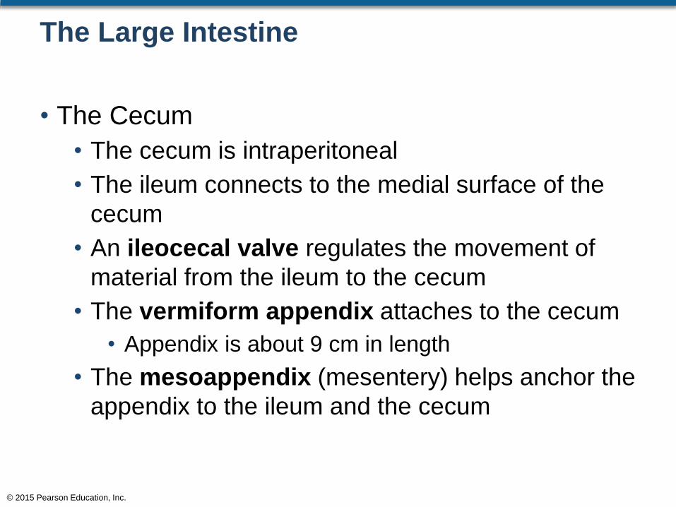
• The Cecum
• The cecum is intraperitoneal
• The ileum connects to the medial surface of the
cecum
• An ileocecal valve regulates the movement of
material from the ileum to the cecum
• The vermiform appendix attaches to the cecum
• Appendix is about 9 cm in length
• The mesoappendix (mesentery) helps anchor the
appendix to the ileum and the cecum
© 2015 Pearson Education, Inc.

Figure 25.16a The Large Intestine
© 2015 Pearson Education, Inc.
Gross anatomy
and regions of the
large intestine
a
Rectum
IleumRectal
artery
Intestinal arteries
and veins
Sigmoid flexure
Taenia coli
Sigmoid arteries
and veins
Haustra
Left colic artery
Inferior
mesenteric
artery
Left colic vein
DESCENDING
COLON
Greater
omentum (cut)
Left colic
(splenic)
flexure
Splenic vein
Superior mesenteric artery
Inferior mesenteric vein
Aorta
Hepatic portal vein
Superior
mesenteric vein
Inferior vena cava
TRANSVERSE
COLON
Right colic
(hepatic) flexure
Middle colic
artery and vein
Right colic
artery and vein
ASCENDING
COLON
Omental appendices
Ileocecal valve
Cecum
Appendix
SIGMOID COLON

The Large Intestine
• The Colon
• The regions of the colon are:
• Ascending colon
• Transverse colon
• Descending colon
• Sigmoid colon
© 2015 Pearson Education, Inc.

The Large Intestine
• The Colon
• Waste material leaves the ileum and enters the
cecum
• Waste material goes “up” the ascending colon
• Around the hepatic flexure
• “Across” the transverse colon
• Around the splenic flexure
• “Down” the descending colon
• Around the sigmoid flexure
• To the sigmoid colon
• Into the rectum
© 2015 Pearson Education, Inc.

Figure 25.16a The Large Intestine
© 2015 Pearson Education, Inc.
Gross anatomy
and regions of the
large intestine
a
Rectum
IleumRectal
artery
Intestinal arteries
and veins
Sigmoid flexure
Taenia coli
Sigmoid arteries
and veins
Haustra
Left colic artery
Inferior
mesenteric
artery
Left colic vein
DESCENDING
COLON
Greater
omentum (cut)
Left colic
(splenic)
flexure
Splenic vein
Superior mesenteric artery
Inferior mesenteric vein
Aorta
Hepatic portal vein
Superior
mesenteric vein
Inferior vena cava
TRANSVERSE
COLON
Right colic
(hepatic) flexure
Middle colic
artery and vein
Right colic
artery and vein
ASCENDING
COLON
Omental appendices
Ileocecal valve
Cecum
Appendix
SIGMOID COLON

The Large Intestine
• The Colon
• The wall of the colon has pouches that allow for
expansion called haustra
• Longitudinal muscles called taeniae coli aid in the
process of peristalsis
• The serosa of the large intestine has numerous
“flaps” of sacs of fat attached to but yet extending
from the intestine called omental appendices
© 2015 Pearson Education, Inc.

Figure 25.16a The Large Intestine
© 2015 Pearson Education, Inc.
Gross anatomy
and regions of the
large intestine
a
Rectum
IleumRectal
artery
Intestinal arteries
and veins
Sigmoid flexure
Taenia coli
Sigmoid arteries
and veins
Haustra
Left colic artery
Inferior
mesenteric
artery
Left colic vein
DESCENDING
COLON
Greater
omentum (cut)
Left colic
(splenic)
flexure
Splenic vein
Superior mesenteric artery
Inferior mesenteric vein
Aorta
Hepatic portal vein
Superior
mesenteric vein
Inferior vena cava
TRANSVERSE
COLON
Right colic
(hepatic) flexure
Middle colic
artery and vein
Right colic
artery and vein
ASCENDING
COLON
Omental appendices
Ileocecal valve
Cecum
Appendix
SIGMOID COLON

The Large Intestine
• The Rectum
• Temporarily stores waste matter
• The last portion of the rectum is the anal canal
• The anal canal consists of anal columns
• The anal canal ends at the anus
© 2015 Pearson Education, Inc.

Figure 25.16a The Large Intestine
© 2015 Pearson Education, Inc.
Gross anatomy
and regions of the
large intestine
a
Rectum
IleumRectal
artery
Intestinal arteries
and veins
Sigmoid flexure
Taenia coli
Sigmoid arteries
and veins
Haustra
Left colic artery
Inferior
mesenteric
artery
Left colic vein
DESCENDING
COLON
Greater
omentum (cut)
Left colic
(splenic)
flexure
Splenic vein
Superior mesenteric artery
Inferior mesenteric vein
Aorta
Hepatic portal vein
Superior
mesenteric vein
Inferior vena cava
TRANSVERSE
COLON
Right colic
(hepatic) flexure
Middle colic
artery and vein
Right colic
artery and vein
ASCENDING
COLON
Omental appendices
Ileocecal valve
Cecum
Appendix
SIGMOID COLON

Figure 25.16c The Large Intestine
© 2015 Pearson Education, Inc.
Detailed anatomy of the rectum and anusc
Rectum
Anal canal
Anal
columns
Internal anal
sphincter
External anal
sphincter
Anus

The Large Intestine
• Histology of the Large Intestine
• Walls are thinner than the walls of the small
intestine
• The walls lack villi
• Has numerous goblet cells
• Has very distinctive intestinal crypts
• Produces lots of mucus to lubricate undigested
material
• Contains large lymphoid nodules
© 2015 Pearson Education, Inc.

Figure 25.18a The Wall of the Large Intestine
© 2015 Pearson Education, Inc.
Diagrammatic view of the colon walla
Taenia coli
Omental appendices
Haustrum
Lymphoid
nodule
Layers of the
Large Intestine
Mucosa
Submucosa
Muscularis externa
Serosa
Muscularis mucosae
Circular layer
Longitudinal layer
(taenia coli)
Simple
columnar
epithelium
Goblet
cells
Intestinal
crypt

The Large Intestine
• Regulation of the Large Intestine
• Movement of waste material to the transverse
colon is slow
• This allows for appropriate reabsorption of water
• Movement through the rest of the large intestine is
rapid (mass movement)
• This forces material into the rectum for later
defecation
• Distension of the rectal wall stimulates the urge to
defecate / internal sphincter opens
• Fecal material moves into the anal canal / external
sphincter opens
© 2015 Pearson Education, Inc.

Figure 25.17 Anterior/Posterior Radiograph of the Colon
© 2015 Pearson Education, Inc.
Left colic (splenic) flexure
Right colic (hepatic) flexure
Transverse colon
Haustra
Ascending colon
Descending colon
Cecum
Sigmoid colon
Rectum

Accessory Glandular Digestive Organs
• The accessory organs of digestion are:
• Salivary glands
• Liver
• Gallbladder
• Pancreas
© 2015 Pearson Education, Inc.

Figure 25.1 Components of the Digestive System
© 2015 Pearson Education, Inc.
Major Subdivisions of
the Digestive Tract
Oral Cavity
Mechanical processing, moistening,
mixing with salivary secretions
Pharynx
Muscular propulsion of materials into
the esophagus
Mouth
Esophagus
Transport of materials to the stomach
Stomach
Chemical breakdown of materials via acid
and enzymes; mechanical processing
through muscular contractions
Small Intestine
Enzymatic digestion and absorption of
water, organic substrates, vitamins, and ions
Large Intestine
Enzymatic digestion and absorption of
water, organic substrates, vitamins, and ions
Anus
Accessory Organs of
the Digestive System
Salivary Glands
Secretion of lubricating fluid
containing enzymes that
break down carbohydrates
Liver
Secretion of bile (important
for lipid digestion), storage
of nutrients, many other
vital functions
Gallbladder
Storage and concentration
of bile
Pancreas
Exocrine cells secrete
buffers and digestive
enzymes; endocrine cells
secrete hormones

Accessory Glandular Digestive Organs
• The Liver
• The largest visceral organ of the body
• The liver is involved in:
• Metabolic regulation
• Hematological regulation
• Bile production
© 2015 Pearson Education, Inc.
Accessory Glandular Digestive Organs
• Metabolic Regulation
• All blood leaving the digestive tract enters the liver
through the hepatic portal system
• Hepatocytes adjust the circulating metabolites
before the blood enters into systemic circulation
© 2015 Pearson Education, Inc.

Accessory Glandular Digestive Organs
• Hematological Regulation
• The liver is the largest blood reservoir of the body
• As blood passes through the liver:
• Phagocytic cells remove old or damaged
erythrocytes
• Liver cells synthesize plasma proteins for blood
clotting (for example)
© 2015 Pearson Education, Inc.

Accessory Glandular Digestive Organs
• Bile Production
• Bile is made by liver cells (hepatocytes)
• Bile is stored in the gallbladder
• Bile is secreted into the duodenum when it is
needed
• Bile emulsifies fat (from the diet) in the small
intestine
• This emulsification process makes it easier for
lipase to do the actual digestion of fat
© 2015 Pearson Education, Inc.

Table 25.1 Major Functions of the Liver
© 2015 Pearson Education, Inc.

Accessory Glandular Digestive Organs
• Anatomy of the Liver
• Falciform ligament
• Marks the boundary between the left and right lobes
• The inferior portion of the falciform ligament
becomes thick and round and is called the round
ligament
• The round ligament used to be the fetal umbilical
vein
• The falciform ligament spreads on the surface of
the liver attaching to the inferior side of the
diaphragm
• This spreading ligament is called the coronary
ligament© 2015 Pearson Education, Inc.

Figure 25.19c Anatomy of the Liver
© 2015 Pearson Education, Inc.
Anatomical landmarks on the anterior surface of the liverc
Right lobe Left lobe
Coronary ligament
Falciform
ligament
Round ligament
Gallbladder
Accessory Glandular Digestive Organs
• Blood Supply to the Liver
• Two blood vessels supply the liver
• Hepatic artery proper
• Hepatic portal vein
© 2015 Pearson Education, Inc.

Figure 22.15a Arteries of the Abdomen
© 2015 Pearson Education, Inc.
LiverCeliac trunk
Right gastric
Ileocolic
Left common iliac
Rectal
Sigmoid colon
Rectum
Right external iliac
Inferior
pancreaticoduodenal
Inferior vena cava
Left gastric
Right gastroepiploic Left gastroepiploic
PancreaticPancreas
Sigmoid
Small intestine
Ascending colon
Superior mesenteric
Inferior mesenteric
Stomach
Superior
pancreaticoduodenal
ABDOMINAL AORTA
Duodenal
Right internal iliac
Major arteries supplying the abdominal viscera
THORACIC AORTA
Splenic
Spleen
Left colic
Common hepatic
Hepatic artery proper
Cystic
Gastroduodenal
Middle colic (cut)
Right colic
Intestinal
a

Accessory Glandular Digestive Organs
• Histological Organization of the Liver
• The liver is divided into approximately 100,000
liver lobules
• Each lobule is separated by the interlobular
septum
• The center of each lobule consists of a vein from
the hepatic portal system
• The hepatocytes are arranged in such a manner
forming cellular lines extending from the central
vein outward
© 2015 Pearson Education, Inc.

Figure 25.20a Liver Histology
© 2015 Pearson Education, Inc.
Diagrammatic view of lobular organization.a
Interlobular
septumBile
duct
Branch of
hepatic portal veinPortal
area
Bile
ductules

Accessory Glandular Digestive Organs
• Histological Organization of the Liver
• Spaces are created between the lines of
hepatocytes; these spaces are called sinusoids
• Sinusoids consist of:
• Capillaries: leading to the central vein
• Kupffer cells: phagocytic cells of the liver
© 2015 Pearson Education, Inc.

Accessory Glandular Digestive Organs
• Histological Organization of the Liver
• Each lobule of the liver has a hexagonal shape
• At each of the six corners is:
• Branch of the hepatic portal vein
• Branch of the bile duct
• Branch of the hepatic artery proper
• The above three branches form the hepatic triad
© 2015 Pearson Education, Inc.

Figure 25.20b Liver Histology
© 2015 Pearson Education, Inc.
Magnified view showing the portal
area and central vein.
b
Bile
duct
Branch of hepatic
artery proper
Hepatocytes
Central
vein
Kupffer
cells
Sinusoid
Bile
canaliculi
Branch of
hepatic
portal
vein

Accessory Glandular Digestive Organs
• Bile Secretion and Transport
• Hepatocytes produce bile
• Bile enters:
• Bile canaliculi
• Bile travels to the bile ducts
• Bile then collects in the left and right hepatic
ducts
• Bile travels through the common hepatic duct
• Bile can travel through the common bile duct to
the duodenum (through the hepatopancreatic
sphincter) or travel through the cystic duct into
the gallbladder for storage
© 2015 Pearson Education, Inc.

Figure 25.21a The Gallbladder and Associated Bile Ducts
© 2015 Pearson Education, Inc.
A view of the inferior
surface of the liver
showing the position
of the gallbladder and
ducts that support
bile from the liver
to the gallbladder
and duodenum.
a
Round ligament
Right hepatic duct
Cystic duct
Gallbladder
Fundus
Body
Neck
Common bile
ductLiver
Duodenum
Stomach
Pancreas
Left hepatic
duct
Left hepatic
artery
Common
hepatic duct
Cut edge of lesser omentum
Hepatic
portal vein
Common hepatic artery
Right gastric
artery

Accessory Glandular Digestive Organs
• The Gallbladder
• The gallbladder is divided into three regions
• Fundus
• Body
• Neck
• The cystic duct leads from the neck of the
gallbladder to the common bile duct
© 2015 Pearson Education, Inc.

Figure 25.1 Components of the Digestive System
© 2015 Pearson Education, Inc.
Major Subdivisions of
the Digestive Tract
Oral Cavity
Mechanical processing, moistening,
mixing with salivary secretions
Pharynx
Muscular propulsion of materials into
the esophagus
Mouth
Esophagus
Transport of materials to the stomach
Stomach
Chemical breakdown of materials via acid
and enzymes; mechanical processing
through muscular contractions
Small Intestine
Enzymatic digestion and absorption of
water, organic substrates, vitamins, and ions
Large Intestine
Enzymatic digestion and absorption of
water, organic substrates, vitamins, and ions
Anus
Accessory Organs of
the Digestive System
Salivary Glands
Secretion of lubricating fluid
containing enzymes that
break down carbohydrates
Liver
Secretion of bile (important
for lipid digestion), storage
of nutrients, many other
vital functions
Gallbladder
Storage and concentration
of bile
Pancreas
Exocrine cells secrete
buffers and digestive
enzymes; endocrine cells
secrete hormones

Accessory Glandular Digestive Organs
• Gallbladder Function
• Storage of bile
• Bile modification
© 2015 Pearson Education, Inc.

Accessory Glandular Digestive Organs
• Bile Storage and Modification
• When the hepatopancreatic sphincter is closed:
• Bile enters the cystic duct and into the gallbladder
• The gallbladder can store 40–70 ml of bile
• Water is continuously removed from the stored bile
thereby concentrating the bile more and more
• If food entering the small intestine is high in fat
content, the small intestine cells will release
cholecystokinin
• Cholecystokinin will cause the gallbladder to
release bile
© 2015 Pearson Education, Inc.

Accessory Glandular Digestive Organs
• Gallbladder (bile release)
• CCK will cause the gallbladder to contract to
release bile
• CCK also causes the hepatopancreatic sphincter
to open
• Bile enters the cystic duct
• Bile enters the common bile duct
• The hepatopancreatic sphincter opens
• Bile enters the duodenum of the small intestine
• Bile will then emulsify fat
© 2015 Pearson Education, Inc.

Figure 25.21a The Gallbladder and Associated Bile Ducts
© 2015 Pearson Education, Inc.
A view of the inferior
surface of the liver
showing the position
of the gallbladder and
ducts that support
bile from the liver
to the gallbladder
and duodenum.
a
Round ligament
Right hepatic duct
Cystic duct
Gallbladder
Fundus
Body
Neck
Common bile
ductLiver
Duodenum
Stomach
Pancreas
Left hepatic
duct
Left hepatic
artery
Common
hepatic duct
Cut edge of lesser omentum
Hepatic
portal vein
Common hepatic artery
Right gastric
artery

Accessory Glandular Digestive Organs
• The Pancreas
• The pancreas is posterior to the stomach
• The pancreas consists of:
• Head: nearest the curvature of the duodenum
• Body: extends toward the spleen
• Tail: rounded end of the pancreas nearest the
spleen
• Pancreatic duct: delivers secretions from the
pancreas to the duodenum (through the
hepatopancreatic sphincter)
© 2015 Pearson Education, Inc.

Figure 25.1 Components of the Digestive System
© 2015 Pearson Education, Inc.
Major Subdivisions of
the Digestive Tract
Oral Cavity
Mechanical processing, moistening,
mixing with salivary secretions
Pharynx
Muscular propulsion of materials into
the esophagus
Mouth
Esophagus
Transport of materials to the stomach
Stomach
Chemical breakdown of materials via acid
and enzymes; mechanical processing
through muscular contractions
Small Intestine
Enzymatic digestion and absorption of
water, organic substrates, vitamins, and ions
Large Intestine
Enzymatic digestion and absorption of
water, organic substrates, vitamins, and ions
Anus
Accessory Organs of
the Digestive System
Salivary Glands
Secretion of lubricating fluid
containing enzymes that
break down carbohydrates
Liver
Secretion of bile (important
for lipid digestion), storage
of nutrients, many other
vital functions
Gallbladder
Storage and concentration
of bile
Pancreas
Exocrine cells secrete
buffers and digestive
enzymes; endocrine cells
secrete hormones

Accessory Glandular Digestive Organs
• Histological Organization of the Pancreas
• Consists of lobules
• Within each lobule are:
• Acinar cells
• Produce digestive enzymes
• Enzymes travel through the pancreatic duct to the
small intestine
• Pancreatic islets
• Produce hormones
• Hormones enter into the bloodstream to travel to
target organs
© 2015 Pearson Education, Inc.

Figure 25.22a The Pancreas
© 2015 Pearson Education, Inc.
Gross anatomy of the pancreas. The head of the pancreas
is tucked into a curve of the duodenum that begins at the
pylorus of the stomach.
a
Abdominal aorta
Celiac trunk
Common hepatic
artery
Gastroduodenal artery
Common bile duct
Superior
pancreatic artery
Superior pancreatico-
duodenal artery
Accessory pancreatic
duct (to lesser
duodenal papilla)
Duodenum
Head of pancreas
Pancreatic duct
(to greater duodenal
papilla) with common
bile duct
Anterior branch
Posterior branch
Inferior pancreatico-
duodenal artery
Superior
mesenteric
artery
Body of
pancreas
Transverse
pancreatic artery
Lobules
Stomach
Splenic
artery
Great
pancreatic
artery
Tail of
pancreas
Caudal
pancreatic
artery

Figure 25.22b The Pancreas
© 2015 Pearson Education, Inc.
Diagrammatic view of the histological
organization of the pancreas showing
exocrine and endocrine regions.
b
Pancreatic
duct
Acinar cells
(exocrine)
Pancreatic islet
(endocrine)
Pancreatic
acini

Figure 25.22c The Pancreas
© 2015 Pearson Education, Inc.
Histology of the pancreas showing exocrine and
endocrine cells.
Pancreatic acini
(exocrine)
LM x 120Pancreas
Pancreatic islet
(endocrine)
Duct
c

Accessory Glandular Digestive Organs
• Pancreatic Enzymes (from Acinar Cells)
• Lipases
• Digest lipids
• Carbohydrases
• Digest carbohydrates
• Nucleases
• Digest nucleic acids
• Proteinases
• Digest protein
© 2015 Pearson Education, Inc.

Accessory Glandular Digestive Organs
• Pancreatic Hormones (from Pancreatic Islets)
• Insulin
• Glucagon
• Somatostatin
© 2015 Pearson Education, Inc.

Accessory Glandular Digestive Organs
• The Regulation of Pancreatic Secretion
• Cholecystokinin
• From the small intestine will cause the pancreas to
release its digestive enzymes
• Secretin
• From the small intestine will cause the pancreas to
release buffers
• When food leaves the stomach and enters the
duodenum, the chyme is mixed with acid in the
stomach. Therefore, acidic chyme is entering the
duodenum. Buffers are used to maintain a normal
small intestine pH of about 7 or 8
© 2015 Pearson Education, Inc.
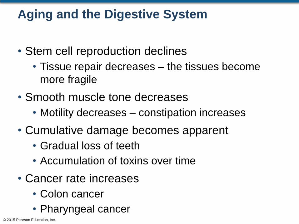
Aging and the Digestive System
• Stem cell reproduction declines
• Tissue repair decreases – the tissues become
more fragile
• Smooth muscle tone decreases
• Motility decreases – constipation increases
• Cumulative damage becomes apparent
• Gradual loss of teeth
• Accumulation of toxins over time
• Cancer rate increases
• Colon cancer
• Pharyngeal cancer© 2015 Pearson Education, Inc.


