Art History Review #5 Fox’s Fabulous 4 th Block Art 1 Spring 2015.
Upload
mariah-stephanie-curtisCategory
view
218download
1
Chapter 14Biology 25: Human BiologyBiology 25: Human Biology
Prof. GonsalvesProf. Gonsalves
Los Angeles City CollegeLos Angeles City College
Based on Mader’s Based on Mader’s Human Human BiologyBiology,7,7thth edition and Fox’s 8 edition and Fox’s 8thth ed ed
PowerpointsPowerpoints


Sensory Receptors
Perceptions of world are created by the Perceptions of world are created by the brain from AP sent from sensory receptors.brain from AP sent from sensory receptors.
Sensory receptors respond to a particular Sensory receptors respond to a particular modality of environmental stimulus.modality of environmental stimulus.
ReceptorsReceptors transduce transduce (change) different (change) different forms of sensation to nerve impulses.forms of sensation to nerve impulses.

Structural Categories of Sensory Receptors
Free:Free: Pain, Pain,
temperature.temperature. Encapsulated:Encapsulated:
Pressure.Pressure. Meissner’s Meissner’s
corpuscles:corpuscles: Touch.Touch.
Rods and cones:Rods and cones: Sight.Sight.
Modified Modified epithelial cells:epithelial cells:
Taste.Taste.

Functional Categories of Sensory Receptors Grouped according to type of stimulus energy Grouped according to type of stimulus energy
they transduce.they transduce. Chemoreceptors:Chemoreceptors:
Chemical stimuli in environment and Chemical stimuli in environment and blood (pH, C0blood (pH, C022).).
Photoreceptors:Photoreceptors: Rods and cones.Rods and cones.
Thermoreceptors:Thermoreceptors: Temperature.Temperature.

Functional Categories of Sensory Receptors Mechanoreceptors:Mechanoreceptors:
Touch and pressure.Touch and pressure. Nociceptors:Nociceptors:
Pain.Pain. Proprioceptors:Proprioceptors:
Body position.Body position.


Sensory Adaptation
Tonic receptors:Tonic receptors: Produce constant Produce constant
rate of firing as rate of firing as long as stimulus is long as stimulus is applied.applied.
Phasic receptors:Phasic receptors: Burst of activity Burst of activity
but quickly reduce but quickly reduce firing rate (adapt).firing rate (adapt).

Cutaneous Sensations
Free nerve endings:Free nerve endings: Temperature: heat and cold.Temperature: heat and cold. More receptors that respond to cold than warm.More receptors that respond to cold than warm. Pain:Pain:
Receptors do not adapt or do slowly.Receptors do not adapt or do slowly.Use substance P or glutamate as NTUse substance P or glutamate as NTCaCa++++ and Na and Na++ enter through channel, enter through channel,
depolarizing the cell. depolarizing the cell. Encapsulated nerve endings:Encapsulated nerve endings:
Touch and pressure.Touch and pressure.Receptors adapt quickly.Receptors adapt quickly.


Neural Pathways
Sensory information from proprioceptors and Sensory information from proprioceptors and cutaneous receptors are carried by large, myelinated cutaneous receptors are carried by large, myelinated nerve fibers.nerve fibers.
Synapses in medulla.Synapses in medulla. 22ndnd order neuron ascends medial lemniscus to order neuron ascends medial lemniscus to
thalamus.thalamus. 33rdrd order neurons project to sensory cortex. order neurons project to sensory cortex.
Lateral spinothalamic tract:Lateral spinothalamic tract:Heat, cold and pain.Heat, cold and pain.
Anterior spinothalamic tract:Anterior spinothalamic tract:Touch and pressure.Touch and pressure.

Receptive Fields
Area of skin whose stimulation results in Area of skin whose stimulation results in changes in the firing rate of the neuron.changes in the firing rate of the neuron.
Area of each receptor field varies inversely Area of each receptor field varies inversely with the density of receptors in the region.with the density of receptors in the region.
Back and legs have few sensory endings.Back and legs have few sensory endings.

Two-Point Touch Threshold Minimum distance at Minimum distance at
which 2 points of which 2 points of touch can be touch can be perceived as perceived as separate.separate.
Measure of distance Measure of distance between receptive between receptive fields.fields.
Indication of tactile Indication of tactile acuity.acuity.

Lateral Inhibition
Sharpening of Sharpening of sensation.sensation.
Sensory neurons in the Sensory neurons in the center areas are center areas are stimulated more than stimulated more than neighboring fields.neighboring fields.
Perceive single touch.Perceive single touch.

Taste
Gustation:Gustation: Sensation of taste.Sensation of taste.
Epithelial cell Epithelial cell receptors clustered in receptors clustered in taste buds.taste buds.
Taste cells are not Taste cells are not neurons, but depolarize neurons, but depolarize upon stimulation and upon stimulation and release chemical release chemical transmitters that transmitters that stimulate sensory stimulate sensory neurons.neurons.
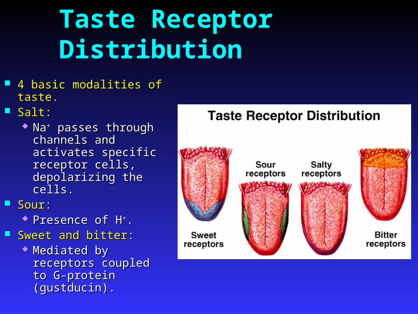
Taste Receptor Distribution
4 basic modalities of taste.4 basic modalities of taste. Salt:Salt:
NaNa++ passes through passes through channels and activates channels and activates specific receptor cells, specific receptor cells, depolarizing the cells.depolarizing the cells.
Sour:Sour: Presence of HPresence of H++..
Sweet and bitter:Sweet and bitter: Mediated by receptors Mediated by receptors
coupled to G-protein coupled to G-protein (gustducin).(gustducin).


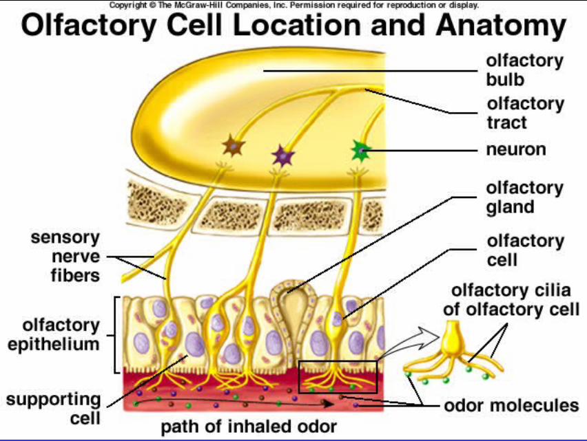
Smell (olfaction)
Bipolar sensory neurons Bipolar sensory neurons located within located within pseudostratified pseudostratified epithelium.epithelium.
Axon projects up into Axon projects up into olfactory bulb of olfactory bulb of cerebrum and dendrite cerebrum and dendrite that terminates in cilia.that terminates in cilia.
Molecules bind to Molecules bind to receptors and act through receptors and act through G-proteins to increase G-proteins to increase cAMP.cAMP.


Vision
Eyes transduce energy in the electrmagnetic Eyes transduce energy in the electrmagnetic spectrum into APs.spectrum into APs.
Only wavelengths of 400 – 700 nm Only wavelengths of 400 – 700 nm constitute visible light.constitute visible light.
Neurons in the retina contribute fibers that Neurons in the retina contribute fibers that are gathered together at the optic disc, are gathered together at the optic disc, where they exit as the optic nerve.where they exit as the optic nerve.


Refraction Light that passes from a Light that passes from a
medium of one density medium of one density into a medium of another into a medium of another density (bent).density (bent).
Refractive index (degree Refractive index (degree of refraction) depends of refraction) depends upon:upon:
Comparative density Comparative density of the 2 media.of the 2 media.
Curvature of Curvature of interface between the interface between the 2 media.2 media.
Refractive index Refractive index of air = 1.00of air = 1.00
Refractive index Refractive index of cornea = 1.38of cornea = 1.38
Image is inverted on Image is inverted on retina.retina.

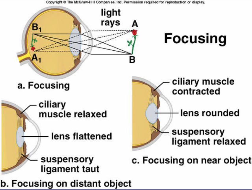
Accommodation
Ability of the eyes Ability of the eyes to keep the image to keep the image focused on the focused on the retina as the retina as the distance between distance between the eyes and object the eyes and object varies.varies.

Changes in the Lens Shape
Ciliary muscle can vary its Ciliary muscle can vary its aperture.aperture.
Distance > 20 feet:Distance > 20 feet: Relaxation places tension on Relaxation places tension on
the suspensory ligament.the suspensory ligament. Pulls lens taut. Pulls lens taut. Lens is least convex.Lens is least convex.
Distance decreases:Distance decreases: Ciliary muscles contract.Ciliary muscles contract. Reduces tension on Reduces tension on
suspensory ligament.suspensory ligament. Lens becomes more Lens becomes more
rounded and more convex.rounded and more convex.

Visual Acuity
Sharpness of vision.Sharpness of vision. Depends upon resolving Depends upon resolving
power:power: Ability of the visual Ability of the visual
system to resolve 2 system to resolve 2 closely spaced dots.closely spaced dots.
Myopia Myopia (nearsightedness):(nearsightedness):
Image brought to Image brought to focus in front of focus in front of retina.retina.
Hyperopia Hyperopia farsightedness):farsightedness):
Image brought to Image brought to focus behind the focus behind the retina.retina.

Visual Acuity
Astigmatism:Astigmatism: Asymmetry of the cornea and/or lens.Asymmetry of the cornea and/or lens. Images of lines of circle appear blurred.Images of lines of circle appear blurred.
Corrected by cylindrical lens.Corrected by cylindrical lens.


Retina
Consists of single-cell-thick Consists of single-cell-thick pigmented epithelium, pigmented epithelium, photoreceptor neurons:photoreceptor neurons: Rods and cones.Rods and cones.
Neural layers are forward Neural layers are forward extension of the brain.extension of the brain. Neural layers face Neural layers face
outward, toward the outward, toward the incoming light.incoming light.
Light must pass through Light must pass through several neural layers several neural layers before striking the rods before striking the rods and cones.and cones.
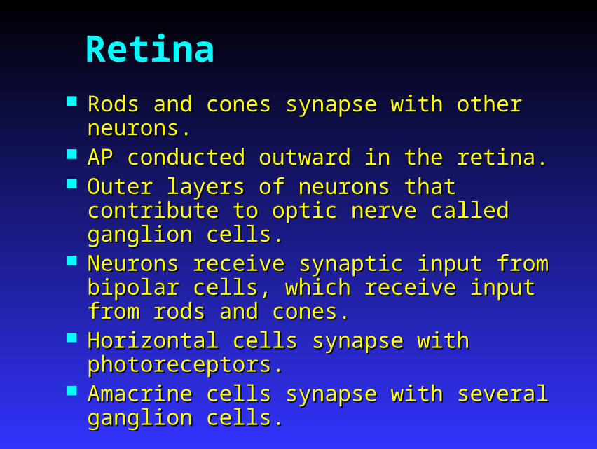
Retina Rods and cones synapse with other neurons.Rods and cones synapse with other neurons. AP conducted outward in the retina.AP conducted outward in the retina. Outer layers of neurons that contribute to optic Outer layers of neurons that contribute to optic
nerve called ganglion cells.nerve called ganglion cells. Neurons receive synaptic input from bipolar cells, Neurons receive synaptic input from bipolar cells,
which receive input from rods and cones.which receive input from rods and cones. Horizontal cells synapse with photoreceptors.Horizontal cells synapse with photoreceptors. Amacrine cells synapse with several ganglion cells.Amacrine cells synapse with several ganglion cells.

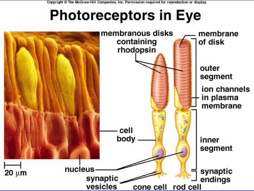


Effect of Light on Rods Rods are activated when light produces Rods are activated when light produces
chemical change in rhodopsin.chemical change in rhodopsin. Bleaching reaction:Bleaching reaction:
Rhodopsin dissociates Rhodopsin dissociates into retinene into retinene (rentinaldehyde) and (rentinaldehyde) and opsin.opsin.
• 11-cis retinene is 11-cis retinene is converted to all-converted to all-trans form.trans form.
Initiates changes in ionic Initiates changes in ionic permeability to produce permeability to produce AP in ganglionic cells.AP in ganglionic cells.
Provide black-and-white vision.Provide black-and-white vision.

Dark Adaptation Gradual increase in photoreceptor Gradual increase in photoreceptor
sensitivity when entering a dark room.sensitivity when entering a dark room. Maximal sensitivity reached in 20 min.Maximal sensitivity reached in 20 min. Increased amounts of visual pigments Increased amounts of visual pigments
produced.produced. Slight increased pigment in cones.Slight increased pigment in cones. Greater increased rhodopsin in rods.Greater increased rhodopsin in rods.
100,00-fold increase in light 100,00-fold increase in light sensitivity in rods.sensitivity in rods.

Electrical Activity of Retinal Cells
Ganglion cells and amacrine cells are only neurons Ganglion cells and amacrine cells are only neurons that produce AP.that produce AP.
In dark, photoreceptors release inhibitory NT that In dark, photoreceptors release inhibitory NT that hyperpolarizes bipolar neurons.hyperpolarizes bipolar neurons.
Light inhibits release of inhibitory NT.Light inhibits release of inhibitory NT. Dark current:Dark current: Rods and cones contain many NaRods and cones contain many Na++ channels that channels that
are open in the dark.are open in the dark. Causes slight membrane depolarization in dark.Causes slight membrane depolarization in dark.

Electrical Activity of Retinal Cells
NaNa++ channels rapidly close in response to light. channels rapidly close in response to light. cGMP required to keep the NacGMP required to keep the Na++ channels open. channels open. Opsin dissociation causes the alpha subunits of Opsin dissociation causes the alpha subunits of
G-proteins to dissociate. G-proteins to dissociate. G-protein subunits bind and activate G-protein subunits bind and activate
phosphodiesterase, converting cGMP to GMP.phosphodiesterase, converting cGMP to GMP. NaNa++ channels close when cGMP converted to channels close when cGMP converted to
GMP.GMP.

Cones and Color Vision Cones less sensitive than rods to light.Cones less sensitive than rods to light. Cones provide color vision and greater Cones provide color vision and greater
visual acuity.visual acuity. High light intensity bleaches out the rods, High light intensity bleaches out the rods,
and color vision with high acuity produced and color vision with high acuity produced by cones.by cones.

Cones and Color Vision Trichromatic theory of color Trichromatic theory of color
vision:vision: 3 types of cones:3 types of cones:
Blue, green and Blue, green and red.red.
According to According to the region of the region of visual spectrum visual spectrum absorbed.absorbed.
Each type of cone contains Each type of cone contains retinene associated with retinene associated with photopsins.photopsins.
Photopsin protein is Photopsin protein is unique for each of unique for each of the 3 cone pigment.the 3 cone pigment.
Each cone absorbs different Each cone absorbs different wavelengths of light.wavelengths of light.

Visual Acuity and Sensitivity
Each eye oriented so that Each eye oriented so that image falls within fovea image falls within fovea centralis.centralis. Fovea only contain cones.Fovea only contain cones. Degree of convergence of Degree of convergence of
cones is 1:1.cones is 1:1. Peripheral regions contain Peripheral regions contain
both rods and cones.both rods and cones. Degree of convergence of Degree of convergence of
rods is much lower.rods is much lower. Visual acuity greatest and Visual acuity greatest and
sensitivity lowest when light sensitivity lowest when light falls on fovea.falls on fovea.
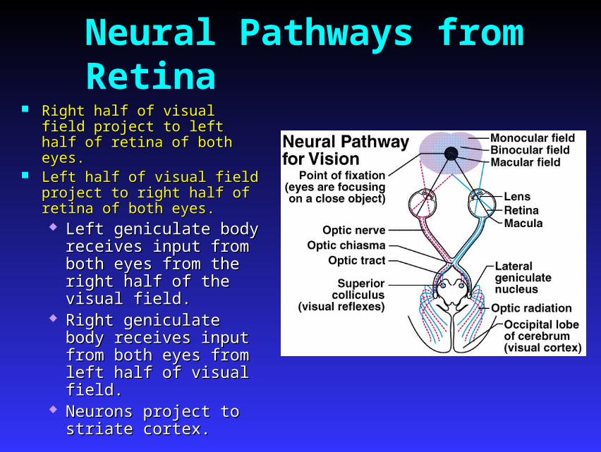
Neural Pathways from Retina Right half of visual field project Right half of visual field project
to left half of retina of both eyes.to left half of retina of both eyes. Left half of visual field project to Left half of visual field project to
right half of retina of both eyes.right half of retina of both eyes. Left geniculate body Left geniculate body
receives input from both receives input from both eyes from the right half of eyes from the right half of the visual field.the visual field.
Right geniculate body Right geniculate body receives input from both receives input from both eyes from left half of eyes from left half of visual field.visual field.
Neurons project to striate Neurons project to striate cortex.cortex.

Eye Movements
Superior colliculus coordinate:Superior colliculus coordinate: Smooth pursuit movements:Smooth pursuit movements:
Track moving objects.Track moving objects. Keep image focused on the fovea.Keep image focused on the fovea.
Saccadic eye movements:Saccadic eye movements: Quick jerky movements.Quick jerky movements. Occur when eyes appear still.Occur when eyes appear still. Move image to different photoreceptors.Move image to different photoreceptors.

Neural Processing of Visual Information Receptive field:Receptive field:
Part of visual field that affects activity of Part of visual field that affects activity of particular ganglion cell.particular ganglion cell.
On-center fields:On-center fields: Responses produced by light in the center of Responses produced by light in the center of
visual fields.visual fields. Off-center fields:Off-center fields:
Responses inhibited by light in the center and Responses inhibited by light in the center and stimulated by light in the surround.stimulated by light in the surround.


Vestibular Apparatus and Equilibrium
Equilibrium (orientation Equilibrium (orientation with respect to gravity) with respect to gravity) is due to vestibular is due to vestibular apparatus.apparatus.
Consists of 2 parts:Consists of 2 parts: Otolith organsOtolith organs
Utricle and Utricle and sacculesaccule
Semicircular canalsSemicircular canals


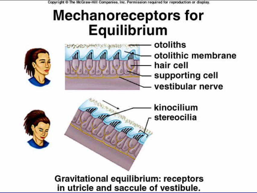
Sensory Hair Cells
Provide information about Provide information about linear acceleration.linear acceleration.
Hair cell receptors:Hair cell receptors: Stereocilia and Stereocilia and
kinocilium:kinocilium: When stereocilia bend When stereocilia bend
toward kinocilium, toward kinocilium, membrane depolarizes membrane depolarizes and releases NT.and releases NT.
When bend away from When bend away from kinocilium, kinocilium, hyperpolarization hyperpolarization occurs.occurs.

Utricle and Saccule
Each have macula with Each have macula with hair cells embedded in an hair cells embedded in an otolithic membrane.otolithic membrane.
Otolothic membrane Otolothic membrane contains crystals of Cacontains crystals of Ca++++ carbonate that resist carbonate that resist change in movement.change in movement.
Utricle:Utricle: More sensitive to More sensitive to
horizontal acceleration.horizontal acceleration. Saccule:Saccule: More sensitive to vertical More sensitive to vertical
acceleration.acceleration.

Semicircular Canals
Provide information about Provide information about rotational acceleration.rotational acceleration.
Project in 3 different planes.Project in 3 different planes. Each canal contains a Each canal contains a
semicircular duct. At the semicircular duct. At the base is the crista ampullaris.base is the crista ampullaris.
Hair cells processes are Hair cells processes are embedded in the cupula.embedded in the cupula.
Endolymph provides inertia Endolymph provides inertia so that the sensory so that the sensory processes will bend in processes will bend in direction opposite to the direction opposite to the angular acceleration.angular acceleration.


Ears and Hearing Sound waves travel in all directions from their source.Sound waves travel in all directions from their source. Waves are characterized by frequency and intensity.Waves are characterized by frequency and intensity.
Frequency:Frequency: Measured in hertz (cycles per second).Measured in hertz (cycles per second). Greater the frequency the higher the pitch.Greater the frequency the higher the pitch.
Intensity:Intensity: Directly related to amplitude of sound waves.Directly related to amplitude of sound waves. Measured in decibels.Measured in decibels.

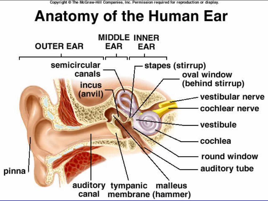
Outer Ear
Sound waves are funneled by the auricle Sound waves are funneled by the auricle into the external auditory meatus.into the external auditory meatus.
External auditory meatus channels sound External auditory meatus channels sound waves to the tympanic membrane.waves to the tympanic membrane. Increases sound wave intensity.Increases sound wave intensity.

Middle Ear Cavity between tympanic Cavity between tympanic
membrane and cochlea. membrane and cochlea. Malleus:Malleus:
Attached to tympanic Attached to tympanic membrane. membrane.
Vibrations of Vibrations of membrane are membrane are transmitted to the transmitted to the stapes.stapes.
Incus:Incus: Anvil.Anvil.
Stapes:Stapes: Attached to oval Attached to oval
window.window. Vibrates in response Vibrates in response
to vibrations in to vibrations in tympanic membrane.tympanic membrane.

Cochlea
Vibrations of stapes and oval window displace Vibrations of stapes and oval window displace perilymph fluid within scala vestibuli.perilymph fluid within scala vestibuli.
Vibrations pass to the scala tympani. Movements Vibrations pass to the scala tympani. Movements of perilymph travel to the base of cochlea where of perilymph travel to the base of cochlea where they displace the round window.they displace the round window.
As sound frequency increases, pressure waves of As sound frequency increases, pressure waves of the perilymph are transmitted through the the perilymph are transmitted through the vestibular membrane and through the basilar vestibular membrane and through the basilar membrane.membrane.
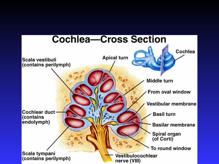

Displacement of basilar membrane is central to pitch discrimination.

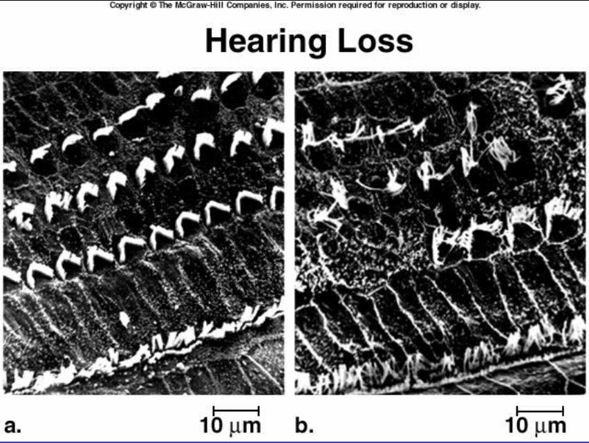
Organ of Corti
Sensory hair cells located on the basilar Sensory hair cells located on the basilar membrane. membrane.

Organ of Corti
Stereocilia of the outer hair Stereocilia of the outer hair cells are embedded in the cells are embedded in the tectorial membrane.tectorial membrane.
When the cochlear duct is When the cochlear duct is displaced, a shearing force is displaced, a shearing force is created, moving and bending created, moving and bending the stereocilia.the stereocilia.
Ion channels open, depolarizing Ion channels open, depolarizing the hair cells, releasing the hair cells, releasing glutamate that stimulates the glutamate that stimulates the sensory neuron.sensory neuron.
Greater bending of stereocilia, Greater bending of stereocilia, the increased frequency of AP the increased frequency of AP produced.produced.