CHAPTER 10 RADIATION RISK ASSESSMENT GUIDANCEinfohouse.p2ric.org/ref/13/12026/ch10.pdf · LLD =...
37
ACRONYMS, SYMBOLS, AND UNITS FOR CHAPTER 10 A(t) = Activity at Time t Bq = Becquerel Ci = Curie CLP = Contract Laboratory Program D = Absorbed Dose DCF = Dose Conversion Factor Per Unit Intake H = Effective Dose Equivalent E H = Dose Equivalent Averaged Over Tissue or T Organ T H = Committed Effective Dose Equivalent Per E,50 Unit Intake H = Committed Dose Equivalent Averaged Over T,50 Tissue T LET = Linear Energy Transfer LLD = Lower Limit of Detection MeV = Million Electron Volts N = Modifying Factor in the Definition of Dose Equivalent pCi = PicoCurie (10 Ci) -12 Q = Quality Factor in Definition of Dose Equivalent RBE = Relative Biological Effectiveness SI = International System of Units Sv = Sievert T = Tissue or Target Organs w = Weighting Factor in the Definition of Effective T Dose Equivalent and Committed Effective Dose Equivalent CHAPTER 10 RADIATION RISK ASSESSMENT GUIDANCE There are many sites contaminated with radioactive substances that are included on the National Priorities List (NPL), and additional sites are expected in future NPL updates. This chapter provides supplemental baseline risk assessment guidance for use at these sites. This guidance is intended as an overview of key differences in chemical and radionuclide assessments, and not as a comprehensive, stand-alone approach for assessing the risks posed by radiation. The reader should be familiar with the guidance provided in Chapters 2 through 9 before proceeding further in Chapter 10. Although the discussions in the previous chapters focus primarily on chemically contaminated sites, much of the information presented is also applicable to the evaluation of radioactively contaminated Superfund sites. For consistency and completeness, the topics discussed in each section of this chapter parallel the topics covered in each of the previous chapters. After a brief introduction to some of the basic principles and concepts of radiation protection (Section 10.1), seven additional areas are addressed: (1) Regulation of Radioactively Contaminated Sites (Section 10.2); (5) Toxicity Assessment (Section 10.6); (2) Data Collection (Section 10.3); (6) Risk Characterization (Section 10.7); and (3) Data Evaluation (Section 10.4); (7) Documentation, Review, and Management (4) Exposure and Dose Assessment (Section and Manager (Section 10.8). 10.5); Tools for the Risk Assessor, Reviewer,
-
Upload
truongnguyet -
Category
Documents
-
view
223 -
download
4
Transcript of CHAPTER 10 RADIATION RISK ASSESSMENT GUIDANCEinfohouse.p2ric.org/ref/13/12026/ch10.pdf · LLD =...
ACRONYMS, SYMBOLS, AND UNITSFOR CHAPTER 10
A(t) = Activity at Time tBq = BecquerelCi = CurieCLP = Contract Laboratory ProgramD = Absorbed DoseDCF = Dose Conversion Factor Per Unit IntakeH = Effective Dose EquivalentE
H = Dose Equivalent Averaged Over Tissue orT
Organ TH = Committed Effective Dose Equivalent PerE,50
Unit IntakeH = Committed Dose Equivalent Averaged Over T,50
Tissue TLET = Linear Energy TransferLLD = Lower Limit of DetectionMeV = Million Electron VoltsN = Modifying Factor in the Definition of Dose EquivalentpCi = PicoCurie (10 Ci)-12
Q = Quality Factor in Definition of Dose EquivalentRBE = Relative Biological EffectivenessSI = International System of UnitsSv = SievertT = Tissue or Target Organsw = Weighting Factor in the Definition of Effective T
Dose Equivalent and Committed Effective Dose Equivalent
CHAPTER 10
RADIATION RISK ASSESSMENTGUIDANCE
There are many sites contaminated with radioactivesubstances that are included on the NationalPriorities List (NPL), and additional sites areexpected in future NPL updates. This chapterprovides supplemental baseline risk assessmentguidance for use at these sites. This guidance isintended as an overview of key differences inchemical and radionuclide assessments, and not as acomprehensive, stand-alone approach for assessingthe risks posed by radiation.
The reader should be familiar with the guidanceprovided in Chapters 2 through 9 before proceedingfurther in Chapter 10. Although the discussions inthe previous chapters focus primarily on chemicallycontaminated sites, much of the informationpresented is also applicable to the evaluation ofradioactively contaminated Superfund sites. Forconsistency and completeness, the topics discussedin each section of this chapter parallel the topicscovered in each of the previous chapters.
After a brief introduction to some of the basicprinciples and concepts of radiation protection(Section 10.1), seven additional areas are addressed:
(1) Regulation of Radioactively ContaminatedSites (Section 10.2); (5) Toxicity Assessment (Section 10.6);
(2) Data Collection (Section 10.3); (6) Risk Characterization (Section 10.7); and
(3) Data Evaluation (Section 10.4); (7) Documentation, Review, and Management
(4) Exposure and Dose Assessment (Section and Manager (Section 10.8).10.5);
Tools for the Risk Assessor, Reviewer,

Page 10-2
DEFINITIONS FOR CHAPTER 10
Absorbed Dose (D). The mean energy imparted by ionizing radiation to matter per unit mass. The special SI unit of absorbeddose is the gray (Gy); the conventional unit is the rad (1 rad = 0.01 Gy).
Becquerel (Bq). One nuclear disintegration per second; the name for the SI unit of activity. 1 Bq = 2.7 x 10 Ci.-11
Committed Dose Equivalent (H). The total dose equivalent (averaged over tissue T) deposited over the 50-year periodT,50
following the intake of a radionuclide.
Committed Effective Dose Equivalent (H). The weighted sum of committed dose equivalents to specified organs and tissues,E,50
in analogy to the effective dose equivalent.
Curie (Ci). 3.7 x 10 nuclear disintegrations per second, the name for the conventional unit of activity. 1 Ci = 3.7 x 10 Bq.10 10
Decay Product(s). A radionuclide or a series of radionuclides formed by the nuclear transformation of another radionuclidewhich, in this context, is referred to as the parent.
Dose Conversion Factor (DCF). The dose equivalent per unit intake of radionuclide.
Dose Equivalent (H). The product of the absorbed dose (D), the quality factor (Q), and any other modifying factors (N). The SIunit of dose equivalent is the sievert (Sv); the conventional unit is the rem (1 rem = 0.01 Sv).
Effective Dose Equivalent (H). The sum over specified tissues of the products of the dose equivalent in a tissue or organ (T)E
and the weighting factor for that tissue.
External Radiation. Radiations incident upon the body from an external source.
Gray (Gy). The SI unit of absorbed dose. 1Gy = 1 Joule kg = 100 rad.-1
Half-Life (physical, biological, or effective). The time for a quantity of radionuclide, i.e., its activity, to diminish by a factor of ahalf (because of nuclear decay events, biological elimination of the material, or both.).
Internal Radiation. Radiation emitted from radionuclides distributed within the body.
Ionizing Radiation. Any radiation capable of displacing electrons from atoms or molecules, thereby producing ions.
Linear Energy Transfer (LET). A measure of the rate of energy absorption, defined as the average energy imparted to theabsorbing medium by a charged particle per unit distance (KeV per um).
Nuclear Transformation. The spontaneous transformation of one radionuclide into a different nuclide or into a different energystate of the same nuclide.
Quality Factor (Q). The principal modifying factor that is employed in deriving dose equivalent, H, from absorbed dose, D;chosen to account for the relative biological effectiveness (RBE) of the radiation in question, but to be independentof the tissue or organ under consideration, and of the biological endpoint. For radiation protection purposes, thequality factor is determined by the linear energy transfer (LET) of the radiation.
Rad. The conventional unit for absorbed dose of ionizing radiation; the corresponding SI unit is the gray (Gy); 1 rad = 0.01 Gy= 0.01 Joule/kg.
Rem. An acronym of radiation equivalent man, the conventional unit of dose equivalent; the corresponding SI unit is theSievert; 1 Sv = 100 rem.
Sievert (Sv). The special name for the SI unit of dose equivalent. 1 Sv = 100 rem.
Slope Factor. The age-averaged lifetime excess cancer incidence rate per unit intake (or unit exposure for external exposurepathways) of a radionuclide.
Weighting Factor (w). Factor indicating the relative risk of cancer induction or hereditary defects from irradiation of a givenT
tissue or organ; used in calculation of effective dose equivalent and committed effective dose equivalent.

Page 10-3
There are special hazards associated with Only summary-level information is presented inhandling radioactive waste and EPA strongly this chapter, and references are provided to a numberrecommends that a health physicist experienced in of supporting technical documents for furtherradiation measurement and protection be consulted information. In particular, the reader is encouragedprior to initiating any activities at a site suspected of to consult Volume 1 of the Background Informationbeing contaminated with radioactive substances. Document for the Draft Environmental ImpactEPA also recommends that the remedial project Statement for Proposed NESHAPS for Radionuclidesmanager (RPM) or on-scene coordinator (OSC) (EPA 1989a) for a more comprehensive discussionshould designate both a chemical risk assessor and a of EPA's current risk assessment methodology forradiation risk assessor. These individuals should radionuclides.work closely with each other and the RPM tocoordinate remedial activities (e.g., site scoping, For additional radiation risk assessmenthealth and safety planning, sampling and analysis) information and guidance, RPMs and otherand exchange information common to both chemical interested individuals can contact the Office ofand radionuclide assessments, including data on the Radiation Programs (ORP) within EPA headquartersphysical characteristics of the site, potentially at 202-475-9630 (FTS 475-9630). Interestedimpacted populations, pathways of concern, and fate individuals also can contact the Regional Radiationand transport models used. At the conclusion of the Program Managers within each of the EPA regionalremedial investigation/feasibility study (RI/FS) offices for guidance and health physics support.process, the RPM should issue a single report thatsummarizes and integrates the results from both thechemical and the radiation risk assessments.
A two-phase evaluation is described for theradiation risk assessment. As discussed in Section10.5, procedures established by the InternationalCommission on Radiological Protection (ICRP 1979)and adopted by EPA in Federal Guidance ReportNo. 11 (EPA 1988) are used to estimate the radiationdose equivalent to humans from potential exposuresto radionuclides through all pertinent exposurepathways at a site. Those estimates of doseequivalent may be used for comparison withestablished radiation protection standards andcriteria. However, this methodology was developedfor regulation of occupational radiation exposuresfor adults and is not completely applicable forestimating health risk to the general population at aSuperfund site. Therefore, a separate methodologyis presented in Section 10.7.2 for estimating healthrisk, based on the age-averaged lifetime excesscancer incidence per unit intake (and per unitexternal exposure) for radionuclides of concern.Radiation risk assessments for Superfund sitesshould include estimates of both the dose equivalentcomputed as described in Section 10.5, and thehealth risk attributable to radionuclide exposurescomputed using the approach described in Section10.7.
10.1 RADIATION PROTECTIONPRINCIPLES ANDCONCEPTS
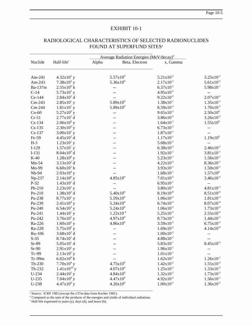
Radioactive atoms undergo spontaneous nucleartransformations and release excess energy in theform of ionizing radiation. Such transformations arereferred to as radioactive decay. As a result of theradioactive decay process, one element istransformed into another; the newly formed element,called a decay product, will possess physical andchemical properties different from those of its parent,and may also be radioactive. A radioactive speciesof a particular element is referred to as aradionuclide or radioisotope. The exact mode ofradioactive transformation for a particularradionuclide depends solely upon its nuclearcharacteristics, and is independent of the nuclide'schemical characteristics or physical state. Afundamental and unique characteristic of eachradionuclide is its radioactive half-life, defined as thetime required for one half of the atoms in a givenquantity of the radionuclide to decay. Over 1,600different radionuclides have been identified to date,with half-lives ranging from fractions of a second tomillions of years. Selected radionuclides of potentialimportance at Superfund sites are listed in Exhibit10-1.
Page 10-4
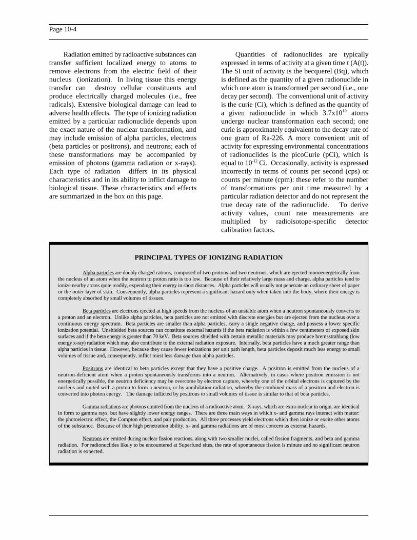
PRINCIPAL TYPES OF IONIZING RADIATION
Alpha particles are doubly charged cations, composed of two protons and two neutrons, which are ejected monoenergetically fromthe nucleus of an atom when the neutron to proton ratio is too low. Because of their relatively large mass and charge, alpha particles tend toionize nearby atoms quite readily, expending their energy in short distances. Alpha particles will usually not penetrate an ordinary sheet of paperor the outer layer of skin. Consequently, alpha particles represent a significant hazard only when taken into the body, where their energy iscompletely absorbed by small volumes of tissues.
Beta particles are electrons ejected at high speeds from the nucleus of an unstable atom when a neutron spontaneously converts toa proton and an electron. Unlike alpha particles, beta particles are not emitted with discrete energies but are ejected from the nucleus over acontinuous energy spectrum. Beta particles are smaller than alpha particles, carry a single negative charge, and possess a lower specificionization potential. Unshielded beta sources can constitute external hazards if the beta radiation is within a few centimeters of exposed skinsurfaces and if the beta energy is greater than 70 keV. Beta sources shielded with certain metallic materials may produce bremsstrahlung (lowenergy x-ray) radiation which may also contribute to the external radiation exposure. Internally, beta particles have a much greater range thanalpha particles in tissue. However, because they cause fewer ionizations per unit path length, beta particles deposit much less energy to smallvolumes of tissue and, consequently, inflict must less damage than alpha particles.
Positrons are identical to beta particles except that they have a positive charge. A positron is emitted from the nucleus of aneutron-deficient atom when a proton spontaneously transforms into a neutron. Alternatively, in cases where positron emission is notenergetically possible, the neutron deficiency may be overcome by electron capture, whereby one of the orbital electrons is captured by thenucleus and united with a proton to form a neutron, or by annihilation radiation, whereby the combined mass of a positron and electron isconverted into photon energy. The damage inflicted by positrons to small volumes of tissue is similar to that of beta particles.
Gamma radiations are photons emitted from the nucleus of a radioactive atom. X-rays, which are extra-nuclear in origin, are identicalin form to gamma rays, but have slightly lower energy ranges. There are three main ways in which x- and gamma rays interact with matter:the photoelectric effect, the Compton effect, and pair production. All three processes yield electrons which then ionize or excite other atomsof the substance. Because of their high penetration ability, x- and gamma radiations are of most concern as external hazards.
Neutrons are emitted during nuclear fission reactions, along with two smaller nuclei, called fission fragments, and beta and gammaradiation. For radionuclides likely to be encountered at Superfund sites, the rate of spontaneous fission is minute and no significant neutronradiation is expected.
Radiation emitted by radioactive substances can Quantities of radionuclides are typicallytransfer sufficient localized energy to atoms to expressed in terms of activity at a given time t (A(t)).remove electrons from the electric field of their The SI unit of activity is the becquerel (Bq), whichnucleus (ionization). In living tissue this energy is defined as the quantity of a given radionuclide intransfer can destroy cellular constituents and which one atom is transformed per second (i.e., oneproduce electrically charged molecules (i.e., free decay per second). The conventional unit of activityradicals). Extensive biological damage can lead to is the curie (Ci), which is defined as the quantity ofadverse health effects. The type of ionizing radiation a given radionuclide in which 3.7x10 atomsemitted by a particular radionuclide depends upon undergo nuclear transformation each second; onethe exact nature of the nuclear transformation, and curie is approximately equivalent to the decay rate ofmay include emission of alpha particles, electrons one gram of Ra-226. A more convenient unit of(beta particles or positrons), and neutrons; each of activity for expressing environmental concentrationsthese transformations may be accompanied by of radionuclides is the picoCurie (pCi), which isemission of photons (gamma radiation or x-rays). equal to 10 Ci. Occasionally, activity is expressedEach type of radiation differs in its physical incorrectly in terms of counts per second (cps) orcharacteristics and in its ability to inflict damage to counts per minute (cpm): these refer to the numberbiological tissue. These characteristics and effects of transformations per unit time measured by aare summarized in the box on this page. particular radiation detector and do not represent the
10
-12
true decay rate of the radionuclide. To deriveactivity values, count rate measurements aremultiplied by radioisotope-specific detectorcalibration factors.
Page 10-5
EXHIBIT 10-1
RADIOLOGICAL CHARACTERISTICS OF SELECTED RADIONUCLIDESFOUND AT SUPERFUND SITESa
Average Radiation Energies (MeV/decay) b
Nuclide Half-life Alpha Beta, Electron x, Gammac
Am-241 4.32x10 y 5.57x10 5.21x10 3.25x10² 0 -2 -2
Am-243 7.38x10 y 5.36x10 2.17x10 5.61x10³ 0 -2 -2
Ba-137m 2.55x10 h -- 6.37x10 5.98x100 -2 -1
C-14 5.73x10 y -- 4.95x10 --3 -2
Ce-144 2.84x10 d -- 9.22x10 2.07x10² -2 -2
Cm-243 2.85x10 y 5.89x10 1.38x10 1.35x101 0 -1 -1
Cm-244 1.81x10 y 5.89x10 8.59x10 1.70x101 0 -3 -3
Co-60 5.27x10 y -- 9.65x10 2.50x100 -2 0
Cr-51 2.77x10 d -- 3.86x10 3.26x101 -3 -2
Cs-134 2.06x10 y -- 1.64x10 1.55x100 -1 0
Cs-135 2.30x10 y -- 6.73x10 --6 -2
Cs-137 3.00x10 y -- 1.87x10 --1 -1
Fe-59 4.45x10 d -- 1.17x10 1.19x101 -1 0
H-3 1.23x10 y -- 5.68x10 --1 -3
I-129 1.57x10 y -- 6.38x10 2.46x10⁷ -2 -2
I-131 8.04x10 d -- 1.92x10 3.81x100 -1 -1
K-40 1.28x10 y -- 5.23x10 1.56x10⁹ -1 -1
Mn-54 3.13x10 d -- 4.22x10 8.36x10² -3 -1
Mo-99 6.60x10 h -- 3.93x10 1.50x101 -1 -1
Nb-94 2.03x10 y -- 1.68x10 1.57x10⁴ -1 0
Np-237 2.14x10 y 4.85x10 7.01x10 3.46x10⁶ 0 -2 -2
P-32 1.43x10 d -- 6.95x10 --1 -1
Pb-210 2.23x10 y -- 3.80x10 4.81x101 -2 -3
Po-210 1.38x10 d 5.40x10 8.19x10 8.51x10² 0 -8 -6
Pu-238 8.77x10 y 5.59x10 1.06x10 1.81x101 0 -2 -3
Pu-239 2.41x10 y 5.24x10 6.74x10 8.07x10⁴ 0 -3 -4
Pu-240 6.54x10 y 5.24x10 1.06x10 1.73x10³ 0 -2 -3
Pu-241 1.44x10 y 1.22x10 5.25x10 2.55x101 -4 -3 -6
Pu-242 3.76x10 y 4.97x10 8.73x10 1.44x10⁵ 0 -3 -3
Ra-226 1.60x10 y 4.86x10 3.59x10 6.75x10³ 0 -3 -3
Ra-228 5.75x10 y -- 1.69x10 4.14x100 -2 -9
Ru-106 3.68x10 d -- 1.00x10 --2 -2
S-35 8.74x10 d -- 4.88x10 --1 -2
Sr-89 5.05x10 d -- 5.83x10 8.45x101 -1 -5
Sr-90 2.91x10 y -- 1.96x10 --1 -1
Tc-99 2.13x10 y -- 1.01x10 --5 -1
Tc-99m 6.02x10 h -- 1.62x10 1.26x100 -2 -1
Th-230 7.70x10 y 4.75x10 1.42x10 1.55x10⁴ 0 -2 -3
Th-232 1.41x10 y 4.07x10 1.25x10 1.33x10¹⁰ 0 -2 -3
U-234 2.44x10 y 4.84x10 1.32x10 1.73x10⁵ 0 -2 -3
U-235 7.04x10 y 4.47x10 4.92x10 1.56x10⁸ 0 -2 -1
U-238 4.47x10 y 4.26x10 1.00x10 1.36x10⁹ 0 -2 -3
Source: ICRP 1983 (except Ba-137m data from Kocher 1981).a
Computed as the sum of the products of the energies and yields of individual radiations.b
Half-life expressed in years (y), days (d), and hours (h).c

Page 10-6
GENERAL HEALTH PHYSICSREFERENCES
Introduction to Health Physics (Cember 1983)
Atoms, Radiation, and Radiation Protection(Turner 1986)
Environmental Radioactivity (Eisenbud 1987)
The Health Physics and Radiological HealthHandbook (Shleien and Terpilak 1984)
The activity per unit mass of a given radionuclide is radiation. The absorbed dose of any radiationcalled the specific activity, and is usually expressed divided by the absorbed dose of a reference radiationin units of becquerels per gram (Bq/g) or curies per (traditionally 250 kVp x-rays) that produces the samegram (Ci/g). The shorter the half-life of the biological endpoint is called the Relative Biologicalradionuclide, the greater is its specific activity. For Effectiveness or RBE. For regulatory purposes, anexample, Co-60 has a radioactive half-life of about arbitrary consensus RBE estimate called the Quality5 years and a specific activity of 4x10 Bq/g, Factor or Q is often used. The dose equivalent (H)13
whereas Np-237 has a half-life of 2 million years and was developed to normalize the unequal biologicala specific activity of 3x10 Bq/g. effects produced from equal absorbed doses of7
Several terms are used by health physicists to defined as:describe the physical interactions of different typesof radiations with biological tissue, and to define the H = DQNeffects of these interactions on human health. One ofthe first terms developed was radiation exposure, where D is the absorbed dose, Q is a quality factorwhich refers to the transfer of energy from a that accounts for the RBE of the type of radiationradiation field of x- or gamma rays to a unit mass of emitted, and N is the product of any additionalair. The unit for this definition of exposure is the modifying factors. Quality factors currently assignedroentgen (R), expressed as coulombs of charge per by the International Commission on Radiologicalkilogram of air (1 R = 2.58x10 C/kg). Protection (ICRP) include values of Q=20 for alpha-4
The term exposure is also defined as the for beta particles, positrons, x-rays, and gamma raysphysical contact of the human body with radiation. (ICRP 1984). These factors may be interpreted asInternal exposure refers to an exposure that occurs follows: on average, if an equal amount of energy iswhen human tissues are subjected to radiations from absorbed, an alpha particle will inflict approximatelyradionuclides that have entered the body via 20 times more damage to biological tissue than ainhalation, ingestion, injection, or other routes. beta particle or gamma ray, and twice as muchExternal exposure refers to the irradiation of human damage as a neutron. The modifying factor istissues by radiations emitted by radionuclides located currently assigned a value of unity (N=1) for alloutside the body either dispersed in the air or water, radiations. The SI unit of dose equivalent is theon skin surfaces, or deposited on ground surfaces. sievert (Sv), and the conventional unit is the rem (1All types of radiation may contribute to internal rem = 0.01 Sv).exposure, whereas only photon, beta, and neutronradiations contribute significantly to externalexposure.
Ionizing radiation can cause deleterious effectson biological tissues only when the energy releasedduring radioactive decay is absorbed in tissue. Theabsorbed dose (D) is defined as the mean energyimparted by ionizing radiation per unit mass oftissue. The SI unit of absorbed dose is the joule perkilogram, also assigned the special name the gray (1Gy = 1 joule/kg). The conventional unit of absorbeddose is the rad (1 rad = 100 ergs per gram = 0.01Gy).
For radiation protection purposes, it is desirableto compare doses of different types of
different types of radiation. The dose equivalent is
particles, Q=10 for neutrons and protons, and Q=1

Page 10-7
EFFECTIVE DOSE EQUIVALENT
The effective dose equivalent, H , is a weighted sum of dose equivalents to all organs and tissues (ICRP 1977, ICRP 1979), defined as:E
H = E w HE T T
T
where w is the weighting factor for organ or tissue T and H is the mean dose equivalent to organ or tissue T. The factor w , which isT T T
normalized so that the summation of all the organ weighting factors is equal to one, corresponds to the fractional contribution of organ or tissueT to the total risk of stochastic health effects when the body is uniformly irradiated. Similarly, the committed effective dose equivalent, H ,E,50
is defined as the weighted sum of committed dose equivalents to all irradiated organs and tissues, as follows:
H = E w HE,50 T T,50
T
H and H thus reflect both the distribution of dose among the various organs and tissues of the body and their assumed relative sensitivitiesE E,50
to stochastic effects. The organ and tissue weighting factor values w are as follows: Gonads, 0.25; Breast, 0.15; Red Marrow, 0.12; Lungs,T
0.12; Thyroid, 0.03; Bone Surface, 0.03; and Remainder, 0.30 (i.e., a value of w = 0.06 is applicable to each of the five remaining organs orT
tissues receiving the highest doses).
The dose delivered to tissues from radiations the same number (but possibly a dissimilarexternal to the body occurs only while the radiation distribution) of fatal stochastic health effects as thefield is present. However, the dose delivered to particular combination of committed organ dosebody tissues due to radiations from systemically equivalents (see the box on this page).incorporated radionuclides may continue long afterintake of the nuclide has ceased. Therefore, internal A special unit, the working level (WL), is useddoses to specific tissues and organs are typically to describe exposure to the short-lived radioactivereported in terms of the committed dose equivalent decay products of radon (Rn-222). Radon is a(H ), which is defined as the integral of the dose naturally occurring radionuclide that is of particularT,50
equivalent in a particular tissue T for 50 years after concern because it is ubiquitous, it is very mobile inintake (corresponding to a working lifetime). the environment, and it decays through a series of
When subjected to equal doses of radiation, significant dose to the lung when inhaled. The WLorgans and tissues in the human body will exhibit is defined as any combination of short-lived radondifferent cancer induction rates. To account for decay products in one liter of air that will result inthese differences and to normalize radiation doses the ultimate emission of 1.3x10 MeV of alphaand effects on a whole body basis for regulation of energy. The working level month (WLM) is definedoccupational exposure, the ICRP developed the as the exposure to 1 WL for 170 hours (1 workingconcept of the effective dose equivalent (H ) and month).E
committed effective dose equivalent (H ), whichE,50
are defined as weighted sums of the organ-specific Radiation protection philosophy encourages thedose equivalents (i.e., E w H ) and organ-specific reduction of all radiation exposures as low asT T
committed dose equivalents (i.e., Ew H ), reasonably achievable (ALARA), in consideration ofT T,50
respectively. Weighting factors, w , are based on technical, economic, and social factors. Further, noT
selected stochastic risk factors specified by the ICRP practice involving radiation exposure should beand are used to average organ-specific dose adopted unless it provides a positive net benefit. Inequivalents (ICRP 1977, 1979). The effective dose addition to these general guidelines, specific upperequivalent is equal to that dose equivalent, delivered limits on radiation exposures and doses have beenat a uniform whole-body rate, that corresponds to established by regulatory authorities as described in
short-lived decay products that can deliver a
5
the following section.

Page 10-8
Additional discussion on the measurement of Uranium Mill Tailings Radiation Controlradioactivity is provided in Sections 10.3 and 10.4, Act (UMTRCA), the Nuclear Wasteand the evaluation of radiation exposure and dose is Policy Act, the Resource Conservation anddiscussed further in Section 10.5. Discussion of Recovery Act (RCRA), and CERCLA.potential health impacts from ionizing radiation is EPA's major responsibilities with regard topresented in Section 10.6. radiation include the development of
10.2 REGULATION OFRADIOACTIVELYCONTAMINATED SITES
Chapter 2 briefly describes the statutes,regulations, guidance, and studies related to thehuman health evaluation process for chemicalcontaminants. The discussion describes CERCLA,as amended by SARA, and the RI/FS process. Sinceradionuclides are classified as hazardous substancesunder CERCLA, this information is also applicableto radioactively contaminated sites. Chapter 2 alsointroduces the concept of compliance with applicableor relevant and appropriate requirements (ARARs)in federal and state environmental laws as requiredby SARA. Guidance on potential ARARs for theremediation of radioactively contaminated sitesunder CERCLA is available in the CERCLACompliance with Other Laws Manual (EPA 1989c).Only a brief summary of regulatory authorities ispresented here.
The primary agencies with regulatory authorityfor the cleanup of radioactively contaminated sitesinclude EPA, the Nuclear Regulatory Commission(NRC), the Department of Energy (DOE), and stateagencies. Other federal agencies, including theDepartment of Transportation (DOT) andDepartment of Defense (DOD), also have regulatoryprograms (but more limited) for radioactivematerials. Also, national and international scientificadvisory organizations provide recommendationsrelated to radiation protection and radioactive wastemanagement, but have no regulatory authority. Thefollowing is a brief description of the main functionsand areas of jurisdiction of these agencies andorganizations.
! EPA's authority to protect public healthand the environment from adverse effectsof radiation exposure is derived fromseveral statutes, including the AtomicEnergy Act, the Clean Air Act, the
federal guidance and standards,assessment of new technologies, andsurveillance of radiation in theenvironment. EPA also has leadresponsibility in the federal governmentfor advising all federal agencies onradiation standards. EPA's radiationstandards apply to many different types ofactivities involving all types of radioactivematerial (i.e., source, byproduct, specialnuclear, and naturally occurring andaccelerator produced radioactive material[NARM]). For some of the EPAstandards, implementation andenforcement responsibilities are vested inother agencies, such as NRC and DOE.
! NRC licenses the possession and use ofcertain types of radioactive material atcertain types of facilities. Specifically, theNRC is authorized to license source,byproduct, and special nuclear material.The NRC is not authorized to licenseNARM, although NARM may be partiallysubject to NRC regulation when it isassociated with material licensed by theNRC. Most of DOE's operations areexempt from NRC's licensing andregulatory requirements, as are certainDOD activities involving nuclear weaponsand the use of nuclear reactors for militarypurposes.
! DOE is responsible for conducting oroverseeing radioactive material operationsat numerous government-owned/contractor-operated facilities.DOE is also responsible for managingseveral inactive sites that containradioactive waste, such as sites associatedwith the Formerly Utilized Sites RemedialAction Program (FUSRAP), the UraniumMill Tailings Remedial Action Program(UMTRAP), the Grand Junction Remedial

Page 10-9
MAJOR FEDERAL LAWS FOR RADIATION PROTECTION
! Atomic Energy Act of 1954, Public Law 83-703 - established the Atomic Energy Commission as the basic regulatoryauthority for ionizing radiation.
! Energy Reorganization Act of 1974, Public Law 93-438 - amended the Atomic Energy Act, and established the NuclearRegulatory Commission to regulate nondefense nuclear activities.
! Marine Protection, Research, and Sanctuaries Act of 1972, Public Law 92-532 - established controls for ocean disposal ofradioactive waste.
! Safe Drinking Water Act, Public Law 93-523 - mandated regulation of radionuclides in drinking water.
! Clean Air Act Amendments of 1977, Public Law 95-95 - extended coverage of the Act's provisions to includeradionuclides.
! Uranium Mill Tailings Radiation Control Act of 1978, Public Law 96-415 - required stabilization and control of byproductmaterials (primarily mill tailings) at licensed commercial uranium and thorium processing sites.
! Low-Level Radioactive Waste Policy Act of 1980, Public Law 96-573 - made states responsible for disposal of LLRWgenerated within their borders and encouraged formation of inter-state compacts.
! Nuclear Waste Policy Act of 1982, Public Law 97-425 - mandated the development of repositories for the disposal ofhigh-level radioactive waste and spent nuclear fuel.
! Low-Level Radioactive Waste Policy Act Amendments of 1985, Public Law 99-240 - amended LLRWPA requirements and
Action Program (GJRAP), and the ! States have their own authority and regulationsSurplus Facilities Management Program for managing radioactive material and waste.(SFMP). DOE is authorized to control all In addition, 29 states (Agreement States) havetypes of radioactive materials at sites entered into agreements with the NRC,within its jurisdiction. whereby the Commission has relinquished to
! Other federal agencies with regulatory the states its regulatory authority over source,programs applicable to radioactive waste byproduct, and small quantities of specialinclude DOT and DOD. DOT has issued nuclear material. Both Agreement States andregulations that set forth packaging, Nonagreement States can also regulate NARM.labeling, record keeping, and reporting Such state-implemented regulations arerequirements for the transport of potential ARARs.radioactive material (see 49 CFR Parts 171 ! The National Council on Radiation Protectionthrough 179). Most of DOD's radioactive and Measurements (NCRP) and thewaste management activities are regulated International Commission on Radiologicalby NRC and/or EPA. However, DOD has Protection (ICRP) provide recommendationsits own program for controlling wastes on human radiation protection. The NCRP wasgenerated for certain nuclear weapon and chartered by Congress to collect, analyze,reactor operations for military purposes. develop, and disseminate information andOther agencies, such as the Federal recommendations about radiation protectionEmergency Management Agency (FEMA) and measurements. The ICRP's function isand the Department of the Interior (DOI), basically the same, but on an internationalmay also play a role in radioactive waste level. Although neither the NCRP nor thecleanups in certain cases. ICRP have regulatory authority, their
recommendations serve as the basis for manyof the general (i.e., not
Page 10-10
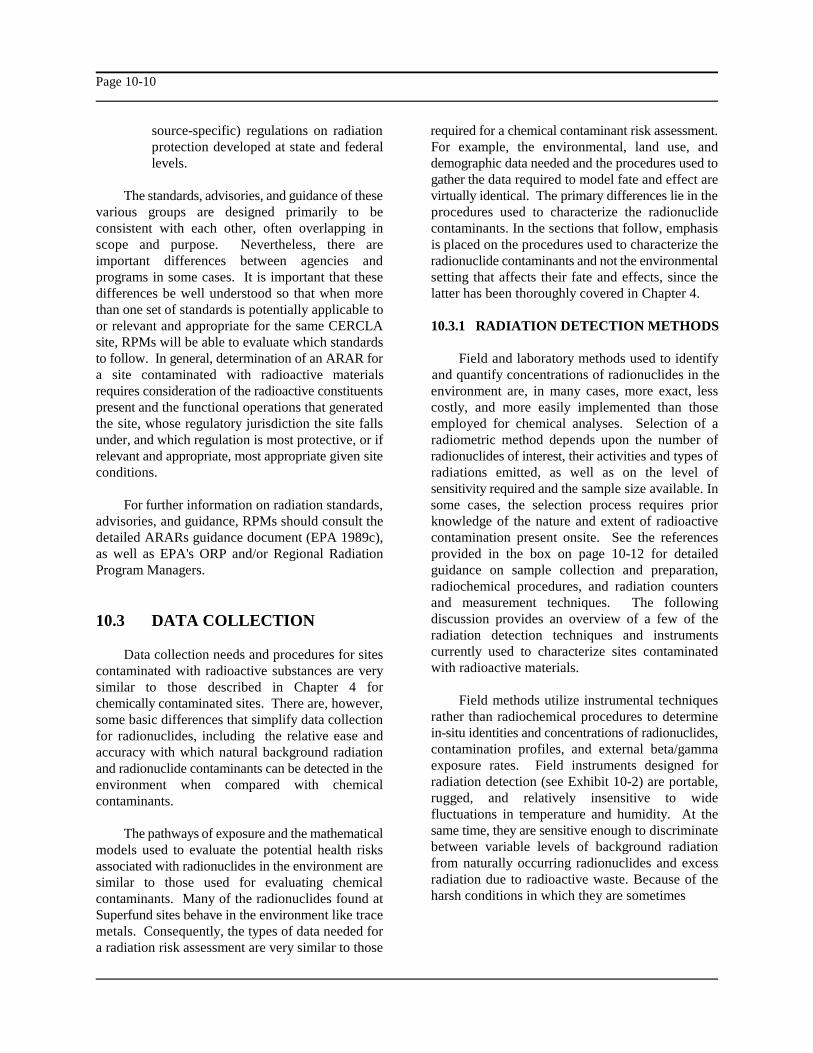
source-specific) regulations on radiation required for a chemical contaminant risk assessment.protection developed at state and federal For example, the environmental, land use, andlevels. demographic data needed and the procedures used to
gather the data required to model fate and effect areThe standards, advisories, and guidance of these virtually identical. The primary differences lie in the
various groups are designed primarily to be procedures used to characterize the radionuclideconsistent with each other, often overlapping in contaminants. In the sections that follow, emphasisscope and purpose. Nevertheless, there are is placed on the procedures used to characterize theimportant differences between agencies and radionuclide contaminants and not the environmentalprograms in some cases. It is important that these setting that affects their fate and effects, since thedifferences be well understood so that when more latter has been thoroughly covered in Chapter 4.than one set of standards is potentially applicable toor relevant and appropriate for the same CERCLAsite, RPMs will be able to evaluate which standardsto follow. In general, determination of an ARAR for Field and laboratory methods used to identifya site contaminated with radioactive materials and quantify concentrations of radionuclides in therequires consideration of the radioactive constituents environment are, in many cases, more exact, lesspresent and the functional operations that generated costly, and more easily implemented than thosethe site, whose regulatory jurisdiction the site falls employed for chemical analyses. Selection of aunder, and which regulation is most protective, or if radiometric method depends upon the number ofrelevant and appropriate, most appropriate given site radionuclides of interest, their activities and types ofconditions. radiations emitted, as well as on the level of
For further information on radiation standards, some cases, the selection process requires prioradvisories, and guidance, RPMs should consult the knowledge of the nature and extent of radioactivedetailed ARARs guidance document (EPA 1989c), contamination present onsite. See the referencesas well as EPA's ORP and/or Regional Radiation provided in the box on page 10-12 for detailedProgram Managers. guidance on sample collection and preparation,
10.3 DATA COLLECTION
Data collection needs and procedures for sitescontaminated with radioactive substances are verysimilar to those described in Chapter 4 forchemically contaminated sites. There are, however,some basic differences that simplify data collectionfor radionuclides, including the relative ease andaccuracy with which natural background radiationand radionuclide contaminants can be detected in theenvironment when compared with chemicalcontaminants.
The pathways of exposure and the mathematicalmodels used to evaluate the potential health risksassociated with radionuclides in the environment aresimilar to those used for evaluating chemicalcontaminants. Many of the radionuclides found atSuperfund sites behave in the environment like tracemetals. Consequently, the types of data needed fora radiation risk assessment are very similar to those
10.3.1 RADIATION DETECTION METHODS
sensitivity required and the sample size available. In
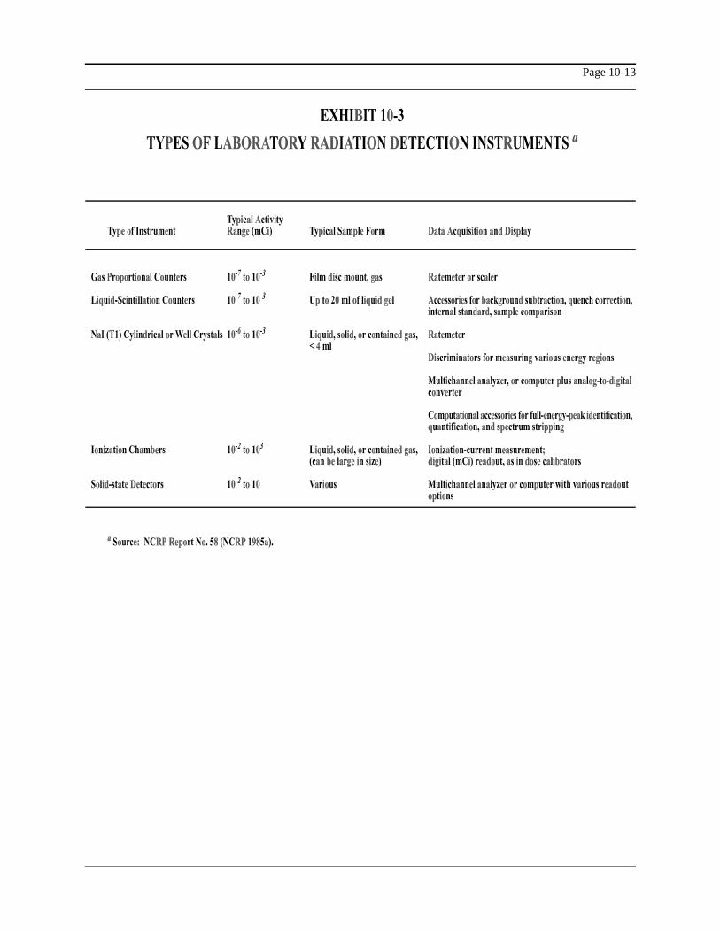
radiochemical procedures, and radiation countersand measurement techniques. The followingdiscussion provides an overview of a few of theradiation detection techniques and instrumentscurrently used to characterize sites contaminatedwith radioactive materials.
Field methods utilize instrumental techniquesrather than radiochemical procedures to determinein-situ identities and concentrations of radionuclides,contamination profiles, and external beta/gammaexposure rates. Field instruments designed forradiation detection (see Exhibit 10-2) are portable,rugged, and relatively insensitive to widefluctuations in temperature and humidity. At thesame time, they are sensitive enough to discriminatebetween variable levels of background radiationfrom naturally occurring radionuclides and excessradiation due to radioactive waste. Because of theharsh conditions in which they are sometimes

Page 10-11

Page 10-12
RADIONUCLIDE MEASUREMENTPROCEDURES
Environmental Radiation Measurements(NCRP 1976)
Instrumentation and Monitoring Methods forRadiation Protection (NCRP 1978)
Radiochemical Analytical Procedures forAnalysis of Environmental Samples (EPA1979a)
Eastern Environmental Radiation FacilityRadiochemistry Procedures Manual (EPA1984a)
A Handbook of Radioactivity MeasurementProcedures (NCRP 1985a)
operated, and because their detection efficiency radiochemical procedure is initiated.varies with photon energy, all field instrumentsshould be properly calibrated in the laboratory For alpha counting, samples are prepared asagainst National Bureau of Standards (NBS) thin-layer (low mass) sources on membrane filters byradionuclide sources prior to use in the field. coprecipitation with stable carriers or on metal discsDetector response should also be tested periodically by electrodeposition. These sample filters and discsin the field against NBS check-sources of known are then loaded into gas proportional counters,activity. scintillation detectors, or alpha spectrometry systems
Commonly used gamma-ray survey meters proportional counter, the sample is immersed in ainclude Geiger-Muller (G-M) probes, sodium iodide counting gas, usually methane and argon, and(NaI(Tl)) crystals, and solid-state germanium diodes subjected to a high voltage field: alpha emissions(Ge(Li)) coupled to ratemeters, scalers, or dissociate the counting gas creating an ionizationmultichannel analyzers (MCAs). These instruments current proportional to the source strength, which isprovide measurements of overall exposure rates in then measured by the system electronics. In acounts per minute, or microRoentgens or microrem scintillation detector, the sample is placed in contactper hour. However, only NaI and Ge(Li) detectors with a ZnS phosphor against the window of awith MCAs provide energy spectra of the gamma photomultiplier (PM) tube: alpha particles inducerays detected and can therefore verify the identity of flashes of light in the phosphor that are converted tospecific radionuclides. Thin window G-M detectors an electrical current in the PM tube and measured.and Pancake (ionization) probes are used to detect Using alpha spectrometry, the sample is placed in abeta particles. Alpha-particle surface monitors holder in an evacuated chamber facing a solid-state,include portable air proportional, gas proportional, surface-barrier detector: alpha particles strike theand zinc sulfide (ZnS) scintillation detectors, which detector and cause electrical impulses, which areall have very thin and fragile windows. The sorted by strength into electronic bins and counted.references in the box on this page provide additional All three systems yield results in counts per minute,information on several other survey techniques and which are then converted into activity units usinginstruments, such as aerial gamma surveillance used detector- and radionuclide-specific calibration
to map gamma exposure rate contours over largeareas.
Laboratory methods involve both chemical andinstrumental techniques to quantify low-levels ofradionuclides in sample media. The preparation ofsamples prior to counting is an importantconsideration, especially for samples containingalpha- and beta-emitting radionuclides that either donot emit gamma rays or emit gamma rays of lowabundance. Sample preparation is a multistepprocess that achieves the following three objectives:(1) the destruction of the sample matrix (primarilyorganic material) to reduce alpha- and beta-particleself-absorption; (2) the separation and concentrationof radionuclides of interest to increase resolution andsensitivity; and (3) the preparation of the sample ina suitable form for counting. Appropriate radioactivetracers (i.e., isotopes of the radionuclides of interestthat are not present in the sample initially, but areadded to the sample to serve as yield determinants)must be selected and added to the sample before a
for measurement (see Exhibit 10-3). In a
Page 10-13

Page 10-14
values. Alpha spectrometry is the only system,however, that can be used to identify specific alpha-emitting radionuclides.
For beta counting, samples are prepared both as conceptual model and the types of information thatthin-sources and as solutions mixed with scintillation may be obtained during a site sampling investigation.fluid, similar in function to a phosphor. Beta- These exhibits apply to radioactively contaminatedemitting sources are counted in gas proportional sites with only minor modifications. For example,counters at higher voltages than those applied for additional exposure pathways for direct externalalpha counting or in scintillation detectors using exposure from immersion in contaminated air orphosphors specifically constructed for beta-particle water or from contaminated ground surfaces maydetection. Beta-emitters mixed with scintillation fluid need to be addressed for certain radionuclides; theseare counted in 20 ml vials in beta-scintillation exposure pathways are discussed further incounters: beta-particle interactions with the fluid subsequent sections. In addition, several of theproduce detectable light flashes. Like alpha parameters identified in these exhibits are not asdetectors, beta detectors provide measurements in important or necessary for radiological surveys. Forcounts per minute, which are converted to activity example, the parameters that are related primarily tounits using calibration factors. It should be noted, the modeling of organic contaminants, such as thehowever, that few detection systems are available for lipid content of organisms, are typically not neededdetermining the identity of individual beta-emitting for radiological assessments.radionuclides, because beta particles are emitted asa continuous spectrum of energy that is difficult tocharacterize and ascribe to any specific nuclide.
It is advisable to count all samples intact in a As is the case with a chemically contaminatedknown geometry on a NaI or Ge(Li) detector system site, the background characteristics of a radioactivelyprior to radiochemical analysis, because many contaminated site must be defined reliably in orderradionuclides that emit gamma rays in sufficient to distinguish natural background radiation andabundance and energy can be detected and measured fallout from the onsite sources of radioactive waste.by this process. Even complex gamma-ray spectra With the possible exception of indoor sources ofemitted by multiple radionuclide sources can be Rn-222, it is often possible to make theseresolved using Ge(Li) detectors, MCAs, and distinctions because the radiation detectionsoftware packages, and specific radionuclide equipment and analytical techniques used are veryconcentrations can be determined. If the sample precise and sensitive. At a chemically contaminatedactivity is low or if gamma rays are feeble, then more site, there can be many potential andrigorous alpha or beta analyses are advised. difficult-to-pinpoint offsite sources for the
10.3.2 REVIEWING AVAILABLE SITEINFORMATION
In Chapter 4, reference is made to reviewing the are, in general, easier to isolate and identify. In fact,site data for chemical contaminants in accordance some radionuclides are so specifically associatedwith Stage 1 of the Data Quality Objectives (DQO) with particular industries that the presence of aprocess (see box on Page 4-4). This process also certain radioactive contaminant sometimes acts as aapplies to radionuclides. For further guidance on the "fingerprint" to identify its source. Additionalapplicability of DQOs to radioactively contaminated information on the sources of natural backgroundsites, consult EPA's Office of Radiation Programs. and man-made radiation in the environment may be
10.3.3 ADDRESSING MODELINGPARAMETER NEEDS
Exhibits 4-1 and 4-2 describe the elements of a
10.3.4 DEFINING BACKGROUNDRADIATION SAMPLING NEEDS
contamination found onsite, confounding theinterpretation of field measurements. With aradioactively contaminated site, however, this is notusually a problem because sources of radionuclides
found in the references listed in the box on the nextpage.
Page 10-15
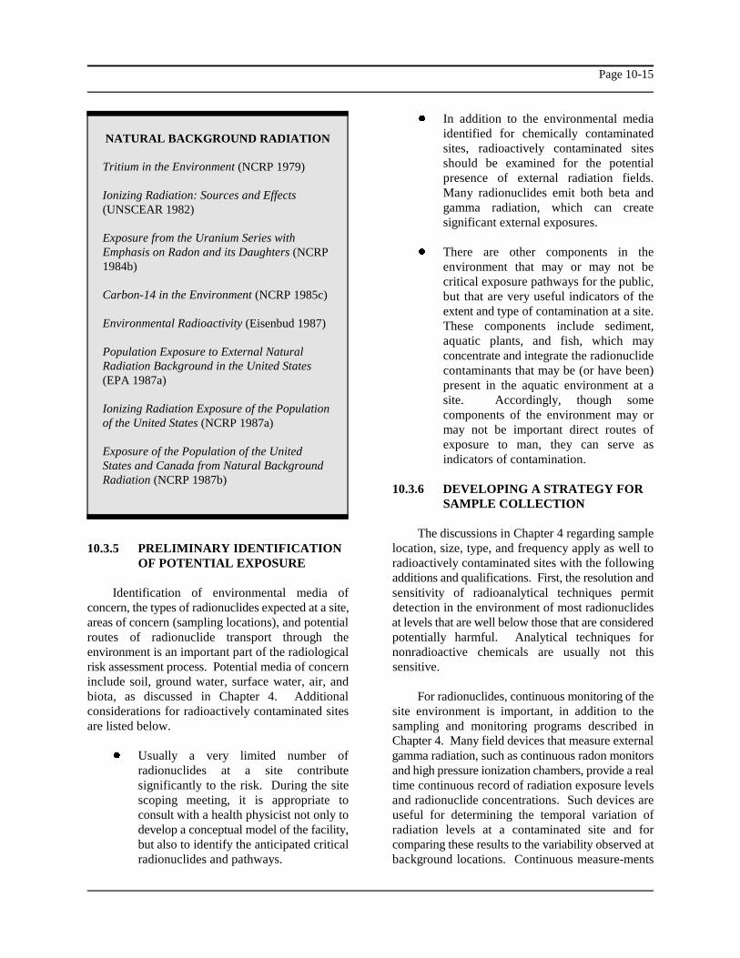
NATURAL BACKGROUND RADIATION
Tritium in the Environment (NCRP 1979)
Ionizing Radiation: Sources and Effects(UNSCEAR 1982)
Exposure from the Uranium Series withEmphasis on Radon and its Daughters (NCRP1984b)
Carbon-14 in the Environment (NCRP 1985c)
Environmental Radioactivity (Eisenbud 1987)
Population Exposure to External NaturalRadiation Background in the United States(EPA 1987a)
Ionizing Radiation Exposure of the Populationof the United States (NCRP 1987a)
Exposure of the Population of the UnitedStates and Canada from Natural BackgroundRadiation (NCRP 1987b)
10.3.5 PRELIMINARY IDENTIFICATIONOF POTENTIAL EXPOSURE
Identification of environmental media of sensitivity of radioanalytical techniques permitconcern, the types of radionuclides expected at a site, detection in the environment of most radionuclidesareas of concern (sampling locations), and potential at levels that are well below those that are consideredroutes of radionuclide transport through the potentially harmful. Analytical techniques forenvironment is an important part of the radiological nonradioactive chemicals are usually not thisrisk assessment process. Potential media of concern sensitive.include soil, ground water, surface water, air, andbiota, as discussed in Chapter 4. Additional For radionuclides, continuous monitoring of theconsiderations for radioactively contaminated sites site environment is important, in addition to theare listed below. sampling and monitoring programs described in
! Usually a very limited number of gamma radiation, such as continuous radon monitorsradionuclides at a site contribute and high pressure ionization chambers, provide a realsignificantly to the risk. During the site time continuous record of radiation exposure levelsscoping meeting, it is appropriate to and radionuclide concentrations. Such devices areconsult with a health physicist not only to useful for determining the temporal variation ofdevelop a conceptual model of the facility, radiation levels at a contaminated site and forbut also to identify the anticipated critical comparing these results to the variability observed atradionuclides and pathways. background locations. Continuous measure-ments
! In addition to the environmental mediaidentified for chemically contaminatedsites, radioactively contaminated sitesshould be examined for the potentialpresence of external radiation fields.Many radionuclides emit both beta andgamma radiation, which can createsignificant external exposures.
! There are other components in theenvironment that may or may not becritical exposure pathways for the public,but that are very useful indicators of theextent and type of contamination at a site.These components include sediment,aquatic plants, and fish, which mayconcentrate and integrate the radionuclidecontaminants that may be (or have been)present in the aquatic environment at asite. Accordingly, though somecomponents of the environment may ormay not be important direct routes ofexposure to man, they can serve asindicators of contamination.
10.3.6 DEVELOPING A STRATEGY FORSAMPLE COLLECTION
The discussions in Chapter 4 regarding samplelocation, size, type, and frequency apply as well toradioactively contaminated sites with the followingadditions and qualifications. First, the resolution and
Chapter 4. Many field devices that measure external

Page 10-16
RADIONUCLIDE MEASUREMENTQA/QC PROCEDURES
Quality Control for EnvironmentalMeasurements Using Gamma-RaySpectrometry (EPA 1977b)
Quality Assurance Monitoring Programs(Normal Operation) - Effluent Streams and theEnvironment (NRC 1979)
Upgrading Environmental Radiation Data(EPA 1980)
Handbook of Analytical Quality Control inRadioanalytical Laboratories (EPA 1987b)
QA Procedures for Health LabsRadiochemistry (American Public HealthAssociation 1987)
provide an added level of resolution for quantifying ! The required number and type of QCand characterizing radiological risk. blanks are fewer for radionuclide samples.
Additional factors that affect the frequency of generally used because radionuclidesampling for radionuclides, besides those discussed samples are less likely to be contaminatedin Chapter 4, include the half-lives and the decay from direct exposure to air than areproducts of the radionuclides. Radionuclides with samples of volatile organics.short half-lives, such as Fe-59 (half-life = 44.5 days),have to be sampled more frequently because Limited guidance is available that specifies fieldrelatively high levels of contamination can be missed QA/QC procedures (see the box on this page). Thesebetween longer sampling intervals. The decay and other issues related to QA/QC guidance forproducts of the radionuclides must also be radiological analyses are discussed further in theconsidered, because their presence can interfere with Section 10.4.the detection of the parent nuclides of interest, andbecause they also may be important contributors torisks.
10.3.7 QUALITY ASSURANCE ANDQUALITY CONTROL (QA/QC)MEASURES
The QA/QC concepts described in Chapter 4also apply to sampling and analysis programs forradionuclides, although the procedures differ.Guidance regarding sampling and measurement ofradionuclides and QA/QC protocols for theiranalyses are provided in the publications listed in thebox on this page.
The QA/QC protocols used for radionuclideanalysis were not developed to meet the evidentialneeds of the Superfund program; however, it is likelythat many of the current radiological QA/QCguidance would meet the intent of Superfundrequirements. Some areas where radiologicalQA/QC guidance may not meet the intent ofSuperfund are listed below.
! The degree of standardization forradiochemical procedures may be lessrigorous in the QA/QC protocols than thatrequired for chemical labs under theContract Laboratory Program (CLP). Inradiochemical laboratories, severaldifferent techniques may be used toanalyze for a specific radionuclide in agiven matrix with comparable results. TheCLP requires all participating chemicallaboratories to use standardizedtechniques.
For example, a "trip" blank is not
10.4 DATA EVALUATION
Chapter 5 describes the procedures fororganizing and evaluating data collected during a sitesampling investigation for use in risk assessment.The ten-step process outlined for chemical dataevaluation is generally applicable to the evaluation ofradioactive contaminants, although many of thedetails must be modified to accommodate differencesin sampling and analytical methods.

Page 10-17
10.4.1 COMBINING DATA FROMAVAILABLE SITEINVESTIGATIONS
All available data for the site should be gathered In both cases, these intercomparison programsfor evaluation and sorted by environmental medium are less comprehensive than the CLP in terms ofsampled, analytical methods, and sampling periods. facility requirements other than analysis ofDecisions should be made, using the process performance evaluation samples, such as laboratorydescribed in Section 5.1, to combine, evaluate space and procedural requirements, instrumentation,individually, or eliminate specific data for use in the training, and quality control. However, until suchquantitative risk assessment. time as radiation measurements become fully
10.4.2 EVALUATING ANALYTICALMETHODS
As with chemical data, radiological data should laboratory accreditation, all analytical results shouldbe grouped according to the types of analyses be carefully scrutinized and not accepted at faceperformed to determine which data are appropriate value.for use in quantitative risk assessment. Analyticalmethods for measuring radioactive contaminants As discussed in Chapter 5 for chemicaldiffer from those for measuring organic and analyses, radioanalytical results that are not specificinorganic chemicals. Standard laboratory procedures for a particular radionuclide (e.g., gross alpha, grossfor radionuclide analyses are presented in references, beta) may have limited usefulness for quantitativesuch as those listed in the box on page 10-12. risk assessment. They can be useful as a screeningAnalytical methods include alpha, beta, and gamma tool, however. External gamma exposure rate data,spectrometry, liquid scintillation counting, although thought of as a screening measurement, canproportional counting, and chemical separation be directly applied as input data for a quantitativefollowed by spectrometry, depending on the specific risk assessment.radionuclides of interest.
Laboratory accreditation procedures for theanalysis of radionuclides also differ. Radionuclideanalyses are not currently conducted as part of the Lower limits of detection (LLDs), orRoutine Analytical Services (RAS) under the quantitation limits, for standard techniques for mostSuperfund CLP. However, these analyses may be radionuclide analyses are sufficiently low to ensureincluded under Special Analytical Services (SAS). the detection of nuclides at activity concentrationsThe EPA Environmental Radioactivity well below levels of concern. There are exceptions,Intercomparison Program, coordinated by the however: some radionuclides with very low specificNuclear Radiation Assessment Division of the activities, long half-lives, and/or low-energy decayEnvironmental Monitoring Systems Laboratory in emissions (e.g., I-129, C-14) are difficult to detectLas Vegas (EMSL-LV), provides quality assurance precisely using standard techniques. To achieveoversight for participating radiation measurement lower LLDs, a laboratory may: (1) use morelaboratories (EPA 1989b). Over 300 federal, state, sensitive measurement techniques and/or chemicaland private laboratories participate in some phase of extraction procedures; (2) analyze larger samplethe program, which includes analyses for a variety of sizes; or (3) increase the counting time of the sample.radionuclides in media (e.g., water, air, milk, and A laboratory may also choose to apply all threefood) with activity concentrations that approximate options to increase detection capabilities. Exhibitlevels that may be encountered in the environment. 10-4 presents examples of typical LLDs using Similar intercomparison programs for analysis of standard analytical techniques. The same specialthermoluminescent dosimeters (TLDs) for external considerations noted for chemical analyses radiation exposure rate measurements are conducted
by the DOE Environmental MeasurementsLaboratory (EML) and the DOE Radiological andEnvironmental Services Laboratory (RESL).
incorporated in the CLP, use of laboratories thatsuccessfully participate in these intercomparisonstudies may be the best available alternative forensuring high-quality analytical data. Regardless of
10.4.3 EVALUATING QUANTITATIONLIMITS

Page 10-18
EXHIBIT 10-4
EXAMPLES OF LOWER LIMITS OF DETECTION (LLD)FOR SELECTED RADIONUCLIDES USING STANDARD ANALYTICAL METHODSa
LLD Isotope Sample Media pCi Bq Methodologyb
Co-60 -Water 10 0.4 Gamma Spectrometry-Soil (dry wt.) 0.1 0.004 Gamma Spectrometry-Biota (wet wt.) 0.1 0.004 Gamma Spectrometryc
-Air 25 0.9 Gamma Spectrometryd
Sr-90 -Water 1 0.04 Radiochemistry
Cs-137 -Water 10 0.4 Gamma Spectrometry0.3 0.01 R a d i o c h e m i s t r y
-Soil (dry wt.) 1 0.04 Gamma Spectrometry0.3 0.01 Radiochemistry
-Biota (wet wt.) 1 0.04 Gamma Spectrometry0.3 0.01 Radiochemistry
-Air 30 1 Gamma Spectrometry
Pb-210 -Water 0.2 0.007 Radiochemistry-Soil (dry wt.) 0.2 0.007 Radiochemistry-Biota (wet wt.) 0.2 0.007 Radiochemistry-Air 5 0.2 Radiochemistry
Ra-226 -Water 100 4 Gamma Spectrometry0.1 0.004 Radiochemistry0.1 0.004 Radon Daughter Emanation
-Soil (dry wt.) 0.1 0.004 Radon Daughter Emanation-Biota (wet wt.) 0.1 0.004 Radon Daughter Emanation-Air 1 0.04 Alpha Spectrometry
Th-232 -Water 0.02 0.0007 Alpha Spectrometry-Soil (dry wt.) 0.2 0.007 Radiochemistry-Biota (wet wt.) 0.02 0.0007 Alpha Spectrometry-Air 0.3 0.01 Alpha Proportional Counter
U-234 -Water 0.02 0.0007 Alpha SpectrometryU-235 -Soil (dry wt.) 0.1 0.004 Alpha SpectrometryU-238 -Biota (wet wt.) 0.01 0.0004 Alpha Spectrometry
-Air 0.2 0.007 Alpha Spectrometry
(continued)

Page 10-19
EXHIBIT 10-4 (continued)
EXAMPLES OF LOWER LIMITS OF DETECTION (LLD)FOR SELECTED RADIONUCLIDES USING STANDARD ANALYTICAL METHODSa
LLD Isotope Sample Media pCi Bq Methodologyb
Pu-238 -Water 0.02 0.0007 Alpha SpectrometryPu-239 -Soil (dry wt.) 0.1 0.004 Alpha SpectrometryPu-240 -Biota (wet wt.) 0.01 0.0004 Alpha Spectrometry
-Air 0.2 0.007 Alpha Spectrometry
Source: U.S. Environmental Protection Agency Eastern Environmental Radiation Facility (EPA-EERF), Department of Energy Environmentala
Measurements Laboratory (DOE-EML), and commercial laboratories. Note that LLDs are radionuclide-, media-, sample size-, and laboratory-specific: higher and lower LLDs than those reported above are possible. The risk assessor should request and report the LLDs supplied bythe laboratory performing the analyses.
Nominal sample sizes: water (1 liter), soil (1 kg dry wt.), biota (1 kg wet wt.), and air (1 filter sample).b
Biota includes vegetation, fish, and meat.c
Air refers to a sample of 300 m of air collected on a filter, which is analyzed for the radionuclide of interest.d 3

Page 10-20
would also apply for radionuclides that are not laboratory conducting the analysis and datadetected in any samples from a particular medium, validation qualifiers assigned by personnel involvedbut are suspected to be present at a site. In these in data validation. These qualifiers pertain tocases, three options may be applied: (1) re-analyze QA/QC problems and generally indicate questionsthe sample using more sensitive methods; (2) use the concerning chemical identity, chemicalLLD value as a "proxy" concentration to evaluate the concentration, or both. No corresponding system ofpotential risks at the detection limit; or (3) evaluate qualifiers has been developed for radioanalyticalthe possible risk implication of the radionuclide data, although certain of the CLP data qualifiersqualitatively. An experienced health physicist might be adopted for use in reporting radioanalyticalshould decide which of these three options would be data. The health physicist should define andmost appropriate. evaluate any qualifiers attached to data for
When multiple radionuclides are present in a Chapter 5, the references on methods listed above,sample, various interferences can occur that may and professional judgment, the health physicistreduce the analytical sensitivity for a particular should eliminate inappropriate data from use in theradionuclide. Also, in some areas of high risk assessment.background radioactivity from naturally occurringradionuclides, it may be difficult to differentiatebackground contributions from incremental sitecontamination. It may be possible to eliminate suchinterferences by radiochemical separation or specialinstrumental techniques.
A sample with activity that is nondetectable or reagent blanks, field blanks, calibration blanks) isshould be reported as less than the appropriate an important component of a proper radioanalyticalsample and radionuclide-specific LLD value. program. Analysis of blanks provides a measure ofHowever, particular caution should be exercised contamination introduced into a sample duringwhen applying this approach to radionuclides that sampling or analysis activities.are difficult to measure and possess unusually highdetection limits, as discussed previously. In most The CLP provides guidance for inorganic andcases where a potentially important radionuclide organic chemicals that are not common laboratorycontaminant is suspected, but not detected, in a contaminants. According to this guidance, if a blanksample, the sample should be reanalyzed using more contains detectable levels of any uncommonrigorous radiochemical procedures and more laboratory chemical, site sample results should besophisticated detection techniques. considered positive only if the measured
If radionuclide sample data for a site are maximum amount detected in any blank. Samplesreported without sample-specific radionuclide containing less than five times the blankquantitation limits, the laboratory conducting the concentration should be classified as nondetects, andanalyses should be contacted to determine the the maximum blank-related concentration should beappropriate LLD values for the analytical techniques specified as the quantitation limit for that chemicaland sample media. in the sample. Though they are not considered to be
10.4.4 EVALUATING QUALIFIED ANDCODED DATA
Various data qualifiers and codes may be analytical procedures for possible sources ofattached to problem data from inorganic and organic contamination.chemical analyses conducted under the CLP asshown in Exhibits 5-4 and 5-5. These includelaboratory qualifiers assigned by the
radionuclide analyses. Based on the discussions in
10.4.5 COMPARING CONCENTRATIONSDETECTED IN BLANKS WITHCONCENTRATIONS DETECTEDIN SAMPLES
The analysis of blank samples (e.g., laboratory
concentration in the sample exceeds five times the
common laboratory contaminants, radionuclidesshould not be classified as nondetects using theabove CLP guidance. Instead, the health physicistshould evaluate all active sample preparation and
Page 10-21
10.4.6 EVALUATING TENTATIVELY 10.4.8 DEVELOPING A SET OFIDENTIFIED RADIONUCLIDES RADIONUCLIDE DATA AND
Because radionuclides are not included on theTarget Compound List (TCL), they may be classifiedas tentatively identified compounds (TICs) under The process described in Section 5.8 forCLP protocols. In reality, however, radioanalytical selection of chemical data for inclusion in thetechniques are sufficiently sensitive that the identity quantitative risk assessment generally applies forand quantity of radionuclides of potential concern at radionuclides as well. One exception is the lack ofa site can be determined with a high degree of CLP qualifiers for radionuclides, as discussedconfidence. In some cases, spectral or matrix previously. Radionuclides of concern should includeinterferences may introduce uncertainties, but these those that are positively detected in at least oneproblems usually can be overcome using special sample in a given medium, at levels significantlyradiochemical and/or instrumental methods. In cases above levels detected in blank samples andwhere a radionuclide's identity is not sufficiently significantly above local background levels. Aswell-defined by the available data set: (1) further discussed previously, the decision to includeanalyses may be performed using more sensitive radionuclides not detected in samples from anymethods, or (2) the tentatively identified medium but suspected at the site based on historicalradionuclide may be included in the risk assessment information should be made by a qualified healthas a contaminant of potential concern with notation physicist.of the uncertainty in its identity and concentration.
10.4.7 COMPARING SAMPLES WITH CLASSBACKGROUND
It is imperative to select, collect, and analyze an quantitative risk assessment is generally unnecessaryappropriate number of background samples to be and inappropriate. Radiation dose and resultingable to distinguish between onsite sources of health risk is highly dependent on the specificradionuclide contaminants from radionuclides properties of each radionuclide. In some cases,expected normally in the environment. Background however, it may be acceptable to group differentmeasurements of direct radiation and radionuclide radioisotopes of the same element that have similarconcentrations in all media of concern should be radiological characteristics (e.g., Pu-238/239/240,determined at sampling locations geologically U-235/238) or belong to the same decay series. Suchsimilar to the site, but beyond the influence of the groupings should be determined very selectively andsite. Screening measurements (e.g., gross alpha, seldom offer any significant advantage.beta, and gamma) should be used to determinewhether more sensitive radionuclide-specificanalyses are warranted. Professional judgmentshould be used by the health physicist to selectappropriate background sampling locations and For sites with a large number of radionuclidesanalytical techniques. The health physicist should detected in samples from one or more media, the riskalso determine which naturally occurring assessment should focus on a select group ofradionuclides (e.g., uranium, radium, or thorium) radionuclides that dominate the radiation dose anddetected onsite should be eliminated from the health risk to the critical receptors. For example,quantitative risk assessment. All man-made when considering transport through ground water toradionuclides detected in samples collected should, distant receptors, transit times may be very long;however, be retained for further consideration. consequently, only radionuclides with long half-lives
INFORMATION FOR USE IN ARISK ASSESSMENT
10.4.9 GROUPING RADIONUCLIDES BY
Grouping radionuclides for consideration in the
10.4.10 FURTHER REDUCTION IN THENUMBER OF RADIONUCLIDES
or radioactive progeny that are formed duringtransport may be of concern for that exposurepathway. For direct external exposures, high-energygamma emitters are of principal concern, whereas

Page 10-22
alpha-emitters may dominate doses from the Chapter 6 describes the procedures forinhalation and ingestion pathways. The important conducting an exposure assessment for chemicalradionuclides may differ for each exposure pathway contaminants as part of the baseline risk assessmentand must be determined on their relative for Superfund sites. Though many aspects of theconcentrations, half-lives, environmental mobility, discussion apply to radionuclides, the termand dose conversion factors (see Section 10.5 for "exposure" is used in a fundamentally different waydiscussion of dose conversion factors) for each for radionuclides as compared to chemicals. Forexposure pathway of interest. chemicals, exposure generally refers to the intake
The total activity inventory and individual toxic chemical, expressed in units of mg/kg-day.concentrations of radionuclides at a Superfund site These units are convenient because the toxicitywill change with time as some nuclides decay away values for chemicals are generally expressed in theseand others "grow in" as a result of radioactive decay terms. For example, the toxicity value used to assessprocesses. Consequently, it may be important to carcinogenic effects is the slope factor, expressed inevaluate different time scales in the risk assessment. units of risk of lifetime excess cancers perFor example, at a site where Ra-226 (half-life = 1600 mg/kg-day. As a result, the product of the intakeyears) is the only contaminant of concern in soil at estimate with the slope factor yields the risk ofsome initial time, the Pb-210 (half-life = 22.3 years) cancer (with proper adjustments made forand Po-210 (half-life = 138 days) progeny will also absorption, if necessary).become dominant contributors to the activity onsiteover a period of several hundred years. Intakes by inhalation, ingestion, and absorption
10.4.11 SUMMARIZING ANDPRESENTING DATA
Presentation of results of the data collection and these internal exposure pathways may becomeevaluation process will be generally the same for systemically incorporated and emit alpha, beta, orradionuclides and chemical contaminants. The gamma radiation within tissues or organs. Unlikesample table formats presented in Exhibits 5-6 and chemical assessments, an exposure assessment for5-7 are equally applicable to radionuclide data, radioactive contaminants can include an explicitexcept that direct radiation measurement data should estimation of the radiation dose equivalent. Asbe added, if appropriate for the radionuclides and discussed previously in Section 10.1, the doseexposure pathways identified at the site. equivalent is an expression that takes into
10.5 EXPOSURE AND DOSEASSESSMENT
This section describes a methodology forestimating the radiation dose equivalent to humansfrom potential exposures to radionuclides through allpertinent exposure pathways at a remedial site.These estimates of dose equivalent may be used forcomparison with radiation protection standards andcriteria. However, this methodology has beendeveloped for regulation of occupational radiationexposures for adults and is not completely applicablefor estimating health risk to the general population.Section 10.7.2, therefore, describes a separatemethodology for estimating health risk.
(e.g., inhalation, ingestion, dermal exposure) of the
are also potentially important exposure pathways forradionuclides, although radionuclide intake istypically expressed in units of activity (i.e., Bq or Ci)rather than mass. Radionuclides that enter through
consideration both the amount of energy deposited ina unit mass of a specific organ or tissue as a result ofthe radioactive decay of a specific radionuclide, aswell as the relative biological effectiveness of theradiations emitted by that nuclide. (Note that theterm dose has a different meaning for radionuclides[dose = energy imparted to a unit mass of tissue]than that used in Chapter 6 for chemicals [dose, orabsorbed dose = mass penetrating into an organism].)
Unlike chemicals, radionuclides can havedeleterious effects on humans without being takeninto or brought in contact with the body. This isbecause high energy beta particles and photons fromradionuclides in contaminated air, water, or soil cantravel long distances with only minimum attenuationin these media before depositing their energy inhuman tissues. External radiation exposures can

Page 10-23
REFERENCES ON EXPOSUREASSESSMENT FOR RADIONUCLIDES
Calculation of Annual Doses to Man fromRoutine Releases of Reactor Effluents (NRC1977)
Radiological Assessment: A Textbook onEnvironmental Dose Analysis (Till and Meyer1983)
Models and Parameters for EnvironmentalRadiological Assessments (Miller 1984)
Radiological Assessment: Predicting theTransport, Bioaccumulation, and Uptake byMan of Radionuclides Released to theEnvironment (NCRP 1984a)
Background Information Document, Draft EISfor Proposed NESHAPS for Radionuclides,Volume I, Risk Assessment Methodology (EPA1989a)
Screening Techniques for DeterminingCompliance with Environmental Standards (NCRP 1989)
result from either exposure to radionuclides at the plans should be implemented to reduce thesite area or to radionuclides that have been possibility of radiation exposures that are in excesstransported from the site to other locations in the of allowable limits.environment. Gamma and x-rays are the mostpenetrating of the emitted radiations, and comprisethe primary contribution to the radiation dose fromexternal exposures. Alpha particles are notsufficiently energetic to penetrate the outer layer ofskin and do not contribute significantly to theexternal dose. External exposure to beta particlesprimarily imparts a dose to the outer layer skin cells,although high-energy beta radiation can penetrateinto the human body.
The quantification of the amount of energydeposited in living tissue due to internal and externalexposures to radiation is termed radiation dosimetry.The amount of energy deposited in living tissue is ofconcern because the potential adverse effects ofradiation are proportional to energy deposition. Theenergy deposited in tissues is proportional to thedecay rate of a radionuclide, and not its mass.Therefore, radionuclide quantities andconcentrations are expressed in units of activity (e.g.,Bq or Ci), rather than in units of mass.
Despite the fundamental difference between theway exposures are expressed for radionuclides andchemicals, the approach to exposure assessmentpresented in Chapter 6 for chemical contaminantslargely applies to radionuclide contaminants.Specifically, the three steps of an exposureassessment for chemicals also apply to radionuclides:(1) characterization of the exposure setting; (2)identification of the exposure pathways; and (3)quantification of exposure. However, some of themethods by which these three steps are carried outare different for radionuclides. The identification of exposure pathways for
10.5.1 CHARACTERIZING THEEXPOSURE SETTING
Initial characterization of the exposure settingfor radioactively contaminated sites is virtually ! In addition to the various ingestion,identical to that described in Chapter 6. One inhalation, and direct contact pathwaysadditional consideration is that, at sites suspected of described in Chapter 6, external exposurehaving radionuclide contamination, a survey should to penetrating radiation should also bebe conducted to determine external radiation fields considered. Potential external exposureusing any one of a number of field survey pathways to be considered includeinstruments (preferably, G-M tubes and NaI(Tl) field immersion in contaminated air, immersiondetectors) (see Exhibit 10-2). Health and safety in contaminated water, and radiation
10.5.2 IDENTIFYING EXPOSUREPATHWAYS
radioactively contaminated sites is very similar tothat described in Chapter 6 for chemicallycontaminated sites, with the following additionalguidance.

Page 10-24
exposure from ground surfaces models have been developed specificallycontaminated with beta- and photon- for evaluating the transport ofemitting radionuclides. radionuclides in the environment and
! As with nonradioactive chemicals, individuals. In general, models developedenvironmentally dispersed radionuclides specifically for radiological assessmentsare subject to the same chemical processes should be used. Such models include, forthat may accelerate or retard their transfer example, explicit consideration ofrates and may increase or decrease their radioactive decay and ingrowth ofbioaccumulation potentials. These radioactive decay products. (Contact ORPtransformation processes must be taken for additional guidance on the fate andinto consideration during the exposure transport models recommended by EPA.)assessment.
! Radionuclides undergo radioactive decaythat, in some respects, is similar to thechemical or biological degradation of One of the primary objectives of an exposureorganic compounds. Both processes assessment is to make a reasonable estimate of thereduce the quantity of the hazardous maximum exposure to individuals and criticalsubstance in the environment and produce population groups. The equation presented inother substances. (Note, however, that Exhibit 6-9 to calculate intake for chemicals may bebiological and chemical transformations considered to be applicable to exposure assessmentcan never alter, i.e., either increase or for radionuclides, except that the body weight anddecrease, the radioactivity of a averaging time terms in the denominator should beradionuclide.) Radioactive decay products omitted. However, as discussed previously,can also contribute significantly to the exposures to radionuclides include both internal andradiation exposure and must be considered external exposure pathways. In addition, radiationin the assessment. exposure assessments do not end with the calculation
! Chapter 6 presents a series of equations in order to estimate radiation dose equivalent.(Exhibits 6-11 through 6-19) forquantification of chemical exposures. The radiation dose equivalent to specifiedThese equations and suggested default organs and the effective dose equivalent due tovariable values may be used to estimate intakes of radionuclides by inhalation or ingestionradionuclide intakes as a first are estimated by multiplying the amount of eachapproximation, if the equations are radionuclide inhaled or ingested times appropriatemodified by deleting the body weight and dose conversion factors (DCFs), which represent theaveraging time from the denominator. dose equivalent per unit intake. As noted previously,However, depending upon the the effective dose equivalent is a weighted sum ofcharacteristics of the radionuclides of the dose equivalents to all irradiated organs andconcern, consideration of radioactive tissues, and represents a measure of the overalldecay and ingrowth of radioactive decay detriment. Federal Guidance Report No. 11 (EPAproducts may be important additions, as 1988) provides DCFs for each of over 700well as the external exposure pathways. radionuclides for both inhalation and ingestion
! Chapter 6 also refers to a number of these DCFs were developed for regulation ofcomputer models that are used to predict occupational exposures to radiation and may not bethe behavior and fate of chemicals in the appropriate for the general population.environment. While those models may besuitable for evaluations of radioactive Radionuclide intake by inhalation and ingestioncontaminants in some cases, numerous is calculated in the same manner as chemical intake
predicting the doses and risks to exposed
10.5.3 QUANTIFYING EXPOSURE:GENERAL CONSIDERATIONS
of intake, but take the calculation an additional step
exposures. It is important to note, however, that

Page 10-25
except that it is not divided by body weight or detonations. Consequently, the exposure and riskaveraging time. For radionuclides, a reference body assessment guidance for radionuclides presented inweight is already incorporated into the DCFs, and this chapter is limited to situations causing chronicthe dose is an expression of energy deposited per exposures to low levels of radioactive contaminants.gram of tissue.
If intake of a radionuclide is defined for aspecific time period (e.g., Bq/year), the doseequivalent will be expressed in corresponding terms(e.g., Sv/year). Because systemically incorporated The preferred method for estimating theradionuclides can remain within the body for long concentration of chemical or radioactiveperiods of time, internal dose is best expressed in contaminants at those places where members of theterms of the committed effective dose equivalent, public may come into contact with them is by directwhich is equal to the effective dose equivalent over measurement. However, this will not be possible inthe 50-year period following intake. many circumstances and it may be necessary,
External exposures may be determined by models to predict contaminant concentrations. Suchmonitoring and sampling of the radionuclide modeling would be necessary, for example: (1)concentrations in environmental media, direct when it is not possible to obtain representativemeasurement of radiation fields using portable samples for all radionuclides of concern; (2) wheninstrumentation, or by mathematical modeling. the contaminant has not yet reached the potentialPortable survey instruments that have been properly exposure points; and (3) when the contaminants arecalibrated can display dose rates (e.g., Sv/hr), and below the limits of detection but, if present, can stilldose equivalents can be estimated by multiplying by represent a significant risk to the public.the duration of exposure to the radiation field.Alternatively, measured or predicted concentrations Numerous fate and transport models have beenin environmental media may be multiplied by DCFs, developed to estimate contaminant concentrations inwhich relate radionuclide concentrations on the ground water, soil, air, surface water, sediments, andground, in air, or in water to external dose rates (e.g., food chains. Models developed for chemicalSv/hr per Bq/m for ground contamination or Sv/hr contaminants, such as those discussed in Chapter 6,2
per Bq/m for air or water immersion). may also be applied to radionuclides with allowance3
The dose equivalents associated with external products. There are also a number of models thatand internal exposures are expressed in identical have been developed specifically for radionuclides.units (e.g., Sv), so that contributions from all These models are similar to the models used forpathways can be summed to estimate the total toxic chemicals but have features that make themeffective dose equivalent value and prioritize risk convenient to use for radionuclide pathway analysis,from different sources. such as explicit consideration of radioactive decay
In general, radiation exposure assessments need radiation risk assessments range in complexity fromnot consider acute toxicity effects. Acute exposures a series of hand calculations to major computerare of less concern for radionuclides than for codes. For example, NRC Regulatory Guide 1.109chemicals because the quantities of radionuclides presents a methodology that may be used torequired to cause adverse effects from acute manually estimate dose equivalents from a variety ofexposure are extremely large and such levels are not exposure pathways (NRC 1977). Examples ofnormally encountered at Superfund sites. Toxic computerized radiological assessment modelseffects from acute radiation exposures are possible include the AIRDOS-EPA code and thewhen humans are exposed to the radiation from EPA-PRESTO family of codes, which are usedlarge amounts of radioactive materials released extensively by EPA to estimate exposures and dosesduring a major nuclear plant accident, such as to populations following atmospheric releases ofChernobyl, or during above-ground weapons radionuclides and releases from a low-level waste
10.5.4 QUANTIFYING EXPOSURE:DETERMINING EXPOSURE POINTCONCENTRATIONS
therefore, to use environmental fate and transport
for radioactive decay and ingrowth of decay
and daughter ingrowth. Available models for use in

Page 10-26
disposal facility, respectively. Guidance on selection much more extensive input data and may includeand use of the various models can be obtained from default parameter values that differ somewhat fromthe EPA Office of Radiation Programs. the values recommended in these exhibits.
Exhibit 6-10, Example of Table Format for Exhibit 6-16 presents the equation andSummarizing Exposure Concentrations, may be used assumptions used to estimate the contaminant intakefor radionuclide contaminants, except that from air. For radionuclides, the dose from inhalationradionuclide concentrations are expressed in terms of of contaminated air is determined as the product ofactivity per unit mass or volume of the the radionuclide concentration in air (Bq/m ), theenvironmental medium (e.g., Bq/kg, Bq/L) rather breathing rate (m per day or year), exposurethan mass. duration (day or year), and the inhalation DCF (Sv
10.5.5 QUANTIFYING EXPOSURE:ESTIMATING INTAKE AND DOSEEQUIVALENT
Section 6.6 presents a description of the uptake. This point is also true for mostmethods used to estimate intake rates of radionuclides, except airborne tritiated water vapor,contaminants from the various exposure pathways. which is efficiently taken into the body throughExhibits 6-11 to 6-19 present the equations and input dermal absorption. In order to account for this routeassumptions recommended for use in intake of uptake, the inhalation DCF for tritium includes ancalculations. In concept, those equations and adjustment factor to account for dermal absorption.assumptions also apply generally to radionuclides,except that the body weight and averaging time termsin the denominators should be omitted. However, asdiscussed previously, the product of thesecalculations for radionuclides is an estimate of theradionuclide intake, expressed in units of activity(e.g., Bq), as opposed to mg/kg-day. In addition, theendpoint of a radiation exposure assessment isradiation dose, which is calculated using DCFs asexplained below. As explained previously, doseequivalents calculated in the following mannershould be used to compare with radiation protectionstandards and criteria, not to estimate risk.
Internal Exposure. Exhibits 6-11, 6-12, 6-14, be important for many organic chemicals; however,6-17, 6-18, and 6-19 present simplified models for dermal uptake is generally not an important route ofthe ingestion of water, food, and soil as pathways uptake for radionuclides, which have small dermalfor the intake of environmental contaminants. The permeability constants. External radiation exposurerecommended assumptions for ingestion rates and due to submersion in water contaminated withexposure durations are applicable to radionuclide radionuclides is possible and is similar to externalexposures and may be used to estimate the intake exposure due to immersion in air. However, becauserates of radionuclides by these pathways. As noted of the shielding effects of water and the generallypreviously, however, these intake estimates for short durations of such exposures, immersion inradionuclides should not be divided by the body water is typically of lesser significance. The productweight or averaging time. These intake rates must be of the radionuclide concentration in water (Bq/m ),multiplied by appropriate DCF values in order to the relevant DCF (Sv/hr per Bq/m ), and the durationobtain committed effective dose equivalent values. of exposure (hours) yields effective dose equivalent.The more rigorous and complex radionuclidepathway models noted previously typically require
3
3
per Bq inhaled). The result of this calculation is thecommitted effective dose equivalent, in units of Sv.
Chapter 6 points out that dermal absorption ofairborne chemicals is not an important route of
External Exposure. Immersion in aircontaining certain beta-emitting and/orphoton-emitting radioactive contaminants can alsoresult in external exposures. Effective doseequivalents from external exposure are calculated asthe product of the airborne radionuclideconcentration (Bq/m ), the external DCF for air3
immersion (Sv/hr per Bq/m ), and the duration of3
exposure (hours).
Exhibits 6-13 and 6-15 illustrate the dermaluptake of contaminants resulting from immersion inwater or contact with soil. This route of uptake can
3
3

Page 10-27
The third external exposure pathway ofpotential significance is irradiation fromradionuclides deposited on the ground surface.Effective dose equivalents resulting from thispathway may be estimated as the product of the soil Exhibit 6-22 presents a sample format forsurface concentration (Bq/m ) of photon-emitting summarizing the results of the exposure assessment.2
radionuclides of concern, the external DCF for The format may also be used for radionuclideground surface exposure (Sv/hr per Bq/m ), and the contaminants except that the entries should be2
duration of exposure (hours). specified as committed effective dose equivalents
10.5.6 COMBINING INTAKES ANDDOSES ACROSS PATHWAYS
The calculations described previously result in and pathways contributing to the total health riskestimates of committed effective dose equivalents may be identified. (Sv) from individual radionuclides via a largenumber of possible exposure pathways. Because a The information should be organized bygiven population may be subject to multiple exposure pathway, population exposed, and currentexposure pathways, the results of the exposure and future use assumptions. For radionuclides,assessment should be organized by grouping all however, it may not be necessary to summarizeapplicable exposure pathways for each exposed short-term and long-term exposures separately aspopulation. Risks from various exposure pathways specified for chemical contaminants.and contaminants then can be integrated during therisk characterization step (see Section 10.7).
10.5.7 EVALUATING UNCERTAINTY
The radiation exposure assessment shouldinclude a discussion of uncertainty, that, at aminimum, should include: (1) a tabular summary ofthe values used to estimate exposures and doses andthe range of these values; and (2) a summary of themajor assumptions of the exposure assessment,including the uncertainty associated with eachassumption and how it might affect the exposure anddose estimates. Sources of uncertainty that must beaddressed include: (1) how well the monitoring datarepresent actual site conditions; (2) the exposuremodels, assumptions, and input variables used toestimate exposure point concentrations; and (3) thevalues of the variables used to estimate intakes andexternal exposures. More comprehensivediscussions of uncertainty associated withradiological risk assessment are provided in theBackground Information Document for the Draft EISfor Proposed NESHAPS for Radionuclides (EPA1989a), Radiological Assessment (Till and Meyer1983), and NCRP Report No. 76 (NCRP 1984a).
10.5.8 SUMMARIZING ANDPRESENTING EXPOSUREASSESSMENT RESULTS
(Sv) and the annual estimated intakes (Bq) for eachradionuclide of concern. The intakes and doseestimates should be tabulated for each exposurepathway so that the most important radionuclides
10.6 TOXICITY ASSESSMENT
Chapter 7 describes the two-step processemployed to assess the potential toxicity of a givenchemical contaminant. The first step, hazardidentification, is used to determine whetherexposure to a contaminant can increase the incidenceof an adverse health effect. The second step,dose-response assessment, is used to quantitativelyevaluate the toxicity information and characterize therelationship between the dose of the contaminantadministered or received and the incidence ofadverse health effects in the exposed population.
There are certain fundamental differencesbetween radionuclides and chemicals that somewhatsimplify toxicity assessment for radionuclides. Asdiscussed in the previous sections, the adverseeffects of exposure to radiation are due to the energydeposited in sensitive tissue, which is referred to asthe radiation dose. In theory, any dose of radiationhas the potential to produce an adverse effect.Accordingly, exposure to any radioactive substancesis, by definition, hazardous.
Dose-response assessment for radionuclides isalso more straightforward. The type of effects and

Page 10-28
REFERENCES ON HEALTH EFFECTSOF RADIATION EXPOSURE
Recommendations of the ICRP (ICRP 1977)
Limits for Intake of Radionuclides by Workers(ICRP 1979)
Influence of Dose and Its Distribution in Timeon Dose-Response Relationships for Low-LETRadiations (NCRP 1980)
The Effects on Populations of Exposure to LowLevels of Ionizing Radiation (NAS 1980)
Induction of Thyroid Cancer by IonizingRadiation (NCRP 1985b)
Lung Cancer Risk from Indoor Exposures toRadon Daughters (ICRP 1987)
Health Risks of Radon and Other InternallyDeposited Alpha-Emitters (National Academyof Sciences 1988)
Ionizing Radiation: Sources, Effects, andRisks (UNSCEAR 1988)
Health Effects Models for Nuclear PowerPlant Accident Consequence Analysis: Low-LET Radiation (NRC 1989)
the likelihood of occurrence of any one of a numberof possible adverse effects from radiation exposuredepends on the radiation dose. The relationshipbetween dose and effect is relatively wellcharacterized (at high doses) for most types ofradiations. As a result, the toxicity assessment,within the context that it is used in this manual, neednot be explicitly addressed in detail for individualradionuclides at each contaminated site.
The sections that follow provide a briefsummary of the human and experimental animalstudies that establish the hazard and dose-responserelationship for radiation exposure. More detaileddiscussions of radiation toxicity are provided inpublications of the National Academy of SciencesCommittee on Biological Effects of IonizingRadiation (BEIR), the United Nations ScientificCommittee on Effects of Atomic Radiation(UNSCEAR), NRC, NCRP, and ICRP listed in thebox on this page.
10.6.1 HAZARD IDENTIFICATION
The principal adverse biological effectsassociated with ionizing radiation exposures fromradioactive substances in the environment arecarcinogenicity, mutagenicity, and teratogenicity.Carcinogenicity is the ability to produce cancer.Mutagenicity is the property of being able to induce , alpha particles) produce high density regions ofgenetic mutation, which may be in the nucleus of ionization. For this reason, they are called high-LETeither somatic (body) or germ (reproductive) cells. (linear energy transfer) particles. Other types ofMutations in germ cells lead to genetic or inherited radiation (e.g., x-rays, gamma rays, and beta defects. Teratogenicity refers to the ability of anagent to induce or increase the incidence of particles) are called low-LET radiations because ofcongenital malformations as a result of permanent the low density pattern of ionization they produce.structural or functional deviations produced during In equal doses, the carcinogenicity and mutagenicitythe growth and development of an embryo (more of high-LET radiations may be an order ofcommonly referred to as birth defects). Radiation magnitude or more greater than those of low-LETmay induce other deleterious effects at acute doses radiations, depending on the endpoint beingabove about 1 Sv, but doses of this magnitude are evaluated. The variability in biological effectivenessnot normally associated with radioactive is accounted for by the quality factor used tocontamination in the environment. calculate the dose equivalent (see Section 10.1).
As discussed in Section 10.1, ionizing radiationcauses injury by breaking molecules into electricallycharged fragments (i.e., free radicals), therebyproducing chemical rearrangements that may lead topermanent cellular damage. The degree of biolog--ical damage caused by various types of radiationvaries according to how spatially close together the ionizations occur. Some ionizing radiations (e.g.
Carcinogenesis. An extensive body ofliterature exists on radiation carcinogenesis in manand animals. This literature has been reviewed mostrecently by the United Nations Scientific Committeeon the Effects of Atomic Radiation (UNSCEAR) andthe National Academy of Sciences AdvisoryCommittee on the Biological Effects of IonizingRadiations (NAS-BEIR Committee) (UNSCEAR

Page 10-29
1977, 1982, 1988; NAS 1972, 1980, 1988). On average, approximately 50 percent of all ofEstimates of the average risk of fatal cancer from the cancers induced by radiation are lethal. Thelow-LET radiation from these studies range from fraction of fatal cancers is different for each type ofapproximately 0.007 to 0.07 fatal cancers per sievert. cancer, ranging from about 10 percent in the case of
An increase in cancer incidence or mortality cancer (NAS 1980, 1988). Females havewith increasing radiation dose has been approximately 2 times as many total cancers as fataldemonstrated for many types of cancer in both cancers following radiation exposure, and maleshuman populations and laboratory animals have approximately 1.5 times as many (NAS 1980).(UNSCEAR 1982, 1988; NAS 1980, 1988). Studiesof humans exposed to internal or external sources ofionizing radiation have shown that the incidence ofcancer increases with increased radiation exposure.This increased incidence, however, is usuallyassociated with appreciably greater doses andexposure frequencies than those encountered in theenvironment. Therefore, risk estimates from smalldoses obtained over long periods of time aredetermined by extrapolating the effects observed athigh, acute doses. Malignant tumors in variousorgans most often appear long after the radiationexposure, usually 10 to 35 years later (NAS 1980,1988; UNSCEAR 1982, 1988). Radionuclidemetabolism can result in the selective deposition ofcertain radionuclides in specific organs or tissues,which, in turn, can result in larger radiation dosesand higher-than-normal cancer risk in these organs.
Ionizing radiation can be consideredpancarcinogenic, i.e., it acts as a completecarcinogen in that it serves as both initiator andpromoter, and it can induce cancers in nearly anytissue or organ. Radiation-induced cancers inhumans have been reported in the thyroid, femalebreast, lung, bone marrow (leukemia), stomach,liver, large intestine, brain, salivary glands, bone,esophagus, small intestine, urinary bladder, pancreas,rectum, lymphatic tissues, skin, pharynx, uterus,ovary, mucosa of cranial sinuses, and kidney(UNSCEAR 1977, 1982, 1988; NAS 1972, 1980,1988). These data are taken primarily from studiesof human populations exposed to high levels ofradiation, including atomic bomb survivors,underground miners, radium dial painters, patientsinjected with thorotrast or radium, and patients whoreceived high x-ray doses during various treatmentprograms. Extrapolation of these data to much lowerdoses is the major source of uncertainty indetermining low-level radiation risks (see EPA1989a). It is assumed that no lower threshold existsfor radiation carcinogenesis.
thyroid cancer to 100 percent in the case of liver
Mutagenesis. Very few quantitative data areavailable on radiogenic mutations in humans,particularly from low-dose exposures. Somemutations are so mild they are not noticeable, whileother mutagenic effects that do occur are similar tononmutagenic effects and are therefore notnecessarily recorded as mutations. The bulk of datasupporting the mutagenic character of ionizingradiation comes from extensive studies ofexperimental animals (UNSCEAR 1977, 1982, 1988;NAS 1972, 1980, 1988). These studies havedemonstrated all forms of radiation mutagenesis,including lethal mutations, translocations, inversions,nondisjunction, and point mutations. Mutation ratescalculated from these studies are extrapolated tohumans and form the basis for estimating the geneticimpact of ionizing radiation on humans (NAS 1980,1988; UNSCEAR 1982, 1988). The vast majority ofthe demonstrated mutations in human germ cellscontribute to both increased mortality and illness(NAS 1980; UNSCEAR 1982). Moreover, theradiation protection community is generally inagreement that the probability of inducing geneticchanges increases linearly with dose and that no"threshold" dose is required to initiate heritabledamage to germ cells.
The incidence of serious genetic disease due tomutations and chromosome aberrations induced byradiation is referred to as genetic detriment. Seriousgenetic disease includes inherited ill health,handicaps, or disabilities. Genetic disease may bemanifest at birth or may not become evident untilsome time in adulthood. Radiation-induced geneticdetriment includes impairment of life, shortened lifespan, and increased hospitalization. The frequencyof radiation-induced genetic impairment is relativelysmall in comparison with the magnitude of detrimentassociated with spontaneously arising geneticdiseases (UNSCEAR 1982, 1988).

Page 10-30
Teratogenesis. Radiation is a well-knownteratogenic agent. The developing fetus is muchmore sensitive to radiation than the mother. The ageof the fetus at the time of exposure is the mostimportant factor in determining the extent and typeof damage from radiation. The malformationsproduced in the embryo depend on which cells,tissues, or organs in the fetus are most activelydifferentiating at the time of radiation exposure.Embryos are relatively resistant to radiation-inducedteratogenic effects during the later stages of theirdevelopment and are most sensitive from just afterimplantation until the end of organogenesis (abouttwo weeks to eight weeks after conception)(UNSCEAR 1986; Brent 1980). Effects on nervoussystem, skeletal system, eyes, genitalia, and skinhave been noted (Brent 1980). The brain appears tobe most sensitive during development of theneuroblast (these cells eventually become the nervecells). The greatest risk of brain damage for thehuman fetus occurs at 8 to 15 weeks, which is thetime the nervous system is undergoing the most rapiddifferentiation and proliferation of cells (Otake1984).
10.6.2 DOSE-RESPONSERELATIONSHIPS
This section describes the relationship of therisk of fatal cancer, serious genetic effects, and otherdetrimental health effects to exposure to low levelsof ionizing radiation. Most important from thestandpoint of the total societal risk from exposures tolow-level ionizing radiation are the risks of cancerand genetic mutations. Consistent with our currentunderstanding of their origins in terms of DNAdamage, these effects are believed to be stochastic;that is, the probability (risk) of these effectsincreases with the dose of radiation, but the severityof the effects is independent of dose. For neitherinduction of cancer nor genetic effects, moreover, isthere any convincing evidence for a "threshold" (i.e.,some dose level below which the risk is zero).Hence, so far as is known, any dose of ionizingradiation, no matter how small, might give rise to acancer or to a genetic effect in future generations.Conversely, there is no way to be certain that a givendose of radiation, no matter how large, has caused anobserved cancer in an individual or will cause one inthe future.
Exhibit 10-5 summarizes EPA's currentestimates of the risk of adverse effects associatedwith human exposure to ionizing radiation (EPA1989a). Important points from this summary tableare provided below.
! Very large doses (>1 Sv) of radiation arerequired to induce acute and irreversibleadverse effects. It is unlikely that suchexposures would occur in theenvironmental setting associated with apotential Superfund site.
! The risks of serious noncarcinogeniceffects associated with chronic exposure toradiation include genetic and teratogeniceffects. Radiation-induced genetic effectshave not been observed in humanpopulations, and extrapolation fromanimal data reveals risks per unit exposurethat are smaller than, or comparable to, therisk of cancer. In addition, the geneticrisks are spread over several generations.The risks per unit exposure of seriousteratogenic effects are greater than therisks of cancer. However, there is apossibility of a threshold, and theexposures must occur over a specificperiod of time during gestation to causethe effect. Teratogenic effects can beinduced only during the nine months ofpregnancy. Genetic effects are inducedduring the 30-year reproductive generationand cancer can be induced at any pointduring the lifetime. If a radiation source isnot controlled, therefore, the cumulativerisk of cancer may be many times greaterthan the risk of genetic or teratogeniceffects due to the potentially longer periodof exposure.

Page 10-31
EXHIBIT 10-5
SUMMARY OF EPA'S RADIATION RISK FACTORSa
Risk Significant Exposure Period Risk Factor Range
Low LET (Gy )-1
Teratogenic:b
Severe mental retardation Weeks 8 to 15 of gestation 0.25-0.55
Genetic:Severe hereditary defects, 30-year reproductive generation 0.006-0.11all generations
Somatic:Fatal cancers Lifetime 0.012-0.12
In utero 0.029-0.10All cancers Lifetime 0.019-0.19
High LET (Gy )-1
Genetic:Severe hereditary defects, 30-year reproductive generation 0.016-0.29all generations
Somatic:Fatal cancers Lifetime 0.096-0.96All cancers Lifetime 0.15-1.5
Radon Decay Products (10 WLM )-6 -1
Fatal lung cancer Lifetime 140-720
In addition to the stochastic risks indicated, acute toxicity may occur at a mean lethal dose of 3-5 Sv with aa
threshold in excess of 1 Sv.
The range assumes a linear, non-threshold dose-response. However, it is plausible that a threshold may existb
for this effect.

Page 10-32
Based on these observations, it appears that the transfer factors and dose conversion factors that mayrisk of cancer is limiting and may be used as the sole not always be entirely applicable to the conditionsbasis for assessing the radiation-related human health being analyzed. For example, the standard doserisks of a site contaminated with radionuclides. conversion factors are based on certain generic
For situations where the risk of cancer induction exposed individual and the chemical and physicalin a specific target organ is of primary interest, the properties of the radionuclides. Also, as is the casecommitted dose equivalent to that organ may be for chemical contaminants, the environmentalmultiplied by an organ-specific risk factor. The transfer factors used in the models may not apply torelative radiosensitivity of various organs (i.e., the all settings.cancer induction rate per unit dose) differs markedlyfor different organs and varies as a function of the Though the risk assessment models may includeage and sex of the exposed individual. Tabulations a large number of radionuclides and pathways, theof such risk factors as a function of age and sex are important radionuclides and pathways are usuallyprovided in the Background Information Document few in number. As a result, it is often feasible tofor the Draft Environmental Impact Statement for check the computer output using hand calculations.Proposed NESHAPS for Radionuclides (EPA 1989a) This type of review can be performed by healthfor cancer mortality and cancer incidence. physicists familiar with the models and their
10.7 RISK CHARACTERIZATION
The final step in the risk assessment process isrisk characterization. This is an integration step inwhich the risks from individual radionuclides andpathways are quantified and combined whereappropriate. Uncertainties also are examined anddiscussed in this step.
10.7.1 REVIEWING OUTPUTS FROMTHE TOXICITY AND EXPOSUREASSESSMENTS
The exposure assessment results should beexpressed as estimates of radionuclide intakes byinhalation and ingestion, exposure rates and durationfor external exposure pathways, and committedeffective dose equivalents to individuals from allrelevant radionuclides and pathways. The riskassessor should compile the supportingdocumentation to ensure that it is sufficient tosupport the analysis and to allow an independentduplication of the results. The review should alsoconfirm that the analysis is reasonably complete interms of the radionuclides and pathways addressed.
In addition, the review should evaluate thedegree to which the assumptions inherent in theanalysis apply to the site and conditions beingaddressed. The mathematical models used tocalculate dose use a large number of environmental
assumptions regarding the characteristics of the
limitations. Guidance on conducting suchcalculations is provided in numerous references,including Till and Meyer (1983) and NCRP ReportNo. 76 (NCRP 1984a).
10.7.2 QUANTIFYING RISKS
Given that the results of the exposureassessment are virtually complete, correct, andapplicable to the conditions being considered, thenext step in the process is to calculate and combinerisks. As discussed previously, the risk assessmentfor radionuclides is somewhat simplified becauseonly radiation carcinogenesis needs to be considered.
Section 10.5 presents a methodology forestimating committed effective dose equivalents thatmay be compared with radiation protection standardsand criteria. Although the product of these doseequivalents (Sv) and an appropriate risk factor (riskper Sv) yields an estimate of risk, the health riskestimate derived in such a manner is not completelyapplicable for members of the general public. Abetter estimate of risk may be computed using age-and sex-specific coefficients for individual organsreceiving significant radiation doses. Thisinformation may be used along with organ-specificdose conversion factors to derive slope factors thatrepresent the age-averaged lifetime excess cancerincidence per unit intake for the radionuclides ofconcern. The Integrated Risk Information System(IRIS) contains slope factor values for radionuclidesof concern at remedial sites for each of the four

Page 10-33
major exposure pathways (inhalation, ingestion, air transport processes and routes of exposure are theimmersion, and ground-surface irradiation), along same for radionuclides and chemicals.with supporting documentation for the derivation ofthese values (see Chapter 7 for more detail on IRIS). In cases where different environmental fate and
The slope factors from the IRIS data base for and radionuclide exposure, the mathematical modelsthe inhalation pathway should be multiplied by the may incorporate somewhat different assumptions.estimated inhaled activity (derived using the methods These differences can result in incompatibilities inpresented in Section 6.6.3 and Exhibit 6-16, the two estimates of risk. One important differencewithout division of the body weight and averaging of this nature is how the cancer toxicity values (i.e.,time) for each radionuclide of concern to estimate slope factors) were developed. For bothrisks from the inhalation pathway. Similarly, risks radionuclides and chemicals, cancer toxicity valuesfrom the ingestion pathway should be estimated by are obtained by extrapolation from experimental andmultiplying the ingestion slope factors by the activity epidemiological data. For radionuclides, however,ingested for each radionuclide of concern (derived human epidemiological data form the basis of theusing the methods presented in Exhibits 6-11, 6-12, extrapolation, while for many chemical carcinogens,6-14, 6-17, 6-18, and 6-19, without division by the laboratory experiments are the primary basis for thebody weight and averaging time). Estimates of the extrapolation. Another even more fundamentalrisk from the air immersion pathway should be difference between the two is that slope factors forcomputed by multiplying the appropriate slope chemical carcinogens generally represent an upperfactors by the airborne radionuclide concentration bound or 95th percent confidence limit value, while(Bq/m ) and the duration of exposure. Risk from the radionuclide slope factors are best estimate values.3
ground surface pathway should be computed as theproduct of the slope factor, the soil concentration In light of these limitations, the two sets of risk(Bq/m ), and the duration of exposure for each estimates should be tabulated separately in the final2
radionuclide of concern. baseline risk assessment.
The sum of the risks from all radionuclides andpathways yields the lifetime risk from the overallexposure. As discussed in Chapter 8, professionaljudgment must be used in combining the risks from Uncertainties in the risk assessment must bevarious pathways, as it may not be physically evaluated and discussed, including uncertainties inpossible for one person to be exposed to the the physical setting definition for the site, in themaximum radionuclide concentrations for all models used, in the exposure parameters, and in thepathways. toxicity assessment. Monte Carlo uncertainty
10.7.3 COMBINING RADIONUCLIDEAND CHEMICAL CANCER RISKS
Estimates of the lifetime risk of cancer to assessments is provided in NCRP Report No. 76exposed individuals resulting from radiological and (NCRP 1984a), Radiological Assessment (Till andchemical risk assessments may be summed in order Meyer 1983), and in the Background Informationto determine the overall potential human health Document for the Draft EIS for Proposed NESHAPshazard associated with a site. Certain precautions for Radionuclides (EPA 1989a).should be taken, however, before summing theserisks. First, the risk assessor should evaluatewhether it is reasonable to assume that the sameindividual can receive the maximum radiological andchemical dose. It is possible for this to occur insome cases because many of the environmental
transport models have been used to predict chemical
10.7.4 ASSESSING AND PRESENTINGUNCERTAINTIES
analyses are frequently performed as part of theuncertainty and sensitivity analysis for radiologicalrisk assessments. A summary of the use ofuncertainty analyses in support of radiological risk

Page 10-34
10.7.5 SUMMARIZING ANDPRESENTING THE BASELINERISK CHARACTERIZATIONRESULTS
The results of the baseline risk characterizationshould be summarized and presented in an effectivemanner to assist in decision-making. The estimatesof risk should be summarized in the context of thespecific site conditions. Information should includethe identity and concentrations of radionuclides,types and magnitudes of health risks predicted,uncertainties in the exposure estimates and toxicityinformation, and characteristics of the site andpotentially exposed populations. A summary tableshould be provided in a format similar to that shownin Exhibit 6-22, as well as graphical presentations ofthe predicted health risks (see Exhibit 8-7).
10.8 DOCUMENTATION,REVIEW, ANDMANAGEMENT TOOLS FORTHE RISK ASSESSOR,REVIEWER, AND MANAGER
The discussion provided in Chapter 9 alsoapplies to radioactively contaminated sites. Thesuggested outline provided in Exhibit 9-1 may alsobe used for radioactively contaminated sites withonly minor modifications. For example, the portionsthat uniquely pertain to the CLP program andnoncarcinogenic risks are not needed. In addition,because radionuclide hazard and toxicity have beenaddressed adequately on a generic basis, there is noneed for an extensive discussion of toxicity in thereport.

Page 10-35
REFERENCES FOR CHAPTER 10
American Public Health Association. 1987. QA Procedures for Health Labs Radiochemistry.
Beebe, G.W., Kato, H., and Land, C.E. 1977. Mortality Experience of Atomic Bomb Survivors, 1950-1974, Life Span Study Report 8. RERFTR 1-77. Radiation Effects Research Foundation. Japan.
Brent, R.L. 1980. Radiation Teratogenesis. Teratology 21:281-298.
Cember, H. 1983. Introduction to Health Physics (2nd Ed.) Pergamon Press. New York, NY.
Department of Energy (DOE). 1987. The Environmental Survey Manual. DOE/EH-0053.
Department of Energy (DOE). 1988. External Dose-Rate Conversion Factors for Calculation of Dose to the Public. DOE/EH-0070.
Department of Energy (DOE). 1989. Environmental Monitoring for Low-level Waste Disposal Sites. DOE/LLW-13Tg.
Eisenbud, M. 1987. Environmental Radioactivity (3rd Ed.) Academic Press. Orlando, FL.
Environmental Protection Agency (EPA). 1972. Environmental Monitoring Surveillance Guide.
Environmental Protection Agency (EPA). 1977a. Handbook of Analytical Quality Control in Radioanalytical Laboratories. Office of Researchand Development. EPA/600/7-77/008.
Environmental Protection Agency (EPA). 1977b. Quality Control for Environmental Measurements Using Gamma-Ray Spectrometry.EPA/500/7-77/14.
Environmental Protection Agency (EPA). 1979a. Radiochemical Analytical Procedures for Analysis of Environmental Samples. EMSL-LV-0539-17.
Environmental Protection Agency (EPA). 1980. Upgrading Environmental Radiation Data. Office of Radiation Programs. EPA/520/1-80/012.
Environmental Protection Agency (EPA). 1984a. Eastern Environmental Radiation Facility Radiochemistry Procedures Manual. Office ofRadiation Programs. EPA/520/5-84/006.
Environmental Protection Agency (EPA). 1984b. Federal Guidance Report No. 10: The Radioactivity Concentration Guides. Office of RadiationPrograms. EPA/520/1-84/010.
Environmental Protection Agency (EPA). 1987a. Population Exposure to External Natural Radiation Background in the United States. Officeof Radiation Programs. EPA ORP/SEPD-80-12.
Environmental Protection Agency (EPA). 1987b. Handbook of Analytical Quality Control in Radioanalytical Laboratories. Office of Researchand Development. EPA/600/7-87/008.
Environmental Protection Agency (EPA). 1988. Federal Guidance Report No. 11: Limiting Values of Radionuclide Intake and Air Concentrationand Dose Conversion Factors for Inhalation, Submersion, and Ingestion. Office of Radiation Programs. EPA/520/1-88/020.
Environmental Protection Agency (EPA). 1989a. Background Information Document, Draft EIS for Proposed NESHAPS for Radionuclides,Volume I, Risk Assessment Methodology. Office of Radiation Programs. EPA/520/1-89/005.
Environmental Protection Agency (EPA). 1989b. Annual Report Fiscal Year 1988 Laboratory Intercomparison Studies for Radionuclides.
Environmental Protection Agency (EPA). 1989c. CERCLA Compliance with Other Laws Manual. Part II. Office of Emergency and RemedialResponse. (OSWER Directive 9234.1-02).
International Commission on Radiological Protection (ICRP). 1977. Recommendations of the ICRP. ICRP Publication 26.
International Commission on Radiological Protection (ICRP). 1979. Limits for Intake of Radionuclides by Workers. ICRP Publication 30.
International Commission on Radiological Protection (ICRP). 1983. Principles for Limiting Exposure of the Public to Natural Sources ofRadiation. ICRP Publication 39.

Page 10-36
International Commission on Radiological Protection (ICRP). 1984. A Compilation of the Major Concepts and Quantities in Use by the ICRP.ICRP Publication 42.
International Commission on Radiological Protection (ICRP). 1985. Principles of Monitoring for the Radiation Protection of the Population.ICRP Publication 43.
International Commission on Radiological Protection (ICRP). 1987. Lung Cancer Risk from Indoor Exposures to Radon Daughters. ICRPPublication 50.
Kato, H. and Schull, W.J. 1982. Studies of the Mortality of A-Bomb Survivors. Report 7 Part 1, Cancer Mortality Among Atomic BombSurvivors, 1950-78. Radiation Research 90:395-432.
Kocher, D. 1981. Radioactive Decay Data Tables: A Handbook of Decay Data for Application to Radiation Dosimetry and RadiologicalAssessments. DOE/TIC-11026.
Miller. 1984. Models and Parameters for Environmental Radiological Assessments. DOE/TIC-11468.
National Academy of Sciences - National Research Council. 1972. The Effects on Populations of Exposures to Low Levels of Ionizing Radiation.(BEIR Report).
National Academy of Sciences - National Research Council. 1980. The Effects on Populations of Exposures to Low Levels of Ionizing Radiation.(BEIR Report).
National Academy of Sciences - National Research Council. 1988. Health Risks of Radon and Other Internally Deposited Alpha-Emitters. (BEIRReport).
National Council on Radiation Protection and Measurements (NCRP). 1989. Screening Techniques for Determining Compliance withEnvironmental Standards. NCRP Commentary No. 3.
National Council on Radiation Protection and Measurements (NCRP). 1976. Environmental Radiation Measurements. NCRP Report No. 50.
National Council on Radiation Protection and Measurements (NCRP). 1978. Instrumentation and Monitoring Methods for Radiation Protection.NCRP Report No. 57.
National Council on Radiation Protection and Measurements (NCRP). 1979. Tritium in the Environment. NCRP Report No. 62.
National Council on Radiation Protection and Measurements (NCRP). 1980. Influence of Dose and Its Distribution in Time on Dose-responseRelationships for Low-LET Radiations. NCRP Report No. 64.
National Council on Radiation Protection and Measurements (NCRP). 1984a. Radiological Assessment: Predicting the Transport,Bioaccumulation, and Uptake by Man of Radionuclides Released to the Environment. NCRP Report No. 76.
National Council on Radiation Protection and Measurements (NCRP). 1984b. Exposure from the Uranium Series with Emphasis on Radon andits Daughters. NCRP Report No. 77.
National Council on Radiation Protection and Measurements (NCRP). 1985a. A Handbook of Radioactivity Measurement Procedures. NCRPReport No. 58.
National Council on Radiation Protection and Measurements (NCRP). 1985b. Induction of Thyroid Cancer by Ionizing Radiation. NCRP ReportNo. 80.
National Council on Radiation Protection and Measurements (NCRP). 1985c. Carbon-14 in the Environment. NCRP Report No. 81.
National Council on Radiation Protection and Measurements (NCRP). 1987a. Ionizing Radiation Exposure of the Population of the United States.NCRP Report No. 93.
National Council on Radiation Protection and Measurements (NCRP). 1987b. Exposure of the Population of the United States and Canada fromNatural Background Radiation. NCRP Report No. 94.
National Council on Radiation Protection and Measurements (NCRP). 1989. Screening Techniques for Determining Compliance withEnvironmental Standards. NCRP Commentary No. 3.
Nuclear Regulatory Commission (NRC). 1977. Calculation of Annual Doses to Man from Routine Releases of Reactor Effluents for the Purposeof Evaluating Compliance with 10 CFR 50, Appendix I. Regulatory Guide 1.109.

Page 10-37
Nuclear Regulatory Commission (NRC). 1979. Quality Assurance Monitoring Programs (Normal Operation) -- Effluent Streams and theEnvironment. NRC Regulatory Guide 4.15.
Nuclear Regulatory Commission (NRC). 1989. Health Effects Models for Nuclear Power Plant Accident Consequence Analysis: Low-LETRadiation, Part II: Scientific Bases for Health Effects Models. NUREG/CR-4214, Rev. 1. Part II.
Otake, M. and Schull W. 1984. Mental Retardation in Children Exposed in Utero to the Atomic Bombs: A Reassessment. Technical ReportRERF TR 1-83. Radiation Effects Research Foundation. Japan.
Schleien, B. and Terpilak, M., (Eds). 1984. The Health Physics and Radiological Health Handbook. (7th Ed.) Nucleon Lectern Assoc., Inc.Maryland.
Till, J.E. and Meyer, H.R. 1983. Radiological Assessment: A Textbook on Environmental Dose Analysis. Prepared for Office of NuclearReactor Regulation. U.S. Nuclear Regulatory Commission. Washington, DC. NUREG/CR-3332.
Turner, J.E. 1986. Atoms, Radiation, and Radiation Protection. Pergamon Press. New York, NY.
United Nations Scientific Committee Report on the Effects of Atomic Radiation (UNSCEAR). 1958. Official Records: Thirteenth Session,Supplement No. 17(A/3838). United Nations. New York, NY.
United Nations Scientific Committee on the Effects of Atomic Radiation (UNSCEAR). 1977. Sources and Effects of Ionizing Radiation. UnitedNations. New York, NY.
United Nations Scientific Committee on the Effects of Atomic Radiation (UNSCEAR). 1982. Ionizing Radiation: Sources and Effects. UnitedNations. New York, NY.
United Nations Scientific Committee on the Effects of Atomic Radiation (UNSCEAR). 1986. Genetic and Somatic Effects of Ionizing Radiation.United Nations. New York, NY.
United Nations Scientific Committee on the Effects of Atomic Radiation (UNSCEAR). 1988. Sources, Effects, and Risks of Ionizing Radiation.United Nations. New York, NY.
Wakabayashi, T., et al. 1983. Studies of the Mortality of A-Bomb Survivors, Report 7, Part III, Incidence of Cancer in 1959-1978, Based onthe Tumor Registry, Nagasaki. Radiat. Res. 93: 112-146.