Chapter 10 Companion site for Basic Medical Endocrinology, 4th Edition Author: Dr. Goodman.
22
Chapter 10 Companion site for Basic Medical Endocrinology, 4th Edition Author: Dr. Goodman
-
Upload
abigail-crawford -
Category
Documents
-
view
223 -
download
0
Transcript of Chapter 10 Companion site for Basic Medical Endocrinology, 4th Edition Author: Dr. Goodman.

Chapter 10Companion site for Basic Medical Endocrinology, 4th Edition
Author: Dr. Goodman
Companion site for Basic Medical Endocrinology, 4th Edition. by Dr. Goodman Copyright © 2009 by Academic Press. All rights reserved.
2
Daily calcium balance in a typical adult.
FIGURE 10.1

Companion site for Basic Medical Endocrinology, 4th Edition. by Dr. Goodman Copyright © 2009 by Academic Press. All rights reserved.
3
A thick ground section of the tibia illustrating cortical compact bone and the lattice of trabeculae of cancellous bone. (From Fawcett, D.W. (1986) A Textbook of Histology, 11th ed., 201. Saunders, Philadelphia.)
FIGURE 10.2

Companion site for Basic Medical Endocrinology, 4th Edition. by Dr. Goodman Copyright © 2009 by Academic Press. All rights reserved.
4
Cross-section through a bony trabecula. The pale blue area indicates mineralized matrix.
FIGURE 10.3

Companion site for Basic Medical Endocrinology, 4th Edition. by Dr. Goodman Copyright © 2009 by Academic Press. All rights reserved.
5
Differentiation and activation of osteoclasts. c-Fms = receptor for macrophage colony stimulating factor. M-CSF = macrophage colony stimulating factor. RANK = receptor activator of NF-kB. RANKL = RANK ligand. OPG = osteoprotegerin. (Modified from Khosla, S. (2001) Minireview: The OPG/RANKL/RANK system. Endocrinology 142: 5050–5055.)
FIGURE 10.4

Companion site for Basic Medical Endocrinology, 4th Edition. by Dr. Goodman Copyright © 2009 by Academic Press. All rights reserved.
6
Daily phosphorus balance in a typical adult.
FIGURE 10.5

Companion site for Basic Medical Endocrinology, 4th Edition. by Dr. Goodman Copyright © 2009 by Academic Press. All rights reserved.
7
Drawing of a section through a human parathyroid gland showing small chief cells and larger oxyphil cells. The cells are arranged in cords surrounded by loose connective tissue. (Modified from Borysenko and Beringer. (1984) Functional Histology, 2nd ed., 316. Little, Brown, Boston.)
FIGURE 10.6

Companion site for Basic Medical Endocrinology, 4th Edition. by Dr. Goodman Copyright © 2009 by Academic Press. All rights reserved.
8
A. Posttranslational metabolism of PTH. The leader sequence (–31 to –6) is removed cotranslationally in the endoplasmic reticulum. The hexapeptide –6 to 1 is removed in the Golgi during packaging of the peptide. PTH 1-84 is the intact hormone. C terminal fragments are generated in the secretory granules just prior to or during secretion. B. The known biologically active portion of PTH, the epitopes required for detection and assay of the intact hormone. Detection antibodies that recognize sequences downstream from the amino terminal tripeptide cannot distinguish between the intact active hormone and its truncated antagonist.
FIGURE 10.7

Companion site for Basic Medical Endocrinology, 4th Edition. by Dr. Goodman Copyright © 2009 by Academic Press. All rights reserved.
9
Effects of PTH on bone. PTH acts on cells of osteoblastic lineage to stimulate production of M-CSF (macrophage colony stimulating factor) and RANKL (receptor activator of N-kappa B ligand), which results in osteoclast formation and activation. Digestion of the bone matrix releases calcium and phosphorus and growth factors that were deposited by osteoblasts when the matrix was laid down, Growth factors stimulate osteoblast precursors to multiply, differentiate, and lay down new bone matrix.
FIGURE 10.8

Companion site for Basic Medical Endocrinology, 4th Edition. by Dr. Goodman Copyright © 2009 by Academic Press. All rights reserved.
10
Effects of PTH on the principal cells in the distal nephron. PTH stimulates insertion of epithelial calcium channels in the luminal membrane and calcium extrusion mechanisms in the basolateral membrane. GS = the stimulatory G protein; AC = adenylyl cyclase; cAMP = cyclic adenosine monophosphate; PKA = protein kinase A.
FIGURE 10.9

Companion site for Basic Medical Endocrinology, 4th Edition. by Dr. Goodman Copyright © 2009 by Academic Press. All rights reserved.
11
Effects of PTH on proximal tubule cells. Phosphorylation of NERF (sodium hydrogen exchange regulatory factor) releases PT (sodium phosphate cotransporter) from anchoring sites in the membrane. PTs migrate in the plane of the membrane to clathrin coated pits where they are internalized and transferred to lysosomes and degraded. PTH also stimulates the expression and activation of the enzyme (P450 1-hydroxylase) that converts 25-0HD3 to 1,25(OH)2D3, the active form of vitamin D (see Figure 10.16). GS = subunit of the stimulatory G protein; AC = adenylyl cyclase; cAMP = cyclic adenosine monophosphate; PKA = protein kinase A; CREB = cyclic AMP response element binding protein.
FIGURE 10.10

Companion site for Basic Medical Endocrinology, 4th Edition. by Dr. Goodman Copyright © 2009 by Academic Press. All rights reserved.
12
Relation between plasma ionized calcium concentration and PTH secretion. (Redrawn and modified from Brown, E.B. (1983) Four parameter model of the sigmoidal relationship between parathyroid hormone release and extracellular calcium concentration in normal and abnormal parathyroid tissue. J. Clin. Endocrinol. Metab. 56: 572–581.)
FIGURE 10.11

Companion site for Basic Medical Endocrinology, 4th Edition. by Dr. Goodman Copyright © 2009 by Academic Press. All rights reserved.
13
Regulation of PTH secretion. () = decrease; (+) = increase.
FIGURE 10.12
Companion site for Basic Medical Endocrinology, 4th Edition. by Dr. Goodman Copyright © 2009 by Academic Press. All rights reserved.
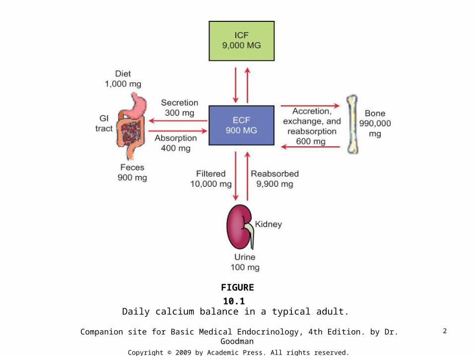
14
Regulation of parathyroid hormone secretion by calcium (Ca2+). Heptihelical calcium receptors on the surface of chief cells communicate with Ca2+ channels, adenylyl cyclase (AC), and phospholipase C (PLC) by way of guanosine nucleotide binding proteins (Gi and Gq). The resulting increase in Ca2+ and decrease in cyclic AMP (cAMP) concentration decreases protein kinase A (PKA) mediated events that lead to secretion. IP3 = inositol trisphosphate. The increase in calcium also accelerates cleavage of PTH to C-terminal fragments.
FIGURE 10.13

Companion site for Basic Medical Endocrinology, 4th Edition. by Dr. Goodman Copyright © 2009 by Academic Press. All rights reserved.
15
Photomicrograph showing the relationship of calcitoninsecreting parafollicular cells to follicles in the thryoid gland of a rat. Arrows point to parafollicular cells. The colloid-filled thyroid follicle is surround by cuboidal epithelial cells. (Courtesy of Dr. John Cooke, Department of Cell Biology, University of Massachusetts Medical School)
FIGURE 10.14

Companion site for Basic Medical Endocrinology, 4th Edition. by Dr. Goodman Copyright © 2009 by Academic Press. All rights reserved.
16
Alternate splicing of calcitonin/calcitonin gene related peptide (CGRP) mRNA gives rise to either calcitonin or CGRP, with no shared sequences of amino acids.
FIGURE 10.15
Companion site for Basic Medical Endocrinology, 4th Edition. by Dr. Goodman Copyright © 2009 by Academic Press. All rights reserved.
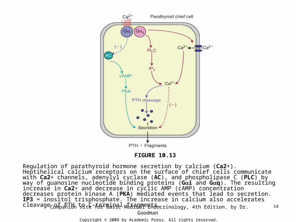
17
Biosynthesis of 1,25 dihydroxycholecalciferol (1,25(OH)2D3).
FIGURE 10.16

Companion site for Basic Medical Endocrinology, 4th Edition. by Dr. Goodman Copyright © 2009 by Academic Press. All rights reserved.
18
Effects of 1,25(OH)2D3 on intestinal transport of calcium. VDR = vitamin D receptor; ECaC = epithelial calcium channels (also called TRPV5 and TRPV6); CaB =calbindin 9.
FIGURE 10.17

Companion site for Basic Medical Endocrinology, 4th Edition. by Dr. Goodman Copyright © 2009 by Academic Press. All rights reserved.
19
Multiple negative feedback loops in the regulation of 1,25 dihydroxycholecalciferol synthesis. Solid arrows indicate stimulation; dashed red arrows represent inhibition.
FIGURE 10.18

Companion site for Basic Medical Endocrinology, 4th Edition. by Dr. Goodman Copyright © 2009 by Academic Press. All rights reserved.
20
Overall regulation of calcium balance by PTH, calcitonin, and 1,25(OH)2D3. Solid green arrows indicate stimulation; dashed arrows represent inhibition.
FIGURE 10.19

Companion site for Basic Medical Endocrinology, 4th Edition. by Dr. Goodman Copyright © 2009 by Academic Press. All rights reserved.
21
Increased plasma calcium concentrations regulate calcium reabsorption in the thick ascending limb of Henle’s loop. In this portion of the nephron calcium passes through the cellular junctions driven by a positive luminal voltage. The calcium receptor signals through the guanosine nucleotide binding protein Gq to activate PLC (phospholipase C) and form DAG (diacylglycerol), which activates PKC (protein kinase C). Back diffusion of potassium through renal outer medullary potassium channels (ROMK) is inhibited, which decreases the positive potential of luminal fluid and limits reabsorption of sodium and chloride. The receptor also signals through Gi, thus inhibits adenylyl cyclase (AC) reduces any cyclic AMP dependent stimulation of the sodium/potassium/2 chloride cotransporter. (PKA = protein kinase A)
FIGURE 10.20

Companion site for Basic Medical Endocrinology, 4th Edition. by Dr. Goodman Copyright © 2009 by Academic Press. All rights reserved.
22
Relation of estrogens to cytokines and growth factors in the overall economy of bones.
FIGURE 10.21