Chapter 1 Cellular Reaction to Injury
16
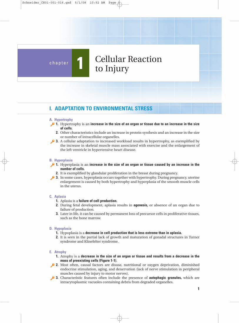
Cellular Reaction to Injury 1 chapter 1 I. ADAPTATION TO ENVIRONMENTAL STRESS A. Hypertrophy 1. Hypertrophy is an increase in the size of an organ or tissue due to an increase in the size of cells. 2. Other characteristics include an increase in protein synthesis and an increase in the size or number of intracellular organelles. 3. A cellular adaptation to increased workload results in hypertrophy, as exemplified by the increase in skeletal muscle mass associated with exercise and the enlargement of the left ventricle in hypertensive heart disease. B. Hyperplasia 1. Hyperplasia is an increase in the size of an organ or tissue caused by an increase in the number of cells. 2. It is exemplified by glandular proliferation in the breast during pregnancy. 3. In some cases, hyperplasia occurs together with hypertrophy. During pregnancy, uterine enlargement is caused by both hypertrophy and hyperplasia of the smooth muscle cells in the uterus. C. Aplasia 1. Aplasia is a failure of cell production. 2. During fetal development, aplasia results in agenesis, or absence of an organ due to failure of production. 3. Later in life, it can be caused by permanent loss of precursor cells in proliferative tissues, such as the bone marrow. D. Hypoplasia 1. Hypoplasia is a decrease in cell production that is less extreme than in aplasia. 2. It is seen in the partial lack of growth and maturation of gonadal structures in Turner syndrome and Klinefelter syndrome. E. Atrophy 1. Atrophy is a decrease in the size of an organ or tissue and results from a decrease in the mass of preexisting cells (Figure 1-1). 2. Most often, causal factors are disuse, nutritional or oxygen deprivation, diminished endocrine stimulation, aging, and denervation (lack of nerve stimulation in peripheral muscles caused by injury to motor nerves). 3. Characteristic features often include the presence of autophagic granules, which are intracytoplasmic vacuoles containing debris from degraded organelles. Schneider_CH01-001-016.qxd 5/1/08 10:52 AM Page 1
Transcript of Chapter 1 Cellular Reaction to Injury
Cellular Reaction to Injury1c h a p t e r
1
I. ADAPTATION TO ENVIRONMENTAL STRESS
A. Hypertrophy1. Hypertrophy is an increase in the size of an organ or tissue due to an increase in the size
of cells.2. Other characteristics include an increase in protein synthesis and an increase in the size
or number of intracellular organelles.3. A cellular adaptation to increased workload results in hypertrophy, as exemplified by
the increase in skeletal muscle mass associated with exercise and the enlargement ofthe left ventricle in hypertensive heart disease.
B. Hyperplasia1. Hyperplasia is an increase in the size of an organ or tissue caused by an increase in the
number of cells.2. It is exemplified by glandular proliferation in the breast during pregnancy.3. In some cases, hyperplasia occurs together with hypertrophy. During pregnancy, uterine
enlargement is caused by both hypertrophy and hyperplasia of the smooth muscle cellsin the uterus.
C. Aplasia1. Aplasia is a failure of cell production.2. During fetal development, aplasia results in agenesis, or absence of an organ due to
failure of production.3. Later in life, it can be caused by permanent loss of precursor cells in proliferative tissues,
such as the bone marrow.
D. Hypoplasia1. Hypoplasia is a decrease in cell production that is less extreme than in aplasia.2. It is seen in the partial lack of growth and maturation of gonadal structures in Turner
syndrome and Klinefelter syndrome.
E. Atrophy1. Atrophy is a decrease in the size of an organ or tissue and results from a decrease in the
mass of preexisting cells (Figure 1-1). 2. Most often, causal factors are disuse, nutritional or oxygen deprivation, diminished
endocrine stimulation, aging, and denervation (lack of nerve stimulation in peripheralmuscles caused by injury to motor nerves).
3. Characteristic features often include the presence of autophagic granules, which areintracytoplasmic vacuoles containing debris from degraded organelles.
Schneider_CH01-001-016.qxd 5/1/08 10:52 AM Page 1

4. In some instances, atrophy is thought to be mediated in part by the ubiquitin-proteo-some pathway of protein degradation. In this pathway, ubiquitin-linked proteins aredegraded within the proteosome, a large cytoplasmic protein complex.
F. Metaplasia is the replacement of one differentiated tissue by another (Figure 1-2).1. Squamous metaplasia
a. Squamous metaplasia is exemplified by the replacement of columnar epithelium atthe squamocolumnar junction of the cervix by squamous epithelium.
b. It can also occur in the respiratory epithelium of the bronchus, in the endometrium,and in the pancreatic ducts.
c. Associated conditions include chronic irritation (e.g., squamous metaplasia of thebronchi with long-term use of tobacco) and vitamin A deficiency.
d. This process is often reversible.2. Osseous metaplasia
a. Osseous metaplasia is the formation of new bone at sites of tissue injury.b. Cartilaginous metaplasia may also occur.
3. Myeloid metaplasia (extramedullary hematopoiesis) is proliferation of hematopoietictissue at sites other than the bone marrow, such as the liver or spleen.
2 BRS Pathology
FIGURE 1-1 Marked atrophy of frontal cortex ofbrain. Note the thinning of the gyri and the widen-ing of the sulci. (From Rubin R, Strayer D, et al.,eds.: Rubin’s Pathology. Clinicopathologic Foun-dations of Medicine, 5th ed. Baltimore, LippincottWilliams & Wilkins, 2008, p. 3. Original source:Okazaki H, Scheithauer BW: Atlas of Neu-ropathology. New York, Gower Medical Publish-ing, 1988. By permission of the author.)
FIGURE 1-2 Squamous metaplasia in theuterine cervix. The columnar epithelium ispartially replaced with squamous epithe-lium. Although this is a benign process, it can become a focus of dysplasia, whichcan lead to malignant change. (Reprintedwith permission from Rubin R, Strayer D,et al., eds.: Rubin’s Pathology. Clinico-pathologic Foundations of Medicine, 5th ed. Baltimore, Lippincott Williams & Wilkins, 2008, p. 7.)
Schneider_CH01-001-016.qxd 5/1/08 10:52 AM Page 2

Chapter 1 Cellular Reaction to Injury 3
II. HYPOXIC CELL INJURY
A. Causes. Hypoxic cell injury results from cellular anoxia or hypoxia, which in turn resultsfrom various mechanisms, including:1. Ischemia (obstruction of arterial blood flow), which is the most common cause2. Anemia, which is a reduction in the number of oxygen-carrying red blood cells3. Carbon monoxide poisoning, which results in diminution in the oxygen-carrying capacity
of red blood cells by chemical alteration of hemoglobin4. Decreased perfusion of tissues by oxygen-carrying blood, which occurs in cardiac failure,
hypotension, and shock5. Poor oxygenation of blood secondary to pulmonary disease
B. Early stage. Hypoxic cell injury first affects the mitochondria, with resultant decreasedoxidative phosphorylation and adenosine triphosphate (ATP) synthesis. Consequences ofdecreased ATP availability include:1. Failure of the cell membrane pump (ouabain-sensitive Na�-K�-ATPase) results in increased
intracellular Na� and water and decreased intracellular K�. This process causes cellularswelling and swelling of organelles.a. Cellular swelling, or hydropic change, is characterized by the presence of large vacuoles
in the cytoplasm.b. Swelling of the endoplasmic reticulum is one of the first ultrastructural changes evident
in reversible injury.c. Swelling of the mitochondria progresses from reversible, low-amplitude swelling to irre-
versible, high-amplitude swelling, which is characterized by marked dilation of theinner mitochondrial space.
2. Disaggregation of ribosomes leads to failure of protein synthesis. Ribosomal disaggrega-tion is also promoted by membrane damage.
3. Stimulation of phosphofructokinase activity results in increased glycolysis, accumulationof lactate, and decreased intracellular pH. Acidification causes reversible clumping ofnuclear chromatin.
C. Late stage1. Hypoxic cell injury eventually results in membrane damage to plasma and to lysosomal
and other organelle membranes, with loss of membrane phospholipids.2. Reversible morphologic signs of damage include the formation of:
a. Myelin figures, whorl-like structures probably originating from damaged membranesb. Cell blebs, a cell surface deformity most likely caused by disorderly function of the
cellular cytoskeleton
D. Cell death. Finally, cell death is caused by severe or prolonged injury.1. The point of no return is marked by irreversible damage to cell membranes, leading to
massive calcium influx, extensive calcification of the mitochondria, and cell death.2. Intracellular enzymes and various other proteins are released from necrotic cells into the cir-
culation as a consequence of the loss of integrity of cell membranes. This phenomenon isthe basis of a number of useful laboratory determinations as indicators of necrosis.a. Myocardial enzymes in serum. These are discussed in more depth in Chapter 10.
(1) Enzymes that have been useful in the diagnosis of myocardial infarction (“heartattack,” see Chapters 3 and 10) include the following:(a) Aspartate aminotransferase (AST, previously known as SGOT)(b) Lactate dehydrogenase (LDH)(c) Creatine kinase (CK, also known as CPK)
(2) These markers of myocardial necrosis vary in specificity for heart damage, as wellas in the time period after the necrotic event in which elevations in the serumappear and persist. The delineation of isoenzyme forms of LDH and CK has beena useful adjunct in adding specificity to these measures.
Schneider_CH01-001-016.qxd 5/1/08 10:52 AM Page 3
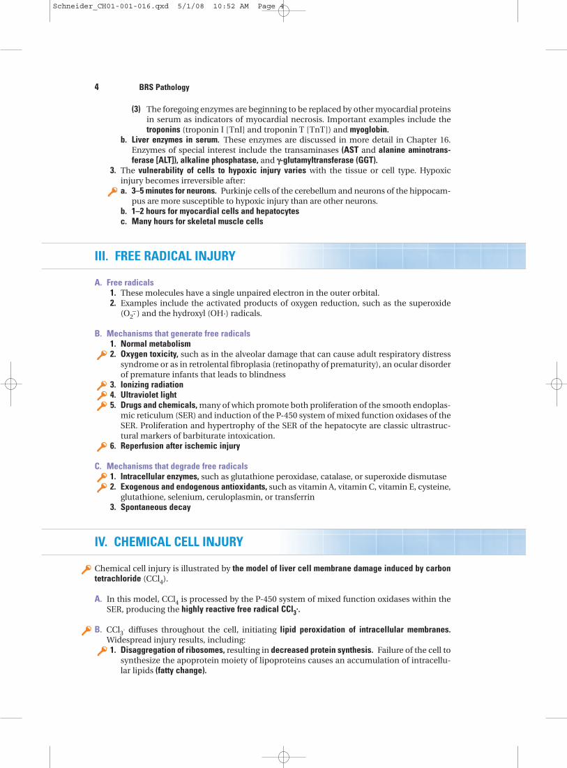
(3) The foregoing enzymes are beginning to be replaced by other myocardial proteinsin serum as indicators of myocardial necrosis. Important examples include thetroponins (troponin I [TnI] and troponin T [TnT]) and myoglobin.
b. Liver enzymes in serum. These enzymes are discussed in more detail in Chapter 16.Enzymes of special interest include the transaminases (AST and alanine aminotrans-ferase [ALT]), alkaline phosphatase, and γγ-glutamyltransferase (GGT).
3. The vulnerability of cells to hypoxic injury varies with the tissue or cell type. Hypoxicinjury becomes irreversible after:a. 3–5 minutes for neurons. Purkinje cells of the cerebellum and neurons of the hippocam-
pus are more susceptible to hypoxic injury than are other neurons.b. 1–2 hours for myocardial cells and hepatocytesc. Many hours for skeletal muscle cells
III. FREE RADICAL INJURY
A. Free radicals1. These molecules have a single unpaired electron in the outer orbital.2. Examples include the activated products of oxygen reduction, such as the superoxide
(O2·�) and the hydroxyl (OH·) radicals.
B. Mechanisms that generate free radicals1. Normal metabolism2. Oxygen toxicity, such as in the alveolar damage that can cause adult respiratory distress
syndrome or as in retrolental fibroplasia (retinopathy of prematurity), an ocular disorderof premature infants that leads to blindness
3. Ionizing radiation4. Ultraviolet light5. Drugs and chemicals, many of which promote both proliferation of the smooth endoplas-
mic reticulum (SER) and induction of the P-450 system of mixed function oxidases of theSER. Proliferation and hypertrophy of the SER of the hepatocyte are classic ultrastruc-tural markers of barbiturate intoxication.
6. Reperfusion after ischemic injury
C. Mechanisms that degrade free radicals1. Intracellular enzymes, such as glutathione peroxidase, catalase, or superoxide dismutase2. Exogenous and endogenous antioxidants, such as vitamin A, vitamin C, vitamin E, cysteine,
glutathione, selenium, ceruloplasmin, or transferrin3. Spontaneous decay
IV. CHEMICAL CELL INJURY
Chemical cell injury is illustrated by the model of liver cell membrane damage induced by carbontetrachloride (CCl4).
A. In this model, CCl4 is processed by the P-450 system of mixed function oxidases within theSER, producing the highly reactive free radical CCl3·.
B. CCl3· diffuses throughout the cell, initiating lipid peroxidation of intracellular membranes.
Widespread injury results, including:1. Disaggregation of ribosomes, resulting in decreased protein synthesis. Failure of the cell to
synthesize the apoprotein moiety of lipoproteins causes an accumulation of intracellu-lar lipids (fatty change).
4 BRS Pathology
Schneider_CH01-001-016.qxd 5/1/08 10:52 AM Page 4
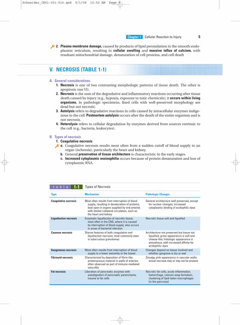
2. Plasma membrane damage, caused by products of lipid peroxidation in the smooth endo-plasmic reticulum, resulting in cellular swelling and massive influx of calcium, withresultant mitochondrial damage, denaturation of cell proteins, and cell death
V. NECROSIS (TABLE 1-1)
A. General considerations1. Necrosis is one of two contrasting morphologic patterns of tissue death. The other is
apoptosis (see VI).2. Necrosis is the sum of the degradative and inflammatory reactions occurring after tissue
death caused by injury (e.g., hypoxia, exposure to toxic chemicals); it occurs within livingorganisms. In pathologic specimens, fixed cells with well-preserved morphology aredead but not necrotic.
3. Autolysis refers to degradative reactions in cells caused by intracellular enzymes indige-nous to the cell. Postmortem autolysis occurs after the death of the entire organism and isnot necrosis.
4. Heterolysis refers to cellular degradation by enzymes derived from sources extrinsic tothe cell (e.g., bacteria, leukocytes).
B. Types of necrosis1. Coagulative necrosis
a. Coagulative necrosis results most often from a sudden cutoff of blood supply to anorgan (ischemia), particularly the heart and kidney.
b. General preservation of tissue architecture is characteristic in the early stages.c. Increased cytoplasmic eosinophilia occurs because of protein denaturation and loss of
cytoplasmic RNA.
Chapter 1 Cellular Reaction to Injury 5
1-1t a b l e
Type Mechanism Pathologic Changes
Coagulative necrosis Most often results from interruption of blood General architecture well preserved, exceptsupply, resulting in denaturation of proteins; for nuclear changes; increasedbest seen in organs supplied by end arteries cytoplasmic binding of acidophilic dyeswith limited collateral circulation, such as the heart and kidney
Liquefactive necrosis Enzymatic liquefaction of necrotic tissue, Necrotic tissue soft and liquefiedmost often in the CNS, where it is caused by interruption of blood supply; also occurs in areas of bacterial infection
Caseous necrosis Shares features of both coagulation and Architecture not preserved but tissue not liquefaction necrosis; most commonly seen liquefied; gross appearance is soft and in tuberculous granulomas cheese-like; histologic appearance is
amorphous, with increased affinity for acidophilic dyes
Gangrenous necrosis Most often results from interruption of blood Changes depend on tissue involved and supply to a lower extremity or the bowel whether gangrene is dry or wet
Fibrinoid necrosis Characterized by deposition of fibrin-like Smudgy pink appearance in vascular walls; proteinaceous material in walls of arteries; actual necrosis may or may not be presentoften observed as part of immune-mediated vasculitis
Fat necrosis Liberation of pancreatic enzymes with Necrotic fat cells, acute inflammation, autodigestion of pancreatic parenchyma; hemorrhage, calcium soap formation, trauma to fat cells clustering of lipid-laden macrophages
(in the pancreas)
Types of Necrosis
Schneider_CH01-001-016.qxd 5/1/08 10:52 AM Page 5

d. Nuclear changes, the morphologic hallmark of irreversible cell injury and necrosis, arecharacteristic. These include:(1) Pyknosis, chromatin clumping and shrinking with increased basophilia(2) Karyorrhexis, fragmentation of chromatin(3) Karyolysis, fading of chromatin material(4) Disappearance of stainable nuclei
2. Liquefactive necrosisa. Ischemic injury to the central nervous system (CNS) characteristically results in liquefac-
tive necrosis. After the death of CNS cells, liquefaction is caused by autolysis.b. Digestion, softening, and liquefaction of tissue are characteristic.c. Suppurative infections characterized by the formation of pus (liquefied tissue debris
and neutrophils) by heterolytic mechanisms involve liquefactive necrosis.3. Caseous necrosis
a. This type of necrosis occurs as part of granulomatous inflammation and is a manifesta-tion of partial immunity caused by the interaction of T lymphocytes (CD4�, CD8�,and CD4-CD8-), macrophages, and probably cytokines, such as interferon-�, derivedfrom these cells.
b. Tuberculosis is the leading cause of caseous necrosis.c. Caseous necrosis combines features of both coagulative necrosis and liquefactive
necrosis.d. On gross examination, caseous necrosis has a cheese-like (caseous) consistency.e. On histologic examination, caseous necrosis has an amorphous eosinophilic
appearance.4. Gangrenous necrosis
a. This type of necrosis most often affects the lower extremities or bowel and is secondaryto vascular occlusion.
b. When complicated by infective heterolysis and consequent liquefactive necrosis, gan-grenous necrosis is called wet gangrene.
c. When characterized primarily by coagulative necrosis without liquefaction, gangrenousnecrosis is called dry gangrene.
5. Fibrinoid necrosisa. This deposition of fibrin-like proteinaceous material in the arterial walls appears smudgy
and acidophilic.b. Fibrinoid necrosis is often associated with immune-mediated vascular damage.
6. Fat necrosis occurs in two forms.a. Traumatic fat necrosis, which occurs after a severe injury to tissue with high fat content,
such as the breastb. Enzymatic fat necrosis, which is a complication of acute hemorrhagic pancreatitis,
a severe inflammatory disorder of the pancreas(1) Proteolytic and lipolytic pancreatic enzymes diffuse into inflamed tissue and lit-
erally digest the parenchyma.(2) Fatty acids liberated by the digestion of fat form calcium salts (saponification, or
soap formation).(3) Vessels are eroded, with resultant hemorrhage.
VI. APOPTOSIS (TABLE 1-2)
A. General considerations1. Apoptosis is a second morphologic pattern of tissue death. (The other is necrosis; see V.)
It is often referred to as programmed cell death.2. This is an important mechanism for the removal of cells. An example is apoptotic removal
of cells with irreparable DNA damage (from free radicals, viruses, cytotoxic immunemechanisms), protecting against neoplastic transformation.
6 BRS Pathology
Schneider_CH01-001-016.qxd 5/1/08 10:52 AM Page 6

3. In addition, apoptosis is an important mechanism for physiologic cell removal dur-ing embryogenesis and in programmed cell cycling (e.g., endometrial cells duringmenstruation).
4. This involutional process is similar to the physiologic loss of leaves from a tree; apopto-sis is a Greek term for “falling away from.”
B. Morphologic features1. A tendency to involve single isolated cells or small clusters of cells within a tissue2. Progression through a series of changes marked by a lack of inflammatory response
a. Blebbing of plasma membrane, cytoplasmic shrinkage, chromatin condensationb. Budding of cell and separation of apoptotic bodies (membrane-bound segments)c. Phagocytosis of apoptotic bodies
3. Involution and shrinkage of affected cells and cell fragments, resulting in small roundeosinophilic masses often containing chromatin remnants, exemplified by Councilmanbodies in viral hepatitis
C. Biochemical events1. Diverse injurious stimuli (e.g., free radicals, radiation, toxic substances, withdrawal of
growth factors or hormones) trigger a variety of stimuli, including cell surface receptorssuch as FAS, mitochondrial response to stress, and cytotoxic T cells.
2. The extrinsic pathway of initiation is mediated by cell surface receptors exemplified byFAS, a member of the tumor necrosis factor receptor family of proteins. This pathway isinitiated by signaling by molecules such as the FAS ligand, which in turn signals a seriesof events that involve activation of caspases. Caspases are aspartate-specific cysteineproteases that have been referred to as “major executioners” or “molecular guillotines.”The death signals are conveyed in a proteolytic cascade, through activation of a chain ofcaspases and other targets. The initial activating caspases are caspase-8 and caspase-9,and the terminal caspases (executioners) include caspase-3 and caspase-6 (among otherproteases).
Chapter 1 Cellular Reaction to Injury 7
1-2t a b l e
Characteristics Necrosis Apoptosis
Etiology Gross irreversible cellular injury Subtle cellular damage, physiologic programmed cell removal
Morphologic changes Involves many contiguous cells Involves single cells or small clusters of cellsIncreased cytoplasmic eosinophilia Cytoplasmic shrinking and increased
due to denaturation of proteins eosinophilic stainingProgressive nuclear condensation Chromatin condensation and fragmentation
and fragmentation with eventual Fragmentation into membrane-bound disappearance of nuclei apoptotic bodies
Preservation of tissue architecture in early stages of coagulative necrosis
Biochemical changes Passive form of cell death not requiring Active form of cell death requiring gene gene involvement or new protein expression, protein synthesis, and energy synthesis consumption
DNA fragmentation is haphazard DNA fragmentation is regular at rather than regular, resulting in an nucleosomal boundaries, resulting inelectrophoretic smudge pattern an electrophoretic “laddered” pattern
Inflammatory reaction Marked inflammatory reaction, liberation No inflammatory reactionof lysosomal enzymes, digestion of cell Apoptotic bodies engulfed by neighboring membranes, and disruption of cells macrophages and epithelial cells
Influx of macrophages due to releaseof chemotactic factors
Removal of debris by phagocytic macrophages
Comparison of Necrosis and Apoptosis
Schneider_CH01-001-016.qxd 5/1/08 10:52 AM Page 7

3. The intrinsic, or mitochondrial, pathway, which is initiated by the loss of stimulation bygrowth factors and other adverse stimuli, results in the inactivation and loss of bcl-2 andother antiapoptotic proteins from the inner mitochondrial membrane. This loss resultsin increased mitochondrial permeability, the release of cytochrome c, and the stimula-tion of proapoptotic proteins such as bax and bak. Cytochrome c interacts with Apaf-1causing self-cleavage and activation of caspase-9. Downstream caspases are activated byupstream proteases and act themselves to cleave cellular targets.
4. Cytotoxic T-cell activation is characterized by direct activation of caspases by granzyme B,a cytotoxic T-cell protease that perhaps directly activates the caspase cascade. The entryof granzyme B into target cells is mediated by perforin, a cytotoxic T-cell protein.
5. Degradation of DNA by endonucleases into nucleosomal chromatin fragments that aremultiples of 180–200 base pairs results in the typical “laddering” appearance of DNA onelectrophoresis. This phenomenon is characteristic of, but not entirely specific for,apoptosis.
6. Activation of transglutaminases crosslinks apoptotic cytoplasmic proteins.7. The caspases consist of a group of aspartic acid-specific cysteine proteases that are acti-
vated during apoptosis. 8. Newer methods such as the TUNEL assay (Terminal Transferase dUTP Nick End Labeling)
are ways to quantitate cleaving of nucleosomes and, thus, apoptosis. Similarly, caspaseassays are coming into use as apoptotic markers. Surely more will follow.
D. Regulation of apoptosis is mediated by a number of genes and their products. Importantgenes include bcl-2 (gene product inhibits apoptosis), bax (gene product facilitates apopto-sis), and p53 (gene product decreases transcription of bcl-2 and increases transcription ofbax, thus facilitating apoptosis).
E. Additionally, complex signaling pathways involving multiple genes and gene products arethe subject of vigorous scientific investigation. Since many pathologic processes are relatedto either stimulation or inhibition of apoptosis (e.g., many forms of cancer), this area ofinquiry promises to yield major understanding that will surely lead to important therapeu-tic applications.
VII. REVERSIBLE CELLULAR CHANGES AND ACCUMULATIONS
A. Fatty change (fatty metamorphosis, steatosis)1. General considerations
a. Fatty change is characterized by the accumulation of intracellular parenchymal triglyc-erides and is observed most frequently in the liver, heart, and kidney. For example, inthe liver, fatty change may be secondary to alcoholism, diabetes mellitus, malnutri-tion, obesity, or poisonings.
2. Imbalance among the uptake, utilization, and secretion of fat is the cause of fatty change,and this can result from any of the following mechanisms:a. Increased transport of triglycerides or fatty acids to affected cellsb. Decreased mobilization of fat from cells, most often mediated by decreased production
of apoproteins required for fat transport. Fatty change is thus linked to the disaggrega-tion of ribosomes and consequent decreased protein synthesis caused by failure ofATP production in CCl4-injured cells.
c. Decreased use of fat by cellsd. Overproduction of fat in cells
B. Hyaline change1. This term denotes a characteristic (homogeneous, glassy, eosinophilic) appearance in
hematoxylin and eosin sections.2. It is caused most often by nonspecific accumulations of proteinaceous material.
8 BRS Pathology
Schneider_CH01-001-016.qxd 5/1/08 10:52 AM Page 8
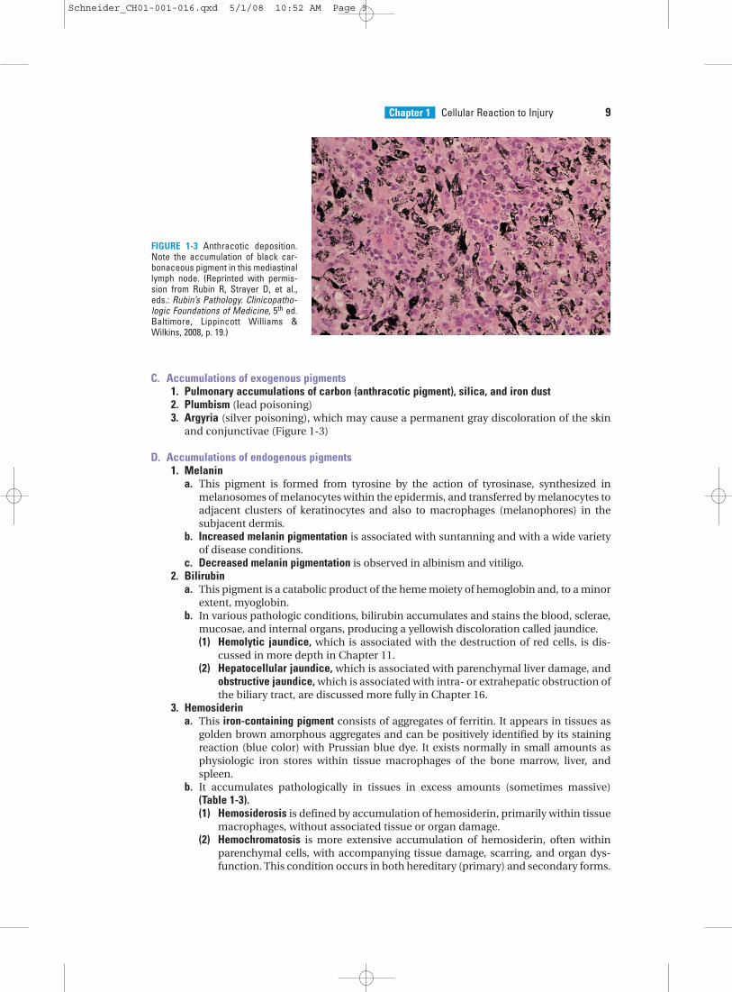
C. Accumulations of exogenous pigments1. Pulmonary accumulations of carbon (anthracotic pigment), silica, and iron dust2. Plumbism (lead poisoning)3. Argyria (silver poisoning), which may cause a permanent gray discoloration of the skin
and conjunctivae (Figure 1-3)
D. Accumulations of endogenous pigments1. Melanin
a. This pigment is formed from tyrosine by the action of tyrosinase, synthesized inmelanosomes of melanocytes within the epidermis, and transferred by melanocytes toadjacent clusters of keratinocytes and also to macrophages (melanophores) in thesubjacent dermis.
b. Increased melanin pigmentation is associated with suntanning and with a wide varietyof disease conditions.
c. Decreased melanin pigmentation is observed in albinism and vitiligo.2. Bilirubin
a. This pigment is a catabolic product of the heme moiety of hemoglobin and, to a minorextent, myoglobin.
b. In various pathologic conditions, bilirubin accumulates and stains the blood, sclerae,mucosae, and internal organs, producing a yellowish discoloration called jaundice.(1) Hemolytic jaundice, which is associated with the destruction of red cells, is dis-
cussed in more depth in Chapter 11.(2) Hepatocellular jaundice, which is associated with parenchymal liver damage, and
obstructive jaundice, which is associated with intra- or extrahepatic obstruction ofthe biliary tract, are discussed more fully in Chapter 16.
3. Hemosiderina. This iron-containing pigment consists of aggregates of ferritin. It appears in tissues as
golden brown amorphous aggregates and can be positively identified by its stainingreaction (blue color) with Prussian blue dye. It exists normally in small amounts asphysiologic iron stores within tissue macrophages of the bone marrow, liver, andspleen.
b. It accumulates pathologically in tissues in excess amounts (sometimes massive)(Table 1-3).(1) Hemosiderosis is defined by accumulation of hemosiderin, primarily within tissue
macrophages, without associated tissue or organ damage.(2) Hemochromatosis is more extensive accumulation of hemosiderin, often within
parenchymal cells, with accompanying tissue damage, scarring, and organ dys-function. This condition occurs in both hereditary (primary) and secondary forms.
Chapter 1 Cellular Reaction to Injury 9
FIGURE 1-3 Anthracotic deposition.Note the accumulation of black car-bonaceous pigment in this mediastinallymph node. (Reprinted with permis-sion from Rubin R, Strayer D, et al.,eds.: Rubin’s Pathology. Clinicopatho-logic Foundations of Medicine, 5th ed.Baltimore, Lippincott Williams &Wilkins, 2008, p. 19.)
Schneider_CH01-001-016.qxd 5/1/08 10:52 AM Page 9
(a) Hereditary hemochromatosis is most often caused by a mutation in the Hfegene on chromosome 6.(i) Hemosiderin deposition and organ damage in the liver, pancreas,
myocardium, and multiple endocrine glands is characteristic, as well asmelanin deposition in the skin.
(ii) This results in the triad of micronodular cirrhosis, diabetes mellitus, andskin pigmentation. This set of findings is referred to as “bronze diabetes.”Laboratory abnormalities of note include marked elevation of the serumtransferrin saturation because of the combination of increased serum ironand decreased total iron-binding capacity (TIBC).
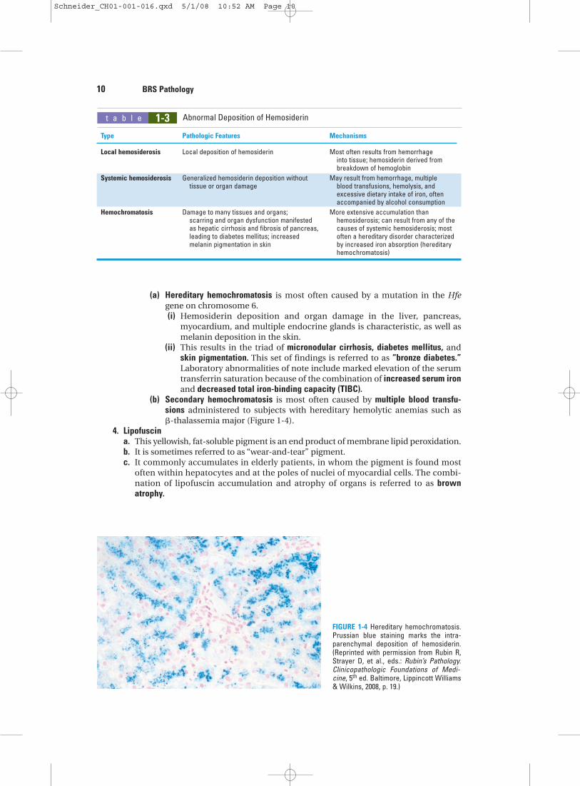
(b) Secondary hemochromatosis is most often caused by multiple blood transfu-sions administered to subjects with hereditary hemolytic anemias such as �-thalassemia major (Figure 1-4).
4. Lipofuscina. This yellowish, fat-soluble pigment is an end product of membrane lipid peroxidation.b. It is sometimes referred to as “wear-and-tear” pigment.c. It commonly accumulates in elderly patients, in whom the pigment is found most
often within hepatocytes and at the poles of nuclei of myocardial cells. The combi-nation of lipofuscin accumulation and atrophy of organs is referred to as brownatrophy.
10 BRS Pathology
FIGURE 1-4 Hereditary hemochromatosis.Prussian blue staining marks the intra-parenchymal deposition of hemosiderin.(Reprinted with permission from Rubin R,Strayer D, et al., eds.: Rubin’s Pathology.Clinicopathologic Foundations of Medi-cine, 5th ed. Baltimore, Lippincott Williams& Wilkins, 2008, p. 19.)
Type Pathologic Features Mechanisms
Local hemosiderosis Local deposition of hemosiderin Most often results from hemorrhage into tissue; hemosiderin derived from breakdown of hemoglobin
Systemic hemosiderosis Generalized hemosiderin deposition without May result from hemorrhage, multiple tissue or organ damage blood transfusions, hemolysis, and
excessive dietary intake of iron, often accompanied by alcohol consumption
Hemochromatosis Damage to many tissues and organs; More extensive accumulation than scarring and organ dysfunction manifested hemosiderosis; can result from any of the as hepatic cirrhosis and fibrosis of pancreas, causes of systemic hemosiderosis; most leading to diabetes mellitus; increased often a hereditary disorder characterized melanin pigmentation in skin by increased iron absorption (hereditary
hemochromatosis)
1-3t a b l e Abnormal Deposition of Hemosiderin
Schneider_CH01-001-016.qxd 5/1/08 10:52 AM Page 10
E. Pathologic calcifications1. Metastatic calcification
a. The cause of metastatic calcification is hypercalcemia.b. Hypercalcemia most often results from any of the following causes:
(a) Hyperparathyroidism(b) Osteolytic tumors with resultant mobilization of calcium and phosphorus(c) Hypervitaminosis D(d) Excess calcium intake, such as in the milk-alkali syndrome (nephrocalcinosis and
renal stones caused by milk and antacid self-therapy)2. Dystrophic calcification
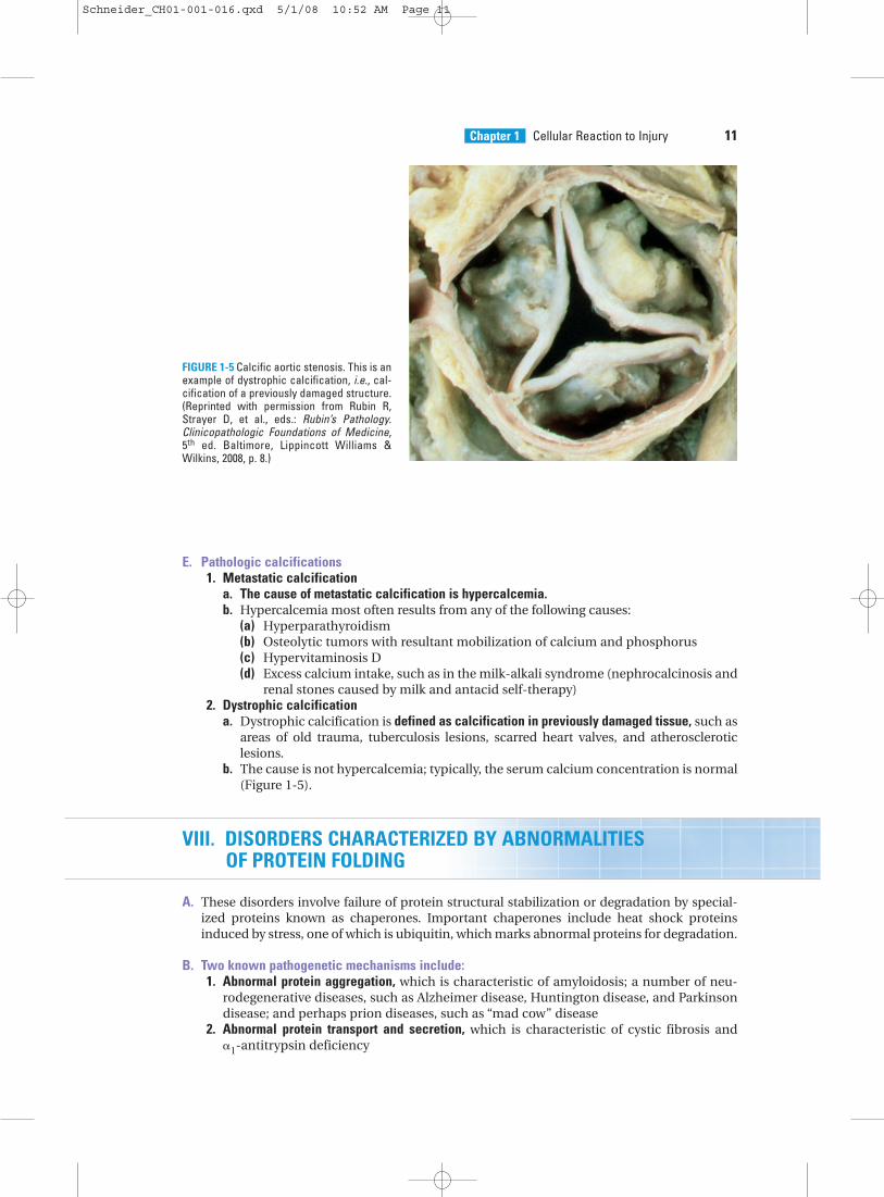
a. Dystrophic calcification is defined as calcification in previously damaged tissue, such asareas of old trauma, tuberculosis lesions, scarred heart valves, and atheroscleroticlesions.
b. The cause is not hypercalcemia; typically, the serum calcium concentration is normal(Figure 1-5).
VIII. DISORDERS CHARACTERIZED BY ABNORMALITIES OF PROTEIN FOLDING
A. These disorders involve failure of protein structural stabilization or degradation by special-ized proteins known as chaperones. Important chaperones include heat shock proteinsinduced by stress, one of which is ubiquitin, which marks abnormal proteins for degradation.
B. Two known pathogenetic mechanisms include:1. Abnormal protein aggregation, which is characteristic of amyloidosis; a number of neu-
rodegenerative diseases, such as Alzheimer disease, Huntington disease, and Parkinsondisease; and perhaps prion diseases, such as “mad cow” disease
2. Abnormal protein transport and secretion, which is characteristic of cystic fibrosis and �1-antitrypsin deficiency
Chapter 1 Cellular Reaction to Injury 11
FIGURE 1-5 Calcific aortic stenosis. This is anexample of dystrophic calcification, i.e., cal-cification of a previously damaged structure.(Reprinted with permission from Rubin R,Strayer D, et al., eds.: Rubin’s Pathology.Clinicopathologic Foundations of Medicine,5th ed. Baltimore, Lippincott Williams &Wilkins, 2008, p. 8.)
Schneider_CH01-001-016.qxd 5/1/08 10:52 AM Page 11
Review Test
12
Directions: Each of the numbered items or incomplete statements in this section is followedby answers or by completions of the statement. Select the one lettered answer or completionthat is best in each case.
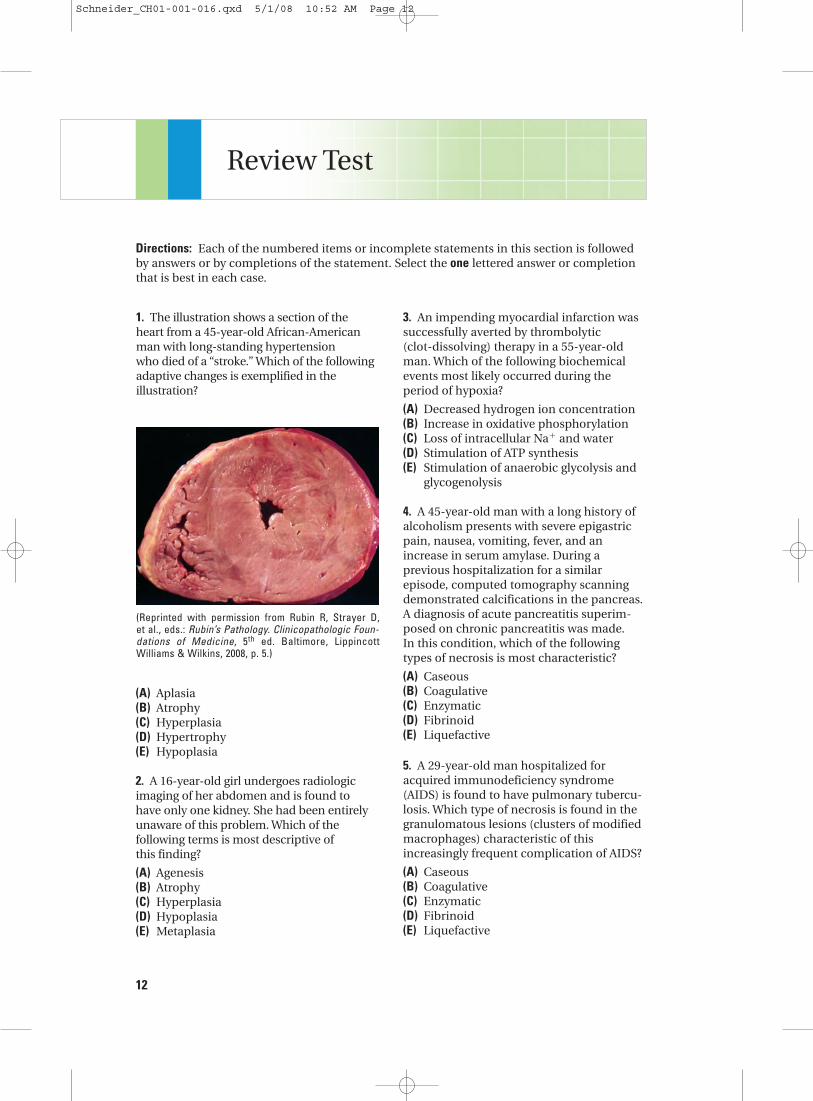
(Reprinted with permission from Rubin R, Strayer D, et al., eds.: Rubin’s Pathology. Clinicopathologic Foun-dations of Medicine, 5th ed. Baltimore, LippincottWilliams & Wilkins, 2008, p. 5.)
1. The illustration shows a section of the heart from a 45-year-old African-Americanman with long-standing hypertension who died of a “stroke.” Which of the followingadaptive changes is exemplified in the illustration?
3. An impending myocardial infarction wassuccessfully averted by thrombolytic (clot-dissolving) therapy in a 55-year-oldman. Which of the following biochemicalevents most likely occurred during theperiod of hypoxia?
(A) Decreased hydrogen ion concentration(B) Increase in oxidative phosphorylation(C) Loss of intracellular Na� and water(D) Stimulation of ATP synthesis(E) Stimulation of anaerobic glycolysis and
glycogenolysis
4. A 45-year-old man with a long history ofalcoholism presents with severe epigastricpain, nausea, vomiting, fever, and anincrease in serum amylase. During a previous hospitalization for a similarepisode, computed tomography scanningdemonstrated calcifications in the pancreas.A diagnosis of acute pancreatitis superim-posed on chronic pancreatitis was made. In this condition, which of the followingtypes of necrosis is most characteristic?
(A) Caseous(B) Coagulative(C) Enzymatic(D) Fibrinoid(E) Liquefactive
5. A 29-year-old man hospitalized foracquired immunodeficiency syndrome(AIDS) is found to have pulmonary tubercu-losis. Which type of necrosis is found in thegranulomatous lesions (clusters of modifiedmacrophages) characteristic of this increasingly frequent complication of AIDS?
(A) Caseous(B) Coagulative(C) Enzymatic(D) Fibrinoid(E) Liquefactive
(A) Aplasia(B) Atrophy(C) Hyperplasia(D) Hypertrophy(E) Hypoplasia
2. A 16-year-old girl undergoes radiologicimaging of her abdomen and is found to have only one kidney. She had been entirelyunaware of this problem. Which of the following terms is most descriptive of this finding?
(A) Agenesis(B) Atrophy(C) Hyperplasia(D) Hypoplasia(E) Metaplasia
Schneider_CH01-001-016.qxd 5/1/08 10:52 AM Page 12
6. A 45-year-old woman is investigated forhypertension and is found to have enlarge-ment of the left kidney. The right kidney issmaller than normal. Contrast studiesreveal stenosis of the right renal artery. The size change in the right kidney is an example of which of the following adaptive changes?
(A) Aplasia(B) Atrophy(C) Hyperplasia(D) Hypertrophy(E) Metaplasia
7. A 56-year-old man recovered from amyocardial infarction after his myocardiumwas entirely “saved” by immediate thrombolytic therapy. If it had been possibleto examine microscopic sections of his heartduring his ischemic episode, which of thefollowing would be the most likely cellularchange to be found?
(A) Karyolysis(B) Karyorrhexis(C) Pyknosis(D) Swelling of the endoplasmic
reticulum
Chapter 1 Cellular Reaction to Injury 13
(Reprinted with permission from Rubin R,Strayer D, et al., eds.: Rubin’s Pathology.Clinicopathologic Foundations of Medicine,5th ed. Baltimore, Lippincott Williams &Wilkins, 2008, p. 23.)
8. A 64-year-old woman presents with fever, chills, headache, neck stiffness, vomiting, and confusion. The Kernig sign(passive knee extension eliciting neck pain)and Brudzinski sign (passive neck flexioneliciting bilateral hip flexion) are both posi-tive. Examination of the cerebrospinal fluidreveals changes consistent with bacterialmeningitis, and brain imaging demonstratesa localized abscess. Which of the followingtypes of necrosis is most characteristic ofabscess formation?
(A) Caseous(B) Coagulative(C) Enzymatic(D) Fibrinoid(E) Liquefactive
9. A 20-year-old man presents with yellow-ing of the sclerae, skin, and oral mucosa.Which of the following accumulationsunderlies these findings?
(A) Bilirubin(B) Hemosiderin(C) Lead(D) Melanin(E) Silver
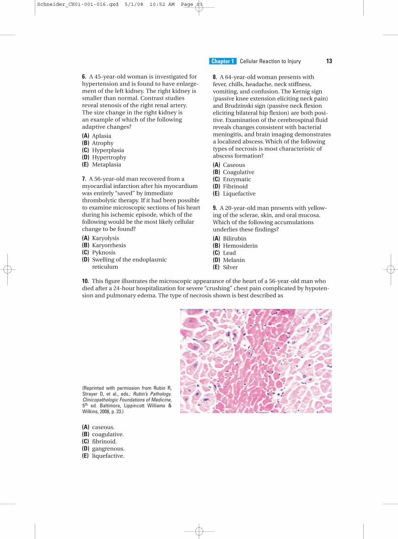
10. This figure illustrates the microscopic appearance of the heart of a 56-year-old man whodied after a 24-hour hospitalization for severe “crushing” chest pain complicated by hypoten-sion and pulmonary edema. The type of necrosis shown is best described as
(A) caseous.(B) coagulative.(C) fibrinoid.(D) gangrenous.(E) liquefactive.
Schneider_CH01-001-016.qxd 5/1/08 10:52 AM Page 13
(A) Accumulation of triglycerides withinhepatocytes
(B) Apoptosis with replacement of damagedcells by lipid-laden macrophages
(C) Bilirubin accumulation with mobiliza-tion of fat by bile salts
(D) Enzymatic fat necrosis with digestion ofliver parenchyma by released enzymes
(E) Irreversible damage to mitochondria
12. A 45-year-old man is referred because ofa recent diagnosis of hereditary hemochro-matosis. Which of the following is a correctstatement about this disorder?
(A) Damage to organs results from abnormal deposition of lead.
(B) It can progress to liver cirrhosis, diabetesmellitus, and skin pigmentation.
(C) Most cases are due to spontaneousmutations.
(D) Skin hyperpigmentation is due to bilirubin accumulation.
(E) The total iron-binding capacity (TIBC) is characteristically increased.
14 BRS Pathology
13. A 60-year-old woman with breast cancerand widespread bony metastases is found tohave calcification of multiple organs. Thecalcifications are best described as
(A) dystrophic with decreased serum calcium.
(B) dystrophic with increased serum calcium.
(C) metastatic with decreased serum calcium.
(D) metastatic with increased serum calcium.
14. A 56-year-old man dies 24 hours afterthe onset of substernal chest pain radiatingdown his left arm to the ulnar aspect of his fingertips. Which of the following morphologic myocardial findings is an indicator of irreversible injury?
(A) Cell blebs(B) Depletion of glycogen(C) Mitochondrial swelling(D) Myelin figures(E) Pyknotic nuclei
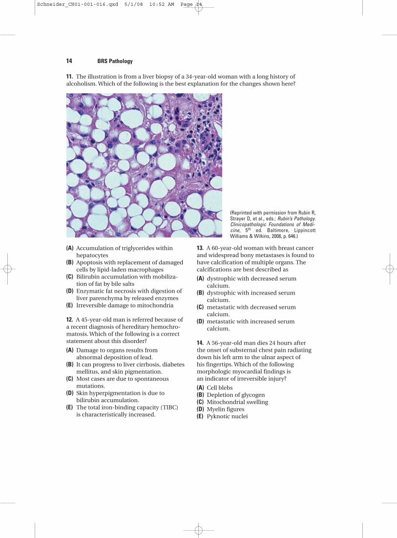
11. The illustration is from a liver biopsy of a 34-year-old woman with a long history of alcoholism. Which of the following is the best explanation for the changes shown here?
(Reprinted with permission from Rubin R,Strayer D, et al., eds.: Rubin’s Pathology.Clinicopathologic Foundations of Medi-cine, 5th ed. Baltimore, LippincottWilliams & Wilkins, 2008, p. 646.)
Schneider_CH01-001-016.qxd 5/1/08 10:52 AM Page 14

Chapter 3 15
1. The answer is D. The illustration shows marked hypertrophy of the left ventricle. Hyper-trophy of this extent, often seen in hypertensive heart disease, is caused by increasedworkload from increased ventricular pressure. This organ enlargement is the result of anincrease in size of the individual muscle cells.
2. The answer is A. The patient has renal agenesis, absence of the kidney due to failure oforgan development. The congenital lack of one kidney differs from atrophy, in which adecrease in the size of an organ results from a decrease in the mass of pre-existing cells.Unilateral renal agenesis is usually a harmless malformation, and the opposite kidney is often enlarged due to compensatory hypertrophy. Bilateral renal agenesis is incompatible with life and is of special interest since it can lead to the Potter progression (see Chapter 17).
3. The answer is E. The sequence of events in hypoxic cell damage is as follows: Hypoxiaresults in failure of oxidative phosphorylation, with resultant depletion of ATP andincrease in AMP and ADP. Anaerobic glycolysis and glycogenolysis are stimulated (not inhibited) through increased phosphofructokinase and phosphorylase activities,respectively. This results in an accumulation of cell lactate, with a decrease in intracellular pH and depletion of cellular glycogen stores. Decreased availability of ATP also results in failure of the Na�K�-ATPase pump, which then leads to increased cell Na� and water and decreased cell K�.
4. The answer is C. Pancreatic enzymatic fat necrosis represents autodigestion by proteolytic and lipolytic enzymes released from damaged parenchymal cells of the pancreas. Fatty acids liberated by the digestion of fat form calcium soaps, a processreferred to as saponification. The precipitated calcium in the soaps can be visualized by radiologic imaging.
5. The answer is A. Caseous necrosis occurs as part of granulomatous inflammation, typified by the lesions of tuberculosis.
6. The answer is B. The decreased size is due to restriction of the blood supply, one of thecauses of atrophy. The increase in size of the opposite kidney is referred to as compensa-tory hypertrophy. Unilateral renal artery stenosis is a well-known cause of secondaryhypertension. In this setting, increased renin excretion and stimulation of the renin-angiotensin system results in a form of hypertension that is potentially curable by surgical correction of the underlying vascular abnormality.
7. The answer is D. If infarction is averted by immediate thrombolytic therapy, indicators of necrosis, such as karyorrhexis, pyknosis, and karyolysis, which represent irreversiblechanges, would not be expected. Swelling of the endoplasmic reticulum from increasedcell water, one of the earliest ultrastructural changes observed in injured cells, isreversible and would be expected.
8. The answer is E. Liquefactive necrosis is characteristic of ischemic injury in the centralnervous system and suppurative infections that cause abscess formation (see Chapter 2).The changes in the cerebrospinal spinal fluid characteristic of bacterial meningitis aredetailed in Chapter 3.
9. The answer is A. Yellowing of the sclerae, skin, and oral mucosa are all characteristic ofjaundice, the accumulation of bilirubin, the catabolic product of the heme moiety ofhemoglobin. Jaundice can occur by diverse mechanisms: hemolytic (see Chapter 11),hepatocellular (see Chapter 16), or obstructive (see Chapter 16).
Answers and Explanations
Schneider_CH01-001-016.qxd 5/1/08 10:52 AM Page 15

10. The answer is B. The figure illustrates general preservation of myocardial architecture withsome fragmentation, more intense cytoplasmic staining corresponding to increased cellu-lar eosinophilia, and loss of nuclei, all of which are characteristic of coagulative necrosis.
11. The answer is A. The figure illustrates fatty change of the liver, which is characterizedby the accumulation of intracellular parenchymal triglycerides. It is seen most frequently in the liver, heart, and kidney and commonly is secondary to alcoholism.Fatty change results from an imbalance between the uptake, utilization, and mobilization of fat from liver cells. Alcoholic fatty liver may be reversible with complete abstinence from alcohol.
12. The answer is B. In advanced form, primary (hereditary) hemochromatosis is character-ized by the triad of cirrhosis, diabetes, and hyperpigmentation, or so-called “bronze dia-betes.” The disease is most often caused by a mutation in the Hfe gene on chromosome 6and is characteristically familial rather than sporadic. The manifestations of the disorderare the result of iron overload and deposition of hemosiderin in tissues such as the liver,pancreas, skin, joints, and pituitary. Laboratory abnormalities of note include increasedserum iron and decreased total iron-binding capacity (TIBC). The skin hyperpigmenta-tion is due largely to increases in melanin and to lesser accumulations of hemosiderin.
13. The answer is D. Metastatic calcification, or deposition of calcium in previously normaltissue, is caused by hypercalcemia. In this patient, tumor metastases to bone withincreased osteolytic activity caused mobilization of calcium and phosphate, resulting inhypercalcemia. Metastatic calcification should be contrasted with dystrophic calcifica-tion, in which the serum calcium concentration is normal and previously damaged tis-sues are the sites of deposition.
14. The answer is E. Myelin figures, cell blebs, mitochondrial swelling, and glycogen deple-tion are all signs of reversible injury. Nuclear changes such as pyknosis, karyorrhexis, andkaryolysis are signs of cell death and are, of course, irreversible.
16 BRS Pathology
Schneider_CH01-001-016.qxd 5/1/08 10:52 AM Page 16