Cellular Adaptation and Cell Injury Lec 4 RKM
70
1 Cellular Adaptation and Cell Injury CLDavis Foundation On the Beach Lecture 4 Part 1a Intracellular and Extracellular Accumulations, Pathologic Calcification, Pigments R K Myers 2011 Uremia, stomach, dog. A band of calcium has been laid down the middle of the gastric mucosa. A, The calcium is stained blue with hematoxylin. H&E stain. B, The calcium is stained black. von Kossa stain. (A and B, Courtesy Dr. M.D. McGavin, College of Veterinary Medicine, University of Tennessee.) (McGavin, M. Donald. Pathologic Basis of Veterinary Disease, 4th Edition. C.V. Mosby Intracellular accumulations • 3 categories – Normal cellular component accumulated in excess: water, lipids, glycogen, carbohydrates – Abnormal substance: exogenous (mineral, infectious agents) or endogenous (abnormal synthesis during t b li ) metabolism) – Pigment • These substances may be in cytoplasm (especially phagolysosomes) or nucleus, may be being produced or just stored, may be transient or permanent, may be harmless or injurious Intracellular accumulations Mechanisms of intracellular accumulations. A, Abnormal metabolism, as in fatty change in the liver; B, mutations causing alterations in i f ldi d i 1 protein folding and transport, as in α1- antitrypsin deficiency; C, deficiency of critical enzymes that prevent breakdown of substrates that accumulate in lysosomes, as in lysosomal storage diseases; and D, inability to degrade phagocytosed particles, as in hemosiderosis and carbon pigment accumulation. (A through D, From Kumar V, Abbas A, Fausto N: Robbins & Cotran pathologic basis of disease, ed 7, Philadelphia, 2005, Saunders.) (McGavin, M. Donald. Pathologic Basis of Veterinary Disease, 4th Edition. C.V. Mosby,
Transcript of Cellular Adaptation and Cell Injury Lec 4 RKM

1
Cellular Adaptation and Cell InjuryCLDavis Foundation
On the BeachLecture 4 Part 1a
Intracellular and Extracellular Accumulations, Pathologic Calcification, Pigmentsg , g
R K Myers 2011
Uremia, stomach, dog. A band of calcium has been laid down the middle of the gastric mucosa. A, The calcium is stained blue with hematoxylin. H&E stain. B, The calcium is stained black. von
Kossa stain. (A and B, Courtesy Dr. M.D. McGavin, College of Veterinary Medicine, University of Tennessee.) (McGavin, M. Donald. Pathologic Basis of Veterinary Disease, 4th Edition. C.V. Mosby
Intracellular accumulations
• 3 categories– Normal cellular component accumulated in excess:
water, lipids, glycogen, carbohydrates
– Abnormal substance: exogenous (mineral, infectious agents) or endogenous (abnormal synthesis during
t b li )metabolism)
– Pigment
• These substances may be in cytoplasm (especially phagolysosomes) or nucleus, may be being produced or just stored, may be transient or permanent, may be harmless or injurious
Intracellular accumulations
Mechanisms of intracellular accumulations.
A, Abnormal metabolism, as in fatty change in the liver; B, mutations causing alterations in
i f ldi d i 1protein folding and transport, as in α1-antitrypsin deficiency; C, deficiency of critical enzymes that prevent breakdown of substrates that accumulate in lysosomes, as in lysosomal storage diseases; and D, inability to degrade phagocytosed particles, as in hemosiderosis and carbon pigment accumulation.
(A through D, From Kumar V, Abbas A, Fausto N: Robbins & Cotran pathologic basis of disease, ed 7, Philadelphia, 2005, Saunders.) (McGavin, M. Donald. Pathologic Basis of Veterinary Disease, 4th Edition.
C.V. Mosby,

2
Lipids:
• Triglycerides. Discussed as fatty change or lipidosis
• Phospholipids. Components of myelin figures in injured and necrotic cells
• Abnormal lipids and carbohydrates in lysosomal storage diseasesg
• Cholesterol/cholesterol esters– Atherosclerosis– Xanthomas– Foamy macrophages in inflammation and injury– Cholesterol granulomas– Nieman-Pick disease, type C.
Lipids:
• Fatty infiltration is not the same as lipidosis
• Increased adipocytes not just intracellular fat.
• For example in connective tissue and muscle fasciculi
• Excess stored lipid leads to increased• Excess stored lipid leads to increased adipocytes numbers from preadipocyte replication
• Seen in fatty replacement of atrophied muscle
• A component of infiltrative lipomas of dogs
The abdominal muscles of this mouse are outlined in blue. These muscles are paler than they should be due to the accumulation of adipocytes in the muscle bundles. This is fatty infiltration, not fatty change. In both there is increased lipid in the tissue, but in fatty infiltration, the fat is within adipocytes, while in fatty change, the parenchymal cells contain more lipid.

3
Fatty infiltration in an infiltrative lipoma of the limb of a dog.
Fatty infiltration in an infiltrative lipoma of the limb of a dog.
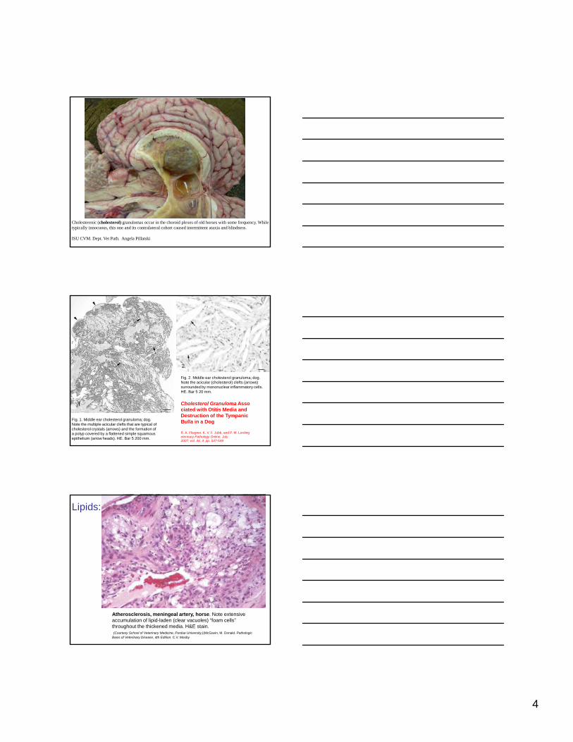
Cholesterenic (cholesterol) granulomas occur in the choroid plexes of old horses with some frequency. These lesions are typically innocuous although they can cause enlargement of lateral ventricles. Nothing about the gross appearance of the masses in the choroid here is diagnostic of this being a cholesterinic granuloma except for the location. Cholesterol crystals occur in sites of old hemorrhage. Inset is cholesterol crystal from a cutaneous mass.
4
Cholesterenic (cholesterol) granulomas occur in the choroid plexes of old horses with some frequency. While typically innocuous, this one and its contralateral cohort caused intermittent ataxia and blindness.
ISU CVM. Dept. Vet Path. Angela Pillatski
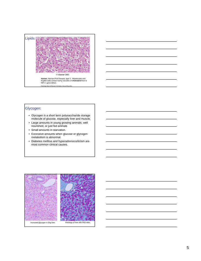
Fig. 1. Middle ear cholesterol granuloma; dog.Note the multiple acicular clefts that are typical ofcholesterol crystals (arrows) and the formation ofa polyp covered by a flattened simple squamousepithelium (arrow heads). HE. Bar 5 200 mm.
Fig. 2. Middle ear cholesterol granuloma; dog.Note the acicular (cholesterol) clefts (arrows) surrounded by mononuclear inflammatory cells. HE. Bar 5 20 mm.
Cholesterol Granuloma Associated with Otitis Media and Destruction of the Tympanic Bulla in a Dog
R. A. Fliegner, K. V. F. Jubb, and P. M. Lordingeterinary Pathology Online, July 2007; vol. 44, 4: pp. 547-549
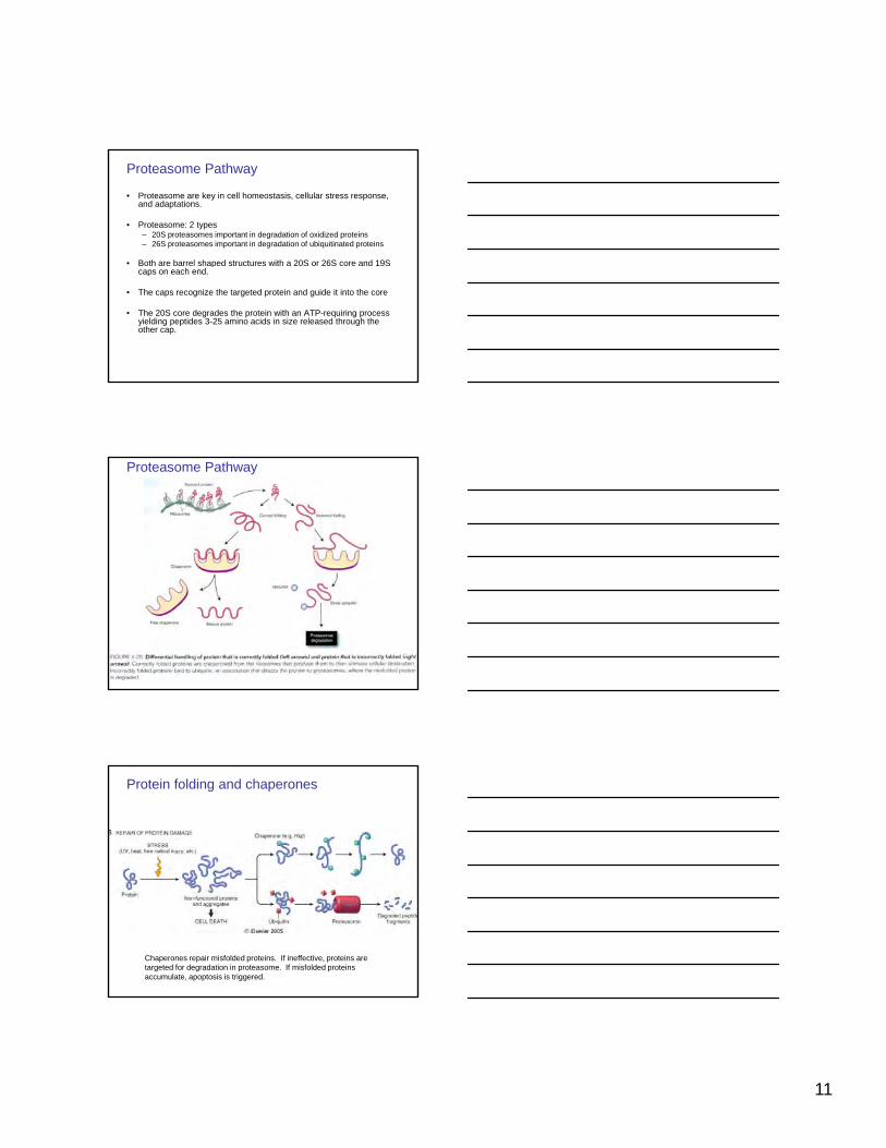
Lipids:
Atherosclerosis, meningeal artery, horse. Note extensive accumulation of lipid-laden (clear vacuoles) “foam cells” throughout the thickened media. H&E stain.(Courtesy School of Veterinary Medicine, Purdue University.)(McGavin, M. Donald. Pathologic
Basis of Veterinary Disease, 4th Edition. C.V. Mosby
5
Lipids:
Human. Nieman-Pick Disease, type 3. Hepatocytes and Kupffer cells contain foamy vacuoles of cholesterol due to NPC-1 gene defect.
Pathologic Basis of Disease, 7th Edition. Elseveir/Saunders
Glycogen:
• Glycogen is a short term polysaccharide storage molecule of glucose, especially liver and muscle.
• Large amounts in young growing animals, well nourished, or just fed animals
• Small amounts in starvationSmall amounts in starvation.
• Excessive amounts when glucose or glycogen metabolism is abnormal.
• Diabetes mellitus and hyperadrenocorticism are most common clinical causes.
Increased glycogen in dog liver. Histology of liver with PAS stain.

6
Glycogen:
Glycogen, liver, dog. A, Ten-percent buffered neutral formalin fixation at 4° C. Glycogen (purplish-red) is uniformly dispersed throughout the cytoplasm of all hepatocytes. Periodic acid–Schiff technique. B, Absolute alcohol (ethanol) fixation at room temperature. The glycogen in each hepatocyte has been pushed to the side
of the cell, so-called polarization of glycogen. Periodic acid–Schiff technique. (A and B, Courtesy Dr. M.D. McGavin,
College of Veterinary Medicine, University of Tennessee.) (McGavin, M. Donald. Pathologic Basis of Veterinary Disease, 4th Edition. C.V. Mosby,
Glycogen:
httpHistology-B408Department of Biological SciencesUniversity of Delaware
://www.udel.edu/biology/Wags/histopage/empage/el/el2.gifMammalian
Normal glycogen rosettes in liver by EM
Glycogen:
• In diabetes glycogen found in hepatocytes (because hepatocytes are highly permeable to glucose in hyperglycemia) and renal proximal tubule cells (due to increased glucose in filtrate) and in B cells of pancreatic islets.
• In glucocorticoid-induced hepatopathy, excessive endogenous or exogenous glucocorticosteroids induce glycogen synthetase. May result in 10x normal amounts of glycogen

7
Glycogen:
Glucocorticoid hepatopathy, liver, dog. A, Extensive accumulation of glycogen in hepatocytes leads to an enlarged and pale brown to beige liver in dogs with glucocorticoid excess from endogenous (Cushing's disease) or exogenous sources. The liver is usually enlarged and the edges rounded. This cut surface would bulge on incision
and not be greasy. B, Note the swollen hepatocytes (arrows) with extensive cytoplasmic vacuolation. H&E stain.
(A, Courtesy Dr. K. Bailey, College of Veterinary Medicine, University of Illinois. B, Courtesy Dr. J. M. Cullen, College of Veterinary Medicine, North Carolina
State University.)(McGavin, M. Donald. Pathologic Basis of Veterinary Disease, 4th Edition. C.V. Mosby,
Dog. Gross. Use histology to help make the MDX.
Slightly pale enlarged liver could be due to: acute cell swelling, glycogen degeneration, or fatty change. Here the patchy vacuolar change is most consistent with glycogen degeneration seen in steroid hepatopathy
Glycogen:
Glycogen metabolism and known enzyme deficiencies (*) leading to glycogen storage diseases.
These may involve one or a few organs or bea few organs or be generalized. Usually liver or skeletal muscle are targets.

8
Glycogen:
Simplified scheme of normal glycogen metabolism and consequences of glycogen storage diseases of hepatic type—Type 1 glucose-6-phosphatase deficiency ( lti i h l i )(resulting in hypoglycemia) and myopathic type (Type V– McArdlels)--resulting in low energy output due to inability of muscle to use glycogen as an energy source derived by glycolysis.
Glycogenosis:
Brahman calf with Type 2 (acid maltase deficiency) glycogenosisFrom D Driemeier
Equine polysaccharide storage myopathy
Equine polysaccharide storage myopathy, semimembranosus muscle, transverse sections, horse. A, Note the increased amount of and irregularly distributed dark-pink staining glycogen. Abnormal aggregates are present both beneath the sarcolemma and within the cytoplasm. Formalin fixation, PAS reaction. B, Severe form. Numerous myofibers contain multiple pale (very light pink)myofibers contain multiple pale (very light pink) subsarcolemmal and intracytoplasmic inclusions of stored polysaccharide. Formalin fixation, H&E stain. C, These inclusions shown in Fig. 15-39, B stain intensely with PAS but are not digested by amylase (not shown) and are characteristic of what is called complex polysaccharide, amylopectin, or polyglucosan. Formalin fixation, PAS reaction. (A, B, and C, Courtesy Dr. B.J. Cooper, College of Veterinary Medicine, Oregon State University.)(McGavin, M. Donald. Pathologic Basis of Veterinary Disease, 4th Edition. C.V. Mosby

9
Equine polysaccharide storage myopathy
•Present in many breeds, especially draft-related
•Clinical signs are variable (recurrent exertional rhabdomyolysis and pelvic limb lameness, et al) and likely due to insufficient energy production in muscles.
•No identified glycolytic or glycogenolytic g y y g y g ydefect identified, unlike other glycogenoses
•Type 2 fibers accumulate excess PAS positive amylase sensitive glycogen
•Ubiquitinated glycogen results in amylase resistant inclusions of glycogen and filamentous protein (amylopectin or polyglucosan or complex polysaccharide)
GYS1 – horse: Subsarcolemmal and amylase sensitive
Comparative Skeletal Muscle Histopathologic and Ultrastructural Features in Two Forms of Polysaccharide Storage Myopathy in HorsesM. E. McCue,A. G. Armién, M. Lucio, J. R. Mickelson,and S. J. ValbergVeterinary Pathology Online, November 2009; vol. 46, 6: pp. 1281-1291
GYS1 + horse: Cytoplasmic and amylase resistant
Protein accumulations:
• Several types occur: droplets, vacuoles, and aggregates with varying causes.
• By EM these may be fibrillar, amorphous, or crystalline
• Defects in protein folding are one cause of protein accumulations and both intracellular and extracellular accumulations (e.g. amyloid).

10
Response to cellular stress:
• Many cellular stress (including heat shock, oxidative damage, infection, toxins) result in damaged misfolded or unfolded (denatured?) proteins accumulating in ER.
• Stress or heat shock response results in production of non-secreted heat shock proteins (e.g. Hsp27 and Hsp90). This response starts in minutes and may last over a day.
• Hsp vary some by organelle and act as chaperones (involved in intracellular p y y g p (protein trafficking, protein folding, assisting with proper protein conformation, and prevention of unwanted protein aggregation)
– Hsp 60, a mitochondrial chaperone– Hsp 27, ubiquitin
• Chaperones , constitutive or induced, rescue misfolded proteins or facilitate degradation involving ubiquitin and proteasomes
• Unfolded protein response is used for ER stress induced by unfolded and misfolded proteins. It can also activate ER capase-12 and cause cell death
RER and Mitochondria:
httpHistology-B408Department of Biological SciencesUniversity of Delaware
://www.udel.edu/biology/Wags/histopage/empage/el/el2.gifMammalian
Normal RER where unfolded protein response is mediated by proteins in and spanning the ER membranes.
Protein folding and chaperones
Chaperones (e.g. Heat shock proteins) protect unfolded or partially folded protein from degradation and guide it to organelle (mitochondrion in this case).
11
Proteasome Pathway
• Proteasome are key in cell homeostasis, cellular stress response, and adaptations.
• Proteasome: 2 types– 20S proteasomes important in degradation of oxidized proteins– 26S proteasomes important in degradation of ubiquitinated proteins
• Both are barrel shaped structures with a 20S or 26S core and 19SBoth are barrel shaped structures with a 20S or 26S core and 19S caps on each end.
• The caps recognize the targeted protein and guide it into the core
• The 20S core degrades the protein with an ATP-requiring process yielding peptides 3-25 amino acids in size released through the other cap.
Proteasome Pathway
Protein folding and chaperones
Chaperones repair misfolded proteins. If ineffective, proteins are targeted for degradation in proteasome. If misfolded proteins accumulate, apoptosis is triggered.

12
Protein folding and chaperones
Protein folding and chaperones
Ubiquitin
• Ubs (small molecules that can be in chains) flag proteins to be degraded ubiquitination
• A cascade of enzymes: Ub activating enzyme (E1) binds Ub and transfers it to Ub conjugating enzyme (E2) which bind to Ub ligating enzymes and add an amino group of lysine
• Multiple cycles of this take place polyubiquitin chainp y p p y q
• De-ubiquitinating enzymes (DUBs) reverse the process.
• Ubiquitination occurs also in normal cell processes like membrane budding, vesicle transport, and protein sorting
• Some protein modifications protect from ubiquitination. E.g. p53 (tumor suppressor protein) is phosphorylated in response to DNA damage and is protected from Ub-mediated degradation

13
Ubiquitin
• Ubiquitination defects may play roles in neurodegenerative disease. E.g. parkin is a ubiquitin ligase and is implicated in Parkinson’s disease.
• Ubiquitination may be involved in neoplasia. E.g. q y p ghuman papilloma viruses of cervical cancer produce E6 protein that inactivates p53
• Impaired ubiquitination may play a role in degenerative changes of aging
• Ub plays role in gene expression, e.g. NFkB inhibitor degradation.
Ubiquitin-Proteasome Pathway
Rubin’s Pathology: Clinicopathologic foundations of medicine. Rubin, R and Strayer DS, Wolters Kluwer/Lippincott Williams and Wilkins
Protein accumulations:
• Hyaline. Any material that is homogeneous, eosinophilic, and amorphous or glassy with H&E (hyaline technically means glassy and transparent). Nonspecific for several changes.
• Intracellular examples of hyaline
– Resorption droplets of protein (“hyaline droplet degeneration” in kidneys) due to proteinuria
– Intestinal epithelium of neonatal pigs with ingested colostrum– Russell bodies in plasma cells– Alcoholic hyalin (Mallory bodies) in human liver with alcohol
damage composed of ubiquitinated keratin filaments
14
Cell hyaline droplets:
Renal tubular epithelium protein resorption droplets—hyaline droplet degeneration.
Cell hyaline droplets:
Russell body in human plasma cell
Noah’s ArkiveEd Friedlander, M.D., [email protected]
Intestinal epithelium of a baby pig with absorbed colostrum
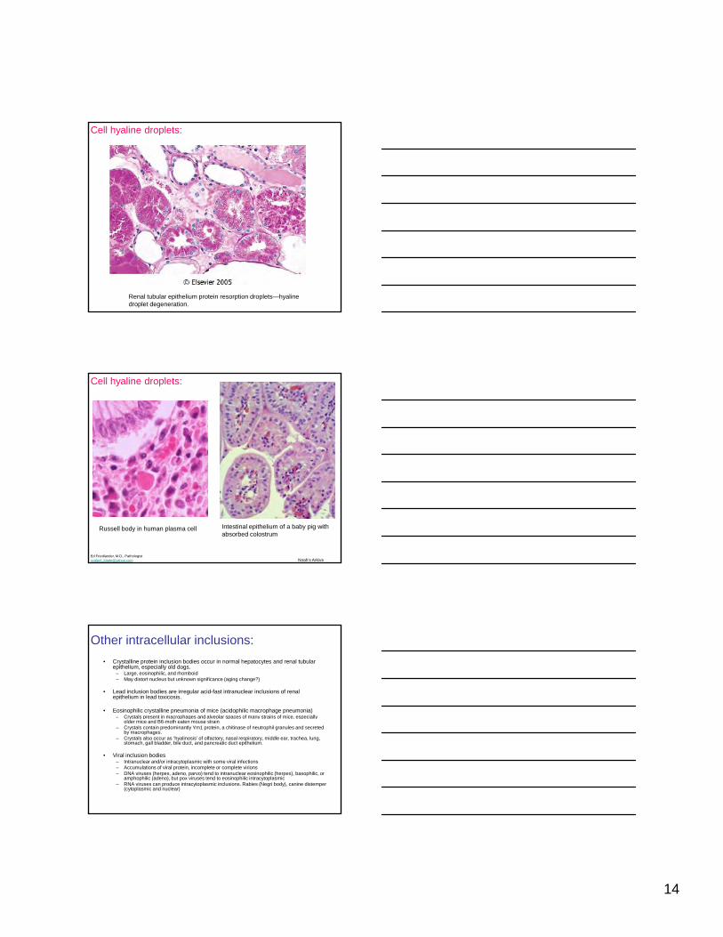
Other intracellular inclusions:
• Crystalline protein inclusion bodies occur in normal hepatocytes and renal tubular epithelium, especially old dogs.
– Large, eosinophilic, and rhomboid– May distort nucleus but unknown significance (aging change?)
• Lead inclusion bodies are irregular acid-fast intranuclear inclusions of renal epithelium in lead toxicosis.
• Eosinophilic crystalline pneumonia of mice (acidophilic macrophage pneumonia)– Crystals present in macrophages and alveolar spaces of many strains of mice, especially y p p g p y p y
older mice and B6-moth eaten mouse strain– Crystals contain predominantly Ym1 protein, a chitinase of neutrophil granules and secreted
by macrophages. – Crystals also occur as ‘hyalinosis’ of olfactory, nasal respiratory, middle ear, trachea, lung,
stomach, gall bladder, bile duct, and pancreatic duct epithelium.
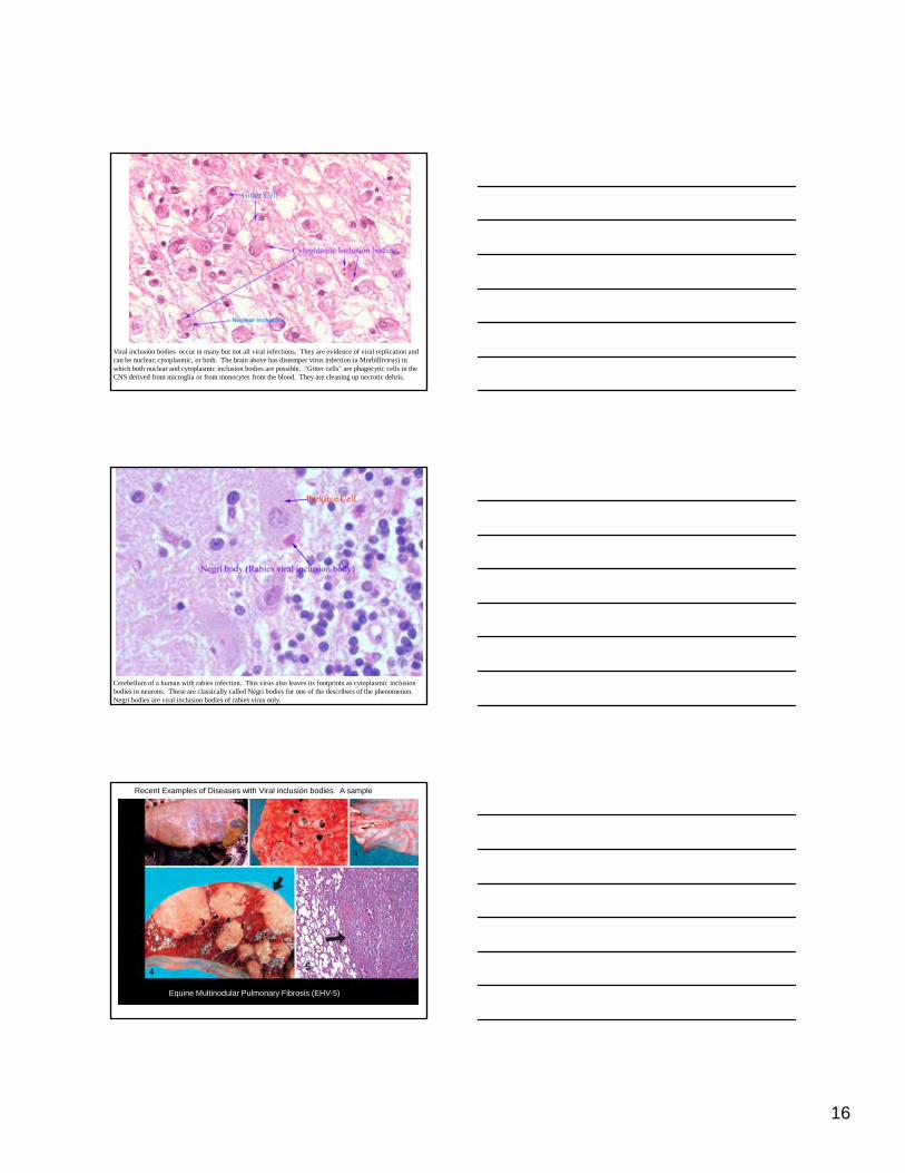
• Viral inclusion bodies– Intranuclear and/or intracytoplasmic with some viral infections– Accumulations of viral protein, incomplete or complete virions– DNA viruses (herpes, adeno, parvo) tend to intranuclear eosinophilic (herpes), basophilic, or
amphophilic (adeno), but pox viruses tend to eosinophilic intracytoplasmic– RNA viruses can produce intracytoplasmic inclusions. Rabies (Negri body), canine distemper
(cytoplasmic and nuclear)

15
Eosinophilic crystalline pneumonia of mice
Multinucleate cell with acicular crystals
Pulmonary adenoma surrounded by acidophilic macrophages
Crystals in bronchioles
Vet Pathol 43:682-688 (2006)© 2006 American College of Veterinary PathologistsEosinophilic Crystalline Pneumonia as a Major Cause of Death in 129S4/SvJae Mice M. J. Hoenerhoff, M. F. Starost and J. M. Ward
Partially birefringent crystals with polarized light
Luna stain of extracellular crystals
IHC with antibodies against Ym1 protein, intracellular and extracellular
Cell droplets and inclusion bodies:
Cell droplets and inclusion bodies. A, Resorption droplets, proteinuria, kidney, dog. The cytoplasm of the proximal tubule epithelial cells are filled with eosinophilic homogeneous droplets—protein that has been resorbed by the cells from the glomerular filtrate. H&E stain. B, Crystalloids, kidney, dog. Note the elongated crystals in the nuclei of the hepatocytes. (A and C, Courtesy Dr. M.D. McGavin, College of Veterinary Medicine, University of Tennessee. B, Courtesy Dr. D.D. Harrington, College of Veterinary Medicine, Purdue University; and Noah's Arkive, College of Veterinary Medicine, The University of Georgia.D, Courtesy Dr. W. Crowell, College of Veterinary Medicine, The University of Georgia; and Noah's Arkive, College of Veterinary Medicine, The University of Georgia. Inset, Courtesy Dr. W. Crowell, College of Veterinary Medicine, The University of Georgia; and Noah's Arkive, College of Veterinary Medicine, The University of
Georgia.) (McGavin, M. Donald. Pathologic Basis of Veterinary Disease, 4th Edition. C.V. Mosby,
Cell droplets and inclusion bodies:
Cell droplets and inclusion bodies. C, Viral inclusion bodies, brain, dog. Note the intranuclear eosinophilic inclusion bodies in glial cells. H&E stain. D, Lead inclusion bodies, kidney, dog. The inclusions in the nuclei of these renal tubular epithelial cells are difficult to see with an H&E stain (arrows). Inset: An acid-fast stain is useful in identifying lead inclusions, which stain red. Ziehl Neelsen stain. (A and C, Courtesy Dr. M.D. McGavin, College of Veterinary Medicine, University of Tennessee. B, Courtesy Dr. D.D. Harrington, College of Veterinary Medicine, Purdue University; and Noah's Arkive, College of Veterinary Medicine, The University of Georgia.D, Courtesy Dr. W. Crowell, College of Veterinary Medicine, The University of Georgia; and Noah's Arkive, College of Veterinary Medicine, The University of Georgia. Inset, Courtesy Dr. W. Crowell, College of Veterinary Medicine, The University of Georgia; and Noah's Arkive, College of Veterinary Medicine, The University of Georgia.) (McGavin, M. Donald. Pathologic Basis of Veterinary Disease, 4th Edition. C.V.
Mosby
16
Viral inclusion bodies occur in many but not all viral infections. They are evidence of viral replication and can be nuclear, cytoplasmic, or both. The brain above has distemper virus infection (a Morbillivirus) in which both nuclear and cytoplasmic inclusion bodies are possible. "Gitter cells" are phagocytic cells in the CNS derived from microglia or from monocytes from the blood. They are cleaning up necrotic debris.
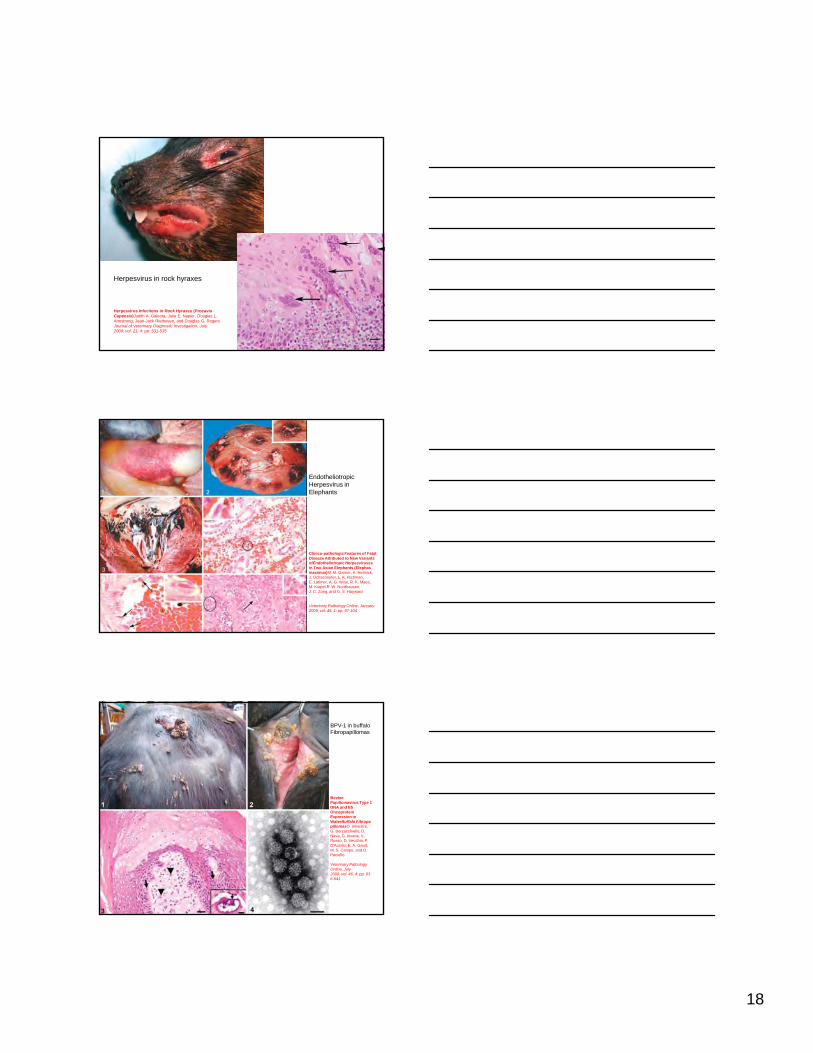
Cerebellum of a human with rabies infection. This virus also leaves its footprints as cytoplasmic inclusion bodies in neurons. These are classically called Negri bodies for one of the describers of the phenomenon. Negri bodies are viral inclusion bodies of rabies virus only.
Recent Examples of Diseases with Viral inclusion bodies. A sample
Equine Multinodular Pulmonary Fibrosis (EHV-5))

17
Fig. 6. EMPF histopathology; horse No. 6. In most cases, there is preservation of an ‘‘alveolar-like’’architecture, which is lined by cuboidal epithelial cells. HE. Bar 5 100 mm.Fig. 7. EMPF histopathology; horse No. 6. Higher magnification of interstitial fibrosis with inflammatory cellswithin lumen of the airspace; the inflammatory cells are primarily neutrophils and macrophages. HE. Bar 5 50 mm.Fig. 8. EMPF histopathology; horse No. 6. Histochemical staining of interstitial fibrosis. The fibrosis (blue) in most animals is well organized around the airspaces. Masson trichrome. Bar 5 50 mm.Fig. 9. EMPF histopathology; horse No. 22. The fibrosis can be arranged in unorganized bands, withoutappreciable preservation of ‘‘alveolar-like’’ airspaces. HE. Bar 5 50 mm.Fig. 10. EMPF histopathology; horse No. 6. Large eosinophilic intranuclear viral inclusion body (arrow) ina large macrophage within the inflammation in the airspaces of the nodular lesions. HE. Bar 5 20 mm.Fig. 11. EMPF lung imprint cytology; horse No. 4. Vacuolated macrophages and neutrophils are intermixed.One macrophage (left) is markedly enlarged and has a single large eosinophilic intranuclear inclusion thatmarginates 2 nucleoli. Wright-Giemsa stain. Bar 5 10 mm.
Equine Multinodular Pulmonary Fibrosis: A Newly Recognized Herpesvirus-Associated Fibrotic Lung DiseaseK. J. Williams,R. Maes, F. Del Piero, A. Lim, A. Wise, D. C. Bolin, J. Caswell,C. Jackson, N. E. Robinson, F. Derksen, M. A. Scott, B. D. Uhal, X. Li,S. A. Youssef, and S. R. BolinVet Pathol, November 2007; vol. 44, 6: pp. 849-862
Fig. 1. Skin of the eyelids, lips, forelimbs, and digits; Western barred bandicoot. Demonstrates nodular and irregular papillomatous thickening.
Cutaneous Papillomatosis and Carcinomatosis in the Western BarredBandicoot (Perameles bougainville)L. Woolford, A. J. O'Hara, M. D. Bennett, M. Slaven,R. Swan, J. A. Friend, A. Ducki, C. Sims, S. Hill, P. K. Nicholls, and K. S. Warren. Veterinary Pathology Online, January 2008; vol. 45, 1: pp. 95-103
Fatal alpha herpesvirus in Alaskan rabbits
An Outbreak of Fatal Herpesvirus Infection in Domestic Rabbits in Alaska. L. Jin, B. A. Valentine, R. J. Baker, C. V. Löhr, R. F. Gerlach, R. J. Bildfell, and M. Moerdyk-Schauwecker
Veterinary Pathology Online, May 2008; vol. 45, 3: pp. 369-374
18
Herpesvirus in rock hyraxes
Herpesvirus Infections in Rock Hyraxes (ProcaviaCapensis)Judith A. Galeota, Julia E. Napier, Douglas L. Armstrong, Jean-Jack Riethoven, and Douglas G. RogersJournal of Veterinary Diagnostic Investigation, July 2009; vol. 21, 4: pp. 531-535
Endotheliotropic Herpesvirus in Elephants
Clinico-pathologic Features of Fatal Disease Attributed to New Variants ofEndotheliotropic Herpesviruses in Two Asian Elephants (Elephas maximus)M. M. Garner, K. Helmick,J. Ochsenreiter, L. K. Richman,E. Latimer, A. G. Wise, R. K. Maes,M. Kiupel,R. W. Nordhausen,J. C. Zong, and G. S. Hayward
Veterinary Pathology Online, January 2009; vol. 46, 1: pp. 97-104
BPV-1 in buffaloFibropapillomas
Bovine Papillomavirus Type 1 DNA and E5DNA and E5 Oncoprotein Expression in WaterBuffalo FibropapillomasO. Silvestre,G. Borzacchiello, D. Nava, G. Iovane, V. Russo, D. Vecchio, F. D'Ausilio, E. A. Gault,M. S. Campo, and O. Paciello
Veterinary Pathology Online, July 2009; vol. 46, 4: pp. 636-641

19
Systemic poxviral disease in red and grey squirrels
Cutaneous and Systemic Poxviral Disease in Red (Tamiasciurus hudsonicus)and Gray (Sciurus carolinensis) SquirrelsD. S. Bangari,M. A. Miller, G. W. Stevenson, H. L. Thacker, A. Sharma, and S. K. Mittal
Veterinary Pathology Online, July 2009; vol. 46, 4: pp. 667-672
Papillomatosis in green sea turtles due to:chelonid fibropapilloma-associated herpesvirus
Postmortem Diagnostic Investigation of Disease in Free-Ranging Marine Turtle Populations: A Review of Common Pathologic Findings and ProtocolsMark Flint,Janet C. Patterson-Kane, Colin J. Limpus,Thierry M. Work, David Blair, and Paul C. Mills
Journal of Veterinary Diagnostic Investigation, November 2009; vol. 21, 6: pp. 733-759
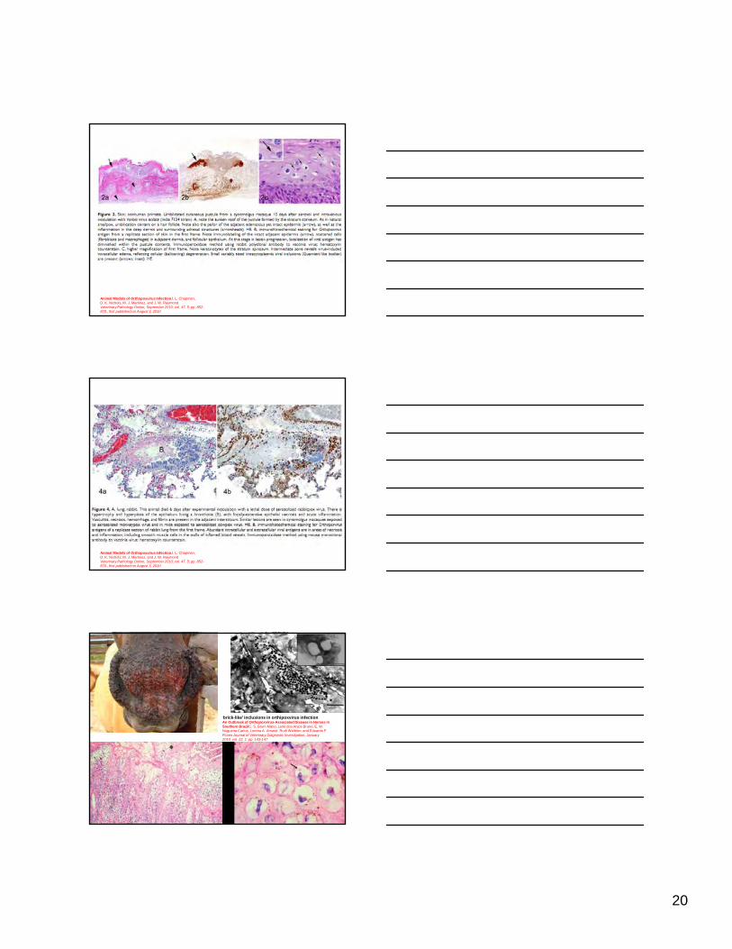
Animal Models of Orthopoxvirus InfectionJ. L. Chapman,D. K. Nichols, M. J. Martinez, and J. W. RaymondVeterinary Pathology Online, September 2010; vol. 47, 5: pp. 852-870., first published on August 3, 2010
20
Animal Models of Orthopoxvirus InfectionJ. L. Chapman,D. K. Nichols, M. J. Martinez, and J. W. RaymondVeterinary Pathology Online, September 2010; vol. 47, 5: pp. 852-870., first published on August 3, 2010
Animal Models of Orthopoxvirus InfectionJ. L. Chapman,D. K. Nichols, M. J. Martinez, and J. W. RaymondVeterinary Pathology Online, September 2010; vol. 47, 5: pp. 852-870., first published on August 3, 2010
‘brick-like’ inclusions in orthipoxvirus infectionAn Outbreak of Orthopoxvirus-Associated Disease in Horses in Southern BrazilC. S. Brum Mário, Leite dos Anjos Bruno, E. W. Nogueira Carlos, Lorena A. Amaral, Rudi Weiblen, and Eduardo F. Flores Journal of Veterinary Diagnostic Investigation, January 2010; vol. 22, 1: pp. 143-147

21
Systemic Iridovirus Infection in the Banggai Cardinalfish (Pterapogon KauderniKoumans 1933)E. Scott Weber III, Thomas B. Waltzek, Devon A. Young, Erica L. Twitchell, Amy E. Gates, Alejandro Vagelli,Guillermo R. Risatti, Ronald P. Hedrick, and Frasca Salvatore, Jr. Journal of Veterinary Diagnostic Investigation, May 2009; vol. 21, 3: pp. 306-320
Extracellular accumulations:
• Extracellular hyaline substances– Hyaline casts in renal tubules in proteinuria– Serum or plasma in blood vessels– Plasma proteins in vessel walls– Old scarsOld scars– Thickened basement membranes– Hyaline membranes of alveoli– Hyaline microthrombi– Amyloid– Fibrinoid change
Kidney from a dog with glomerular disease in which protein has leaked into the filtrate. The reabsorption of the protein into the tubules results in protein droplet accumulation seen as hyaline droplets. The protein within the tubular lumen is also "hyalin." Remember hyalin is a generic term for a staining feature that correlates with specific substances.

22
Amyloid: a chemically diverse group of extracellular proteins histologically and ultrastructurally similar
• All are Congo red positive (Congophilic)• Group of glycoproteins that are beta-pleated sheets and
form 10 nm fibrils
Amyloid: Classification. Systemic
• Reactive systemic: the most common in animals– Secondary to chronic antigen stimulation: chronic infections, neoplasia
– Deposits in kidney (glomerulus, medulla, basement membranes), liver (space of Disse), spleen (germinal centers), lymph nodes (germinal centers), adrenal cortex
– Glomerular deposits are most important due to causing proteinuria
Protein is AA from serum amyloid associated an acute phase protein– Protein is AA from serum amyloid associated, an acute phase protein secreted from liver
• Primary amyloid: most common form in humans– Due to immunocyte dyscrasias: plasma cell tumors, myelomas
– Deposits in same organs as reactive systemic with same consequences
– Protein is AL immunoglobulin light chains
• Hereditary Amyloidosis in humans has a variety of forms
Amyloid: Classification. Local
• Islet Amyloid– Pancreatic islets of cats, raccoons, nonhuman primates, and humans– Leads to beta and alpha cell degeneration– Associated with mature onset diabetes– Protein is IAPP islet amyloid polypeptide, a hormone
• Nasal septum and turbinates of horsesNasal septum and turbinates of horses• Dermis and subcutis of horses• Plasma cell tumors• Amyloid odontogenic tumors of cats• Endocrine tumors: thyroid c cell tumors• Meningeal and cerebral vessels of old dogs
– Similar to beta-amyloid found in Alzheimer's disease
• Prion diseases (misfolded PrPsc)

23
Amyloid: Pathogenesis.
Amyloid:
Amyloidosis, kidney, cross section, dog. Note the blue-black foci, which are glomeruli-containing amyloid stained by Lugol's iodine.
(Courtesy Dr. M.D. McGavin, College of Veterinary Medicine, University of Tennessee.)(McGavin, M. Donald. Pathologic Basis of Veterinary Disease, 4th Edition. C.V. Mosby,
Amyloid:
Amyloidosis, kidney, dog. A, The renal glomerulus contains large amounts of pale homogeneous eosinophilic material, which is amyloid. H&E stain. B, The amyloid in the glomeruli stains orange. Congo red stain. (A and B, Courtesy Dr. M.D. McGavin, College of Veterinary Medicine, University of Tennessee.)(McGavin, M. Donald. Pathologic Basis of Veterinary Disease, 4th Edition. C.V. Mosby,
24
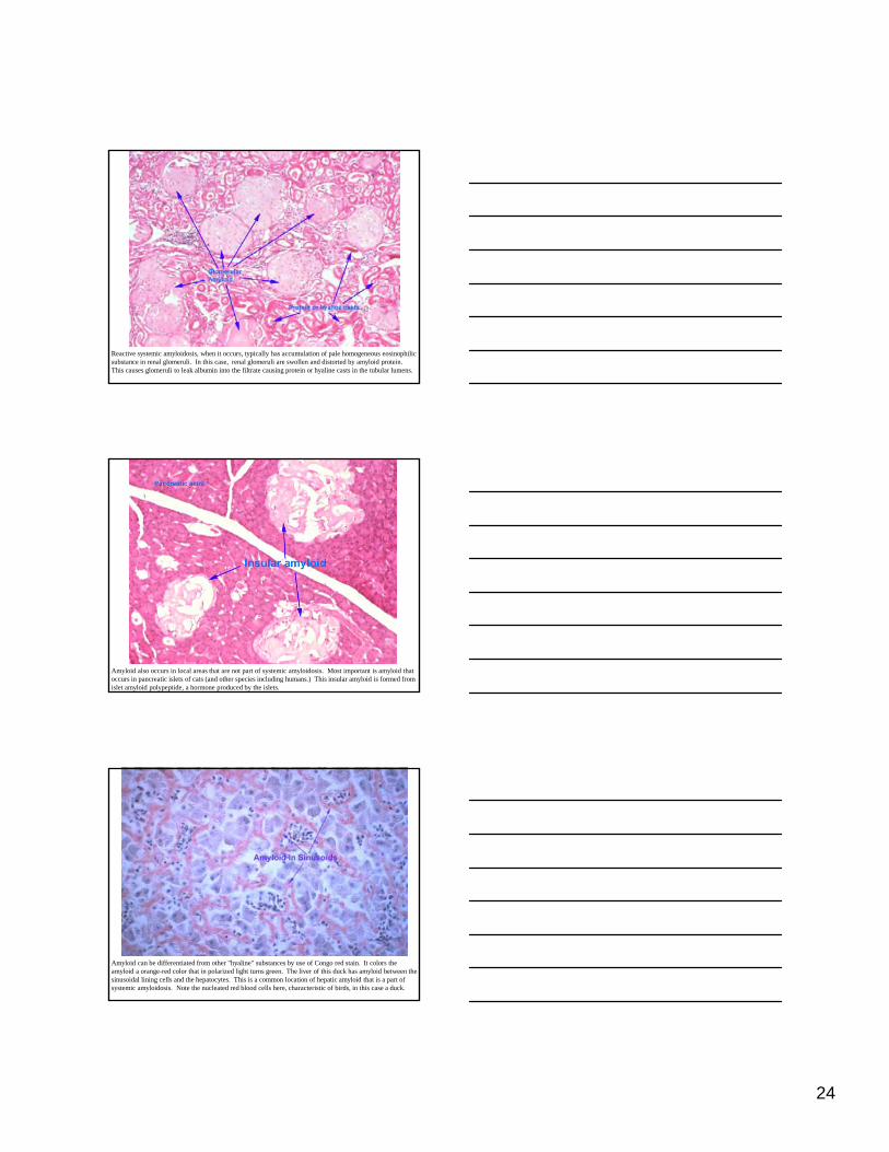
Reactive systemic amyloidosis, when it occurs, typically has accumulation of pale homogeneous eosinophilic substance in renal glomeruli. In this case, renal glomeruli are swollen and distorted by amyloid protein. This causes glomeruli to leak albumin into the filtrate causing protein or hyaline casts in the tubular lumens.
Amyloid also occurs in local areas that are not part of systemic amyloidosis. Most important is amyloid that occurs in pancreatic islets of cats (and other species including humans.) This insular amyloid is formed from islet amyloid polypeptide, a hormone produced by the islets.
Amyloid can be differentiated from other "hyaline" substances by use of Congo red stain. It colors the amyloid a orange-red color that in polarized light turns green. The liver of this duck has amyloid between the sinusoidal lining cells and the hepatocytes. This is a common location of hepatic amyloid that is a part of systemic amyloidosis. Note the nucleated red blood cells here, characteristic of birds, in this case a duck.

25
Amyloid: other examples
Amyloid producing odontogenic tumor, cat. Note large lakes of Congophilic material surrounded by odontogenic epithelium. The exact nature of the amyloid in this tumor has not been confirmed but there is some evidence in humans and animals that it derives from keratin elements of epithelial cells.
Amyloid: other examples
Horse with Recurrent uveitis
Amyloid: other examples
Horse with Recurrent uveitisAmyloid-like protein on ciliary body neuroepithelium

26
Amyloid: other examples
Horse with Recurrent uveitisAmyloid-like protein on ciliary body neuroepithelium with polarized light
Fibrinoid Change:
• Fibrinoid degeneration or necrosis (misnomer as the change is extracellular)– Deposition of immunoglobulin, complement, plasma
proteins (including fibrinogen) in blood vessel walls– Not seen grossly, but may result in thrombosis and
hemorrhageg– Microscopically has intense red to pink segmentally or
circumferential staining of tunica intima and/or media– May be accompanied by cellular and nuclear debris
and inflammatory cells– Immune mediated diseases or direct injury to
endothelial cells, basement membranes, and muscles by viruses or toxins
Fibrinoid change.
“Beagle pain syndrome” arteritis and periarteritis.
27
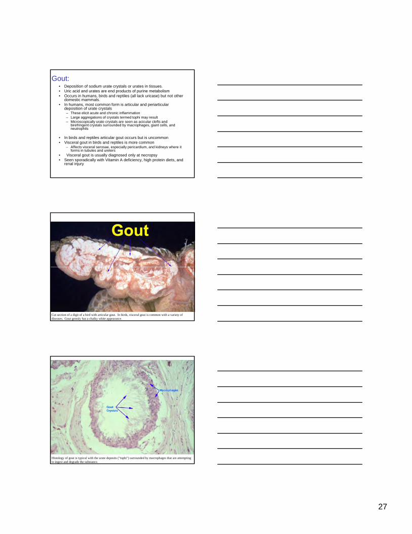
Gout:• Deposition of sodium urate crystals or urates in tissues.• Uric acid and urates are end products of purine metabolism• Occurs in humans, birds and reptiles (all lack uricase) but not other
domestic mammals.• In humans, most common form is articular and periarticular
deposition of urate crystals– These elicit acute and chronic inflammation– Large aggregations of crystals termed tophi may result– Microscopically urate crystals are seen as acicular clefts and
birefringent crystals surrounded by macrophages, giant cells, and neutrophils
• In birds and reptiles articular gout occurs but is uncommon• Visceral gout in birds and reptiles is more common
– Affects visceral serosae, especially pericardium, and kidneys where it forms in tubules and ureters
• Visceral gout is usually diagnosed only at necropsy• Seen sporadically with Vitamin A deficiency, high protein diets, and
renal injury
Cut section of a digit of a bird with articular gout. In birds, visceral gout is common with a variety of diseases. Gout grossly has a chalky white appearance.
Histology of gout is typical with the urate deposits ("tophi") surrounded by macrophages that are attempting to ingest and degrade the substance.

28
Visceral Gout
Visceral gout pericardium and cavity, hawk and turtle
Renal gout: boa
Extracellular accumulations:Pseudogout– Deposits of calcium pyrophosphate crystals
– Humans. Autosomal dominant trait. Chalky white deposits in joints.
– Occurs rarely in dogs with unknown pathogenesis
Calcium pyrophosphate crystals in human synovial fluid
Melamine and calcium oxylate containing crystals
Vet Pathol 45:417-426 (2008)© 2008 American College of Veterinary PathologistsENVIRONMENTAL PATHOLOGYCharacterization of Melamine-containing and Calcium Oxalate Crystals in Three Dogs with Suspected Pet Food–induced NephrotoxicosisM. E. Thompson, M. R. Lewin-Smith, V. F. Kalasinsky, K. M. Pizzolato, M. L. Fleetwood, M. R. McElhaney and T. O. Johnson
Melamine containing crystals in kidney with
Oil red O 72 hrs.
Alizarin red non staining
Von Kossa non staining
29
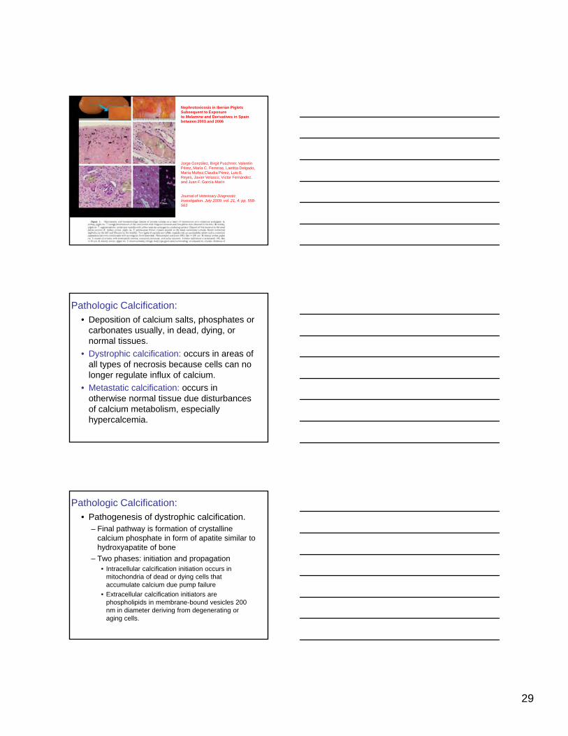
Nephrotoxicosis in Iberian Piglets Subsequent to Exposure to Melamine and Derivatives in Spain between 2003 and 2006
Jorge González, Birgit Puschner, Valentín Pérez, María C. Ferreras, Laetitia Delgado,María Muñoz,Claudia Pérez, Luis E. Reyes, Javier Velasco, Víctor Fernández,and Juan F. García-Marín
Journal of Veterinary Diagnostic Investigation, July 2009; vol. 21, 4: pp. 558-563
Pathologic Calcification:• Deposition of calcium salts, phosphates or
carbonates usually, in dead, dying, or normal tissues.
• Dystrophic calcification: occurs in areas of all types of necrosis because cells can no yplonger regulate influx of calcium.
• Metastatic calcification: occurs in otherwise normal tissue due disturbances of calcium metabolism, especially hypercalcemia.
Pathologic Calcification:• Pathogenesis of dystrophic calcification.
– Final pathway is formation of crystalline calcium phosphate in form of apatite similar to hydroxyapatite of bone
– Two phases: initiation and propagation• Intracellular calcification initiation occurs in
mitochondria of dead or dying cells that accumulate calcium due pump failure
• Extracellular calcification initiators are phospholipids in membrane-bound vesicles 200 nm in diameter deriving from degenerating or aging cells.

30
Pathologic Calcification:• Extracellular initiation concentrates calcium in
vesicles by membrane-facilitated calcification.– Calcium binds to phospholipids in vesicle membrane– Membrane phosphatases generate phosphate groups
the bind to calcium– Repeating cycles of calcium and phosphate binding
which increase local accumulations especially nearwhich increase local accumulations especially near the membrane
– Structural changes of the calcium and phosphate groups generating microcrystals that propagate and perforate membranes.
– Propagation depends on concentration of calcium and phosphate ions, presence of inhibitors, proteins in extracellular space such as matrix proteins.
Pathologic Calcification:• Dystrophic calcification common sites.
– Myocardium– TB granulomas– Dead parasites– Skin
• Calcinosis circumscripta seen as focal mineral and macrophagic inflammation in sites of chronic trauma or inmacrophagic inflammation in sites of chronic trauma or in large breed dogs
• Calcinosis cutis occurs in more diffuse areas of dermis of dogs with hyperadrenocorticism
• Dystrophic calcification is relatively permanent and may be harmless or cause mechanical interference (e.g. heart valves)
Pathologic Calcification:• Metastatic calcification is widespread and
usually due to hypercalcemia with mineral being deposited in normal tissues.
• Common locations are:– Lung, stomach, kidney, blood vessels
Sites where acid is lost and an internal alkaline– Sites where acid is lost and an internal alkaline compartment prone to calcium salt precipitation
• Significance as indicator of hypercalcemia– Extracellular calcium deposits may cause no harm or
lead to severe organ injury, e.g. nephrocalcinosis

31
Pathologic Calcification:• Common causes of hypercalcemia in animals
– Renal failure which results in phosphorus retention, induction of renal secondary hyperparathyroidism and hypercalcemia in some species. Uremia associated deposits in gastric mucosa, kidney, and alveolar septa
– Vitamin D toxicosis. Ingestion of calcinogenic plants in herbivores (Cestrum diurnum) usually results in aorta, heart, and lung deposits. Rodenticides containing cholecalciferol in dogs and cats result in deposits in intestinal mucosa, vessel walls, lung, and kidneys
Pathologic Calcification:• Common causes of hypercalcemia in
animals– Parathormone (PTH) and PTH-related protein
cause bone resorption and hypercalcemia• PTH from functional parathyroid tumors (rare)• Hypercalcemia of malignancy due to elevated ype ca ce a o a g a cy due o e e a ed
levels of PTH-related protein. Seen most commonly in dogs with lymphoma and with anal sac gland carcinomas.
– Bone destruction from primary or metastatic neoplasms leads to accelerated bone turnover and hypercalcemia
Pathologic Calcification: Morphology• Lesions have similar appearance whether
dystrophic or metastatic
• Gross lesions are white and gritty
• Microscopic lesions.H&E basophilic amorphous granules or– H&E basophilic, amorphous granules or clumps, intracellular or extracellular
– Special stains • von Kossa marks phosphate and carbonate groups
and is not specific for calcium but calcium is usually present
• Alizarin red for calcium

32
This is the heart from a horse with monensin toxicosis. This substance in high enough levels in some species causes coagulation necrosis of the cardiac myocytes. These necrotic myocytes become calcified as a consequence of the necrosis, which is dystrophic calcification.
You are looking at peripelvic fat from a fat cow. Fat in this area can be damaged by dystocias (difficult birth). Damaged fat becomes saponified and calcified, a process known as enzymatic necrosis of fat or fat necrosis.
These are lymph nodes from a cow with tuberculosis. These are granulomas with caseous necrosis and calcification. The calcification is especially dense here. This calcification is why tubercular lesions often show up on radiographs.

33
In metastatic calcification, here due to vitamin D toxicosis in a rabbit, blood vessel walls can also be mineralized. Aorta is white and mottled here due to calcification.
Calcification in the aorta of an cow with Vitamin D toxicosis. This can be dystrophic as well as metastatic calcification.
Mineral in the alveolar septa of a dog with chronic renal failure is stained slightly purple but is hard to discern. A special stain helps show that this material is mineral in the following slide.

34
Mineral in the alveolar septa of a dog with chronic renal failure is stained black by Von Kossa stain, a stain that is used to detect subtle areas of mineral in tissues.
In metastatic calcification, basement membranes of the renal tubules, as seen here, are a common place for calcium to occur.
Calcinosis cutis occurs in dogs with hyperadrenocorticism, either endogenous or exogenous. Pathogenesis is somewhat unclear, with some terming it idiopathic calcification and other regarding it as dystrophic.
Noah’s Arkive
35
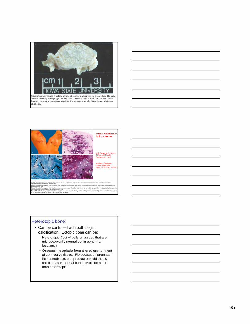
Calcinosis circumscripta is nodular accumulation of calcium salts in the skin of dogs. The salts are surrounded by macrophages histologically. The white color is due to the calcium. These lesions occur most often at pressure points of large dogs, especially Great Danes and German shepherds.
Arterial Calcificationin Race Horses
L. G. Arroyo, M. A. Hayes,J. DeLay, C. Rao, B. Duncan, and L. Viel
Fig. 1. Pulmonary artery trunk and main branches; 2-year-old Thoroughbred horse. Severe calcification of the main branches showing thickening and deformation of the tunica intima (arrowheads). Fig. 2. Affected pulmonary artery branch; Horse. Extensive areas of calcification (dark purple) within the tunica media of the arterial wall. Arrow indicates the endothelium. HE (10x). Fig. 3. Affected pulmonary artery branch; Horse. Fragmented, thinned, and calcified elastic fibers (arrowheads), surrounded by a disorganized lattice network of collagen fibers bundles (light blue). Elastin-trichrome (100x). Fig. 4. Affected pulmonary artery branch; Horse. Large round-to-oval cells with clear cytoplasm and large nuclei (arrowheads), occasional multinucleation, seen at the periphery of the calcified zones. Ca = calcified area. HE (40x). .
Veterinary Pathology Online, September 2008; vol. 45, 5: pp. 617-625
Heterotopic bone: • Can be confused with pathologic
calcification. Ectopic bone can be:– Heterotopic (foci of cells or tissues that are
microscopically normal but in abnormal locations)
– Osseous metaplasia from altered environment of connective tissue. Fibroblasts differentiate into osteoblasts that product osteoid that is calcified as in normal bone. More common than heterotopic

36
Ectopic bone:
Ectopic bone, lung, dog, A nodule of mature bone in the connective tissue of the lung. H&E stain. (Courtesy Dr. M.D. McGavin, College of Veterinary Medicine, University of Tennessee.) (McGavin, M. Donald. Pathologic Basis of Veterinary Disease, 4th Edition. C.V. Mosby,
Pigments:
• A heterogeneous and often unrelated group of substances imparting an unusual color systemically or locally
• Recognition of the change may, or may not be, clinically or pathologically , y p g yimportant
• Broadly classified into exogenous and endogenous
Carbon:Exogenous pigments
Anthracosis, lung, aged dog. A, The fine black foci are peribronchiolar deposits of carbon. The animal was exsanguinated at euthanasia to remove the blood from the lung to render the carbon deposits more visible. B, Carbon (black) inhaled into the alveoli has been phagocytosed by macrophages and transported to the peribronchial region. H&E stain. (A and B, Courtesy Dr. M.D. McGavin, College of Veterinary Medicine, University of Tennessee.) (McGavin, M. Donald. Pathologic Basis of Veterinary Disease, 4th Edition. C.V. Mosby,

37
When carbon particles (e.g. from cigarette smoke or air pollution) are inhaled into the lung, they are ingested by macrophages that aggregate together and can be seen grossly as small black foci, termed anthracosis or anthracotic pigment. The carbon is persistent in the macrophages as it is not easily degraded, but by itself is not particularly pathogenic. Anthracosis is only one type of pneumoconiosis.
Carotenoid pigments.
Fat soluble lipochrome pigments from plants including beta-carotene, a Vitamin A precursor
Exogenous pigments
Carotenosis, kidney and the perirenal fat, Jersey ox. Accumulation of carotenoids in the adipocytes has colored the fat yellow to dark yellow. (Courtesy Dr. M.D. McGavin, College of Veterinary Medicine, University of Tennessee.(McGavin, M. Donald. Pathologic Basis of Veterinary Disease, 4th Edition. C.V. Mosby
Carotenoid
Horse. Epicardial and mesenteric fat is bright yellow due to this pigment.

38
Exogenous pigments
Tetracycline staining, teeth, young dog. The teeth of this dog have been stained yellow by the tetracycline ingested during their development. (Courtesy Dr. M.D. McGavin, College of Veterinary Medicine, University of Tennessee.)(McGavin, M. Donald. Pathologic Basis of Veterinary Disease, 4th Edition. C.V. Mosby,
Tetracycline staining of decidual teeth only.
Noah’s Arkive
Exogenous pigments
Liver from a cow infected with liver flukes. These flukes (seen at the left being held on a rubber glove) migrate through the liver and cause fibrosis and leave black parasite pigment behind. This is a type of hematin and is sometimes called "fluke exhaust.”
Figure 2-84C: Liver from a cow infected with liver flukes. These flukes leave black parasite pigment behind. This is a type of hematin and is sometimes called "fluke exhaust."

39
Pigments: Endogenous• Melanin
– Normally present in skin, retina, iris– Also normal in pia-arachnoid os black animals
(e.g. Suffolk sheep) and oral mucosa of some breeds (e.g. Chow and Jersey cattle)
– Melanin is secreted by melanocytesMelanin is secreted by melanocytes• Dendritic cells in basal layer of skin that transfer
melanosomes to keratinocytes through dendritic processes.
• Melanin forms a cap (microparasol) over the nucleus and protects against UV light
• Melanin formed by oxidation of tyrosine, which requires tyrosine (a copper containing molecule)
Pigments: Endogenous• Melanin
– Copper deficiency causes faded color– Albinism due to defect in tyrosinase
(melanocytes are normal)– Melanin is secreted by melanocytes
• Dendritic cells of ectoderm origin in basal layer of skin that transfer melanosomes (membrane boundskin that transfer melanosomes (membrane bound granules) to keratinocytes through dendritic processes.
• Melanin formed by oxidation of tyrosine, which requires tyrosine (a copper containing molecule)
• Melanin forms a cap (microparasol) over the nucleus and protects against UV light
• Melanin functions also as a free radical sink
Pigments: Endogenous• Melanin increases
– Skin hyperpigmentation in chronic inflammation and endocrinopathies
– Melanocytic tumors– Pigmented basal tumors– Congential melanosis
• Lungs and aortic intima of cattle, sheep, pigs• Leptomeninges• No adverse effect of these melanin deposits, except as
aesthetics and requiring differential diagnoses
• Melanin decrease– Many conditions due to immunomediated loss in skin
(vitiligo)– Albinism, congenital partial or complete inability to
produce pigmented melanosomes

40
Pigments: Endogenous
• Melanin decrease– Many conditions due to immune mediated loss in skin
(vitiligo)– Albinism: usually congenital partial or complete
inability to produce pigmented melanosomesmany different types
Genes Associated with Albinism
Gene Type of Albinism
Tyrosinase gene OCA1 (OCA1A and OCA1B)
P gene OCA2
TRP1 gene OCA3
HPS gene Hermansky-Pudlak Syndrome
CHS gene Chediak Higashi Syndrome
OA1 gene X-linked ocular albinismhttp://albinism.med.umn.edu/facts.htm#class. RA King et al
Melano-phagolysosomes are organized into supranuclear "caps" within keratinocytes. Note melanized dendritic melanocytes and adjacent keratinocytes with the supranuclear "caps". Melanin silver stained (Fontana–Masson) section of heavily melanized human epidermis. Bar: 50 m. Role of Cytoplasmic Dynein in Perinuclear Aggregation of Phagocytosed Melanosomes and Supranuclear Melanin Cap Formation in Human KeratinocytesH Randolph Byers, Soniya Maheshwary, Dana M Amodeo and Sarah G Dykstra
This black mass on the ventrum of this bovine is a neoplasm composed of melanocytes bearing large numbers of melanin granules. We term these generically melanomas, but malignant melanoma if we know it is malignant or melanocytoma if it is benign, which this one was

41
Melanin:
Congenital melanosis, lung, pig. Melanin deposits are subpleural and extend into the substance of a lung. The lesion has no pathological significance. (Courtesy Dr. M.D. McGavin, College of Veterinary Medicine, University of Tennessee.) (McGavin, M. Donald. Pathologic Basis of Veterinary Disease, 4th Edition. C.V.
Mosby,
Melanin:
Congenital melanosis, leptomeninges, Suffolk sheep. The leptomeninges have scattered black areas of melanin. This is normal in black-faced sheep.(Courtesy Dr. M.D. McGavin, College of Veterinary Medicine, University of Tennessee.)(McGavin, M. Donald. Pathologic
Basis of Veterinary Disease, 4th Edition. C.V. Mosby,
Pigments: Endogenous• Melano-macrophage centers (MMCs) are pigment
containing aggregates in heterothermic vertebrates, especially fish– Found in spleen and liver and foci of chronic
inflammation elsewhere– These cells contain a variety of pigments, including
melanins, lipofuscin, and hemosiderin– Can be depositories of resistant intracellular bacteriaCan be depositories of resistant intracellular bacteria– Function as iron capture in hemolytic disease, antigen
trapping and presentation, sequestration of cellular degradation and toxic materials such as melanins, free radicals, catabolic breakdown products.
– May also be a site of melanogenesis– In fish MMCs increase in size and frequency in
environmental stressAgius C, Roberts RJ.
Melano-macrophage centres and their role in fish pathology.J Fish Dis. 2003 Sep;26(9):499-509. Review

42
Pigments: Endogenous
Melano-macrophage cells in the hemopoietic
Agius C, Roberts RJ.
Melano-macrophage centres and their role in fish pathology.J Fish Dis. 2003 Sep;26(9):499-509. Review
cells in the hemopoietic tissue of the kidney of an old brown trout. They are more numerous thanin younger fish. Usually they increase infrequency in association with tissue catabolismsuch as the degeneration of hemopoietic tissue as seen in the center field
Pigments: Endogenous. Lipofuscin-ceroid
• Lipofuscin is a common, brown, granular, lipid, intracellular material, also known as “wear and tear” pigment.
C id h i il• Ceroid has similar appearance.
• Simplistically:– Lipofuscin is oxidized endogenous lipids
– Ceroid is oxidized exogenous lipids
Pigments: Endogenous. Lipofuscin-ceroid

43
Pigments: Endogenous. Lipofuscin• Lipofuscin accumulates with age and in
pathologic conditions• Age related most obvious in post-mitotic
cells: cardiac myocytes and neurons, but also slowly dividing cells: hepatocytes and glial cells Lipofuscin is divided betweenglial cells. Lipofuscin is divided between dividing cells.
• The un degradable end product of autophagocytosis of effete cell components (especially mitochondria-lots of oxidative injury)
Pigments: Endogenous. Ceroid• Found in response to:
– Severe malnutrition– Vitamin E deficiency– Cancer cachexia– Irradiation
I h it d id li f i– Inherited ceroid-lipofuscinoses
• Accumulates in:– Kupffer cells– Hepatocytes, skeletal and smooth muscle– Neurons in ceroid lipofuscinoses
Morphology of Lipofuscin-ceroid• Gross.
– Large amounts may tint heart and brain brown (brown atrophy)
– Ceroid tints intestines of dogs brown (intestinal lipofuscinosis)
– Lemon yellow to orange tint of nutritional panniculitis in cats, foals, and pig due to Vit E deficiency
• Micro.– H&E varies from light golden brown to dark brown– Perinuclear in neurons, cardia cells, and skeletal
muscle because it is intra lysosomal– Nutritional panniculitis has extracellular globules
ingested by macrophages and giant cells.
44
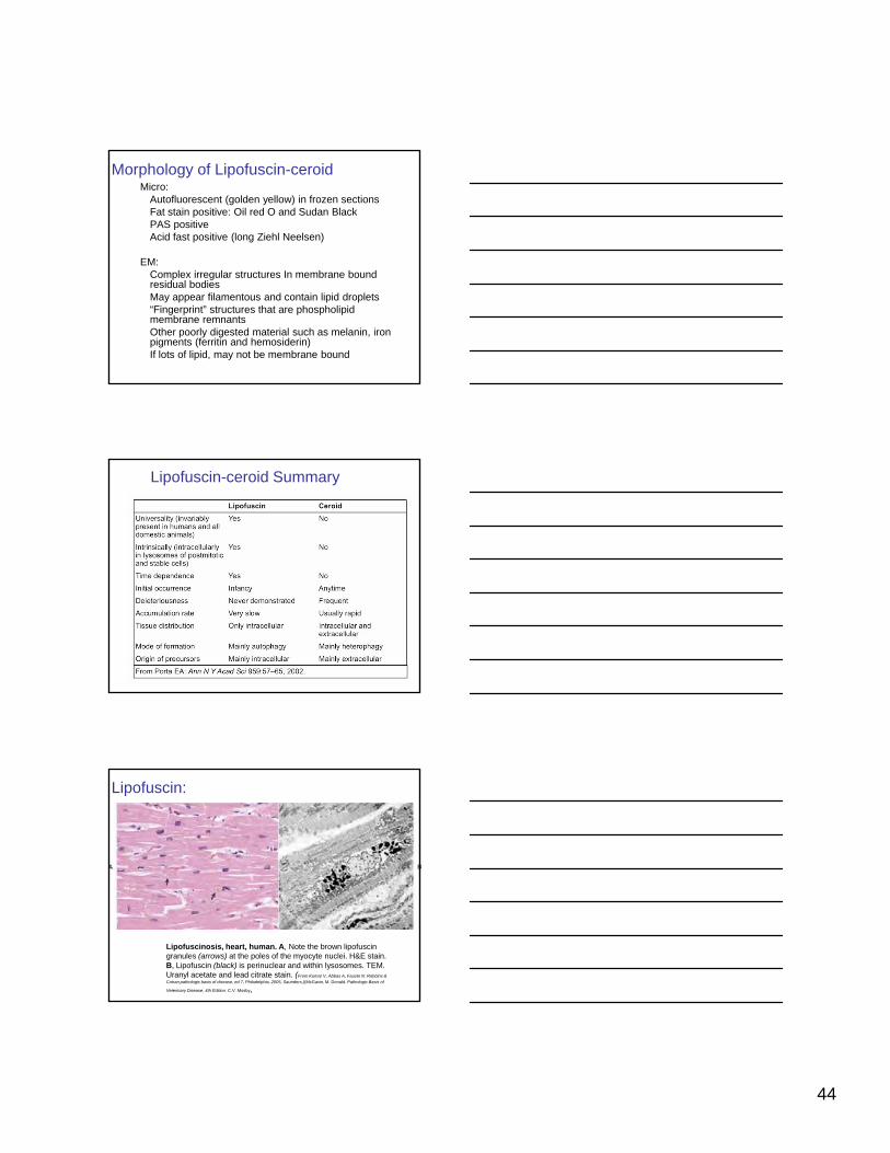
Morphology of Lipofuscin-ceroidMicro:
Autofluorescent (golden yellow) in frozen sectionsFat stain positive: Oil red O and Sudan BlackPAS positiveAcid fast positive (long Ziehl Neelsen)
EM: Complex irregular structures In membrane bound residual bodiesMay appear filamentous and contain lipid droplets“Fingerprint” structures that are phospholipid membrane remnantsOther poorly digested material such as melanin, iron pigments (ferritin and hemosiderin)If lots of lipid, may not be membrane bound
Lipofuscin-ceroid Summary
Lipofuscin:
Lipofuscinosis, heart, human. A, Note the brown lipofuscin granules (arrows) at the poles of the myocyte nuclei. H&E stain. B, Lipofuscin (black) is perinuclear and within lysosomes. TEM. Uranyl acetate and lead citrate stain. (From Kumar V, Abbas A, Fausto N: Robbins & Cotran pathologic basis of disease, ed 7, Philadelphia, 2005, Saunders.)(McGavin, M. Donald. Pathologic Basis of
Veterinary Disease, 4th Edition. C.V. Mosby,
45
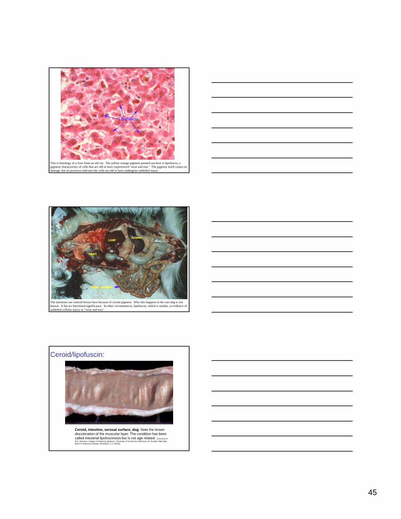
This is histology of a liver from an old cat. The yellow orange pigment pointed out here is lipofuscin, a pigment characteristic of cells that are old or have experienced "wear and tear." The pigment itself causes no damage, but its presence indicates the cells are old or have undergone sublethal injury.
The intestines are colored brown here because of ceroid pigment. Why this happens in the rare dog is not known. It has no functional significance. In other circumstances, lipofuscin, which is similar, is evidence of sublethal cellular injury or "wear and tear."
Ceroid/lipofuscin:
Ceroid, intestine, serosal surface, dog. Note the brown discoloration of the muscular layer. The condition has been called intestinal lipofuscinosis but is not age related. (Courtesy Dr. M.D. McGavin, College of Veterinary Medicine, University of Tennessee.) (McGavin, M. Donald. Pathologic Basis of Veterinary Disease, 4th Edition. C.V. Mosby

46
Hematogenous pigments:Unoxygenated hemoglobin results in blue color or cyanosis
In cyanide toxicosis, cytochrome oxidase cannot use oxygen and the venous
Cyanosis, feet, cat. The footpads of the paw on the left are bluish due to unoxygenated hemoglobin, the result of a partial obstruction of the iliac artery at the aortic bifurcation by a saddle thrombus. Normal control paw on the right. (Courtesy Dr. M.D. McGavin, College of Veterinary Medicine, University of Tennessee. (McGavin, M. Donald. Pathologic Basis of Veterinary Disease, 4th Edition. C.V. Mosby
hemoglobin will be the same color as arterial hemoglobin
Hematogenous pigments:CO poisoning results in “bright cherry red” blood due to carboxyhemoglobinhaving a higher affinity for heme than oxygen.
Carbon monoxide (CO) poisoning, brain, human. The blood in the brain is cherry red from the carboxyhemoglobin formed by the inhalation of CO in exhaust gases. (Courtesy Dr. J.C. Parker, School of Medicine, University of Louisville.)(McGavin, M. Donald. Pathologic Basis of Veterinary Disease, 4th Edition. C.V. Mosby
Hematogenous pigments:Methemoglobin is an oxide in which ferrous iron is converted to ferric iron resulting in “chocolate brown” blood. Due to nitrites (in nitrate-accumulating plantaccumulating plant poisoning) and reported with acetaminophen, naphthalene, local anesthetics, and chlorates. Can be congenital in humans.
Methemoglobinemia, experimental nitrite poisoning, hind leg, pig. Left, The methemoglobin in the blood has discolored the blood and muscle chocolate brown. Right, Normal control. (Courtesy Dr. L. Nelson, College of Veterinary Medicine, Michigan State University.)(McGavin, M. Donald. Pathologic Basis of Veterinary Disease, 4th Edition. C.V. Mosby

47
Hematogenous pigments:Hemoglobin released from intravascular hemolysis stains plasma pink. Excreted by kidney, it accumulates in lumens and stains kidney dark red tokidney dark red to black and urine red. Myoglobin may do similar coloring.
Acute hemolysis from chronic copper poisoning, kidney and urine, sheep. The dark bluish color of the kidney and the dark red of the urine are caused by hemoglobin excreted via the kidney. (Courtesy Dr. M.D. McGavin, College of Veterinary Medicine, University of Tennessee. (McGavin, M. Donald. Pathologic Basis of Veterinary Disease, 4th Edition. C.V. Mosby
You are looking at renal tubules from a sheep with hemolytic anemia. These are hemoglobin casts and hemoglobin pigment particles due to released hemoglobin that has passed into the filtrate. Grossly the kidney would be dark red to black because of this pigment.
Hematogenous pigments: HematinFormalin pigment (acid formalin hematin)
Microscopic artefact occurring in blood rich lesions in which acid solutions of formalin are used. It interferes with interpretation. Buffer formalin to neutral or at least above pH6.
Occasionally seen in lesions with acid centers such as abscesses even when BNF is used.
Parasite hematins also occur. See extracellular pigments.

48
Histology of lung that had autolyzed and was placed in non-buffered formalin. Acid formalin causes hematin pigment to form. This pigment is an artefact but can be confused with hemosiderin, melanin, and other dark pigments. Proper formalin fixation will prevent its formation except in areas of hypoxic hemorrhage where it might form antemortem.
Hematogenous pigments: Hemosiderin• Iron is formed in two
forms, both protein-iron complexes– Ferritin is in all tissues
and most cell types, but especially liver, spleen, bone marrow, andbone marrow, and skeletal muscle and is present in lysosomes or cytosol
– Hemosiderin is formed from intracellular aggregates of partially degraded protein shells of ferritin
Internal iron cycle.
Hematogenous pigments: Hemosiderin• Excess iron from RBC breakdown, senescent or
hemolytic, or reduced erythropoiesis is stored mainly in spleen.– Rare causes of iron excess are iron injections,
multiple blood transfusions, and rarely excessive gut absorption.
• Local iron storage is the result of hemorrhage or sites of RBC breakdown– Chronic passive congestion of lung (heart failure
cells) and liver– Hemosiderin is formed in sites of hemorrhage– Can form in sites of iron injections

49
Hematogenous pigments: Hemosiderin• Significance depends on location and amount
– Excess hemosiderin in spleen and liver occurs with hemolytic disease (e.g. AIHA or hemotropic parasites)
– Local hemosiderin indicates chronic hemorrhage• E.g. ethmoid hematoma of horses
• Hemosiderosis is excess hemosiderin occurring with iron overload
Increased absorption of dietar iron in h mans– Increased absorption of dietary iron in humans– Impaired use of iron– Hemolytic anemia– Transfusions
• Hemochromatosis. Extreme amounts of hemosiderin resulting in liver, pancreas, and heart damage.– In humans a genetic disease of increased iron absorption– Rarely occurs with transfusions
Hematogenous pigments: Hemosiderin
Hemosiderosis, liver, human being. A, Hemosiderin is present as fine golden brown granules in hepatocytes H&E stain. B, Granules of hemosiderin are stained dark blue by the Prussian blue reaction, which is specific for iron. Prussian blue reaction.(A and B, From Kumar V, Abbas A, Fausto N: Robbins & Cotran pathologic basis of
disease, ed 7, Philadelphia, 2005, Saunders.) (McGavin, M. Donald. Pathologic Basis
of Veterinary Disease, 4th Edition. C.V. Mosby,
Spleen of an animal with hemolytic anemia. The macrophages shown above have large amounts of hemosiderin, an iron breakdown product of hemoglobin.

50
Heart and lung from a dog with heartworm disease. Heartworms in the right ventricle sometimes release and go to lung where they cause areas of hemorrhage and necrosis. The areas of hemorrhage become brown as the macrophages phagocytose the red cells, degrade them, and temporarily retain the iron as hemosiderin. Areas of dark brown hemosiderosis are outlined in blue here.
Chronic passive congestion, lung, dog. A, Alveolar macrophages containing hemosiderin (blue) are present in the alveoli. Prussian blue reaction. B, The lungs have chronic passive congestion attributed to chronic left side heart failure. They are moderately firm and yellow-brown due to alveolar macrophages containing hemosiderin. Inflammatory mediators produced by these macrophages have induced fibroplasia and thus in the long term there has been extensive formation of interstitial collagen. This collagen is the reason the lungs have failed to collapse following incision of the diaphragm, which releases the negative pressure in the pleural cavity (note the rib impressions in the lung). (A, Courtesy Dr. M.D. McGavin, College of Veterinary Medicine, University of Tennessee. B, Courtesy College of Veterinary Medicine, University of Illinois.) (McGavin, M. Donald. Pathologic Basis of Veterinary Disease, 4th Edition. C.V. Mosby,
Hematogenous pigments: Bilirubin
• Bilirubin is a yellow-brown pigment derived from breakdown of heme, the porphyrin of hemoglobin– Breakdown occurs in macrophages of spleen, liver,
bone marrow, and sites of hemorrhage– Senescent RBCs (life spans: 70 days for cat, 150
days for cattle and horses) phagocytosed by monocyte macrophage system cellsmonocyte macrophage system cells.
– After phagocytosis, iron is removed and stored, porphyrin ring broken down to bilirubin, which is released into blood and bound to albumin and travels to liver by blood, enters space of Disse and is taken up by microvilli of hepatocytes.
– In hepatocytes, bilirubin is conjugated to bilirubin diglucuronide and excreted into bili canaliculus.

51
Synthesis and excretion of bili bi
Histology and Cell Biology. An introduction ot Pathology. Kierszenbaum, AL, Mosby
bilirubin
Bilirubin metabolism:Schematic diagram of bilirubin metabolism and elimination (as depicted in human beings). 1, Normal bilirubin production from heme (0.2 to 0.3 g per day) is derived primarily from the breakdown of senescent circulating erythrocytes, with a minor contribution from degradation of tissue heme-containing proteins. 2, Extrahepatic bilirubin is bound to serum albumin and delivered to the liver. 3 Hepatocellular uptake and3, Hepatocellular uptake and 4, glucuronidation in the endoplasmic reticulum generate bilirubin monoglucuronides and diglucuronides, which are water soluble and readily excreted into bile.5, Gut bacteria deconjugate the bilirubin and degrade it to colorless urobilinogens. The urobilinogens and the residue of intact pigments are excreted in the feces, with some reabsorption and excretion into urine.
(From Kumar V, Abbas AK, Fausto N: Robbins & Cotran pathologic basis of disease, ed 7, Philadelphia, 2005, Saunders.) (McGavin, M.
Donald. Pathologic Basis of Veterinary Disease, 4th Edition. C.V. Mosby,
Hematogenous pigments: Icterus
• Icterus or jaundice is yellow staining of tissue by bilirubin• Results from imbalance of production (increased) and
excretion (decreased)• One classification scheme or hyperbilirubinemia:
– Prehepatic: hemolysis results in increased amounts of unconjugated (indirect) bilirubin that exceed hepatocyte ability to take it upH ti h ti i j lt i l f j t d d– Hepatic: hepatic injury results in release of unconjugated and conjugated bilirubin into blood (several mechanisms possible)
– Posthepatic: obstruction of the biliary system, either intrahepatic or extrahepatic (bile duct obstruction) causes reflux of conjugated bile into blood and increased conjugated (direct) bilirubin.
• Unconjugated bilirubin (attached to albumin) cannot be cleared by kidney, but conjugated bilirubin can

52
Bilirubin metabolism: Degradation of heme and
Prehepatic
icterus
Post hepatic
Hepatic
Hematogenous pigments: Icterus
• Major mechanisms of icterus– Excess production due to increased RBC breakdown
– Reduced uptake of bilirubin from the plasma by hepatocytes
– Impaired or absent conjugation in hepatocytes, sometimes an inherited and congenital anomalyg y
– Hepatic necrosis allows leak of bilirubin into blood
– Decreased excretion of conjugated bilirubin from hepatocytes into bile canaliculus
– Reduced flow of bile from liver
Hematogenous pigments: Icterus
• Gross.– Systemically distributed bilirubin is apparent in some
tissues such as skin, sclera, intima of large vessels, mucous membranes of oral cavity and urogenital tract, and alimentary tract as well as omentum, mesentery, and adipose tissue.
– Can be confused with carotenoid coloration of fat in Jersey and Guernsey cattle, horses, and nonhuman primates
• Micro. Not detected, but can be seen in bile ducts
53
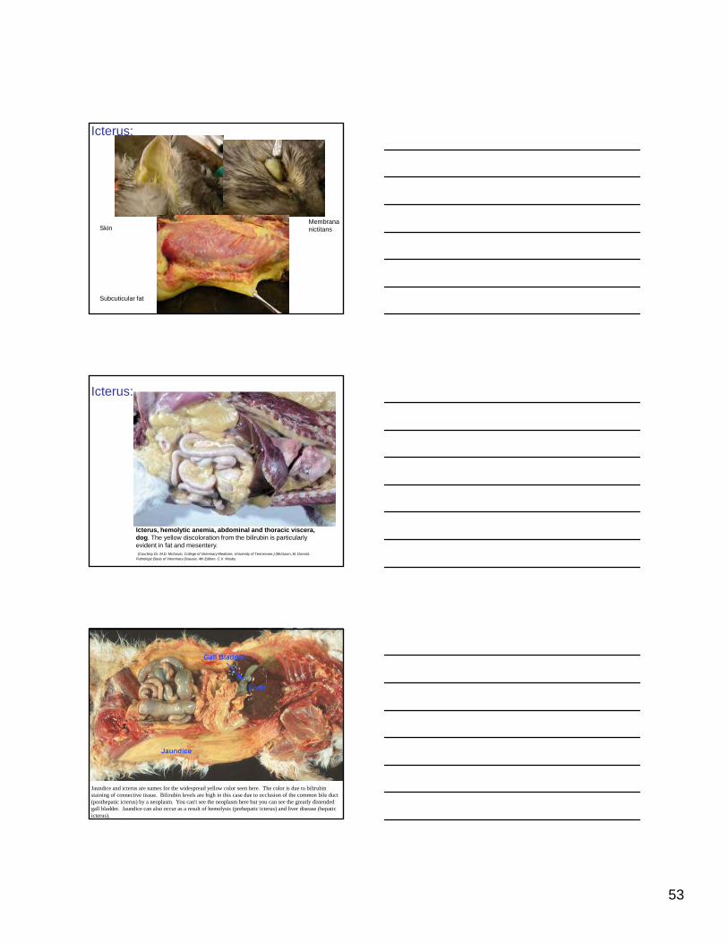
Icterus:
SkinMembrana nictitans
Subcuticular fat
Icterus:
Icterus, hemolytic anemia, abdominal and thoracic viscera, dog. The yellow discoloration from the bilirubin is particularly evident in fat and mesentery.(Courtesy Dr. M.D. McGavin, College of Veterinary Medicine, University of Tennessee.) (McGavin, M. Donald.
Pathologic Basis of Veterinary Disease, 4th Edition. C.V. Mosby
Jaundice and icterus are names for the widespread yellow color seen here. The color is due to bilirubin staining of connective tissue. Bilirubin levels are high in this case due to occlusion of the common bile duct (posthepatic icterus) by a neoplasm. You can't see the neoplasm here but you can see the greatly distended gall bladder. Jaundice can also occur as a result of hemolysis (prehepatic icterus) and liver disease (hepatic icterus).

54
Bile stasis (cholestasis) will turn a liver green after it has been fixed with formalin as seen here in this liver that is filled with bile. The inset shows the yellow brown pigment in bile canaliculi. The canaliculi are the normal route of bile secretion, but bile is usually not seen histologically unless there is occlusion of biliary outflow somewhere along the tract. Animals with this much bile stasis are often icteric.
Icterus:
Icterus. A, Icterus, liver, cat. Note the enlarged liver with rounded edges and yellow-orange color caused by retained bilirubin. B, Bile casts in bile canaliculi. Acute hemolytic anemia, babesiosis, liver, cow. The bile casts are the result of a high rate of bilirubin excretion by the liver secondary to intravascular hemolysis. H&E stain. (A, Courtesy the College of Veterinary Medicine, University of Illinois. B, Courtesy Dr. M.D. McGavin, College of Veterinary Medicine, University of Tennessee.) (McGavin, M. Donald. Pathologic Basis of Veterinary Disease, 4th Edition. C.V. Mosby
Kernicterus in a dog- yellow discoloration
of thalamic and subthalamic nuclei
Kernicterus in an Adult DogC. R. Sangster, C. K. Stevenson, B. A. Kidney, D. L. Montgomery, and A. L. Allen
Veterinary Pathology Online, May 2007; vol. 44, 3: pp. 383-385

55
Hematogenous pigments: Porphyria and porphyrins
• Porphyria. A group of inborn or acquired disturbances of porphyrin metabolism
• Congenital erythropoietic porphyria– Occurs in calves, cats, and pigs
– Defect in heme synthesis caused by deficiency of uroporphyrinogen III cosynthetaseuroporphyrinogen III cosynthetase.
– Called “pink tooth” because of porphyrin coloration in dentin and bone
– Teeth and bones of young animals are reddish and those of adults dark brown
– These fluoresce with UV light
– Can result in photodynamic dermatitis
Congenital porphyria is a metabolic defect in the synthesis of heme. Here the teeth are stained pink due to this disease. Bones are also pink tinged
There are lesions in the lungs from this foal (we know it is either a foal or a small horse because of the size marker and the relative small lungs). What are some lesion rule outs at this point?
Hematogenous pigments: Porphyrin
Next frame for another lesion.
Chromodacryorrhea (‘red tears’) in rats is due to production of a protoporphyrin (4 pyrrole rings without a metal) in the Harderian gland. It is seen in many conditions and it fluoresces pink when dry. Rat tears also contain lipids and melatonin.

56
Accumulations, pigments, etc: Some Examples:
Skin of rabbit with actinomycosis.
Splendore-Hoeppli phenomenon.
Accumulations, pigments, etc: Some Examples:
Rat with panarteritis (periarteritis) nodosa.
Hemoglobin crystals, hemosiderin, and fibrinoid (necrosis)
Accumulations, pigments, etc: Some Examples:
Rat with panarteritis (periarteritis) nodosa and progressive glomerulopathy.
Congo red stain: normal elastin in internal and external elastic membranes
57
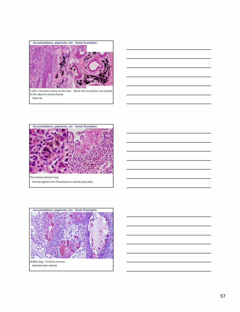
Accumulations, pigments, etc: Some Examples:
Collie. Ulcerative lesion on the nose. Basal cell vacuolation was present in the adjacent stratum basale.
Tattoo ink.
Accumulations, pigments, etc: Some Examples:
Non human primate lung.
Hematin pigment from Pneumonyssus simicola (lung mite).
Accumulations, pigments, etc: Some Examples:
Rabbit lung. Found in airways. Aspirated plant material.
58
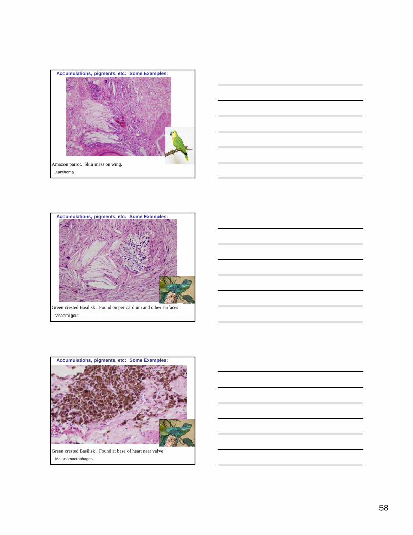
Accumulations, pigments, etc: Some Examples:
Amazon parrot. Skin mass on wing.
Xanthoma
Accumulations, pigments, etc: Some Examples:
Green crested Basilisk. Found on pericardium and other surfaces
Visceral gout
Accumulations, pigments, etc: Some Examples:
Green crested Basilisk. Found at base of heart near valve
Melanomacrophages.

59
Accumulations, pigments, etc: Some Examples:
Mass on lemon butterfly fish.
Accumulations, pigments, etc: Some Examples:
Mass on lemon butterfly fish. Polarized light
Mixed chromatophoroma. Iridophores and melanophores
Disseminated meningeal melanoma
Disseminated Melanoma in a Dog with Involvement of Leptomeninges and Bone MarrowD. Y. Kim,A. B. Royal, and J. A. Villamil Veterinary Pathology Online, January 2009; vol. 46, 1: pp. 80-83

60
Accumulations, pigments, etc: Some Examples:
Non human primate with diarrhea. Lung
Accumulations, pigments, etc: Some Examples:
Non human primate with diarrhea. Lung
Kaolin aspiration. Polarized light. And mite pigment
Questions for Lecture 4
About the Questions.Cell Injury Questions from Lectures 1-4 come from two sources. Those designated with this red # and ISU/Aub at the bottom of the page were created and contributed by residents and faculty
Questions from ISU/Aub Contributed by:
Dr. Alicia OlivierDr. Katherine Gibson-CorleyDr. Brandon PlattnerDr. Darin MadsonDr. Jodi SmithDr. Rachel DerscheidDr Charlie Johnson
y yof Iowa State University and Auburn University listed as a group.
Answers for these appear at the end of the question pages. Page numbers refer to Pathologic Basis of Veterinary Disease.
Other questions came from the AFIP web site.
Dr. Charlie JohnsonDr. Molly MurphyDr. Tatjana LazicDr. Ann PredgenDr. Aaron LehmkuhlDr. Angela PillatzkiDr. Lisa PohlmanDr. Pete ChristophersonDr. Brandon BrunsonDr. Leah Ann KuhntDr. Kellye Sue JoinerDr. Beth SpanglerDr. Elizabeth Whitley

61
11. Proteins targeted for degradation in the proteosome are first
conjugated to:
A. Tenacin
B. Vinculin
C. Ubiquitin
D. SyndecanD. Syndecan
E. Chaperonin
11. C: Pathologic Basis of disease, 7th ed., 2005, p. 2005
31. In eukaryotic cells, the ATP-dependent proteolytic complex that prevents accumulation of misfolded and damaged proteins by breaking
them down is:
A. Ubiquitin
B. Aggresome
C. 26S proteasome p
D. Proteolysosome complex
E. Heat-shock protein-chaperone complex
31. C Nature, V426, 2003 2004
5. Which of the following does not stimulate proteasome-mediated
protein degradation?
1. Insulin
2. Glucocorticoids
3. Thyroid hormone
4. Tumor necrosis factor
A. 1
B. 1, 2
C. 1, 2, 3
D. 2, 3
E. 3, 4
5. A. Robbins and Cotran, p. 10 2008

62
26. Which protein forms senile cerebral amyloid plaques in the brains of
dogs?
A. Transthyretin
B. ß2-microglobulin
C. Amyloid precursor protein
D. Immunoglobulin light chainsD. Immunoglobulin light chains
E. Serum amyloid associated protein
26. C: Pathologic Basis of disease, 7th ed., 2005, p. 260 Vet Pathol, 36:202-211, 1999; Vet Pathol 33:230-234, 1996 2005
30. Place the steps in the pathogenesis of dystrophic calcification in the correct
sequence:
1. Phosphatases generate phosphates groups and bind calcium 2. A structural change occurs in the arrangement of calcium 3. Calcium ions bind to phospholipids in the vesicle membrane 4. Calcium and phosphate binding cycle is repeated, forming deposit
A. 1 -> 2 -> 3 -> 4 3B. 2 -> 3 -> 1 -> 4 C. 3 -> 2 -> 4 -> 1 D. 3 -> 1 -> 4 -> 2 E. 1 -> 3 -> 4 -> 2
30. D: Robbins and Cotran, Pathological Basis of Disease, p. 41, 2005. 2006
47. All of the following can cause metastatic calcification EXCEPT
A. Lymphoma
B. Renal failure
C. Osteosarcoma
D. Cestrum diurnumD. Cestrum diurnum
E. Mycobacterium bovis
47. E. Pathologic Basis of Veterinary Disease, p. 48-49 2007

63
48. All of the following are true regarding reactive amyloid (AA)
EXCEPT
A. Induced by IL-1 and IL-6
B. Formed from SAA secreted by the liver
C. Form found in equine nasal amyloidosis
D. Most commonly deposited in the space of Disse in birdsD. Most commonly deposited in the space of Disse in birds
E. Form found in hereditary amyloidosis in Shar-Pei dogs and
Abyssinian cats
48. C. Pathologic Basis of Veterinary Disease, p. 46, 474 2007
45. Concerning amyloidosis which of the following statements is true:
1. The most common form of amyloidosis in animals is immunocyte dyscrasia
2. A common site of amyloid deposition in old dogs is the kidney 3. The heart is the most frequent site of reactive amyloidosis 4. Deposition of amyloid-beta has not been documented in dogs4. Deposition of amyloid beta has not been documented in dogs 5. Bence-Jones proteins are present in the urine with reactive
amyloidosis
A. 1, 2 B. 2 C. 3 D. 2, 4 E. 1, 5
45. B. Pathologic Basis of Veterinary Disease, p. 46 2008
15. All are used in the diagnosis of amyloidosis EXCEPT:
A. Thioflavin T
B. Shtrasburg method
C. Crystal violet method
D. Potassium permanganate techniqueD. Potassium permanganate technique
E. Phospho tungsten acid hematoxylin
15. E: Vet Pathol 42:132-139, 2005; Thompsons’ Special Veterinary Pathology. 2006

64
35. Synthesis of a defective enzyme with reduced activity or a reduced
normal enzyme include all of the following consequences EXCEPT:
A. Block of a metabolic pathway and decreased amount of end product
B. Accumulation of a substrate or intermediate substrates
C. Failure to inactivate a tissue-damaging substrate g g
D. Accumulation of overabundant end product
E. Albinism
35. D: Pathologic Basis of disease, 7th ed., 2005, p. 153 2005
26. What is the function of the ubiquitin-proteasome pathway
A. Apoptosis
B. Protein degradation
C. Intracellular signaling
D. Receptor mediated signaling
E. Alternative method of complement activation
26. B. Robbins and Cotran, p. 10 2007
#5
Lipofuscin (PBVD ed.4, pp. 52-53)
1. has a deleterious effect on the cell.2. accumulation is intracellular and extracellular.3. accumulates with vitamin E deficiency.4. is the result of autophagocytosis of cellular constituents.5 has staining characteristics similar to ceroid
ISU/Aub
5. has staining characteristics similar to ceroid.
A. 1,2B. 1,2,3C. 2,3,4D. 3,4,5E. 4,5
65
#8
In diabetes, glycogen is found in which of the following tissues? (PBVD ed.4, 2006, p. 42)
1. Renal proximal tubules2. Hepatocytes3. Skeletal muscle4 Pancreatic beta cells
ISU/Aub
4. Pancreatic beta cells
A. 1,2.B. 1, 2, 3.C. 1, 2, 3, 4.D. 1, 3, 4.E. 1, 2, 4.
#20
Which of the following statements are TRUE regarding autophagic vacuoles? (PBVD ed.4, p. 33)
1. Common in sub-lethally injured cells2. Common in liver3. Common in renal tubular epithelium4 Once formed are present for the remainder of the cell’s life
ISU/Aub
4. Once formed are present for the remainder of the cell s life5. Are seen as basophilic cytoplasmic vacuoles
A. 1 and 2B. 1, 2 and 3C. 1, 2, 3 and 4D. 2, 3 and 4E. 2 and 3
#25
The most common site of localized amyloid deposition in the cat is: (PBVD ed.4, p. 46)
A. Renal medulla
B. Spleen
ISU/Aub
C. Liver
D. Lymph nodes
E. Pancreatic islets
66
#27
Which of the following result in visceral gout in birds?
1. Vitamin A deficiency 2. Renal disease3. High protein diet4. Zinc deficiency5 Chronic zinc toxicosis
ISU/Aub
5. Chronic zinc toxicosis
A. 1, 2B. 3, 4C. 1, 2, 3D. 1, 2, 3, 4 E. 1, 2, 3, 4, 5
#36
In nitrite poisoning, the blood is (PBVD ed.4, p.54)
A. Bright cherry red
B. Dark purple
C Chocolate brown
ISU/Aub
C. Chocolate brown
D. Blue-green
E. Normal colored
#56
In regards to carotenoid, which are true (PBVD ed.4, p. 50)
1. Fat soluble pigment of plant origin2. Can be seen microscopically3. Can accumulate in adipose tissue4. Normally occur in testicular cells5 Precursor is beta carotene
ISU/Aub
5. Precursor is beta carotene
A. 1 and 2B. 3C. 1, 3 , 4, 5D. 3 and 4

67
#58
Congenital porphyria of calves, cats, and pigs is caused by a defect in which enzyme: (PBVD ed.4, p. 59)
A. Heme oxygenase.
B. Uroporphyrinogen III cosynthetase.
ISU/Aub
C. Biliverdin reductase.
D. Glucuronide transferase.
#63
Which of the statements is true of gout as it pertains to birds? (PBVD ed.4, pp 46-47)
1. Composed of sodium urate crystals2. End product of purine metabolism 3. Termed tophi in tissue4 Articular gout is more common
ISU/Aub
4. Articular gout is more common
A. 1, 2, and 3B. 4C. 1, 3 , and 4D. 3 and 4
#67
Causes of metastatic calcification observed in veterinary medicine include: (PBVD ed.4, pp. 48-49)
1. Renal disease2. Vitamin D toxicosis3. Primary hyperparathyroidism4 Increased PTH related protein from some malignant neoplasms
ISU/Aub
4. Increased PTH-related protein from some malignant neoplasms5. Destruction of bone from primary or metastatic neoplasms
A. All of the aboveB. None of the aboveC. 1,2,3,4D. 1,2,3E. 4,5

68
#73
Plasma cells termed “Mott cells” have large eosinophilic cytoplasmic bodies. What are these bodies composed of? (PBVD ed.4, p 43)
A. Abundant Golgi
B. Phagocytic debris
ISU/Aub
C. Autophagic vacuoles
D. Gamma globulin
E. Glycogen
#79
Which of the following are characteristic of acute cyanide poisoning? (PBVD ed.4, p.54)
1. Hematin accumulates within the cytoplasm2. Hemoglobin is released from lysed erythrocytes3. Cyanide binds to cytochrome oxidase4 Venous blood is bright red
ISU/Aub
4. Venous blood is bright red5. Occurs following ingestion of nitrate-accumulating plants
A. 1,2.B. 2, 3C. 3,4.D. 1,2,3.E. 3,4,5.
#83
Telomere length in dividing cells is maintained by the enzyme telomerase. Which statements are TRUE regarding telomerase?
1. Telomerase is active in most somatic tissues2. Telomerase is a DNA:protein complex3. Telomerase adds a random nucleotide sequence to the ends of
chromosomes
ISU/Aub
chromosomes4. If telomerase is not active, cells eventually exit from the cell cycle5. Telomerase is active in most immortalized tumor cells
A. 1, 2, 3B. 3, 4,5C. 4, 5D. 2, 4E. 1, 5

69
#95
All are characteristics of ceroid pigment EXCEPT: (PBVD ed.4, p.52)
A. Periodic acid-Schiff-positive.
B. May be intracellular and extracellular.
ISU/Aub
C. Does not have a deleterious effect on cell.
D. Accumulates in the cells in response to vitamin E deficiency.
E. Accumulates in the cells in response to cachexia from cancer.
#105
The cytokines most incriminated in reactive systemic amyloidosis are: (PBVD ed.4, p. 46)
A. IL-6 and IL-10.
B. IL-3 and IL-6.
ISU/Aub
C. IL-1 and IL-6.
D. IL-1 and IL-10.
E. IL-1 and IL-4.
#106
Mineralization of otherwise normal lung may occur as a direct or indirect sequel to which of the following conditions? (PBVD ed.4, pp. 47-48)
1. Chronic hyperphosphatemia.2. Cholecalciferol-containing rodenticide toxicosis.3. Hyperadrenocorticism.4 Adenocarcinoma of the anal sac apocrine glands
ISU/Aub
4. Adenocarcinoma of the anal sac apocrine glands.5. Primary hyperparathyroidism.
A. 1,2,3.B. 1,3,5.C. 2,3,4.D. 1,2,4,5.E. 3,4,5.

70
#135
Mechanisms of icterus development include all of the following except:
A. Reduced plasma uptake
B. Reduced albumin binding
C Excess bilirubin production
ISU/Aub
C. Excess bilirubin production
D. Decreased hepatic excretion of conjugated bilirubin
E. Hepatic necrosis with reduced hepatic biliary outflow
#137
All of the following statements concerning melanin are true EXCEPT:
A. Albino animals lack tyrosine.
B. Copper-deficient animals may have lower levels of melanin.
C Hypermelanosis in skin is associated with chronic injury and
ISU/Aub
C. Hypermelanosis in skin is associated with chronic injury and endocrinopathies.
D. Formed by the oxidation of tyrosine by the copper containing enzyme tyrosinase.
E. Pigment is transferred by dendritic processes to keratinocytes in the stratum spinosum and stratum granulosum.
Answers to ISU/Aub questions for lecture 4.
• 5. E
• 8. E
• 20. B
• 25. E
• 27. C
• 36. C
• 56. C
• 105. C
• 106. D
• 135. B
• 137. A
ISU/Aub
• 58. B
• 63. A
• 67. A
• 73. D
• 79. C
• 83. C
• 95. C