Cerebral A-V fistula excision in a parturient
49
Cerebral A-V fistula excision in a parturient Verghese Cherian, MB, BS, MD, DA, FFARCSI Associate Professor of Anesthesiology Director, Division of General Anesthesia Sonia Vaida, MD Professor of Anesthesiology and Obstetrics and Gynecology Vice Chair for Research Director, Division of Obstetric Anesthesia Arne Budde, MD, DEAA Associate Professor of Anesthesiology Director, Division of Neuroanesthesia Department of Anesthesiology Milton S Hershey Medical Center Penn State University College of Medicine None of the authors has conflicts of interest to disclaim
Transcript of Cerebral A-V fistula excision in a parturient

Cerebral A-V fistula excision in a parturient
Verghese Cherian, MB, BS, MD, DA, FFARCSI Associate Professor of Anesthesiology
Director, Division of General Anesthesia
Sonia Vaida, MD Professor of Anesthesiology and Obstetrics and Gynecology
Vice Chair for Research Director, Division of Obstetric Anesthesia
Arne Budde, MD, DEAA
Associate Professor of Anesthesiology Director, Division of Neuroanesthesia
Department of Anesthesiology
Milton S Hershey Medical Center Penn State University College of Medicine
None of the authors has conflicts of interest to disclaim

Case Presentation • 25 y, 33wks gestation • 65kg, 172cm • ‘worst headache of her life’
– Left sided headache – Associated nausea & vomiting
• Visual disturbances, No diplopia • Right nasal inferior quadrant anopia • No significant past medical history
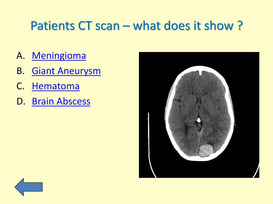
Patients CT scan – what does it show ?
A. Meningioma B. Giant Aneurysm C. Hematoma D. Brain Abscess

Sorry – I disagree
Try again

CORRECT !!
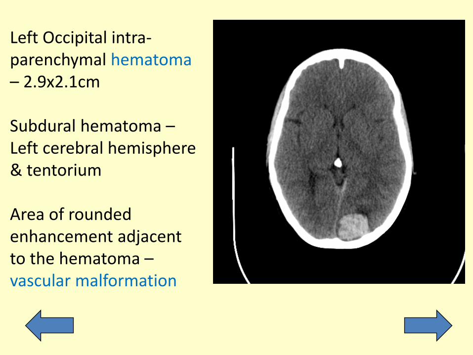
Left Occipital intra-parenchymal hematoma – 2.9x2.1cm Subdural hematoma – Left cerebral hemisphere & tentorium Area of rounded enhancement adjacent to the hematoma – vascular malformation
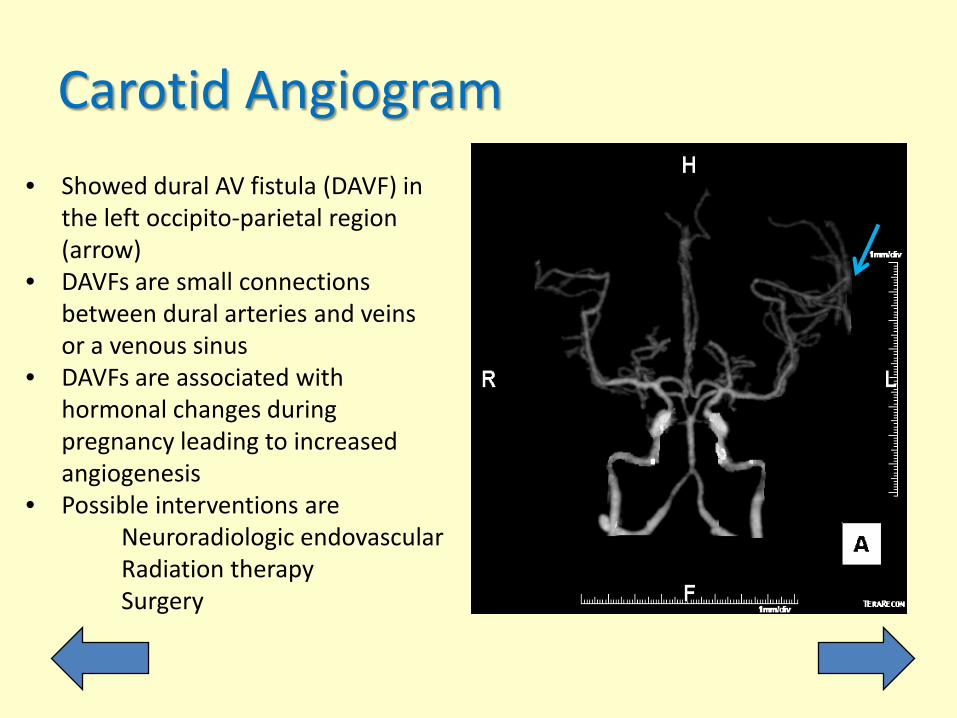
Carotid Angiogram • Showed dural AV fistula (DAVF) in
the left occipito-parietal region (arrow)
• DAVFs are small connections between dural arteries and veins or a venous sinus
• DAVFs are associated with hormonal changes during pregnancy leading to increased angiogenesis
• Possible interventions are Neuroradiologic endovascular Radiation therapy Surgery

Which statement about intracranial vascular malformations is true for a pregnant patient?
A. Increased risk for hemorrhage from AV Malformation B. Increased risk for rupture of Cerebral Aneurysm C. Lower risk of re-bleeding from AVM D. Pregnancy related mortality of ICH is < 1%

Sorry – I disagree
Try again

CORRECT !!
Pregnancy & ICH/SAH
• SAH – ruptured aneurysm (65%); AVMs (35%) • Increased risk of aneurysm rupture
– Increased blood volume & cardiac output – Hormonal changes to the arterial wall
• Pregnancy does NOT increase the risk of hemorrhage from AVM, but risk of re-bleed is 25%, compared to 3-6% in 1 year in a non-pregnant woman
• ICH accounts for 7% of all pregnancy-related mortality • Maternal mortality is approx. 20 %

Treatment plan for our patient
Patients was not considered a candidate for endovascular treatment.
Surgical plan was left parieto-occipital craniotomy and resection of dural AV
fistula with intra-operative angiogram.

Anesthetic Management A few common unique concern between OB Anesthesia and Neuro Anesthesia are mainly based on Organ Blood Flow: Brain vs. Uterus • Monitoring • Positioning • Ventilation • Anesthesia Maintenance • Other pharmacologic concerns
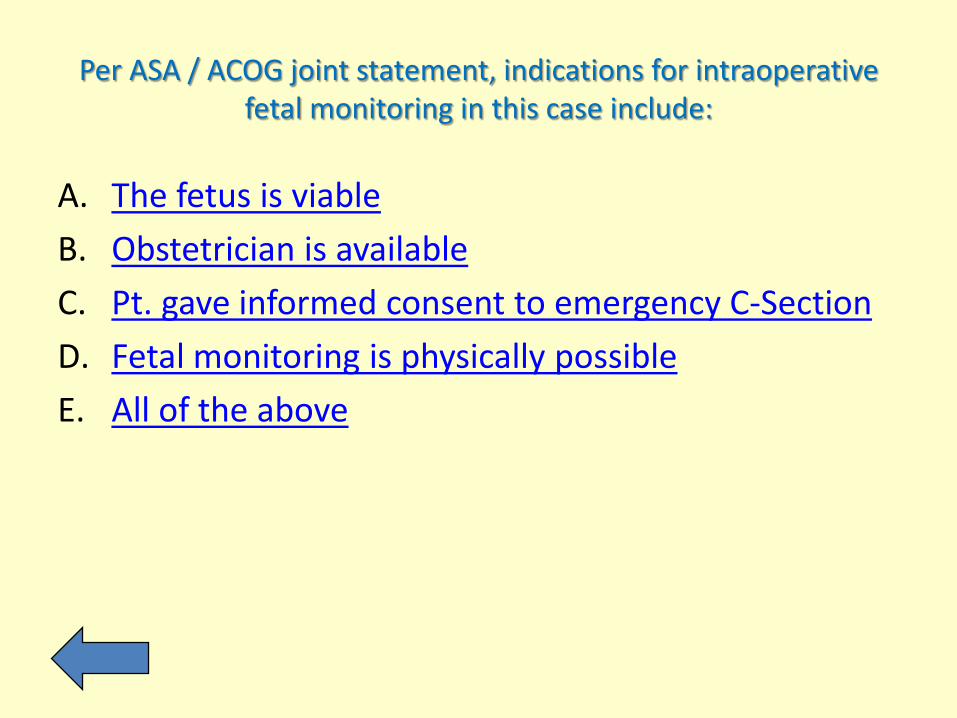
Per ASA / ACOG joint statement, indications for intraoperative fetal monitoring in this case include:
A. The fetus is viable B. Obstetrician is available C. Pt. gave informed consent to emergency C-Section D. Fetal monitoring is physically possible E. All of the above

Sorry – I disagree
Try again

CORRECT !!
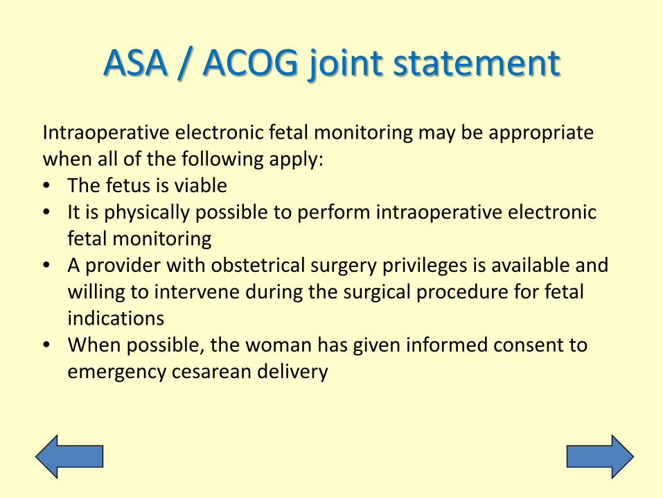
ASA / ACOG joint statement
Intraoperative electronic fetal monitoring may be appropriate when all of the following apply: • The fetus is viable • It is physically possible to perform intraoperative electronic
fetal monitoring • A provider with obstetrical surgery privileges is available and
willing to intervene during the surgical procedure for fetal indications
• When possible, the woman has given informed consent to emergency cesarean delivery
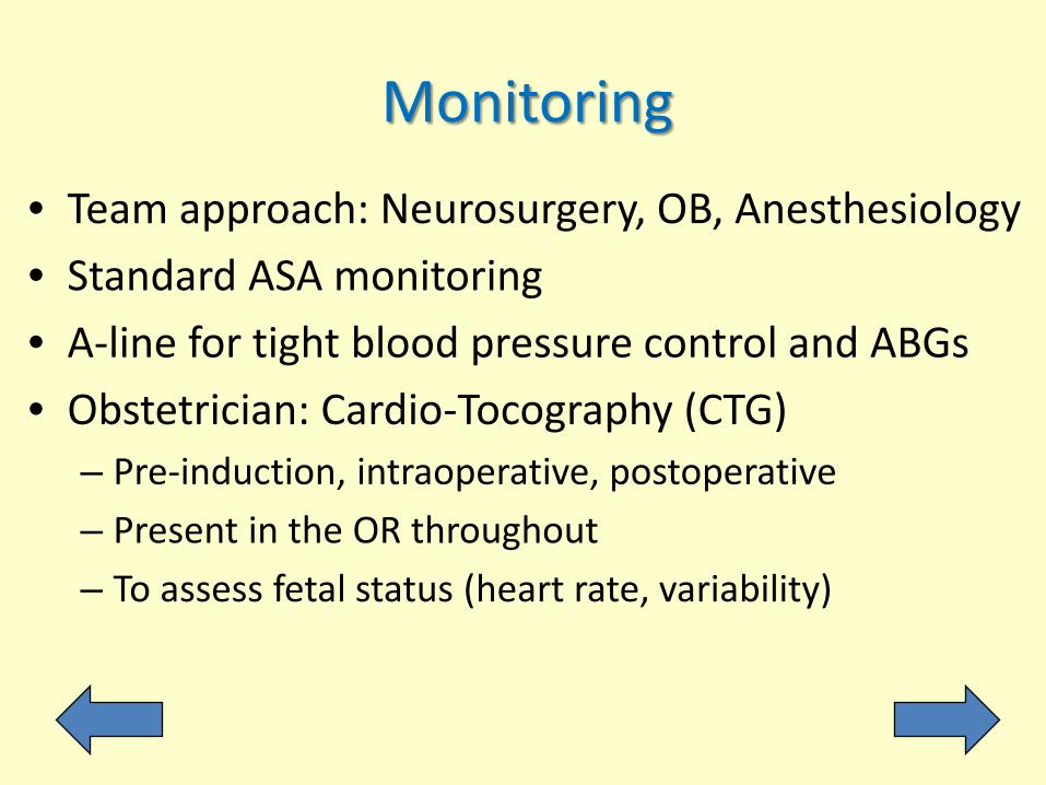
Monitoring
• Team approach: Neurosurgery, OB, Anesthesiology • Standard ASA monitoring • A-line for tight blood pressure control and ABGs • Obstetrician: Cardio-Tocography (CTG)
– Pre-induction, intraoperative, postoperative – Present in the OR throughout – To assess fetal status (heart rate, variability)
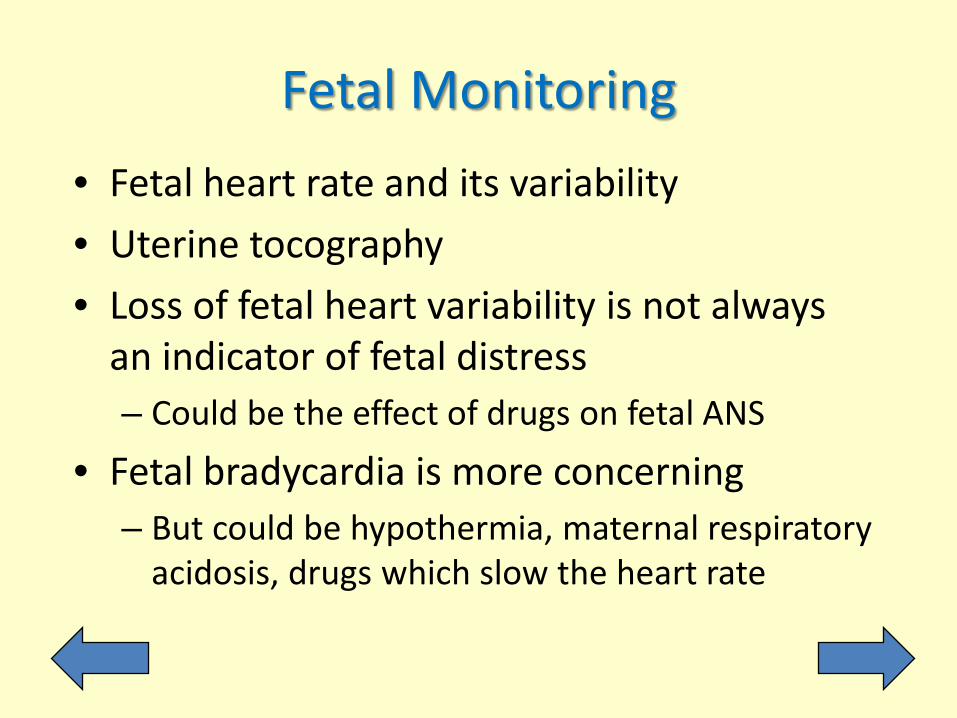
Fetal Monitoring • Fetal heart rate and its variability • Uterine tocography • Loss of fetal heart variability is not always
an indicator of fetal distress – Could be the effect of drugs on fetal ANS
• Fetal bradycardia is more concerning – But could be hypothermia, maternal respiratory
acidosis, drugs which slow the heart rate
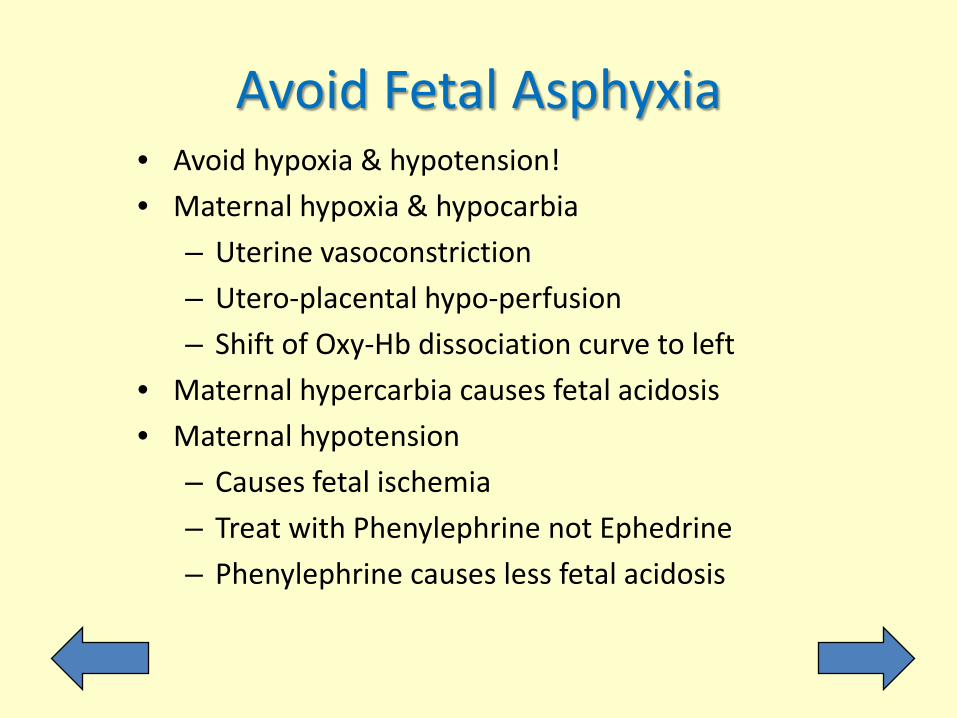
Avoid Fetal Asphyxia • Avoid hypoxia & hypotension! • Maternal hypoxia & hypocarbia
– Uterine vasoconstriction – Utero-placental hypo-perfusion – Shift of Oxy-Hb dissociation curve to left
• Maternal hypercarbia causes fetal acidosis • Maternal hypotension
– Causes fetal ischemia – Treat with Phenylephrine not Ephedrine – Phenylephrine causes less fetal acidosis
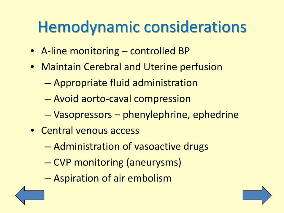
Hemodynamic considerations • A-line monitoring – controlled BP • Maintain Cerebral and Uterine perfusion
– Appropriate fluid administration – Avoid aorto-caval compression – Vasopressors – phenylephrine, ephedrine
• Central venous access – Administration of vasoactive drugs – CVP monitoring (aneurysms) – Aspiration of air embolism

What would be the best position in our case scenario to avoid aorto-caval compression ?
A. Supine B. Prone C. Left lateral decubitus D. Right lateral decubitus

Sorry – I disagree
Try again

CORRECT !!
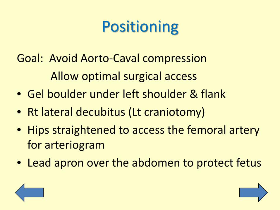
Positioning
Goal: Avoid Aorto-Caval compression Allow optimal surgical access • Gel boulder under left shoulder & flank • Rt lateral decubitus (Lt craniotomy) • Hips straightened to access the femoral artery
for arteriogram • Lead apron over the abdomen to protect fetus
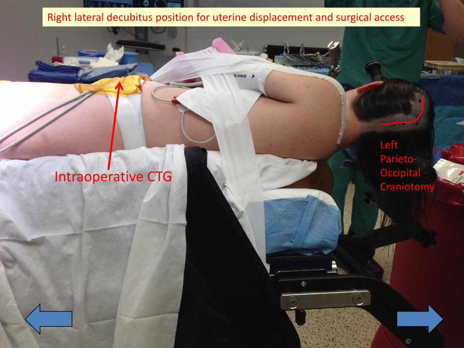
Left Parieto-Occipital Craniotomy
Intraoperative CTG
Right lateral decubitus position for uterine displacement and surgical access

Which of the following comparison between Propofol/Remifentanil TIVA and Volatile Agents is NOT correct?
A. Volatile Agents may interfere more with MEP / SSEP B. TIVA may improve neonatal neuro-behavioral
performance C. Volatile Agents may add a tocolytic effect in
premature labor D. Volatile Agents may increase CBF, CBV E. TIVA always causes increase in ICP

Sorry – I disagree
Try again

CORRECT !!

• TIVA or “half-TIVA” may be preferred if neurophysiologic monitoring is planned
• Inhalational agents may result in increased latency and amplitude of electrical signals at MAC > 1.0
• MAC > 0.7 - 1 should be avoided due to increase of cerebral blood flow and increase in ICP
• TIVA is associated with reduced neonatal neuro-behavioral performance compared to volatile anesthetics after C-Section
• Volatile anesthetics add tocolytic effect if premature labor occurs
Anesthetic Management

Anesthetic Management The following approach was chosen for our patient: • Induction
– Propofol, fentanyl, rocuronium • Maintenance
– Propofol 50-100 μg/kg/min – Remifentanil 0.1-0.2 μg/kg/min – Desflurane 0.3 MAC + FIO2 -0.7
• Lidocaine 2% at the Mayfield pin sites • Dexamethasone + Ondansetron • IV acetaminophen 1 gm
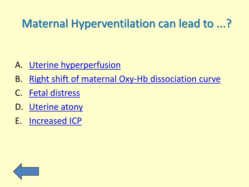
Maternal Hyperventilation can lead to ...?
A. Uterine hyperperfusion B. Right shift of maternal Oxy-Hb dissociation curve C. Fetal distress D. Uterine atony E. Increased ICP

Sorry – I disagree
Try again

CORRECT !!
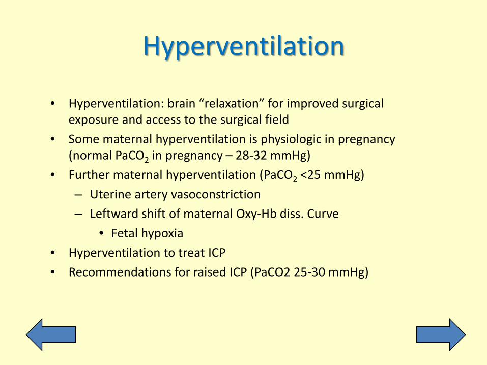
Hyperventilation
• Hyperventilation: brain “relaxation” for improved surgical exposure and access to the surgical field
• Some maternal hyperventilation is physiologic in pregnancy (normal PaCO2 in pregnancy – 28-32 mmHg)
• Further maternal hyperventilation (PaCO2 <25 mmHg) – Uterine artery vasoconstriction – Leftward shift of maternal Oxy-Hb diss. Curve
• Fetal hypoxia • Hyperventilation to treat ICP • Recommendations for raised ICP (PaCO2 25-30 mmHg)
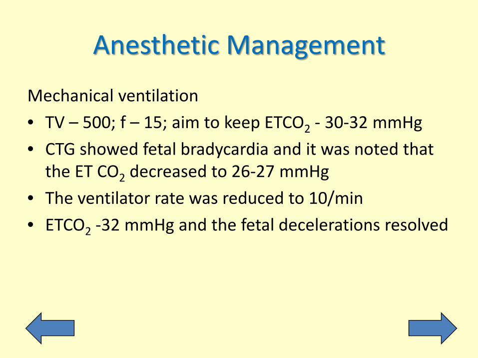
Anesthetic Management
Mechanical ventilation • TV – 500; f – 15; aim to keep ETCO2 - 30-32 mmHg • CTG showed fetal bradycardia and it was noted that
the ET CO2 decreased to 26-27 mmHg • The ventilator rate was reduced to 10/min • ETCO2 -32 mmHg and the fetal decelerations resolved
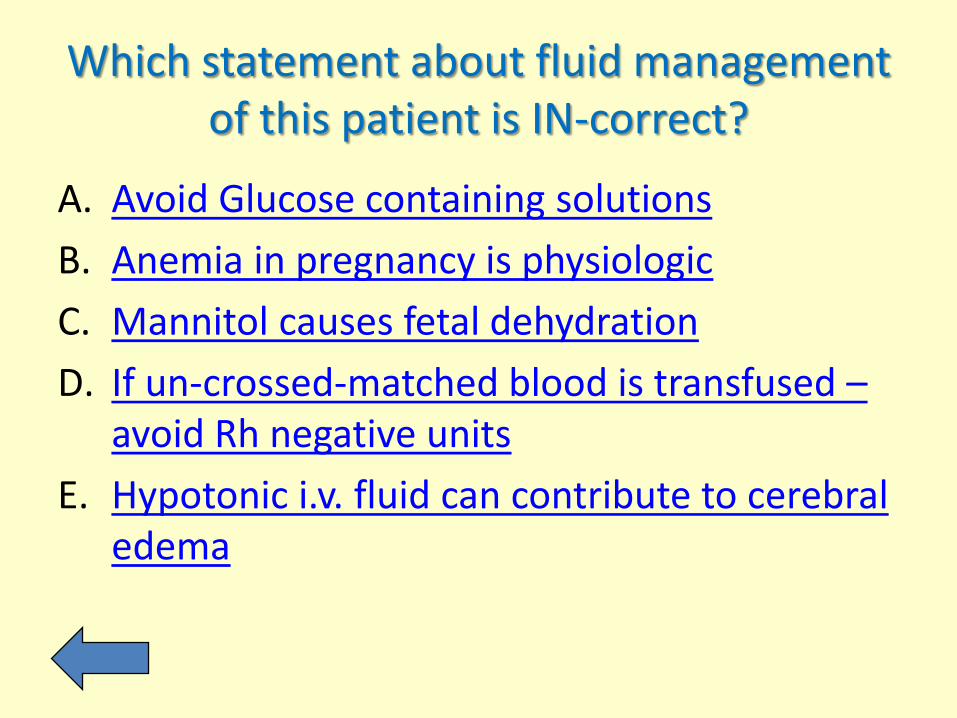
Which statement about fluid management of this patient is IN-correct?
A. Avoid Glucose containing solutions B. Anemia in pregnancy is physiologic C. Mannitol causes fetal dehydration D. If un-crossed-matched blood is transfused –
avoid Rh negative units E. Hypotonic i.v. fluid can contribute to cerebral
edema

Sorry – I disagree
Try again

CORRECT !!

Raised ICP – Fluids and Diuretics • Mannitol
– accumulates in the fetus – fetal hyper-osmolality – fetal dehydration – reduced fetal lung fluid – 0.5 mg/kg appears safe
• Furosemide – Additive effect, to be used cautiously
• IV fluids – Isotonic, glucose-free – Minimize the expansion of cerebral edema

Transfusion • Keep in mind: - physiologic (dilutional) anemia of pregnancy - fibrinogen is normally increased in pregnancy • If massive bleeding – high pre-defined FFP:RBC ratio to
avoid coagulopathy
• If no time to obtain typed/cross-matched blood, type-O, Rh-negative blood is recommended.
(Rh-negative to avoid Rh sensitization in childbearing aged women)
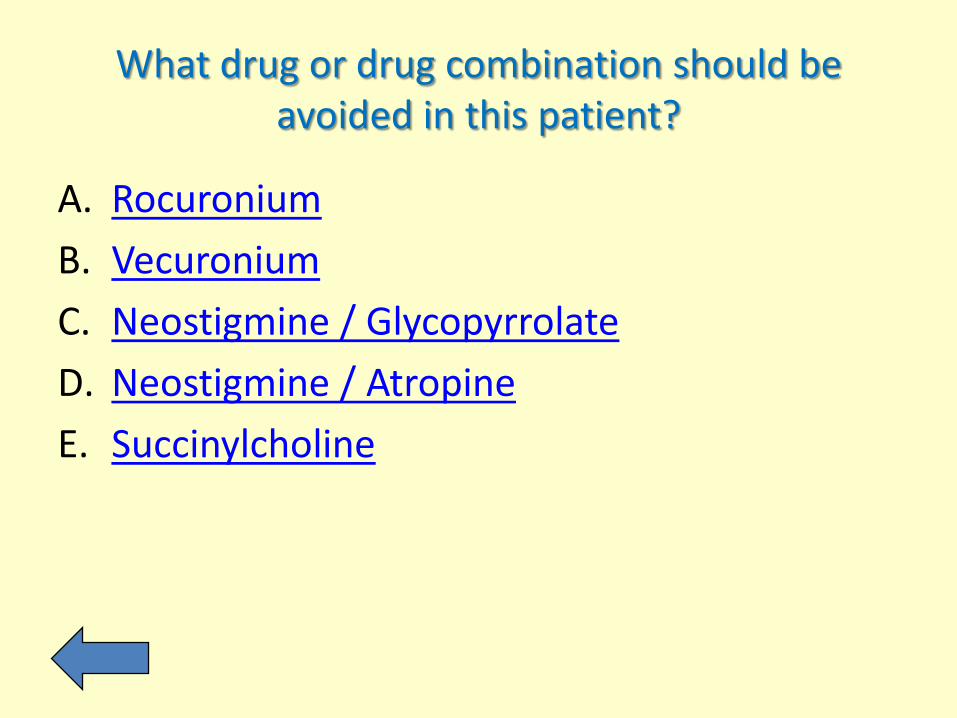
What drug or drug combination should be avoided in this patient?
A. Rocuronium B. Vecuronium C. Neostigmine / Glycopyrrolate D. Neostigmine / Atropine E. Succinylcholine

Sorry – I disagree
Try again

CORRECT !!
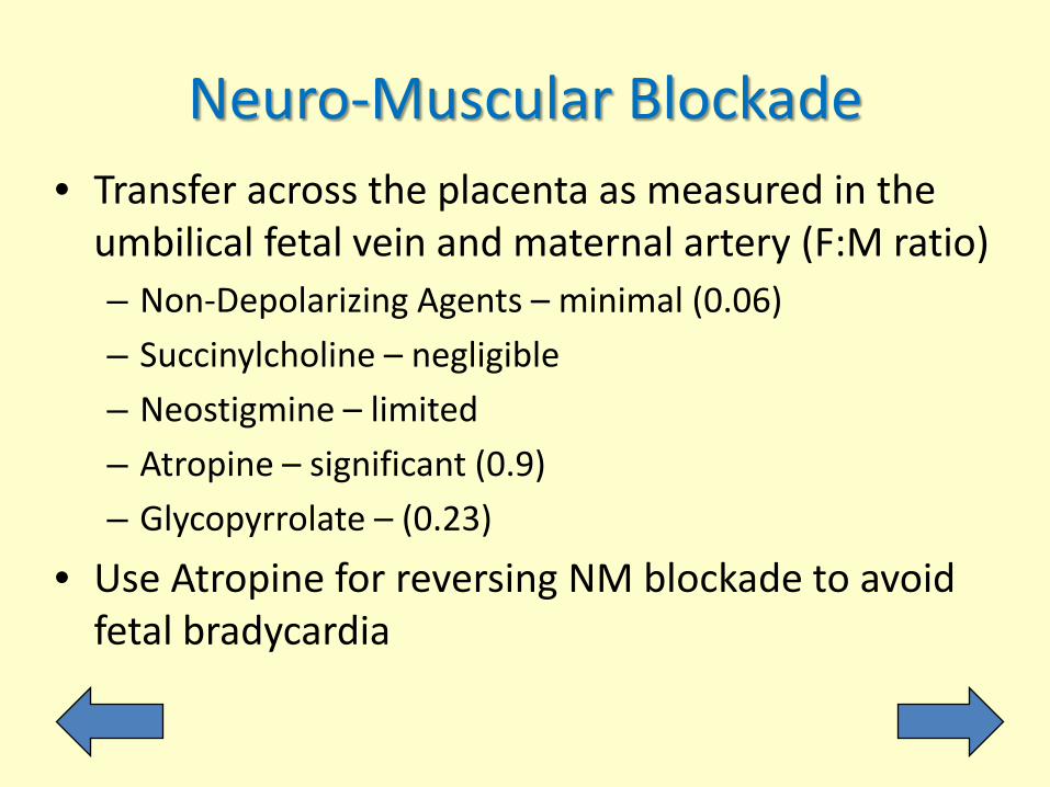
Neuro-Muscular Blockade • Transfer across the placenta as measured in the
umbilical fetal vein and maternal artery (F:M ratio) – Non-Depolarizing Agents – minimal (0.06) – Succinylcholine – negligible – Neostigmine – limited – Atropine – significant (0.9) – Glycopyrrolate – (0.23)
• Use Atropine for reversing NM blockade to avoid fetal bradycardia

Recovery and follow-up
• Extubated in the OR • Transferred to Neuro ICU on nasal oxygen
with monitoring • In the NICU, fetal monitoring was continued • Discharged home on 3rd post-op day • Uneventful C-Section at 38 wks under Spinal
Anesthesia
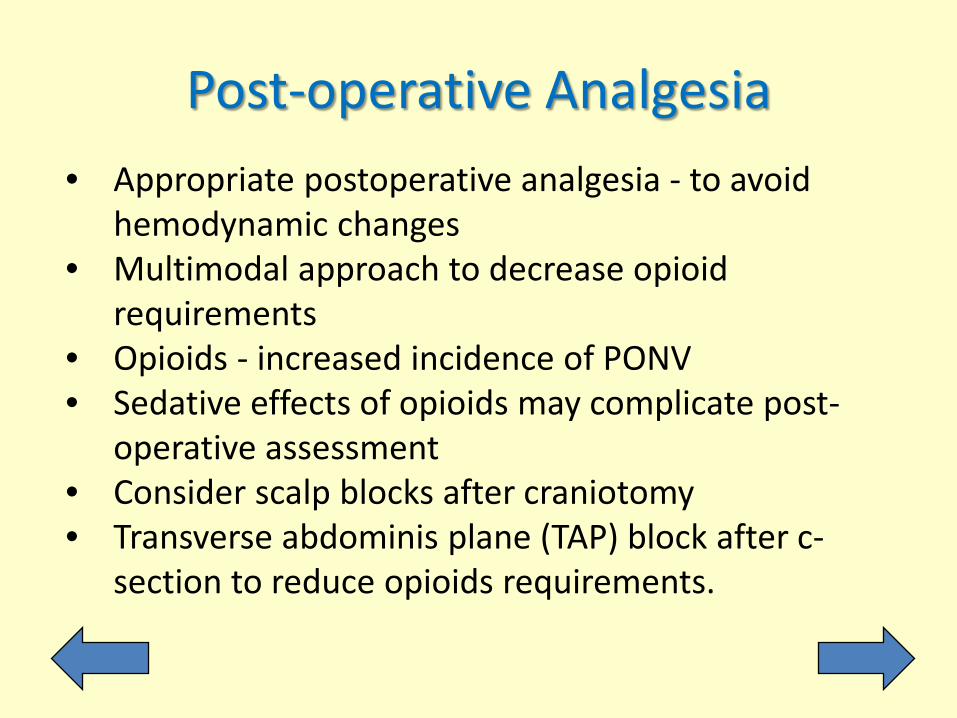
Post-operative Analgesia • Appropriate postoperative analgesia - to avoid
hemodynamic changes • Multimodal approach to decrease opioid
requirements • Opioids - increased incidence of PONV • Sedative effects of opioids may complicate post-
operative assessment • Consider scalp blocks after craniotomy • Transverse abdominis plane (TAP) block after c-
section to reduce opioids requirements.

Summary
• Optimize & Maintain – Normal maternal physiology – Utero-placental blood flow & oxygen delivery
• Avoid – Unwanted drug effects on the fetus – Stimulating the myometrium – Awareness during GA
References • Reitman E, Flood P. Anesthetic considerations for non-obstetric surgery during pregnancy.
BJA 2011; 107: i72-i78. • Kazemi P, Villar G, Flexman AM. Anesthetic management of neurosurgical procedures
during pregnancy: a case series. J Neurosurg Anesthesiology 2014; 26: 234-240. • Gupta A and Periakaruppan A: Intracranial dural arteriovenous fistulas: A Review. Indian J
Radiol Imaging. Feb 2009; 19(1): 43–48. • Wang LP, Paech MJ. Neuroanesthesia for the pregnant woman. Anesthesia and Analgesia
2008; 107: 193-200. • www.asahq.org: STATEMENT ON NONOBSTETRIC SURGERY DURING PREGNANCY.
Committee of Origin: Obstetrical Anesthesia (Approved by the ASA House of Delegates on October 21, 2009).
• Veeser M, Hofmann T, Roth R, Klöhr S, Rossaint R, Heesen M: Vasopressors for the management of hypotension after spinal anesthesia for elective caesarean section. Systematic review and cumulative meta-analysis. Acta Anaesthesiol Scand. 2012 Aug;56(7):810-6.