Centrosomes in Cytokinesis, Cell Cycle Progression and Ciliogenesis
128
University of Massachuses Medical School eScholarship@UMMS GSBS Dissertations and eses Graduate School of Biomedical Sciences 2004-09-08 Centrosomes in Cytokinesis, Cell Cycle Progression and Ciliogenesis: a Dissertation Agata Jurczyk University of Massachuses Medical School Follow this and additional works at: hps://escholarship.umassmed.edu/gsbs_diss Part of the Amino Acids, Peptides, and Proteins Commons , and the Cells Commons is material is brought to you by eScholarship@UMMS. It has been accepted for inclusion in GSBS Dissertations and eses by an authorized administrator of eScholarship@UMMS. For more information, please contact [email protected]. Repository Citation Jurczyk, A. Centrosomes in Cytokinesis, Cell Cycle Progression and Ciliogenesis: a Dissertation. (2004). University of Massachuses Medical School. GSBS Dissertations and eses. Paper 73. DOI: 10.13028/rt2g-d989. hps://escholarship.umassmed.edu/gsbs_diss/ 73
Transcript of Centrosomes in Cytokinesis, Cell Cycle Progression and Ciliogenesis

University of Massachusetts Medical SchooleScholarship@UMMS
GSBS Dissertations and Theses Graduate School of Biomedical Sciences
2004-09-08
Centrosomes in Cytokinesis, Cell CycleProgression and Ciliogenesis: a DissertationAgata JurczykUniversity of Massachusetts Medical School
Follow this and additional works at: https://escholarship.umassmed.edu/gsbs_diss
Part of the Amino Acids, Peptides, and Proteins Commons, and the Cells Commons
This material is brought to you by eScholarship@UMMS. It has been accepted for inclusion in GSBS Dissertations and Theses by an authorizedadministrator of eScholarship@UMMS. For more information, please contact [email protected].
Repository CitationJurczyk, A. Centrosomes in Cytokinesis, Cell Cycle Progression and Ciliogenesis: a Dissertation. (2004). University of MassachusettsMedical School. GSBS Dissertations and Theses. Paper 73. DOI: 10.13028/rt2g-d989. https://escholarship.umassmed.edu/gsbs_diss/73

A Disserttion Presented
Agata Jurczyk
Submitted to the Faculty of the
University of Massachusetts Graduate School of Biomedical Sciences , Worcester
In paral fulfilment of the requirements for the degree of
DOCTOR OF PHILOSOPHY
September, 8th 2004
INTERDESCIPLINARY GRADUATE PROGRAM

COPYRIGHT NOTICE
Work presented in this dissertation has appeared in the following publications:
1. Adam Gromley, Agata Jurczyk, James Siliboure, Ensar Halilovic , Mette
Mogensen Irna Groisman, Maureen Blomberg, and Stephen Doxsey. (2003) A
novel human protein of the maternal centroles is required for the final stages of
cytokinesis and entry into S phase. The Journal of Cell Biology, 161 (3):535-545
(A. Gromley and A. Jurczyk contributed equally to ths work).
2. Agata Jurczyk, Adam Gromley, Sambra Redick, Jovenal San Augustin, George
Witman, Gregory Pazour, Dorien J.M. Peters and Stephen Doxsey. (2004)
Pericentr forms a complex with intrafagellar transport proteins and polycystin 2
and is required for priary cilia assembly. The Joural of Cell Biology, 166
(5):637-643.
3. Agata Jurczyk, Adam Gromley, Sambra Redick and Stephen Doxsey.
Pericentrin is required for the final stages of cytokiesis and entr into S phase
though its association with centrolin. In preparation for publication.

(Signature)_____________________________William Theurkauf, Ph.D., Chair of Committee
(Signature)_____________________________Craig Peterson, Ph.D., Member of Committee
(Signature)_____________________________Dannel McCollum, Ph.D., Member of Committee
(Signature)_____________________________Greenfield Sluder, Ph.D., Member of Committee
(Signature)_____________________________ Stephen Doxsey, Ph.D., Thesis Advisor
(Signature)_____________________________ Anthony Carruthers, Ph.D.,Dean of the Graduate School of Biomedical Sciences

ACKNOWLEDGMENTS
I would like to thank my advisor Stephen Doxsey for his mentorship, good advice and
understanding of all my family emergencies. He always was flexible enough to let me
joggle between home and work, what was not easy at times , but also not impossible.
Special thans to all members of Doxsey and Theurkauf Labs for all the thoughtfl
discussions during joined laboratory meetings and helpful suggestion on my thesis
writing. Sorr, guys, but I stil did not get the rationale for use of the arcles. Special
thans to Sam, even though we did not work together too long, she help me a lot with
many of my experients presented in ths thesis. Thans to Adam for all of his
contrbutions to my thesis and for all the arguments. Jim' s expertse in biochemistry was
instrmental to most of my presented biochemistr, he did the "impossible" (at least for
me) cloning ofpericentr B. Than you Jim for all the apple cakes that you always had
handy for everybody s birdays. I also would lie to thank all my commttee members
for giving me advice and helpful feedback on my work. Finally, I would like to than my
famly and friends for always being there for me, I could not have done it without you!
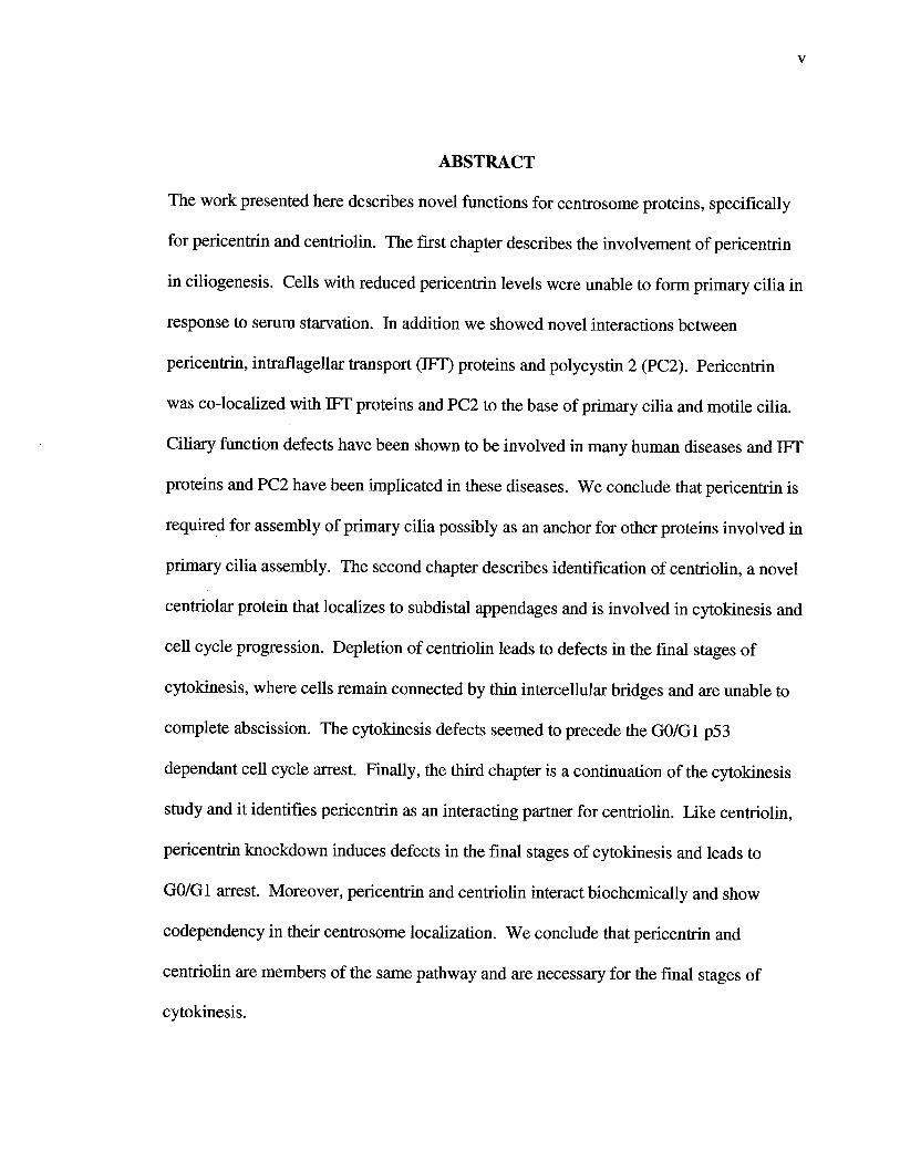
ABSTRACT
The work presented here describes novel functions for centrosome proteins, specifically
for pericentrn and centrolin. The first chapter describes the involvement of pericentrn
in ciliogenesis. Cells with reduced pericentrn levels were unable to form primary cilia in
response to serum staration. In addition we showed novel interactions between
pericentrn, intrafagellar transport (1F) proteins and polycystin 2 (PC2). Pericentrn
was co-localized with 1F proteins and PC2 to the base of priary cilia and motile cilia.
Ciliar function defects have been shown to be involved in many human diseases and
proteins and PC2 have been implicated in these diseases. We conclude that pericentrn is
required for assembly of primary cilia possibly as an anchor for other proteins involved in
primary cilia assembly. The second chapter describes identification of centrolin, a novel
centrolar protein that localizes to subdista appendages and is involved in cytokinesis and
cell cycle progression. Depletion of centrolin leads to defects in the fmal stages of
cytokinesis , where cells remain connected by th intercellular bridges and are unable to
complete abscission. The cytokinesis defects seemed to precede the GO/G 1 p53
dependant cell cycle arest. Finally, the third chapter is a continuation of the cytokinesis
study and it identifies pericentrn as an interacting partner for centrolin. Like centrolin
pericentrn knockdown induces defects in the final stages of cytokiesis and leads
GO/G1 arest. Moreover, pericentrn and centrolin interact biochemically and show
codependency in their centrosome localization. We conclude that pericentrn and
centrolin are members of the same pathway and are necessar for the final stages of
cytokinesis.

TABLE OF CONTENTS
COPYRIGHT
APPROVAL
ACKNOWLEDGEMENTS
ABSTRACT
TABLE OF CONTENTS
LIST OF FIGURES Vll
LIST OF ABREVIA TIONS V11
INTRODUCTION
CHAPTER I
CHAPTER n
CHAPTER il
DISCUSSION 100
REFERENCES 105
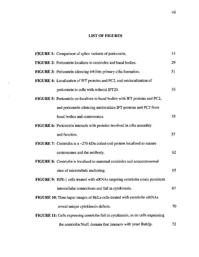
LIST OF FIGURES
FIGURE 1: Comparson of splice varants of pericentr.
FIGURE 2: Pericentrn localizes to centroles and basal bodies.
FIGURE 3: Pericentrn silencing inibits priary cilia formation.
FIGURE 4: Localization of 1F proteins and PC2, and mislocalization of
pericentrn in cells with reduced 1F20.
FIGURE 5: Pericentrn co-localizes to basal bodies with 1F proteins and PC2
and pericentrn silencing mislocalizes 1F proteins and PC2 from
basal bodies and centrosomes.
FIGURE 6: Pericentrn interacts with proteins involved in cilia assembly
and function.
FIGURE 7: Centrolin is a -270-kDa coiled-coil protein localized to mature
centrosomes and the midbody.
FIGURE 8: Centrolin is localized to maternal centroles and noncentrosomal
sites of microtubule anchoring.
FIGURE 9: RPE-1 cells treated with siRNAs targeting centrolin retain persistent
intercellular connections and fail in cytokinesis.
FIGURE 10: Time-lapse images of HeLa cells treated with centrolin siRAs
reveal unique cytokinesis defects.
FIGURE 11: Cells expressing centrolin fail in cytokinesis, as do cells expressing
the centrolin Nud1 domain that interacts with yeast Bub2p.
Vll
FIGURE 12: Cytokinesis failure in Xenopus embryos injected with centrolin
Antibodies.
FIGURE 13: siRAs targeting centrolin induces G 1/GO arest.
FIGURE 14: Pericentrin B localization to the contractile ring and the midbody
durng cytokinesis.
FIGURE 15: Pericentrin B exhibits co-dependency with centrolin at the centrosome
and co-localizes with it at the midbody.
FIGURE 16: Pericentrin and centrolin interact biochemically.
FIGURE 17: Pericentrin B co-localizes with endobrevin at the midbody ring
durg cytokinesis.
FIGURE 18: Pericentr B RNAi induces a late stage cytokinesis defects.
FIGURE 19: Pericentr B disruption causes GO/G 1 arest.
V11
LIST OF ABREVITIONS
DHClb - dynein heavy chain 1b
DIC - differential interference
y- TURC
- y-
tubulin ring complex
GCP2/3 - y-tubulin complex proteins 2 and 3
1FT - intrafagellar transport
IP - imunoprecipitations
MEN - mitotic exit network
MKLPI - M phase phosphoprotein 1
NEB - nuclear envelope breakdown
PCM - pericentrolar material
Pent - pericentrn
PC1I2 - polycystin 1 and 2 proteins
PKD1/2 - polycystic kidney disease genes 1 and 2
RPEI - retial pigmented epithelial cell line 1
SIN - septetion initiation network
siRNA - small interfering RNA
SNARE - soluble N-ethylmaleimide-sensitive factor attachment protein receptor
TACC - transformng acidic coiled-coil protein
INTRODUCTION
Centrosome Structure and Functions
The centrosome is a tiny organelle approximately 1- m in size located in the middle of
most animal cells. Over a century ago, Boveri, described the centrosome as an intensely
haematoxylin staining body surrounded by a radiating aster (Boveri, 1914). It is now
known that the centrosome contains a pair of centrioles surrounded by a dense
pericentrolar matr from which the micro tubules are nucleated. Like the DNA, the
centrosome is duplicated once durig the cell cycle in a semi conservative fashion. That
is, the mother centrole maintaining its existing tubulin serves as a template for the newly
generated one , which consists of both old and newly synthesized tubulin (Kochanski and
Borisy, 1990). Centrosome duplication begins at the GlIS boundar and it is completed
by mitosis when it serves as a spindle pole. As the cell completes mitosis each daughter
cell acquires one centrosome (Reviewed by (Sluder and Hinchcliffe, 2000)).
The two centroles of the centrosome are not identical and they are called mother and
daughter. The mother centrole can be distinguished by two sets of nine appendages at its
distal end that are responsible for the anchorage of astral microtubules (Bornens, 2002;
Mogensen et al., 2000). A subset of centrosome proteins were localzed to those
appendages and they include ninein (Mogensen et al., 2000) and centrolin (Gromley et
al., 2003). There are some cells that lack centroles (higher plants, some meiotic cells

and embryonic systems) and these cells are able to organize acentrolar microtubule-
organizing centers and a functional spindle, probably by using microtubule motors and
strctural proteins instead (Compton , 1998). When present, the centroles act in
dominant fashion to organize microtubules and spindles. The pericentrolar material
(PCM) surounds both centrioles formng an interconnected lattice like strcture which
provides a scaffold for proteins involved in microtubule nucleation (Dictenberg et al.
1998; Moritz et al. , 1995a; Vogel et al. , 1997). Microtubules are nucleated from the
PCM by the small and large y-tubulin ring complexes
(y-
TuRC), both capable of
nucleating microtubules (Zheng et al., 1995). Some organisms, like yeast, contan only
the small complex (Knop et al., 1997), whereas higher organsms, like mamals, contain
both complexes (Gunawardane et al. , 2000; Murphy et aI. , 1998).
In addition to its role in microtubule organization and nucleation, in recent years, the
centrosome was shown to playa very important role in cytokinesis, centrole duplication
cell cycle progression and cilogenesis (Gromley et al. , 2003; Hinchcliffe et al., 2001;
Jurczyk et al. , 2004; Keryer et al., 2003b; Khodjakov and Rieder, 2001). The best
ilustration of the importance of centrosome in cell cycle came from the experiments
where centro somes were removed either by microsurgery or laser ablation. Such cells
were still able to organze spindles and undergo presumably normal mitosis. However
half of these cells had problems completing cytokinesis and subsequently they arested
interphase of the next cell cycle (Hinchcliffe et al., 2001; Khodjakov and Rieder, 2001).
Another study correlated the final events of cytokinesis with the movement of the

maternal centrole to the intracellular bridge, which was necessar for abscission (Piel et
al. 2001). This idea came from budding yeast, where the spindle pole body (equivalent
of the mamalian centrosome) has to move to the bud, bringing into contact the GDP-
bound form of Tem1 and the guanine-nucleotide exchange factor, Lte1 to trgger
cytokinesis and exit from mitosis (Bardin et al. , 2000). Although the movement of the
centrole in the last stages of cytokinesis is similar in yeast and mamalian systems, the
molecular pathway in mamals has not been identified. Our work presented in chapter n
(Gromley et al. , 2003), identified centrolin as one of the possible molecular players in
this process. Centrolin contais a domain that is homologous to yeast Nud1/Cdc11
proteins implicated in exit from cytokiesis (reviewed in (Hinchcliffe, 2003)). More
work is necessar to identify additional molecular players if they are conserved between
yeast and mamals.
Other strctues associated with centroles are centrolar satelltes, which consist of
electron-dense spherical granules, about 70-100 nm in diameter. They are localized
around the centrosomes of many vertebrate cells and they can move along microtubules
in a dynein-dependent way (Balczon et al., 1994; Damerman and Merdes, 2002; Kubo
et al., 1999). PCM- 1localizes to satelltes and it was shown to control centrosome
assembly and microtubule organization (Damermann and Merdes, 2002).

Perieentrins
The centrosome proteins of pericentrin famy are encoded by alternatively spliced
products of one gene (Flory and Davis , 2003). Pericentrns are localized to the PCM
(Doxsey et al., 1994; Flory and Davis, 2003), where they are transported on microtubules
from the cytoplasm by the minus end directed motor, dynein, specifically though
interactions with the dynein light intermediate chain (Purohit et al. , 1999; Tynan et al.
2000). The best characterized isoforms of pericentr include pericentrn A
(approximately 250 kDa) and pericentr B/kendrin (approximately 380 kDa) (Fory and
Davis, 2003; Li et al. , 2001). Pericentrn A is homologous to pericentrn B thoughout
the entie protein sequence (Fory et al. , 2000) (Fig. 1). The C-terminus of pericentrn B
is unique and it contains a calodulin-binding region homologus to cerevisiae
Spc 11 Op, pombe Pcp 1 p (Fory et al. , 2002; Flory et al. , 2000), and drosophila
PLP/CP309 (Kawaguchi and Zheng, 2003; Marinez-Compos et al., 2004). The yeast and
Drosophila ortologs seem to be importnt for calmodulin-dependent attachment of
tubulin to the centrosome (Kawaguchi and Zheng, 2003; Knop and Schiebel, 1997;
Nguyen et al. , 1998; Sundberg et al. , 1996). Pericentrn B , together with CG-
NAP/AKA450, seem to mediate ths attachment in human cells (Takahashi et al. , 2002).
Although, recent data showed that pericentr A can also mediate the attachment of
tubulin in human cells though its interactions with components of y-tubulin ring complex
proteins GCP2 and GCP3 (Zimmerman et al. , 2004). The effect of pericentrns and its
ortologs on microtubule nucleation and organzation is controversial and probably
depends on the level of protein depletion. In Drosophila both y-tubulin and the

pericentrn B ortologue - CP309 are necessar for microtubule nucleation (Kawaguchi
and Zheng, 2003). Specific depletion of pericentrn B had no effect on microtubule
organization in mamalian cells (Li et al., 2001). However Xenopus aster formation
was inhibited with pericentrn B antibody (Takahashi et al. , 2002) and a decrease in the
density of nucleated micro tubules in pericentrn B knockdowns was reported for
pericentrn A and B depleted human cells (Damerman and Merdes, 2002). Pericentr
A and B was shown to be importt for spindle organzation and function (Doxsey et al.
1994; Zimmerman et al., 2004). It was postulated that pericentrn Band CG-
NAP/AKA450 may play redundant roles in mamalian cells and that is why no effect
on microtubule nucleation is observed when only one protein is depleted (Kawaguchi and
Zheng, 2003). It is also possible that residual protein in the knockdowns is sufficient to
sustai its activity or that different isoforms of pericentrn have specific and overlapping
functions. More work needs to be done in order to fully understad the differences
between pericentrn isoforms.
In addition to its role in microtubule and spindle function, pericentr ortologue in
Drosophila was shown to be important for cilia formation and function (Marez-
Compos et al., 2004). Other centrosome proteins including PCM- l (Kubo et al., 1999),
tubulin (Muresan et al., 1993) and centrn (Gavet et al., 2003) have been localized to
basal bodies, however their involvement in cilogenesis has not been determed. Cilia
play roles in motility and sensory reception. Their dysfunction contrbutes to cila-related
diseases and it wil be discussed below.
Cilogenesis
Cila and flagella are specialzed organelles which are anchored in the cell by the basal
body and whose axonemes protrde from the cell surrounded by the plasma membrane of
the cell (Rosenbaum and Witman, 2002). Cila can be classified as motile or nonmotile
based on their function. The motile cilia include flagella that propel cells and epithelial
border cila that beat synchronously and sti the extracellular fluid. Nonmotile cila, also
called priar or rudimenta, are found on alost every anmal cell. The priarcilium of renal epithelial cells serve as a flow mechano-sensor, which bend in response to
fluid flow and induces Ca2+ inux (praetorius and Spring, 2001). Polycystin 1 (PCl) and
2 (PC2) co-localze to the cila (pazour et al., 2002b; Yoder et al. , 2002) and function as
mechanotransducers that increase intracellular calcium in response to fluid flow
(McGrath et al., 2003; Nauli et al., 2003). PC 1 and PC2 are gene products of (Polycystic
Kidney Disease) PKDl and PKD2, respectively, and they are mutated in polycystic
kidney disease (Mochizuki et al. , 1996). PC2 forms a complex with PC 1 to generate a
unique calcium-permeable channel (Hanaoka et al., 2000). Polycystin-mediated
mechanotransduction is also necessar for the establishment of the left-right asymmetry
of visceral organs in mouse embryos (McGrath et al., 2003). In this case, the cilia are
located at the center of the node and produce movement that creates leftward fluid flow
(McGrath et al., 2003). This nodal flow is critical for the asymmetrc gene expression
that establishes the left-right asymmetr (McGrath et al. , 2003).

In terms of microtubule arangement, nonmotile cilia are nonmotile because they are
missing alost all strctures other than the nine outer doublets (9+0 arangement).
Motile cila contain the central pai of micro tubules (9+2 arangement) in addition to
dynein arms. The centrole at the base of cilium is commonly referred to as the basal
body and it grows satellites from its sides, a root from its base, and a cilium from its apex
(Sorokin, 1968). The satelltes and rootlets help to fix the position of the basal body
beneath the cell surface. However, rootlets are frequently formed by motie cila and
rarely by primar cila (Sorokin, 1968). The first steps in the formation of the cilium are
characterized by the appearance of the vesicles at the distal end of a basal body and the
cilar bud beneath it (Sorokin , 1962). The vesicle then lengthened and fused with the
plasma membrane (Sorokin, 1962).
The priar cilium arses directly off the micro tubules of mother centrole, whereas
motile cilia, such as the ones of the epithelial border of lungs, in addition to the centrole
templated growth can also form from strctures called 'deutrosomes ' (Soroki , 1968).
Deutrosomes are electron dense strctures that resemble centroles and up to 14
pro centroles can assemble, regularly spaced, about one central deutrosome (Sorokin
1968). Matue centroles are then separated from deuterosomes and migrate toward the
plasma membrane where they initiate cilia growth (Sorokin, 1968).

Intraflagellar Transport
Growth of the ciliar fiber occurs at the tips and axoneme subunits are delivered to the
tips by a process called intrafagellar transport (1F). This transport was first observed a
decade ago in the green alga Chlamydomonas at the laboratory of Joel Rosenbaum at
Yale (Kozminski et al., 1993). 1F cares protein particles between the outer doublet
microtubules and the membrane of flagella or cilia and it has been described in many
organsms including Chlamydomonas, Tetrahymena, C. elegans, sea urchin and mouse
(Rosenbaum, 2002; Rosenbaum and Witman, 2002). The 1F parcle consists of two
protein complexes, complex A (550 kDa) and complex B (710-760 kDa) (Cole et al.,
1998; Piperno et al., 1998). The relationship between complex A and B are not clear. It
was postulated that complex B proteins may be involved with anterograde transport
(toward the tip of cilia) and flagellar assembly and complex A proteins in retrograde
(from the tip to basal body) (Scholey, 2003). All of the pericentr-IF interactions
presented in chapter I include members of the complex B. The paricles are moved to the
tip of the cila by a plus-end-directed motor, kinesin-ll (Kozminski et al. , 1995; Walther
et al., 1994). Mutants with defects in kinesin-ll were shown to lose their flagella when
shifted to restrctive temperature (Walther et al., 1994). The retrograde movement of
paricles along the cilia is supported by cytoplasmic dynein Ib (pazour et al., 1998; Porter
et al., 1999) (reviewed in (Rosenbaum and Witman, 2002)). Chlamydomonas with a
deletion of DHClb had short flagella fIled with 1F proteins (pazour et al., 1999).

J.,
Mutations in 1F complex A or B or either one of the 1F motor complexes result in cells
that lack flagella or have short, defective flagella. For example, inducible RNAi of
proteins in Trypanosomes leads to the assembly of a shorter flagellum, that is
proportonal to smaller cell size (KoW et al., 2003). In this study the 1F depletion had
an effect only on the newly formed flagellum, the older one was not affected. This is in
agreement with earlier reports demonstrating that cilary micro tubules are stable
strctues which are able to withstand treatment by both nocodazole and colchicine, and
are not in a dynamc equilibrium with cytoplasmic subunits (Tilney and Gibbins, 1968).
However, more recently it has been shown that flagellar axonemal microtubules in sea
urchin, Chlamydomonas and sensory cila in C. elegans are dynamc strctures (Fujiwara
et al., 1999; Song and Dentler, 2001; Stephens, 1997; Stephens, 2000). The effect of
protein depletion on cilia length was also seen in many organisms including
Trypanosomes (KoW et al., 2003), Chlamydomonas (Brazelton et al., 2001; Kozminski et
aI., 1995), mouse (Murcia et al., 2000; Pazour et al., 2000), C. elegans (Cole et al. , 1998;
Perkins et al., 1986), and humans (pazour IF20 unpublished data).
In mamalian systems, defects in the 1F process as well as polycystins are associated
with polycystic kidney disease (pazour and Rosenbaum, 2002). The Chlamydomonas
1F88 ortologue in the mouse encoded by the Tg737 gene when mutated leads to
autosomal recessive polycystic kidney disease (Moyer et al. , 1994). In addition, the
mouse Tg737 gene product, polars, when mutated shows defects in assembly of the
primar cila in kidney (Pazour et aI. , 2000). One of the featues connecting both
and polycystin models of the kidney disease is defective or inconspicuous cila (pazour et
aI. , 2000; Thomson et al., 2003). Moreover, PC2 was shown to localize to the primary
cilia on kidney cells (pazour et al., 2002b) and since cilia are not present (or are
trncated) in the 1F88/polars mutats, ths suggests that functional PC2 containing ion
channels are also not assembled. Taken together, these observations suggest that kidney
priar cilia are important for proper kidney cell function and defects in the priar cilia
can lead to polycystic kidney disease.
Cytokinesis
Cytokinesis is defined as division of cytoplasm into two daughter cells following mitosis.
This process begins in anaphase with furrow ingression which proceeds until the
cytoplasm is constrcted to a narow bridge called midbody. The final stages of
cytokinesis consist of resolution of the midbody and abscission resulting in two daughter
cells. The cleavage fuow regression occurs as a result of constrction of actomyosin
ring, which is composed of actin and myosin II (Carer, 1967; Fujiwara et al., 1978). The
mechanism by which abscission is completed remains largely unkown. Chromosome
passenger proteins have been shown to be importat for cytokiesis , since their
disruption leads to cytokinesis failure resulting in multinucleated cells (Adams et al.,
2001; Bischoff and Plowman, 1999; Caralho et al., 2003; Gassman et al. , 2004; Lens et
al., 2003; Li et al., 1999; Mackay et al., 1998; Romano et al., 2003; Sampath et aI. , 2004;
Vagnarell and Earshaw, 2004).

After mitotic cells have been nearly separated by the ingression of contractile ring the
final separation of the two daughter cells requires a number of proteins and evens. The
proteins implicated in the final stages of cytokinesis include members of vesicular
trafficking and membrane fusion. Vesicle fusion has been well characterized in plant cell
division where a strctue called the phragmoplast is formed in the middle of the cell by
targeted secretion of post-golgi vesicles. Moreover, C.elegans embryos show defects in
late stages of cytokinesis upon treatment with brefeldin A, which inibits post-golgi
vesicle secretion (Skop et al., 2001).
More recent studies in mamalian cells of the SNARE (Soluble N-ethylmaleimde-
sensitive factor Attachment protein REceptor) complex showed the importce of vesicle
fusion during cytokinesis (Low et al., 2003). Overexpression of the components of
SNAR complex , specifically syntaxin-2 or endobrevin, result in cytokinesis faiure and
formation of binucleate cells. This complex of protein mediates the fusion of vesicles
and its target membrane (J ah and Sudhof, 1999). Completion of the late events of
cytokinesis also requires septins (Field and Kellogg, 1999). One of the members of
septin complex , CDCrel- l was shown to interact with the syntaxin, t-SNAR protein
(Beites et al., 1999). Thus, septi complexes may also facilitate the cytokinesis by
mediating the daughter cells membrane fusion (reviewed in (Schweitzer and D'Souza-
Schorey, 2004)).

Studies in both the budding and fission yeast implicated the involvement of exocyst
complex in cytokiesis. Exocyst complex is known to taget secretory vesicles to their
site of secretion (Hsu et al., 2004; Wang et al. , 2004). In the budding yeast, members of
the exocyst complex are localzed to the mother-bud neck suggesting a role in cytokinesis
(Finger et al., 1998). Cells overexpressing the exocyst component SEC15 accumulate
vesicles at the site of cytokiesis in S. cerevisiae (Salmnen and Novick, 1989). In
pombe the exocyst components are also localized to the site of cytokinesis. Moreover
the exocyst mutants accumulate vesicles at the septum and are defective in cleaving the
cells (Wang et al., 2002a).
Kiesin-related proteins, CHOl/MKLPl (Mitotic Kinase-Like Protein 1), RAB6-KIL
and MPPI (M Phase Phosphoprotein 1) are also essential for cytokinesis. Centralspindlin
complex consistig of MKLP- l/CHOI and the GTPase Activation Protein (GAP), was
shown to form a ring inside the actomyosin contractile ring, which controls furrow
ingression by regulation of the GTPase RhoA protein (Saint and Somers, 2003).
CHOl/MKLPl was also shown to block mitotic progression (Nislow et al., 1990) in
addition to disrupting the microtubule bundling at the midzone (Matuliene and Kuriyama,
2002). RAB6-KI was necessar for the final stages of cytokinesis (Fontijn et al.,
2001; Hil et aI. , 2000). Overexpression ofRAB6-KI resulted in cell death and cells
connected by a midbody remnant (Hil et al., 2000). In addition, antibody injection
experients lead to failure in cytokinesis after cleavage furrow ingression. RNA
interference of MPPI resulted in cytokinesis failure followed by apoptosis (Abaza et al.,

2003). How the kiases function to regulate cytokiesis is not known, however,
CHOl/MKLPl was shown to bind the ARF GTPases , which are involved in endosomal
cycling (Boman et al. , 1999; Schweitzer and D'Souza-Schorey, 2002). Therefore,
membrane vesicle trafficking could be one way to facilitate daughter membrane fusion
events.
Figure 1. Comparison of splice variants of perieentrin. Pericentrn B also known
kendrin is a bigger isoform ( -380 kDa) and pericentr A is a smaller isoform (-250
kDa). Boxes indicate exons present in both pericentrn A and B and lines indicate the
location of exons present in pericentrn B but not in pericentrn A. Arow heads on
pericentrn B indicate the positions of the siRNAs tagets used for silencing.

FIGURE 1
eDNA for human pericentri B (kendri)
cDNA for mouse perieentri A

Chapter I
Pericentrin Forms a Complex with Intraflagellar
Transport Proteins and Polycystin 2 and is required for
Primary Cilia Assembly

Abstract
Priar cilia are nonmotile microtubule strctures that assemble from basal bodies by a
process called intrafagellar transport (1F) and are associated with several human
diseases. Here we show that the centrosome protein pericentrn co-localizes with
proteins to the base of primary and motile cilia. Imunogold electron microscopy
demonstrates that pericentrn is on or near basal bodies at the base of cilia. Pericentrn
depletion by RNA interference disrupts basal body localization of 1F proteins and the
cation chanel polycystin-2 (PC2), and inibits priary cila assembly in human
epithelial cells. In addition silencing of several other centrosome proteins including
PCM- l and members of the y-tubulin ring complex proteins were able to inhbit primar
cilia formation. Ninein was the only centrosome protein that when silenced did not inhibit
priar cilia formation. Conversely, silencing of 1F20 mislocalizes pericentrn from
basal bodies and inhbits priar cila assembly. Pericentrn is found in spermatocyte
fractions, and 1F proteins are found in isolated centrosome fractions. Pericentrn
antibodies co-immunoprecipitate 1F proteins and PC2 from several cell lines and
tissues. We conclude that pericentrn, IFs and PC2 form a complex in vertebrate cells
that is required for assembly of priar cila and possibly motile cilia and flagella.

Introduction
Centro somes serve as microtubule organizing centers in interphase and mitotic cells and
playa role in cytokinesis and cell cycle progression (Doxsey, 2001). They are also the
precursors of priar cila, nonmotile sensory organelles found on most vertebrate cells.
Cilar dysfunctions are associated with several human diseases (pazour and Rosenbaum,
2002; Rosenbaum and Witman, 2002). Priar cilia in vertebrate cells appear to arse
from the mother centrole of the centrosome within a membrane sheath, which forms
from cytoplasmic vesicles and ultimately fuses with the plasma membrane (Sorokin
1968). The intimate relationship between the centrosome and the primary cilium
suggests that functions and components may be shared between these strctures.
Priar cilia assembly occurs by a process called intrafagellar transport (1F) (Han et
aI., 2003; Kozminski et al. , 1993; Pazour and Rosenbaum, 2002; Rosenbaum, 2002;
Rosenbaum and Witman, 2002). Interference with 1F protein function results in loss or
reduction of primary cila (pazour et al. , 2002a; Pazour et al., 2000; Pazour and
Rosenbaum, 2002). Priary cilia possess cation channels and receptors that appear to
activate signal transduction pathways that control cellular function (pazour et al., 2002a;
pazour and Rosenbaum, 2002; pazour and Witman, 2003). PC2 is a calcium-selective
channel on primary cilia associated with polycystic kidney disease (Somlo and Ehrlich,
2001). It appears to be activated by mechanical movement of priary cilia in response to
fluid flow (Nauli et al., 2003) and controls the assembly of primary cila (Thomson et al.,
2003; Watnick et al., 2003). However, little is known about the mechanism by which
proteins and PC2 are organzed at the centroleslbasal bodies (terms used
interchangeably).
A role for centrosome proteins in primary cila formation has recently been established.
Mutants of a Drosophila protein that shares homology with the vertebrate centrosome
proteins pericentrn (pcnt) (Fory and Davis, 2003; Zimerman et al. , 2004) and
AKA450 (Keryer et al., 2003a) disrupt formation of mechanosensory and chemosensory
cila (Marnez-Compos et al. , 2004). Drosophila mutats that affect 1F also disrupt
formation of Drosophila sensory cila (Han et al., 2003). However, the molecular
mechanism by which centrosomes and centrosome proteins modulate priar cilia
assembly has not been determned. In this study, we show that Pcnt forms a complex with
1F proteins and PC2 in vertebrate cells and tissues, and that Pcnt depletion by small
interfering RNAs (siRA) disrupts centrole association of IFs and PC2 and inhibits
priar cila formation.
Results and Discussion
In ths study, we have studied a larger isoform ofPcnt using specific siRAs and
antibodies unless otherwise noted (Flory and Davis, 2003; Zimmerman et al. , 2004).
Imunofluorescence imaging demonstrated that Pcnt parially overlapped with
centrolin, a protein associated with the mother centrole at centro somes (Fig. 2 a)
(Gromley et aI., 2003). In addition, Pcnt associated with both centroles at the base of

priary cilia (Fig. 2 b-c) and motile cila (Fig. 2 d). Higher resolution imunogold
electron microscopy demonstrated that Pcnt was on or near the centroles of motile cilia
(Fig. 2 e).
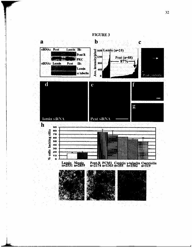
To test the role of Pcnt in cila organization we depleted protein levels by siRNA. We
observed a 75-90% reduction in protein levels and a dramatic reduction in centrosome
levels of Pcnt in most cells (Fig. 3 a-c, arow in c), when compared with cells treated with
control siRNAs targeting lamns A/C (Fig. 3 a-b) or cells that did not respond to siRA
treatment (Fig. 3 c, lower cell). In contrast, centrosome localization of'Y tubulin was only
slightly affected under these conditions (Fig. 3 c, upper cell). Priar cilia were induced
in RPEI cells treated with siRAs targeting Pcnt and were detected with antibodies to
polyglutamylated tubulins (GT335) (Gromley et al., 2003) and by differential
interference microscopy (DIC). In most cells treated with siRAs targetig Pcnt, priary
cilia failed to assemble (Fig. 3 e, g, h), while control cells treated with siRAs targeting
lamin or ninein assembled normal full-length primary cila (Fig. 3 d, f, h). In addition
silencing of other centrosome proteins including centrolin
, y-
tubulin, PCM- l and centrn
also inhibited the priar cila formation (Fig. 3 h).
To address the mechansm of ciliar loss in cells with reduced Pcnt, we examned
centrole function, strcture and composition. Consistent with previous results from our
group and others (Damerman and Merdes , 2002; Marinez-Compos et aI., 2004;
Zimmerman et al., 2004), we found that microtubule organization and nucleation were
k,,,.

not significantly disrupted. In addition, centrole ultrastrcture was normal (Fig. 3 i- , n
= 45 centrosomes). Centroles were sometimes separated (Fig. 3 e , g), but ths was also
observed following functional abrogation of proteins that did not affect primary cila (e.
ninein).
Since vertebrate priar cila formation and function requires 1F proteins (Murcia et al.,
2000; pazour et al., 2000) and the cation chanel PC2 (Nauli et al. , 2003; pazour et al.,
2002b; Rosenbaum and Witman, 2002; Somlo and Ehrlich, 2001), we reasoned that Pcnt
might cooperate with these proteins in priar cilia organzation. To test this , we fIrst
determned the precise localzation of these proteins. IF57 and 1F88 localized
priarly to the distal end of the mother centrole near the base of the priar cilium and
to the tips and in spots along the length of priar cilia (Fig. 4 a-b). Localization of these
IFs to the distal portion of the mother centrole was consistent with known sites of 1FT
protein localization in Chlamydomonas reinhardtii (Cole et aI., 1998; Deane et al. , 2001).
IF20 was found on the proximal portion of mother centrole and the lateral aspect of the
daughter centrole (Fig. 4 c), an area thought to be involved in interconnecting the two
centroles. PC2 localized priarly to the mother centrole underlying the primar cilium
(Fig. 4 d). In mouse tracheal epithelial cells, 1F proteins parally localzed with Pcnt
sites at the base of the motile cila where basal bodies are found (Fig. 4 e, 1F20).
We next addressed the centrolar anchorig mechansm of Pcnt, IFs and PC2. We found
that Pcnt was dependent on 1F proteins for localization to basal bodies using cells that

stably express siRNAs targeting 1F20 (Follit and Pazour, unpublished observations).
These cells showed reduced centrolar IF20 and lacked primar cilia (Fig. 4 g- , h),
compared with cells of the parent line (Fig. 4 f- , h). In cells with reduced centrole-
associated IF20, we observed a similar reduction in Pcnt levels (Fig. 4 g, g , i). In a
reciprocal experient, we found that IFs and PC2 were dependent on Pcnt for centrole
localzation. Pcnt localized to both centroles at the base of cila, parally co-localzed
with 1F proteins (Fig. 5 a , IF57) and totally overlapped with PC2 (Fig. 5 c ). Pcnt
silencing reduced the levels of centrolar Pcnt (Fig. 5 b, b" , c, c" upper cell), 1F57 (Fig.
5 b' - ) and PC2 (Fig. 4 c ' -c " upper cell). In contrast, adjacent nontransfected cells or
cells treated with lam siRNAs had robust staining for 1F57 and PC2 (Fig. 5 a ' -a , c
" lower cell). These results show that Pcnt and IFs are co-dependent in their
localzation to basal bodies of priar cilia.
Previous studies showed that 1F protein complexes and Pcnt complexes had simar S
values on sucrose gradients (17-18S) (Dictenberg et al. , 1998; Pazour et al. , 2002a). To
determne whether Pcnt interacted with 1F proteins , we isolated 1F complexes by a
multistep procedure (San Agustin and Witman, 2001) and found that Pcnt A and B
appeared to be in the same fractions with 1F88 in the final gel fitration column (Fig. 6
a). Based on recent data showing that 1F71 is present on centro somes and spindle poles
(Iomini et al., 2004), we analyzed centrosome preparations (Doxsey et al. , 1994) for the
presence of 1F proteins. 1F88 (Fig. 6 b) were present in pooled fractions contaning
centrosome proteins (y tubulin, Pcnt) but not in pooled fractions lacking centrosomes.

Imunoprecipitation of Pcnt using antibodies (that recognze both small, Pcnt A and
large, Pcnt B isoforms) raised to two independent domais pulled down endogenous
IF88 from two cilated cell lines (Fig. 6 c, two upper panels), ectopically expressed
GST-GFP-IF20 (Fig. 6 c, lower panel) and endogenous PC2 from mitotic cells (Fig. 6
d). We observed no co-imunoprecipitation of any 1F protein or PC2 when Pcnt
antibody was omitted (Fig. 6 d, Bd) or substituted with a nonimmune IgG (Fig. 6 c , e
IgG). a-tubulin antibody (dMla) was used as negative control for pcnt
imunoprecipitation. We observed no co-imunoprecipitation of a-tubulin with pcnt
and in the same imunoprecipitation reactions we were able to pull down endogenous
1F88 and pcnt (Fig. 6 f). In reciprocal experients, we found that PC2
immunoprecipitation pulled down endogenous IF57 from cilated cells (Fig. 6 e) and
that 1F57 pulled down 1F88 (Fig. 6 c). Taken together, these biochemical data suggest
that Pcnt, PC2 and 1F proteins form a complex in the cytoplasm of vertebrate cells.
The data in this manuscript show that Pcnt binds 1F proteins and PC2 and is required for
priar cilia formation in human cells. This suggests a model in which Pcnt recruits
protein complexes involved in cilia assembly and calcium signaling to centroles at the
base of priary cila (and perhaps flagella). Since Drosophila PcntJAKA450 and
were shown separately to function in primary cilia assembly (Han et al., 2003), it is
possible that Pcnt has a conserved function in 1F organization during cila formation in
both Drosophila and vertebrate cells.

1F does not appear to playa role in assembly or function of Drosophila sperm flagella
(Han et aI. , 2003) as seen in other organisms (Rosenbaum and Witman, 2002). Thus, it is
unlikely that defects in flagellar motilty in Drosophila PcntJ AKA450 mutants
(Marnez-Compos et al. , 2004) are a consequence of disruption of the Pcnt-
interaction. However, both vertebrate Pcnt (Dictenberg et al., 1998; Takahashi et al.
2002; Zimerman et al. , 2004) and Drosophila PcntJAKA450 (Kawaguchi et al., 2003)
interact with complexes containing 'Y tubulin , and 'Y tubulin has recently been shown to be
required for flagellar motility in trpanosomes (McKean et al. , 2003). Thus, it is possible
that disruption of the interaction between PcntJ AKA450 and 'Y tubulin complexes could
account for lack of motility in Drosophila flagella. Another possibilty is that the
observed strctural alterations in centroles in spermatocytes from Drosophila
PcntJAKA450 mutants (Marinez-Campos et al. , 2004) could contrbute to defects in
both cilia and flagella. However, in ths study we did not detect changes in centrole
strctue in cells depleted of Pcnt. Given the recent fmdings that Pcnt and other
centrosome proteins are integral components of cila and flagella and that 1F proteins
and PC2 are integral components of centro somes and spindle poles (lomini et al. , 2004) it
is likely that pertrbation of proteins in one of these comparents affects the function of
the other. Since defects in centrosome and spindle pole are well documented in cells with
abrogated Pcnt and Pcnt ortologs, they could contrbute to defects in centrosome
derivatives such as cila and flagella. It is clear from ths discussion that a better
understanding of the precise role of Pcnt in cilia and flagella assembly/function wil
require additional studies.
On a final note, it is interesting that centrosomes in Drosophila PcntJ AKA450 mutants
are disorganized but appear to assemble normal mitotic spindles (Marnez-Compos et al.
2004). It is possible that residual functional protein remaining in Drosophila mutants is
sufficient for spindle function. However, recent results from vertebrate cells indicate that
there are several forms of Pcnt (Flour and Davis, 2003), and that a smaller form of the
protein is required for spindle organization and function, possibly though its role in
anchoring ytubulin complexes or 1F proteins at spindle poles (Zimmerman et al., 2004).
A larger Pcnt isoform that shares homology with Drosophila PcntJ AKA450 does not
have a dramatic effect on spindle organzation (Zimerman et aI., 2004). It is likely that
the multiple Pcnt isoforms contrbute to a multitude of cellular functions.
Materials and methods
Cells, siRNAs, 1FT isolation and primary cila formation
Cells used in ths study, RPEI (Morales et al., 1999), a mouse inner medullar collecting duct
(IMCD3), primary cells isolated from Tg737 wild tye mouse (488) (pazour et al. , 2000) and
fresWy-isolated priar mouse trachea cells, were grown as described in ATCC. Trachea
dissected from mice in PBS were opened and scraped with a wooden applicator stick. Released
ciliated epithelial cells were spun onto cover slips and fIXed in - C methanol. siRAs (21-
nucleotide, Dhara on Research, Inc.) targeting Pcnt B (Gene Ban Acc. No. XM 036857,

nucleotides 301-319), Pcnt A/ or ninein (Damermann and Merdes, 2002) and lamn A/C
(Gromley et al., 2003), were delivered to cells at 200 nM (Oligofectame, Invitrogen). We also
used a stable RPEI cell line expressing IF20-specific siRNAs
(GGAAGAGTGCAAGACTTT) (Follt and Pazour, in preparation).
1F protein fractions were prepared as described (San Agustin and Witman, 2001) using
additional protease inhbitors A (Complete Mini tablets, Roche Diagnostics, Mannheim
Germany) in lysis buffer. The two testes were homogenized in 500 Jt of modified
homogenization buffer (35 mM NaHC03, 2 mM Na2HP04, 70 mM KCI, 74 mM NaCl
(McGrady, 1979J, 1 mM DTT, 0. 16% digitonin, 10 mM 4-amnobenzamdine, 2 mM EDTA, 50
~M leupeptin, 15 ~M pepstatin, 100 ~M TLCK, 1 ~g/ml aprotinin, Complete protease inhbitor
cocktail tablet (Sigma), pH 6.9). The clarfied testis extract was layered on top of a 12-ml 5-20%
sucrose gradient that had the same buffer composition as the homogenization buffer but without
digitonin, and then centrfuged in a Beckman L5-75 ultracentrfuge (36,000 rpm, SW41 rotor, 4
C). The cumulative centrfugal effect (ro t) was set at 6.4 x 10¹¹ radians /sec. About 24 fractions
(500 ~l each) were then collected, electrophoresed in SDS-PAGE gels, transferred to PVDF
membranes and probed with anti-1F antibodies. 1F-containing sucrose gradient fractions of
testis extracts from 6 mice were pooled , concentrated (Centrplus 10, Amicon), and applied to a
Superose 6 column (HR lOX30, AP Biotech,) that had been equilbrated with column buffer (30
mM Na2C03, 2 mM NaH P04, pH 6.9, 78 mM KCI, 70 mM NaCI, 2 mM EDTA, 1 mM DTT).
Fractionation flow rate was 0.2 mlmin. Fractions (500 ~l) were collected, electrophoresed in 5%

SDS-PAGE gels, transferred to PVDF membranes , and probed with anti-pericentrn and anti
1F88 antibodies.
Priary cilia were induced following siRA treatment (72 hours) by culturing RPEI
cells in medium with 0.25% serum and siRAs for 48 h and identified using GT335
antibody and DIC microscopy.
Immunofluorescence, electron microscopy and RT -PCR
Cells were prepared for imunofluorescence, imaged , deconvolved (Metaorph;
Universal Imaging Corp.) and displayed as two-dimensional projections of thee-
dimensional reconstrctions to visualze the entire cell volume as described in (Gromley
et al. , 2003). We used methanol as fixative, then confIrmed using formaldehyde fixation
as previously shown (Dictenberg et al., 1998). Imunogold electron microscopy was
performed as described (Doxsey et al. , 1994). RT -PCR for amplification of Pcnt B
(forward prier 5' -AACACTCTCCA TGATTGCCC and reverse 5'
TACCCTCCCAATCTTTGCTG) and ex tubulin was performed as described (Gromley et
al., 2003).
Immunoprecipitation, Western Blotting and Antibodies
Cells were lysed in lysis buffer consisting of 50mM Tris HCI (pH 7.5), 10 mM Na HP0
(pH 7.2), 1 mM EDTA , 150 mM NaCI , 1 % IGEPAL CA-1630 and Complete Mini
tablets. Testes lysates were prepared as described (San Agustin and Witman, 2001).

'S-
;-.
Antibodies were added to fresWy prepared cell extracts and incubated at 4 C overnght.
Protein A/G Plus-Agarose (Santa Cruz Biotechnology) or Glutathione Sepharose 4B
(Amersham Pharacia Biotech AB, Upsala Sweden) were washed in lysis buffer, added
to the cell extracts and incubated 2 hat 4 C. The beads were washed and resuspended in
sample buffer. 5% SDS-PAGE gels were used to detectPcnt and PC2, 10 % gels to
detect IFs. Controls included cell extracts incubated with rabbit IgG or beads alone.
No bands were seen with control IgGs under any of these conditions or when control
IgGs were used at concentrations lO-fold higher than experiental samples. Cell
extracts used in this study for the pericentr 1F interactions came from cells grown in
25% serum for 48 h to induce cilia formation. We used affinity-purified antibodies
against: the N- and C- termni ofPcnt A/ (pcN, PcC) (Dictenberg et al. , 1998; Doxsey
et al., 1994), Pcnt B (Fory et al. , 2000), 1F proteins (pazour et al. , 2002a), PC2
(Scheffers et al. , 2002), centrolin (Gromley et aI. , 2003), GT335 (Wolff et al., 1992),
tubulin (Sigma-Aldrch), and lam A/C (Cell Signalng Technology).

Figure 2. Perieentrin localizes to centrioles and basal bodies. (a)
Imunofluorescence image of a centrosome in RPEI cells co-staned for Pcnt (green)
and centrolin (red, bar, 1 ~m). (b, c) DIC (b) and immunofluorescence (c) images of a
primar cilum (arow) in RPEI cell stained for Pcnt (green) and centroles/priar
cilium (GT335, red). Bar, 5 ~m for b, c. (d) Imunofluorescence image of a cilated
epithelial cell from mouse trachea showing Pcnt (green) at the base of motile cilia (DIC,
bar, 5 ~m). (e) Imunogold electron microscopic image of a ciliated cell (as in d) after
incubation with antibodies to Pcnt and secondar antibodies bound to 5 nm gold (bar, 250
nm). Stephen Doxsey contrbuted the pericentrn electron microscopic image and Agata
Jurczyk contrbuted the rest.
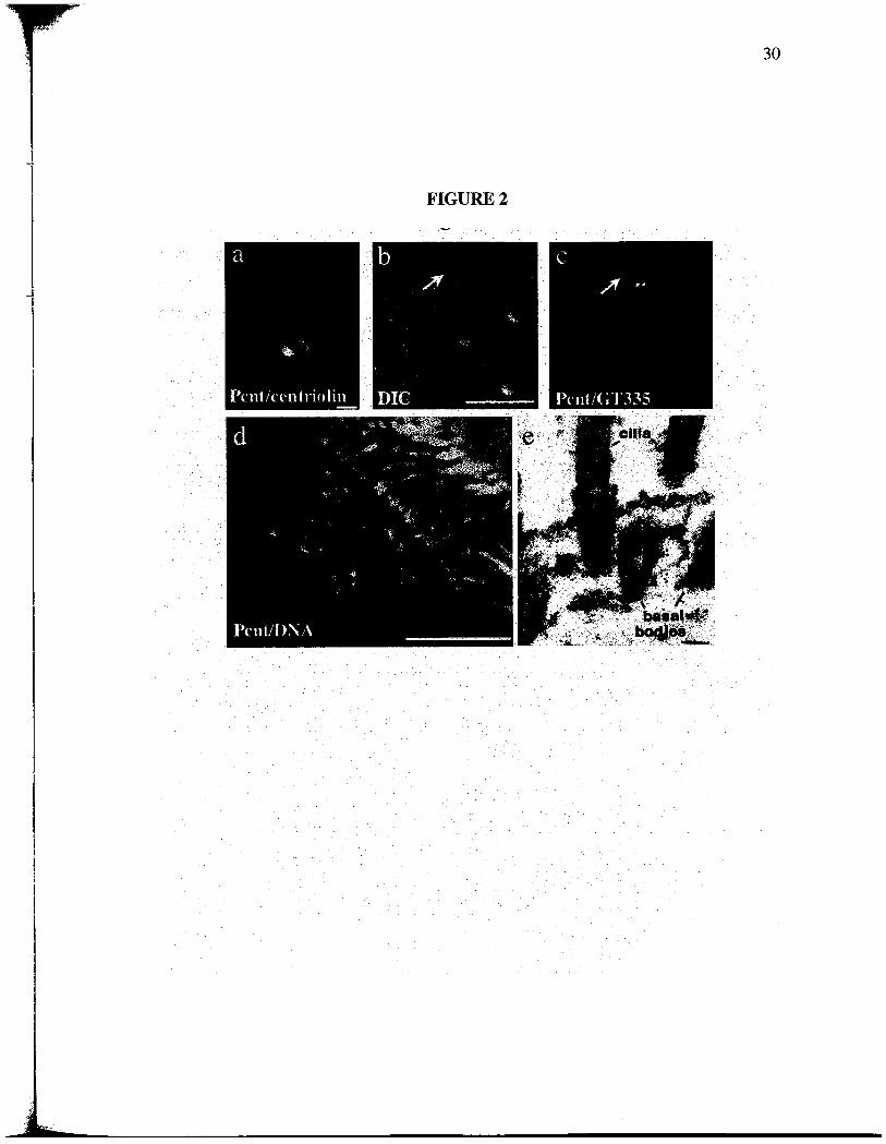
FIGURE 2

~~~
Figure 3. Pent silencing inhibits primary cila formation. (a) Pcnt and lamn protein
levels (a, Western blot) following siRNA as indicated. a tubulin or PKC, loading
controls. (b) Fluorescence intensity of individual centro somes (bars) after treatment with
siRNAs tageting Pcnt or lamn. Centro somal Pcnt is reduced to levels below the lowest
control levels (lamn) in 87% of cells. (c) Imunofluorescence image ofRPEl cells after
Pcnt silencing showing reduced centrosomal Pcnt in one cell (green, arow) and normal
level in the other. 'Y tubulin (red) is not significantly afected. Low (d, e) and high
magnification (f, g) immunofluorescence images of cilia and centroles staned with
GT335 after treatment with Pcnt (e, g) or lamn (d, f) siRAs. Bar in e, 5 ~m for d, e, or
in f, 1 ~m for f, g. DNA, blue. (h) Graph showing percent of cells that lack cila after
treatment with indicated siRNAs. Bars represent average of 3 experients. P value,
stadard T -test. (i-k) Electron micrographs showing centrole strctue in cells with
reduced Pcnt (bar in k, 200 nm for i-k). Gregory Hendricks did the electron microscopy
for figures 3 i-k and Agata Jurczyk contrbuted the rest.
...
S 80:o 70
15 40- 30= 20
FIGURE 3
Pcnt,.B PCMICentrn y-tubulinCetrolinn=2174 n=1303 n=355 n=1502 n=519
-'1
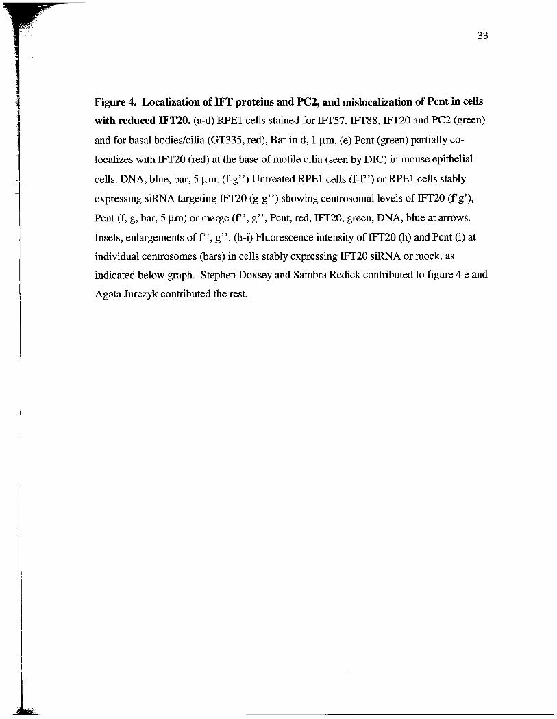
Figure 4. Localization of 1FT proteins and PC2, and misloealation of Pcnt in cells
with reduced IFT20. (a-d) RPEI cells staned for IFT57, IF88, IF20 and PC2 (green)
and for basal bodies/cilia (GT335, red), Bar in d, 1 ~m. (e) Pcnt (green) parially co-
localzes with IF20 (red) at the base of motie cilia (seen by DIC) in mouse epithelial
cells. DNA, blue, bar, 5 ~m. (f- ) Untreated RPEI cells (f- ) or RPEI cells stably
expressing siRA targeting 1F20 (g- ) showing centrosomallevels of IF20 (f'g
Pcnt (f, g, bar, 5 ~m) or merge (f" , g , Pcnt, red, 1F20, green, DNA, blue at arows.
Insets, enlargements of f" , g . (h-i) Fluorescence intensity of 1F20 (h) and Pcnt (i) at
individual centrosomes (bars) in cells stably expressing IF20 siRNA or mock, as
indicated below graph. Stephen Doxsey and Sambra Redick contrbuted to figure 4 e and
Agata Jurczyk contrbuted the rest.

FIGURE 4
hCentrosome intensit, 1F0
1000
o IF20 Mock
iCentrosome intensit, Pcnt1000
60 .
wno

Figure 5. Pcnt co-localzes to basal bodies with 1FT proteins and PC2, and Pent
silencing misloealizes 1FT proteins and PC2 from basal bodies and eentrosomes. (a-
) 1F57 (a-b" red) and PC2 (c-c , red) are mislocalized from basal bodies in RPEI cells
with reduced Pcnt (b- , arows, c-c , green, small arows) but not in RPE 1 cells treated
with lamn siRAs (a-a , bar, 10 fl) or in the cell with control level ofPcnt (c, c
bottom). Insets higher magnification of a , b" , c" as indicated by arows. DNA , blue.
Agata Jurczyk contrbuted the entie figure.

FIGURE 5

;:;
Figure 6. Pent interacts with proteins involved in cilia assembly and function. (a)
Pooled 1F fractions from a sucrose gradient from mouse testes were applied to an FPLC
Colum and fractions were loaded on SDS gels and probed with Pcnt antibodies or IF88
antibody. (b) Pooled peak centrosome fractions from sucrose gradients (+) containing
tubulin, Pcnt and IF88 as indicated and pooled noncentrosome fractions (-). (c, upper
panel) Pcnt N- and C-termnal antibodies (pcN, PcC) independently imunoprecipitated
endogenous IF88 from lysates of cilated 488 cells. IgG, nonimune rabbit IgG, lysates
(Lys) showing IF88 at right. Pcnt imunoprecipitation confIrmed, right. (c, middle
panel) PcN/C imunoprecipitated 1F88 from ciliated RPEI cells , as did antibodies to
1F88 and IF57 but not rabbit IgG. (c, bottom panel) PcN and PcC pull down a GST-
GFP-IF20 fusion protein from a cell line stably overexpressing the protein, as does a
glutathione column (1F20), but not nonimune IgG. Blots were probed with anti-GFP
antibodies, immunoprecipitation with Pcnt (right panel), IB , immunoblot antibody. (d)
PcN imunoprecipitated PC2 from mitotic RPEI cells while beads alone do not (Bds).
Pcnt imunoprecipitation confIrmed by immunoblot (Pcnt B). (e) PC2 antibody, but not
Rabbit IgG, imunoprecipitated 1F57 from cilated 488 cells. IF57, upper band.
Lower band, antibody heavy chain. (f) PcC/N imunoprecipitated endogenous 1F88
from lysated of ciliated RPEI cells and not a-tubulin. Lysate (Lys) indicate the starng
material, supernatants of the imunoprecipitations are shown to the left of each
immunoprecipitations. A hal of the imunoprecipitations were loaded and 3% of
immunoprecipitations are shown in the supernatatnts and lysates with an exception in d.
in figure 6 d all the imunoprecipitations were loaded and 7% of immunoprecipitations
are shown in the lysates. Jovanal San Agustin contrbuted figure 6 a, Keith Mikule
provided centrosome fractions for figure 6 b, and Adam Gromley contrbuted figure 6 d
and Agata Jurczyk contrbuted figures 6 b-c , e , and f.

-.-
FIGURE 6
1FT fractionsIFT88
567.89101.112l3J4
..
w - .
*'''.-,;
- 92
Pent A andS1234 5 6 789101112 13 14
; . *' .. . -. .
. J' -214kD
16UkD
cilated cells IP 488 tii ;ii;:
PeC/N IgG
IFT88
t;7
&;'~~~
i'I! f!0.:;P, IFT88
GFP-IF20
f ciltedce& IP IP Lvs SupRd Sup88 SupPcC/N
:;;,, ~~~
i;;
:;: ~~~
Centrosomefractions
'-ytubulinPcntA
IFT88
PC PN I
. . . .. . +:
Pent B
PeNPeCIgG
, '
, Pent B
IgG::LYS
1FT 88
RPEI

Chapter
A Novel Human Protein of the Maternal Centriole is
required for the Final Stages of Cytokinesis and Entry
into S phase
o;t'1"'.
\ " ;':', : " -
-:1
Abstract
Centrosomes nucleate microtubules and contrbute to mitotic spindle organzation and
function. They also parcipate in cytokinesis and cell cycle progression in ways that are
poorly understood. Here we describe a novel human protein caled centrolin that
localizes to the maternal centrole and functions in both cytokinesis and cell cycle
progression. Centrolin silencing induces cytokiesis failure by a novel mechansm
whereby cells remai interconnected by long intercellular bridges. Most cells continue to
cycle, reenter mitosis, and form multicellular syncytia. Some ultimately divide or
undergo apoptosis specifcally durng the protracted period of cytokinesis. At later times,
viable cells arest in G lIGO. The cytokinesis activity is localized to a centrolin domain
that shares homology with Nudl p and Cdc11 p, budding and fission yeast proteins that
anchor regulatory pathways involved in progression though the late stages of mitosis.
The Nudl p-like domain of centrolin binds Bub2p, another component of the budding
yeast pathway. We conclude that centrolin is required for a late stage of vertebrate
cytokinesis, Perhaps the final cell cleavage event, and plays a role in progression into S
phase.

Introduction
Centro somes are the major microtubule-nucleating organelles in most vertebrate cells
(Doxsey, 2001b). 1n mitosis, they contrbute to spindle organzation and function, and in
interphase, they organize microtubule arays that serve as tracks for transportng proteins
organelles, and chromosomes. The centrosome also anchors regulatory molecules and
may serve as a central site that receives, integrates, and transmits signals that regulate
fundamental cellular functions. The core of the centrosome is comprised of a pai of
centroles , microtubule barels that appear to anchor microtubules (Chretien et al. , 1997;
Piel et al., 2000). Each centrole is surrounded by pericentrolar material or centrosome
matrix, which nucleates the growt of new microtubules and seems to be organized by
the centroles (Bobinnec et al. , 1998). Although best known for their role in microtubule
nucleation, recent data suggest that centrosomes also play key roles in cytokinesis and
cell cycle progression.
A role for centrosomes in defming the site of cell cleavage during cytokinesis has been
suggested for some tie (Rappaport, 1986). Recent studies with vertebrate cells provide
evidence for a direct link between centrosome activity and completion of cytokinesis.
Elimnation of centrosomes from interphase cells by removal with a microneedle
(Hinchcliffe et al., 2001) or from mitotic cells by laser ablation (Khodjakov and Rieder
2001) caused cytokiesis defects, arest, or failure. In another study, it was shown that
during the final stages of cytokinesis, the maternal centrole moved to the intercellular
bridge, the microtubule-filled interconnection between nascent daughter cells (piel et al.,
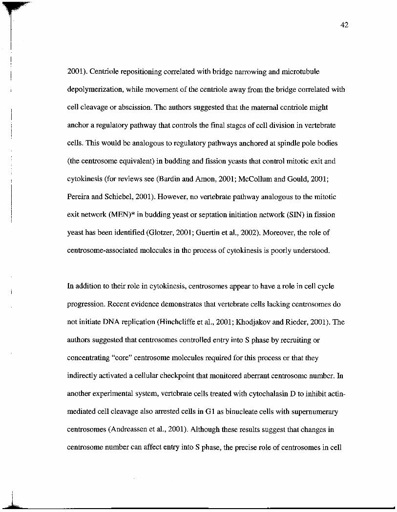
2001). Centrole repositioning correlated with bridge narowing and microtubule
depolymerization, while movement of the centrole away from the bridge correlated with
cell cleavage or abscission. The authors suggested that the maternal centrole might
anchor a regulatory pathway that controls the final stages of cell division in vertebrate
cells. Ths would be analogous to regulatory pathways anchored at spindle pole bodies
(the centrosome equivalent) in budding and fission yeasts that control mitotic exit and
cytokinesis (for reviews see (Bardin and Amon, 2001; McCollum and Gould, 2001;
Pereira and Schiebel, 2001). However, no vertebrate pathway analogous to the mitotic
exit network (MEN)* in budding yeast or septation initiation network (SIN) in fission
yeast has been identified (Glotzer, 2001; Guertn et al. , 2002). Moreover, the role of
centrosome-associated molecules in the process of cytokinesis is poorly understood.
In addition to their role in cytokinesis , centrosomes appear to have a role in cell cycle
progression. Recent evidence demonstrates that vertebrate cells lackig centrosomes do
not initiate DNA replication (Hinchcliffe et al. , 2001; Khodjakov and Rieder, 2001). The
authors suggested that centrosomes controlled entr into S phase by recruiting or
concentrating "core" centrosome molecules required for this process or that they
indirectly activated a cellular checkpoint that monitored aberrant centrosome number. In
another experimental system, vertebrate cells treated with cytochalasin D to inhibit actin-
mediated cell cleavage also arested cells in G 1 as binucleate cells with supernumerar
centro somes (Andreassen et al., 2001). Although these results suggest that changes in
centrosome number can affect entr into S phase , the precise role of centrosomes in cell

1il
cycle progression in vertebrate cells wil require identification of the molecular
components and pathways that control these events.
In this paper, we identify a novel component of the vertebrate maternal centrole called
centrolin. Abrogation of centriolin function by small interfering RNA (siRNA) silencing,
overexpression, or antibody inhibition produces cytokinesis failure and G lIGO arest, just
as seen when centrosomes are experientaly eliminated from cells. Centrolin silencing
produces a novel cytokinesis phenotype in which dividing cells remain interconnected by
long strands of cytoplasm and fai to cleave. The cytokinesis activity lies in a centrolin
domain that is homologous to the MEN/SIN components Nudl p/Cdc 11 p and binds the
Nudlp-interacting GTPase activating protein Bub2p. We conclude that centrolin is
required for a distinct step in the final stages of vertebrate cytokinesis and can influence
entr into S phase.
Results
Identification and cloning of a novel protein localized to the maternal centriole and
intercellular bridge
U sing sera from patients with the autoimune disease scleroderma that react with
centrosomes (Doxsey et al., 1994), we screened a human placenta gt11 cDNA expression
librar to identify genes encoding the autoantigens. Of the 3106 clones screened, only
one of 1.7 kb was identied, indicating that the mRNA for ths molecule was rare. The
full-length cDNA was obtaned, and the protein encoded by the cDNA was called

'i'
--r
centrolin (see below and Materials and methods). The amno acid sequence of centrolin
predicted a protein with several coiled-coil regions interrpted by noncoiled domais
(Lupas, 1991) (Fig. 7 a). Two domains within the centrolin sequence shared homology
with human oncogenic transformng acidic coiled-coil proteins (TACCs), which localize
to centro somes and are implicated in microtubule stabilzation and spindle function (Lee
et al., 2001). Another domain of centrolin was homologous to human stathn, an
oncogenic protein involved in microtubule destabilization (Andersen, 2000). The carboxy
terminus of centrolin was identical to CEPII0, a natualy occurrg fusion to the
fibroblast growth factor receptor that localizes to centrosomes, is oncogenic , and is of
unkown function (Guasch et al., 2000). The relationship of CEPIlO to centrolin is
unkown, although we did not identify a cDNA corresponding to this protein in our
librar screens. A region of centrolin near the amno termnus shared homology with
Nudlp and Cdc11p, budding and fission yeast spindle pole body proteins that anchor
components of the yeast MEN and SIN, respectively, and are required for completion of
mitosis and cytokinesis (Bardin and Amon, 2001; McColluffand Gould, 2001; Pereira
and Schiebel, 2001 ;Guertin et al., 2002). The 120-amo acid region of shared homology
between the Nudl domain, Nudlp, and Cdcllp al have leucine-rich repeats and are
predicted to form B helix strctues in tertar strctue prediction programs. Antibodies
raised against recombinant centrolin recognized a band of -270 kD on Western blots of
isolated centrosome fractions, whereas preimune sera showed no specific bands (Fig. 7
b, Centrosome fractions). In vitro translation and overexpression of the protein in
mammalian cells using the full-length cDNA produced a protein with a molecular weight

"':1
- -
I;I
simlar to the endogenous protein (Fig. 7 b, In vitro translated and Overexpressed) and to
a protein predicted from the cDNA sequence (see Materials and methods).
Imunofluorescence microscopy demonstrated that centrolin was localzed to
centro somes in a wide varety of species, including human , monkey, hamster, mouse, and
Xenopus (Figs. 7 and 8). Centrosome localization was confIrmed by showing that an HA-
tagged centrolin protein ectopically expressed in COS cells localized to centrosomes
(Fig. 8 a). The endogenous protein was present on the centrosome thoughout the cell
cycle. In late G lIearly S phase, centro somes begin to duplicate, and by G2/M, duplication
is usually completed. Durng the duplication process , centrolin was present on only one
of the two duplicating centrosomes, although other proteins, such as y-tubulin, were
found on both (Fig. 7 c, G2 cell). Beginning at late G2/prometaphase, dim staining was
observed next to a brightly staied centrosome. By metaphase , when centrosomes
become "mature," both centro somes had equally high levels of centrolin and were more
brightly stained than at any other cell cycle stage. This demonstrates that centrolin is a
marker for centrosome maturation, a characteristic shared with cenexin (Lange and Gull
1995) and ninein (Mogensen et al., 2000). At the metaphase to anaphase transition,
centrolin staining diminished at centro somes and reached its lowest levels by late
anaphase/telophase. Durng cytokiesis, centrolin sometimes appeared as one or two
dots adjacent to the intercellular bridge, suggesting that the centrosome/centrole had
moved to this site (Fig. 7 c , Telo early). This staining pattern was consistent with recent
time-lapse imaging experients showing that the maternal centrole translocates to the
intercellular bridge during cytokinesis (Piel et al., 2001). Centrolin next appeared as

diffusely organized material within the intercellular bridge and ultiately became,Ii
concentrated at the midbody (Fig. 7 c, Telo late). The organzation of centriolin at the
centrosome was more precisely determied by serum starving cells to induce growth of a
priar cilium from the maternal centrole (Vorobjev and Chentsov, 1982). In these
cells, centrolin staning was confined to the maternal centrole underlying the cilium
(Fig. 8 b). lmunogold electron microscopy on centrosome fractions (Doxsey et al.
1994; Blomberg and Doxsey, 1998) confIrmed localization to the maternal centrole (Fig.
8 e, M) and fuer demonstrated that the protein was concentrated on subdistal
appendages, specialized substrctues of the maternal centrole implicated in microtubule
anchoring (Fig. 8, c-e) (Chretien et al. , 1997; Piel et al. , 2000). Based on its centrolar
localzation, the protein was named centrolin. Centrolin was also found at
noncentrosomal apical bands of material in specialized epithelial cells that lack proteins
involved in microtubule nucleation and appear to anchor the minus ends of microtubules
(Mogensen et al., 1997) (Fig. 8, f and g).
Centriolin silencing by siRA induces cytokiesis failure and a novel cytokiesis
phenotype
To determne the function of centrolin, we reduced its levels using siRNAs (Fire et al.
1998; Elbashi et al., 2001). Treatment of telomerase-immortalized diploid human retinal
pigment epithelial (RPE- l) cells (Morales et al., 1999) with centroli-specific siRNAs
caused a significant reduction in centrolin mRA levels (Fig. 9 a). Although we were
unable to examne protein levels by Western blottig of whole celllysates due to the rare

; -.:-j,:
nature of ths and other centrosome auto antigens (Doxsey et al., 1994),
imunofluorescence staning demonstrated that centrolin was undetectable, or greatly
reduced, at centrosomes in most cells (86%; 012). Quantitative analysis showed that
immunofluorescence signals at individual centro somes were significantly below those in
cells treated with controllamn A/C siRNA, despite severe disruption of the nuclear
lamina in the latter (Fig. 9 b) (Elbashiet al., 2001). Midbody staining of centrolin was
also reduced in cells treated with siRNAs targeting centrolin. Because centrolin shares
homology with proteins known to affect microtubule organization and cytokiesis, we
examed cells with reduced centrolin for defects in these functions. The most obvious
cellular change detected in RPE- l cells with reduced centrolin was a dramatic increase
in the percentage of late-stage mitotic cells (-70-fold increase; Fig. 9 c). In addition , we
observed an increase in the percentage of binucleate cells in thee different cell lines,
suggesting that a certain proporton of cells failed to cleave (Fig. 9 d; see below). The
incidence of binucleate cells was significantly greater than controls, although somewhat
lower than that observed for some other proteins involved in cytokiesis (Matuliene and
Kuryama, 2002; Meraldi et al., 2002; Mollinar et al., 2002). A simiar cytokinesis
phenotype was observed with a second set of siRNAs targeting a different centrolin
sequence and with morpholino antisense DNA oligonucleotides targeting centrolin. The
dramatically high percentage of cells in late mitotic stages suggested a unique cytokinesis
defect in these cells. When carefully analyzed by immunofluorescence microscopy, cells
with reduced centrolin appeared to be arested or delayed in the fmal stages of
cytokinesis. Most cells retaned intercellular bridges of varing length and thckness (Fig.

il_9, m and n , arowheads). In some cases, cells remained connected even though one or
both of the future daughter cells had reentered mitosis (Fig. 9 n, M). Some cells failed to
cleave, formng syncytia with two, thee, of four cells remaining interconnected (Fig. 9
m and n). Durng the early stages of cytokinesis , midbodies appeared normal. A more
complete understanding of the mechansm of cytokinesis failure was obtained by imaging
live HeLa cells treated with centrolin-specific siRNAs (Fig. 10; see Videos 1-
available at htt ://www. i/content/fullJ jcb.200301105/DCl). As expected,
control cells (lamn siRNA) performed a distinct cell cleavage event with normal timing
(average 2 h after mitosis) and immediately flattened and crawled apar (Fig. 10 a). Cells
silenced for centrolin progressed normally though mitosis (Fig. 9, g-j; Fig. 10 e) and
sometimes cleaved normally, but most failed to cleave or cleaved after prolonged periods
of time (up to 23.2 h after metaphase; Fig. 10, b- and f). These cells arested or delayed
in a unique post-telophase state. Most were unusualy elongated , each with a persistent
intercellular bridge of varable diameter that was often dynamc. Bridges alternated
between thin theads of interconnecting cytoplasm to very thick interconnections of large
diameter that appeared able to produce membrane ruffles (Fig. 10 b, 5:50, arow).
Midbodies were not detected within persistent interconnections between cells, suggesting
that they were lost sometime durng the protracted period spent in cytokinesis.
Interconnected cells sometimes coalesced to form single cells and then quickly moved
apar again (Fig. 10 d). They sometimes made multiple failed attempts at cleavage, but in
no case did we observe a cell that formed a stable binucleate. This suggested that
binucleate cells observed in fixed cells (Fig. 9 d) were transient intermediates in a process
-=- - .
Jf - -fJ,
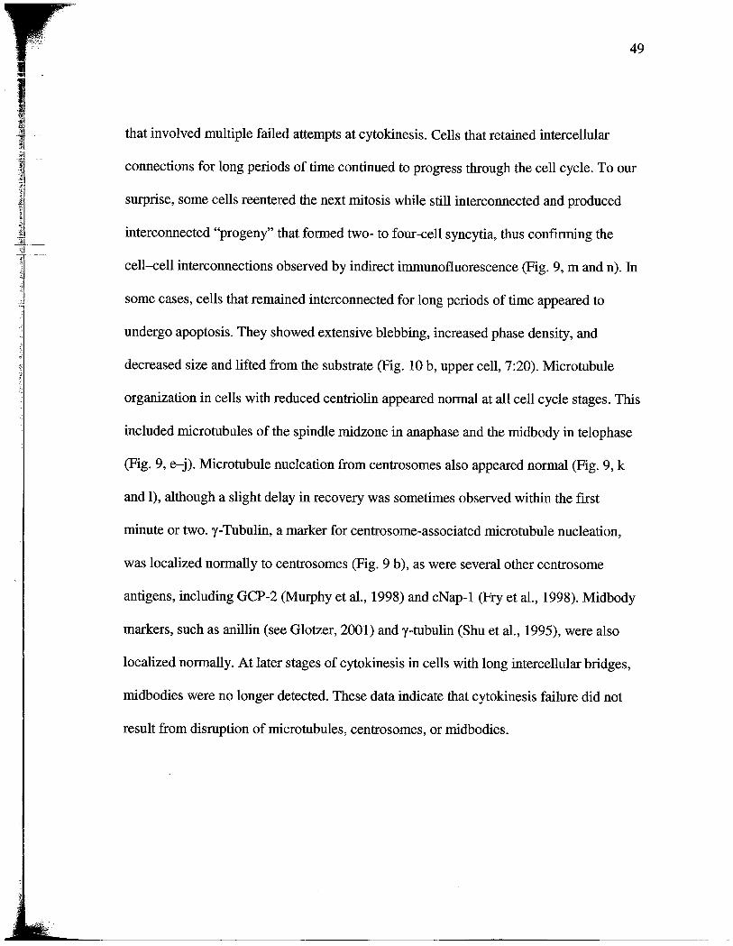
that involved multiple failed attempts at cytokinesis. Cells that retaied intercellular
connections for long periods of tie continued to progress though the cell cycle. To our
surprise, some cells reentered the next mitosis while still interconnected and produced
interconnected "progeny" that formed two- to four-cell syncytia, thus confIrmng the
cell-cell interconnections observed by indirect immunofluorescence (Fig. 9, m and n).
some cases, cells that remained interconnected for long periods of time appeared to
undergo apoptosis. They showed extensive blebbing, increased phase density, and
decreased size and lifted from the substrate (Fig. 10 b, upper cell, 7:20). Microtubule
organzation in cells with reduced centrolin appeared normal at all cell cycle stages. This
included microtubules of the spindle midzone in anaphase and the midbody in telophase
(Fig. 9, e-j). Microtubule nucleation from centrosomes also appeared normal (Fig. 9, k
and I), although a slight delay in recovery was sometimes observed with the fIrst
minute or two.
y-
Tubulin, a marker for centrosome-associated microtubule nucleation
was localized normaly to centrosomes (Fig. 9 b), as were several other centrosome
antigens, including GCP-2 (Murphy et al. , 1998) and cNap- l (Pry et al., 1998). Midbody
markers, such as anlln (see Glotzer, 2001) and y-tubulin (Shu et al., 1995), were also
localzed normally. At later stages of cytokinesis in cells with long intercellular bridges,
midbodies were no longer detected. These data indicate that cytokinesis failure did not
result from disruption of microtubules, centrosomes, or midbodies.
- - - -- --- ..- - - - - -- - - - - - .: - - - .:=
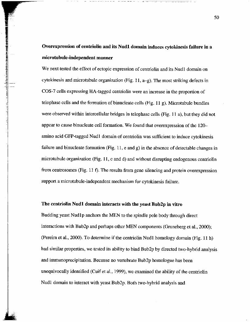
Overexpression of eentriolin and its Nudl domain induces cytokinesis failure in a
microtubule-independent manner
We next tested the effect of ectopic expression of centrolin and its Nudl domain on
cytokinesis and microtubule organization (Fig. 11 , a-g). The most strking defects in
COS-7 cells expressing HA -tagged centrolin were an increase in the proporton
-"1 telophase cells and the formation of binucleate cells (Fig. 11 g). Microtubule bundles
were observed within intercellular bridges in telophase cells (Fig. 11 a), but they did not
appear to cause binucleate cell formation. We found that overexpression of the 120-
amno acid GFP-tagged Nudl domain of centrolin was sufficient to induce cytokinesis
failure and binucleate formation (Fig. 11 , e and g) in the absence of detectable changes in
microtubule organzation (Fig. 11 , c and d) and without disrupting endogenous centrolin
from centrosomes (Fig. 11 f). The results from gene silencing and protein overexpression
support a microtubule-independent mechansm for cytokinesis faiure.
The eentriolin Nudl domain interacts with the yeast Bub2p in vitro
Budding yeast Nudl p anchors the MEN to the spindle pole body though direct
interactions with Bub2p and perhaps other MEN components (Gruneberg et al., 2000);
(Pereira et al. , 2000). To determne if the centriolin Nudl homology domain (Fig. 11 h)
had simar properties, we tested its abilty to bind Bub2p by directed two-hybrid analysis
and immunoprecipitation. Because no vertebrate Bub2p homologue has been
unequivocally identified (Cuif et al., 1999), we examed the abilty of the centrolin
Nudl domain to interact with yeast Bub2p. Both two-hybrid analysis and

lJ n
;t-
immunoprecipitation from yeast cells coexpressing the two proteins revealed a strong and
specific interaction between the centrolin Nudl domain and Bub2p (Fig. 11, i and j). No
signal was observed when either protein was used alone, and no binding was detected
between the centrolin Nudl domain and the budding yeast MEN component Bfal p,
consistent with interaction observations in budding yeast (pereira and Schiebel , 2001).
Cleavage failure is observed in Xenopus embryos injected with centriolin antibodies
A thd approach was used to examne centrolin function. When affinity-purfied
anticentrolin antibodies (Fig. 7 b) were microinjected into one cell of two-cell Xenopus
embryos (Doxsey et al., 1994), the injected cell failed to cleave , or cleaved a few times
and then arested; uninjected cells or preimune IgG-injected cells divided normally
(Fig. 12, a and d). Centrolin antibody-injected cells arested with two nuclei and two
well-organzed microtubule asters, indicating that karokinesis and microtubule
organzation were normal, but cells failed to complete the final event of mitosis, cell
cleavage (Fig. 12 c). Preimune IgG-injected cells had a single nucleus with one or two
microtubule asters, depending on their cell cycle stage, as would be expected for cells
that had undergone normal cell cleavage (Fig. 12 b). Taken together, the results from
gene silencing, antibody injection, and protein overexpression in several experimental
systems all demonstrate that centrolin plays an important role in the late stages of
cytokinesis.

Jt -
siRNA-induced gene silencing of eentriolin causes Gl/GO arrest
Cytokinesis defects and delays induced by centrolin silencing were observed at early
times after treatment of RPE- l cells (18-24 h). At later ties (48-72 h after treatment), a
reduction in the mitotic index was observed, suggesting that the cells were arested at
some other stage of the cell cycle. This was directly tested by treating cells with
nocodazole to induce mitotic arest and quantifying mitotic cells in DAPI stained
preparations. Under these conditions, most lamn siRNA-treated control RPE- l cells
were arested in mitosis (71 %), whereas only a small fraction of centrolin siRA-
treated cells arested at this cell cycle stage (-:1 %). To determne the cell cycle stage of
arest, cells were analyzed by flow cytometr. In the presence of nocodazole, control
cells showed a signifcant shit from the G 1 peak to the G2/M peak (Fig. 13 a, red). In
contrast, cells treated with siRNAs targeting centrolin did not signifcantly shift into the
G2/M peak in the presence ofnocodazole but remained largely in Gl (Fig. 13 a, blue).
The inabilty to undergo a nocodazole induced shift into the G2/M peak was a featue
shared by cells driven into GO by serum starvation (Fig. 13 b, blue). The proportion of
cells in S phase was either unaltered or slightly decreased in cells silenced for centrolin
both in the presence ofnocodazole (centrolin, 13%; lamn, 23%) or in its absence
(centrolin, 13%; lamn, 19%). These results demonstrate that cells with reduced
centrolin arest before S phase, possibly in G liS, G 1, or GO. Ki-67 staning was also
used to examne the stage of cell cycle arest. Ki-67 is an antibody directed against a
nuclear protein that stains cycling cells or cells arested in cycle (e. , GlIS or S phase;
(Gerdes et al., 1984)) but not cells that are quiescent (GO) or differentiated. As expected,
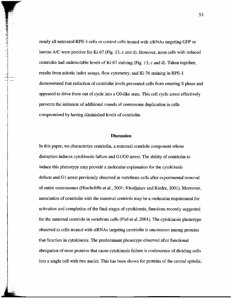
1f-
.:. _
nearly all untreated RPE- l cells or control cells treated with siRNAs targeting GFP or
lamins A/C were positive for Ki-67 (Fig. 13, c and d). However, most cells with reduced
centrolin had undetectable levels of Ki-67 staining (Fig. 13, c and d). Taken together
results from mitotic index assays, flow cytometry, and Ki-76 staining in RPE-
demonstrated that reduction of centrolin levels prevented cells from entering S phase and
appeared to drive them out of cycle into a GO-like state. This cell cycle arest effectively
prevents the initiation of additional rounds of centrosome duplication in cells
compromised by having diminished levels of centrolin.
Discussion
In ths paper, we characterize centrolin, a maternal centrole component whose
disruption induces cytokinesis failure and G l/GO arest. The ability of centrolin to
induce ths phenotype may provide a molecular explanation for the cytokinesis
defects and G 1 arest previously observed in vertebrate cells after experiental removal
of entie centrosomes (Hinchcliffe et al., 2001; Khodjakov and Rieder, 2001). Moreover
association of centrolin with the maternal centrole may be a molecular requirement for
activation and completion of the fmal stages of cytokinesis, functions recently suggested
for the maternal centrole in vertebrate cells (Piel et al. 2001). The cytokinesis phenotype
observed in cells treated with siRAs targeting centrolin is uncommon among proteins
that function in cytokiesis. The predominant phenotye observed after functional
abrogation of most proteins that cause cytokinesis failure is coalescence of dividing cells
into a single cell with two nuclei. This has been shown for proteins of the central spindle,

regulatory proteins that control cytokinesis, microtubule-associated proteins, and the
centrosome proteins y-tubulin and centrn (Shu et aI. , 1995; Kaiser et al., 2002; Manabe
et al., 2002; Matuliene and Kuriyama, 2002; Meraldi et al. , 2002; Mollnar et al., 2002;
Salisbury et al., 2002; Seong et al., 2002). In contrast, the predominant cytokinesis
phenotye observed in cells with reduced centrolin is failure to resolve persistent
intercellular bridges between dividing cells. The apparent consequences of this event are
generation of multcellular syncytia, continuous attempts at cell cleavage that include
binucleate cells as intermediates , completion of cytokinesis, perhaps arficially, by
traction-mediated cytokiesis (Buron and Taylor, 1997), and apoptotic cell death.
Although we have not determned the precise mechanism of cytokinesis failure in cells
with abrogated centrolin function, we are testing whether membrane is incorporated
normally into the cleavage site (Finger and White, 2002; Thompson et al. , 2002).
Persistent intercellular bridges have also been observed between dividing vertebrate cells
overexpressing the Cdc-14A phosphatase (Kaiser et al. , 2002). This uncommon
phenotype has thus far been observed only with proteins (centrolin and Cdc14A) that
share homology with components of yeast regulatory pathways that control mitotic exit
and cytokiesis (MEN/SIN) and localize to the same intracellular sites as the yeast
proteins (centrosome/spindle pole bodies). Persistent intercellular connections may also
be present in cells lacking centro somes and may reflect a simlar type of cytokinesis
defect (Doxsey, 2001a). Although the mechanism of cytokinesis failure appears to be
different in SIN mutants, the outcome is the same, production of bi and multinucleate
cells. These observations support the idea that vertebrate cells may possess regulatory

:iF-
pathways that function in much the same way as those in yeast to coordinate the final
stages of cytokiesis with chromosome segregation. Several domains of centrolin share
sequence homology with proteins implicated in tumorigenesis, including the TACCs and
oncoprotein 18/stathn. In ths paper, we show that the domain homologous to
Nudlp/Cdcllp can induce aneuploidy though cytokinesis failure. This event could
generate fuer genetic instability though the organzation of multipolar spindles by
supernumerary centro somes and could thus facilitate the accumulation of activated
oncogenes and loss of tumor suppressor genes (pihan et al., 2001; Doxsey, 2002). We are
curently investigatig the role of centrolin in human oncogenesis. Our results suggest
that minor changes in centrosome composition, such as loss of centrolin, can arest cells
in G I/GO. Because cells with reduced centrolin have difficulty completing the final
stages of cytokiesis, it is tempting to suggest that G l/GO arest is a consequence of
improper cytokinesis. However, it has been suggested that G 1 arest in cells lacking
centro somes may also result from the loss of core centrosome components , improper
spindle algnment, or the presence of excess DNA in binucleate cells (Andreassen et al.
2001; Hinchcliffe et al., 2001; Khodjakov and Rieder, 2001; Meraldi et al. , 2002). We are
currently testing a model in which the loss of core centrosome components activates a
checkpoint involved in monitoring "centrosome integrty" and thus prevents centrosome
duplication by aresting cells in GlIGO, as we and others have proposed (Doxsey, 2001a;
Quintye and Schroer, 2002).

I O
Materials and methods
Cell culture and transfeetions
'=1
The cells used priarly in this study were diploid, telomerase-immortlized human RPE-
1 cells (hTERT-RPE- ls; CLONTECH Laboratories, Inc.) (Morales et al. , 1999). Other
cells included HeLa, COS- , hTERT-HME- l (human mamary epithelia), U20S, and
Xenopus tissue culture (XTC) cells. All were grown as previously described (American
Type Cultue Collection). COS-7 cells were transfected as previously described
(Lipofectane; Invitrogen).
Antibodies
Amo acids 268-903 of centrolin were fused with GST (CLONTECH Laboratories,
Inc. ), overexpressed in Escherichia coli and purified as previously described (Doxsey et
al. , 1994). Antibodies raised in rabbits were affmity purified by passing sera over a GST
column to remove anti-GST antibodies and then over a SDT-centrolin column.
Antibodies to the following proteins were also used in this study: lamn A/C (Cell
Signaling Technology), (l- and y-tubulins (Sigma-Aldrich), LexA (Santa Cruz
Biotechnology, Inc. ), GAL4 transactivation domai (TAD) (CLONTECH Laboratories,
Inc.), Ki-67, and HA (BD Biosciences).
Immunofluorescence and electron microscopy
Cells were prepared for imunofluorescence, imaged , and deconvolved (Metamorph;
Universal Imaging Corp.), and centrosomes were quantified as previously described

.II
ifl.B -
(Dictenberg et al. , 1998). All immunofluorescence images are two-dimensional
projections of thee-dimensional reconstrctions to ensure that al stained material was
visible in two-dimensional images. Imunnogold electron microscopy was performed as
previously described (Doxsey et al. , 1994) using centrosome fractions from HeLa cells
(Blomberg and Doxsey, 1998) and antibodies to centriolin followed by antibodies
coupled to 5-nm gold paricles (Amersham Biosciences). Pilar cells were prepared and
staned as previously described (Mogensen et al., 1997).
Centriolin cloning
A cDNA of - 7 kb was identified by screening a human placenta expression librar with
serum from individuals with scleroderma (Doxsey et al. , 1994). The nucleotide sequence
was compared with others (blastn) in the human genome database (National Center for
Biotechnology Information) and revealed a sequence with 99% identity on chromosome
9 q34. 11-34. 13. Genscan predicted an - kb gene comprising 40 exons. PCR primers
were used to obtan an -7 -kb cDNA in a human testes cDNA librar. The 5' end,
obtained by rapid amplification of cDNA ends (RACE), was identical to the predicted
sequence. A full-length HA-tagged centrolin was obtaied by inserting and HA tag
(YPYDVPDYASL) 5' to the RACE fragment and ligating the HA-centrolin cDNA to
the original fragment. The full length centrolin cDNA contaned 6 975 nucleotides with
an ORF of 2 325 amno acids and predicted a molecular mass of 269 kD, consistent with
the molecular mass of endogenous centrolin (Fig.7).

..!-
Regions flanng the ORF had a translational star (Kozak sequence), polyadenylation
sequence, poly-A tail, and multiple upstream and down-stream stop codons. The
constrct was inserted into pcDNA 3. 1 Zeo (+) (lnvitrogen) using BamI and Not!
restrction sites. Centrolin was translated in vitro (TNT; Promega) and expressed in
cultued cells using conventional procedures (Lipofectaine). Centrolin amno acids
435-623 and 1385-1658 were 24% identical47% simlar and 20% identical41 % simlar
to the COOH-termnal half ofTACCs, respectively. Amino acids 879-913 were 40%
identical51 % simlar to amno acids 72-106 of human stathn. Amno acids 126-234
were 31-35% identical47-50% simlar to Nudlp and Cdc11p.
siRNA and morpholino antisense
siRNAs tageting centrolin, lam A/C , and GFP mRNAs were made as complimentary
single-stranded 19-mer siRNAs with 3' dTdT overhangs (Dharacon Research),
deprotected , annealed, and delivered into cells using a 400 Jl stock (Oligofectamne;
Invitrogen). The nucleotides targeted were as follows: in centrolin, 117-136 and 145-
163; in lamin A/C, 608-630; and in pEGFP-Cl (CLONTECH Laboratories , Inc.), 233-
252. Fluorescein-conjugated morpholino antisense DNA oligonucleotides (Gene Tools)
targeting the sta codon of centrolin were introduced into cells using the EPEI agent
(Gene Tools). The inverse sequence was used as control.
Time-lapse imaging
HeLa cells plated on coverslips (25 mm diameter) were treated with siRNAs targeting
centrolin for 50 h. They were placed in chamber (PDMI-2; Harard Apparatus) in

J' '1;'
. .::-
':1
complete medium with CO exchange (0.5 liters/min) at 37 C. Cells were imaged every
10 min for 12-20 h using a 20x or 40x phase contrast lens with a green interference fiter
on an inverted microscope (Olympus IX -70). Images were captured on a CoolSnap HQ
CCD camera (Roper Scientific) and concatenated using Metamorph software (Universal
Imaging Corp.
Primary cilum formation and microtubule nucleation
Priar cila were induced by culturing hTERT-RPE- l cells in medium with 0.25%
serum for 48 h and identified using the GT335 antibody raised to polyglutamylated (X-
and B-tubulins (Bobinnec et al., 1998). Microtubule nucleation was performed as
previously described (Prohit et al. , 1999) by treatment with nocodazole (1 mg/ml) for 1
h at 37 , fIXing cells at varous times after washing out drug, and then staining for
microtubules.
RT -PCR
Centrolin mRNA levels were assayed by RT -PCR using 10 Jl mRNA (OneStep RT-
PCR; QIAGEN; (X-tubulin served as an internal control in the same reaction). All
products were sequenced.
Yeast two-hybrid analysis and imunoprecipitations
A fragment containing amno acids 127-233 of centrolin was cloned into EcoRI and SalI
sites ofpGADT7 (CLONTECH Laboratories, Inc. ) to produce a fusion with the GAL

)J TAD. Constrcts pEG202 (LxA), pGP69 (LexA-Bub2), and pGP122 (LxA-BFAl) and
the yeast strain SGY37 were from Elmar Schiebel (Peterson Institute for Cancer
Research, Manchester UK). SGY37 , which contains a LacZ reporter gene under the
control of a LexA operator, was transformed with plasmid DNA using LiAc (Ito et al.,
1983), and transformants were selected for on dropout medium. We used
semiquantitative galactosidase assays with CPRG (cWorophenol red-
galactopyranoside; Roche) as a substrate per the manufacturer s instrctions.
':%
Coimunoprecipitation of LexA and GAL TAD fusion proteins were performed as:'i:J.'
previously described (Schram et al. , 2001).
Flow eytometry
Cells treated with siRA for 50-70 h were treated with 100 ng/ml for 12 h, removed from
plates, and fIXed in methanol. Cells staned with propidium iodide were analyzed by flow
cytometr (FACSCAN; Becton Dickinson) using Flojo software (Tree Star, Inc.
GeneBanklMBLIDDBJ accession numbers
The GenBankMBL/DDBJ accession number for centrolin is AF513978 and for lamn
NC is X0344.
Onlne supplementa material
Supplemental videos (Videos 1-3) are available at
htt:/www.jcb.orglcgilcontent/full/jcb.200301105/DCl. The supplemental videos show

that cells silenced for centriolin delay in cytokinesis , remain interconnected by persistent
intercellular bridges, re-enter mitosis while still interconnected, and form multicellular
syncyta. Some cells divide normaly while others undergo apparent apoptosis
characterized by blebbing, rounding up, dimnution in size, and loss of substrate
attachment.
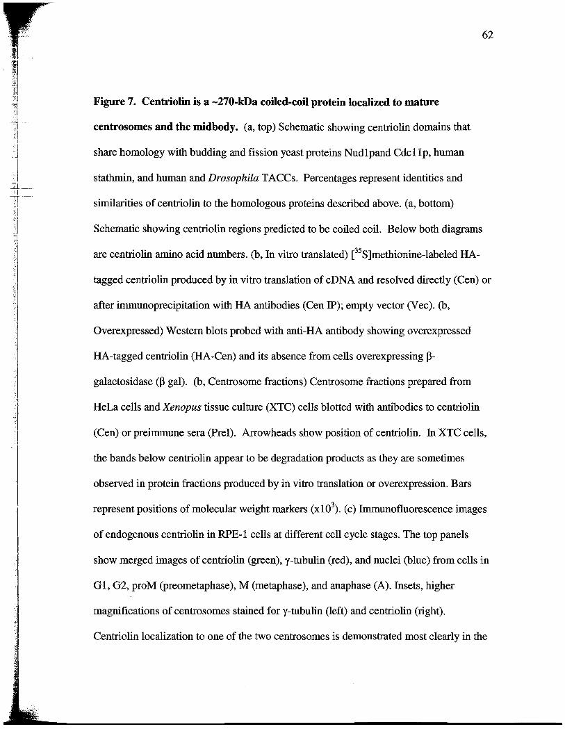
.'1
. !
--1
Figure 7. Centriolin is a -270-kDa coiled-coil protein localized to mature
eentrosomes and the midbody. (a, top) Schematic showing centrolin domains that
share homology with budding and fission yeast proteins Nudl pand Cdc IIp, human
stathn, and human and Drosophila TACCs. Percentages represent identities and
simlarties of centrolin to the homologous proteins described above. (a, bottom)
Schematic showing centrolin regions predicted to be coiled coil. Below both diagrams
are centrolin amno acid numbers. (b, In vitro translated) e S)methionine-labeled HA-
tagged centrolin produced by in vitro translation of cDNA and resolved directly (Cen) or
after imunoprecipitation with HA antibodies (Cen IP); empty vector (V ec). (b,
Overexpressed) Western blots probed with anti-HA antibody showing overexpressed
HA-tagged centrolin (HA-Cen) and its absence from cells overexpressing
galactosidase CP gal). (b, Centrosome fractions) Centrosome fractions prepared from
HeLa cells and Xenopus tissue culture (XTC) cells blotted with antibodies to centrolin
(Cen) or preimune sera (Pre!). Arowheads show position of centrolin. In XTC cells
the bands below centrolin appear to be degradation products as they are sometimes
observed in protein fractions produced by in vitro translation or overexpression. Bars
represent positions of molecular weight markers (xl0 ). (c) Imunofluorescence images
of endogenous centrolin in RPE- l cells at different cell cycle stages. The top panels
show merged images of centrolin (green), y-tubulin (red), and nuclei (blue) from cells in
G 1, G2, proM (preometaphase), M (metaphase), and anaphase (A). Insets, higher
magnifications of centro somes stained for y-tubulin (left) and centrolin (right).
Centrolin localization to one of the two centrosomes is demonstrated most clearly in the

G2 cell. Middle panels (Telo early) and bottom panels (Telo late) show separate images
of y-tubulin staining (left) and centrolin (right).
y-
Tubulin marks both mother and
daughter centroles, which are separated in some cases. Centrolin staining is confmed to
one of two centroles, which sometimes appears at the intercellular bridge (Telo early).
Centroles lacking centrolin are indicated by arowheads in right panels. At later stages
of telophase (Telo late), centrolin is also on the midbody. All imunofluorescene images
(here and elsewhere) are two-dimensional projections of thee-dimensional
reconstrctions to ensure that all stained material is visible. C, centrole; MB, midbody.
Bar in c , 10 Jl. Centrolin sequencing was performed by Cytomi and Adam Gromley
contrbuted the rest.

FIGURE 7feJ
NucUpICdC11p TACC Stathmin
(35%/50%) (24%/47",1 (40%151
127 233 435 623 879-913
1385 325
Coiled coils.
265 434

Figure 8. Centriolin is localized to maternal centrioles and noneentrosomal sites of
microtubule anchoring. (a) HA-tagged centriolin oveexpressed in COS-7 cells localzes
to the centrosome (anti- , green) at the convergence of microtubules (red, anti-a-
tubulin). (b) An RPE- l cell imunostained with an antibody to polyglutamylated tubulin
(GT335) (Bobinnec et al., 1998) to label centroles and the priary cilum (red), and for
centrolin (green), which is localized to the maternal centrole (m) associated with the
priar cilum (yellow in merge) but not on the daughter centrole (d). n, nucleus. (c-e)
Election micrographs showing specific imunogold labeling of centrolin on sub distal
appendages found on maternal (e, bottom, M) but not daughter centroles (e, top, D). c
and d are longitudinal and cross sections, respectively, though maternal centroles, and e
is a longitudinal section though both centroles. Arowheads show strations
characteristic of sub distal appendages. (f and g) Centrolin is found at noncentrosomal
sites of microtubule anchoring in pilar cells of the mouse cochlea (arows) (Mogensen et
al., 1997). (t) Schematic representation of pilar cell. (g) Centrolin immunofluorescence
staning overlaid with phase contrast image. Centrosome and associated cilium are
shown schematically at top of cell in f. Bars: (a and g) 10 Jl; (b) 2 J.; (c-e) 100 nm.
Adam Gromley contrbuted figure 8 a, Agata Jurczyk contrbuted figure 8 b, Chrs
Powers and Maureen Blomberg contrbuted the electron microscope images, and Mette
Mogensen contrbuted figure 8 f and g.
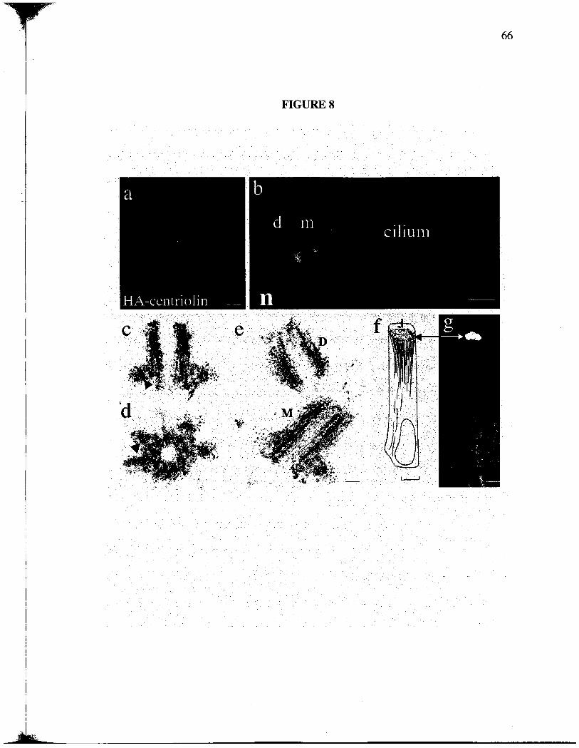
FIGURE 8

Figure 9. RPE- l cells treated with siRNAs targeting eentriolin retain persistent
intercellular connections and fail in cytokiesis. (a) RT-PCR analysis shows that
centrolin mRNA is reduced in RPE- l cells treated with centrolin specific siRNAs (top
right) but is unaffected in cells treated with siRNAs tageting lamn AlC (top left
sequence identity confIrmed). Control (a-tubulin) RT-PCR was performed in the same
reaction mitures with centrolin and lamn (bottom panels). (b) Imunofluorescence
images (top right; a-tubulin, red; centrolin, green) and quantication of centrolin levels
in centrosomes from cells treated with siRNAs as indicated. The top right panes shows a
cell with undetectable centrolin at the centrosomeJcentrole, and top left panel shows a
cell that is unaffected by the treatment and stans for centrolin (green/white). Graph
shows the average centrolin fluorescence intensity/pixel at individual centrosomes (bars)
in cells treated with lamn or centrolin siRNAs. The centrosome fluorescence in most
centrolin siRA-treated cells (83%) was blow the lowest values observed in control
cells. (c) Graph showing a dramatic increase in the percentage of cells in
telophase/cytokinesis after siRNA targeting of centrolin (-70-fold). N, total number of
cells counted (c and d). (d) Graph showing the increased percentage ofbinuc1eate cells in
HeLa, U20S, and RPE- l cell lines after treatment with centrolin siRNAs (4-15-fold
greater than controls). In c and d, values represent data from single experients
representative of thee t four experients. The tie analyzed was as follows: HeLa, 48 h
and 72 h; U20S, 72 h; RPE- l, 24 h. (e-l) Microtubule organization (e-j) and microtubule
nucleation (k and I) are not detectable altered in cells treated with centrolin siRNA (f, h
j, and I) compared with cells treated with lamn siRNA (e, g, I, and k). Arows indicate

position of centrosomes; no centrosome staining is observed in centrolin siRA-treated
cells. Insets in k and I are enlargements of centrolin staning at centro somes (or
microtubule convergence sites) at arows. Cells in interphase (e and f, lower cell),
prometaphase/metaphase (proM/; g and h), and telophase (i and j) were labeled for
microtubules (red), centrosomes (green/yellow), and nuclei (blue). MB, midbody. (m and
n) Cells treated with siRNAs targeting centrolin were stained for microtubules (red) and
DNA (blue). Arowheads indicate contiguous connections between two or more cells.
(m) Thee interconnected cells form a syncytium. (n) One daughter of an interconnected
pai of cells has reentered mitosis (M). Bar in n: (e-j) 5 11m; (k.1, and insets) 3.5 Jl; (m
and n) 15 Jl. The siRA for these experients were done by Adam Gromley and Agata
Jurczyk, al the counts and imaging were performed by Adam Gromley.

FIGURE 9
RT -PCR: siRNA:lamin centrolin
Centrolin
tub linr:.

Figure 10. Time-lapse images of HeLa cells treated with eentriolin siRNAs reveal
unique cytokiesis defects. (a) A cell treated with control siRAs targeting lamn
moves apar, forms visible midbody (arow), and completes the final cleavage event with
normal timing (1-3 h after metaphase). (b-d) Centriolin siRNA-treated cells remain
attached for extended periods of tie though persistent intercellular bridges and
sometimes do not show visible mdbodies. (b) A cell that remains attached by a long
intercellular bridge for at least 8 h. The cell cleaves, both daughter cells round up, and at
least one appears to undergo apoptotic cell death (upper cell, 7:20, extensive blebbing
and decrease in size). (c) A dividing cell that has not completed cleavage reenters the
next mitosis. One cell rounds up and is drawn to the other. The other rounds up and both
undergo the early stages of cytokinesis to form a total of four cells; these progeny often
remain attached by intercellular bridges formng syncytia. (d) Cell showing thee failed
attempts at cell cleavage over a 9. h time period. (e) Cells treated with siRAs targeting
centrolin progress from nuclear envelope breakdown (NB) to anaphase with normal
timing, simiar to lamn siRNA controls. Vertcal bars represent recordings form single
cells (e and f). (f) Centrolin siRNA -treated cells are delayed in cytokiesis (-70%)
compared with controllamn siRA-treated cells, a value consistent with a 70-80%
silencing efficiency. Results represent recordings of individual cells from several
independent experiments. Time in hours and minutes is included in each panel in a-
Bar, 10 Jl. Timelapse videos (Videos 1-3) of the series of images shown in a-c are
available at htt//www.jcb/cgi/content/full/jcb.200301105/DCl. Agata Jurczyk
contrbuted this figure.

FIGURE 10
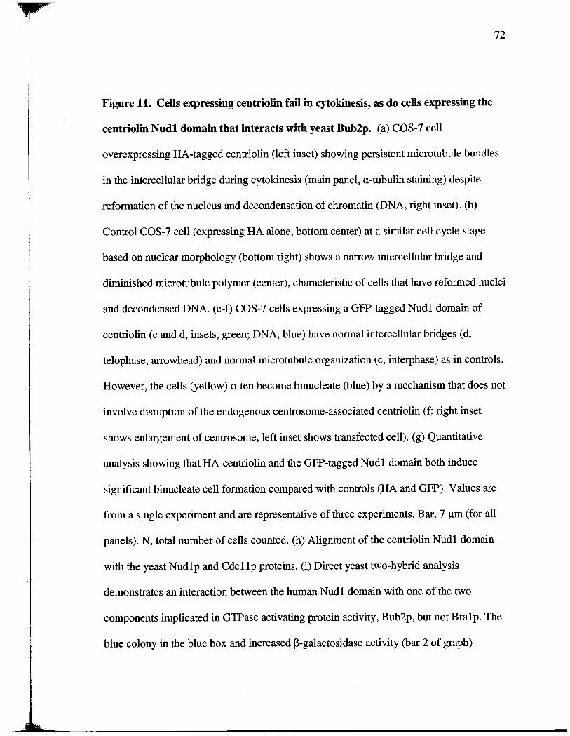
Figure 11. Cells expressing eentriolin fail in cytokiesis, as do cells expressing the
eentriolin Nudl domain that interacts with yeast Bub2p. (a) COS-7 cell
overexpressing HA-tagged centrolin (left inset) showing persistent microtubule bundles
in the intercellular bridge durng cytokinesis (main panel, a-tubulin staining) despite
reformation of the nucleus and decondensation of chromatin (DNA, right inset). (b)
Control COS-7 cell (expressing HA alone, bottom center) at a similar cell cycle stage
based on nuclear morphology (bottom right) shows a narow intercellular bridge and
dimnished microtubule polymer (center), characteristic of cells that have reformed nuclei
and decondensed DNA. (c-t) COS-7 cells expressing a GFP-tagged Nudl domai of
centrolin (c and d, insets, green; DNA, blue) have normal intercellular bridges (d,
telophase, arowhead) and normal microtubule organization (c, interphase) as in controls.
However, the cells (yellow) often become binucleate (blue) by a mechanism that does not
involve disruption of the endogenous centrosome-associated centrolin (f; right inset
shows enlargement of centrosome, left inset shows transfected cell). (g) Quantitative
analysis showing that HA-centrolin and the GFP-tagged Nudl domain both induce
significant binucleate cell formation compared with controls (HA and GFP). Values are
from a single experient and are representative of thee experients. Bar, (for all
panels). N, total number of cells counted. (h) Alignment of the centrolin Nudl domain
with the yeast Nud 1 p and Cdc 11 p proteins. (i) Direct yeast two-hybrid analysis
demonstrates an interaction between the human Nudl domain with one of the two
components implicated in GTPase activating protein activity, Bub2p, but not Bfalp. The
blue colony in the blue box and increased p-galactosidase activity (bar 2 of graph)

demonstrate a specific interaction between the human centrolin Nudl domain (hNudl)
and yeast Bub2p. Bar LexA-BUB2 x TAD; bar 2, Lex A-BUB x TAD-hNudl; bare 3,
LexA-BFAl xTAD; bar 4, TAD-hNudl x LexA-BFAl; bar 5, TAD-hNudl x LexA. (j)
Specific coimunoprecipitation of HA-tagged hNudl and LexA-tagged yeast Bub2p
from yeast cells. Coprecipitation was observed using antibodies to either protein and only
when both were coexpressed (top middle panel in each group). Lane TAD-HA-hNudl
x LexA; lane 2 , TAD-HA-hNudl x Bub2p-LexA; lane 3 , TAD-HA x Bub2p-LexA.
Adam Gromley contrbuted the figures a- , James Silboure contrbuted figure i , and
Ensar Halilovic contrbuted figue j.

FIGURE 11
HA-centrolin30(j(n==2835) Nudl-GFP
-- -
IIi;;:!5
(n=3420)GFP
(n=253)
QIi
- - - -
s: V- G -G ,W t 7 . E D (IE G - - -. . - - N I: J) 3. X. B: P - L D 1.
TAD hNudlaQ
HAIP
-"" "".._"
Blot:LeALeA (Supnt)
LexAJP== JL (Supnt)LeA

Figure 12. Cytokiesis failure in Xenopus embryos injected with eentriolin
antibodies. (a) Xenopus two cell embryos injected into one cell with -50 nl
anticentrolin antibodies (2 mg/ml affinity purfied) fail to cleave (arows), whereas
noninjected cells (side opposite arows) and cells injected with control rabbit IgG (2
mg/ml, top) cleave normaly. (b and c) Imunofluorescence images showing two
microtubule asters near a single nucleus (N) in a cell from an embryo injected with
control IgG (b), and two nuclei and two asters in a cell from a centrolin antibody-
injected cell (c , micro tubules stained with anti-a-tubulin). Bar, 10 (for b and c). (d)
Quantification of results from injection experiments showing an approximately eight fold
increase in cleavage failure (representative of thee experiments). n, number of embryos
examed. Irna Groisman contrbuted figue a, and Adam Gromley contrbuted the rest.
;I_
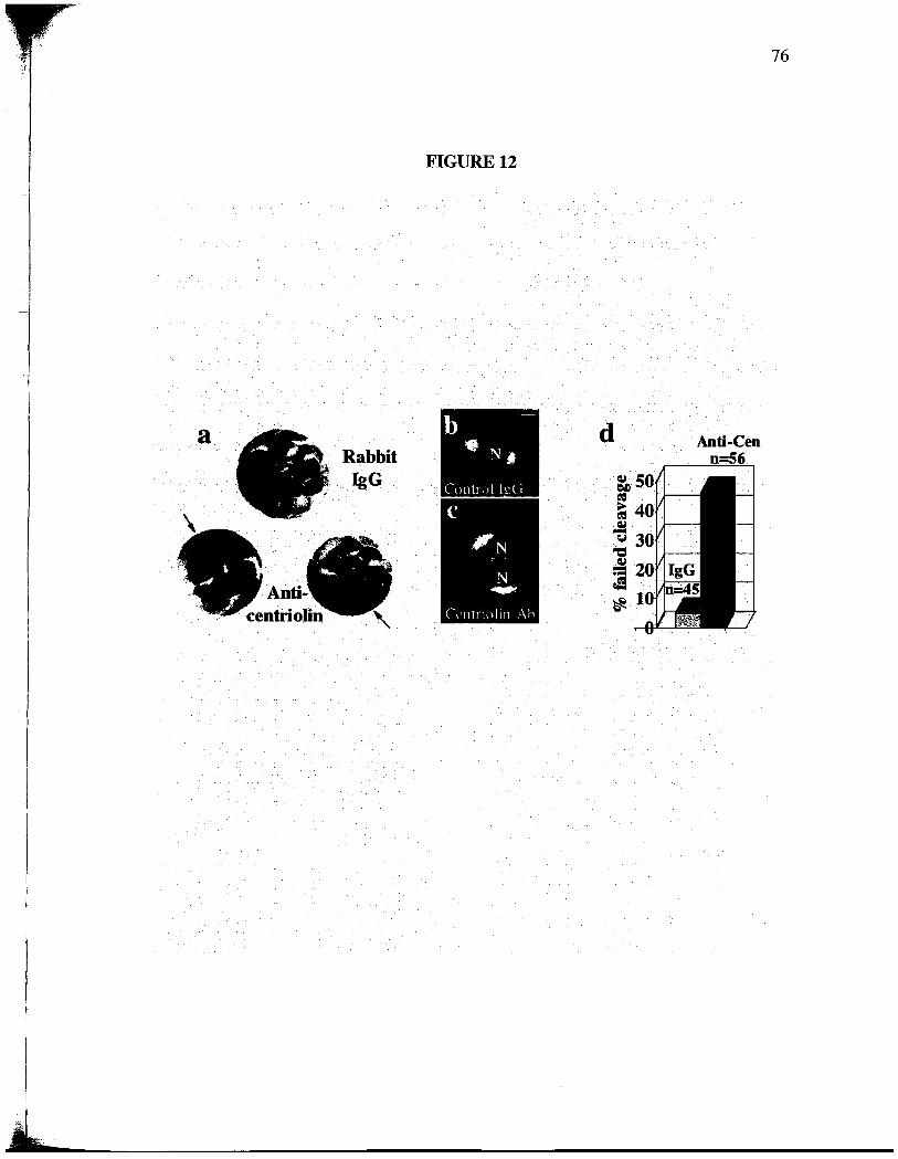
FIGURE 12
- N,CUlIl!!)l h?(I
, - - -
"'J
"'
Anti..
. .
eDmo

Figure 13. siRNAs tageting eentriolin induces G lIGO arrest.
(a) RPE- l cells treated with centrolin siRNAs (blue) do not shift into the G2/M peak
after nocodazole treatment as seen for controllamn siRNA-treated cells (red). (b) The
inability of cells treated with centrolin siRNAs to shift into the G2/M peak is simlar to
that observed in serum-starved cells (blue, serum stared; red, not starved). (c) Ki-
staining of control cells (left cell , green) occurs together with centrolin staining (left cell,
red at arow); both are gone in centrolin siRNA-treated cells (right, arowhead). Bar, 7.
m. (d) Cells treated with siRNAs targeting centrolin lack Ki-67 staiing (-70%),
whereas most control cells stain positively (GFP siRAs). a, b, and d are representative
experients from five experients for a, two for b, and thee for d. n, total number cell
counted. Adam Gromley contrbuted figure d, and Agata Jurczyk contrbuted the rest.

FIGURE 13
Centrioli
100-DOC
o 0
20040 0200 400
Relative. DNA .Ouorescence Relative DNA Ouorescence
. .
r-. CJIo. .
t2.

Chapter III
Pericentrin is Required for the Final Stages of
Cytokinesis and Entry into S Phase Through Its
Association with Centriolin
Abstract
Centrosomes serve as microtubule organzing centers of anal cells and they also playa
role in cytokinesis and cell cycle progression. However, the molecular pathway
implicatig centrosomes in cytokinesis has not been described. Previously we identified
a novel centrole protein called centrolin that is required for the final stage of
cytokinesis. Here we show the centrosome protein pericentr B localizes to the region of
contractile ring during early stages of cytokinesis and later to the midbody. At the
midbody and at the centrosome, pericentrn B co-localizes with centrolin and both
proteins show codependency for their centrosome localization. Centrolin and pericentrn
interact biochemically and produce indistiguishable phenotypes upon protein depletion.
Disruption of pericentrn B by RNAi results in late stage cytokinesis defects identical to
the ones observed for centrolin knockdowns. In addition, in cells with an intact p53
pathway, pericentr depletion results in GO/G 1 arest indistinguishable from centrolin
silencing. Recently centrolin was shown to interact with the members of exocyst and
SNARE complexes, which are required for exocytosis and completion of cytokinesis.
We show that pericentrn B co-localizes with endobrevin, a member of SNARE complex
at the unique midbody ring. We conclude that pericentrn B , centrolin and the
membrane secretion and fusion proteins are necessar for the final stages of cytokinesis
and cell cycle progression.
Introduction
Centro somes are responsible for organizing interphase microtubules and mitotic spindles.
They have been also implicated in cytokinesis and cell cycle progression. Cells lackig
centrosome exhibited defects in cytokinesis and in progression though the subsequent
cell cycle (Hinchcliffe et al., 2001; Piel et al. , 2001). Depletion of one of the centrosome
protein, centrolin, resulted in similar phenotype as was observed in the cells that lacked
centro somes entirely. Both RPE- l and HeLa cells treated with centrolin siRNA had
diffculty completing the final steps of cytokiesis. After the contractile rig was
assembled, alost 50 % of the cells remaied connected by a thin intracellular bridge
(Gromley et al., 2003). Subsequently, depending on their p53 status, the cells either
became multinucleated and died, or if their p53 pathway was intact, they arested in G 1.
The cytokinesis defect observed in centrolin knockdowns took place in the late stages of
cytokinesis, since the ingression of the contractile ring appeared to be normal (Gromley
et al., 2003). Other proteins that were shown to be required during late stages of
cytokinesis include members of the vesicular traffickig and membrane fusion proteins.
Interestingly, centrolin has been shown to interact with the members of exocyst and
SNAR complexes, which are required for exocytosis and completion of cytokinesis
(Gromely, 2004). Our most recent work demonstrate the involvement of both exocyst
and SNAR complexes in cytokinesis in mamalan cells, since disruption of either of
their members by siRA result in late stage cytokinesis defects (Low at al. , 2003;
Gromely, 2004). Moreover, this study identified a new, unique ring strcture in the

midbody of animal cell composed of the centrosome protein centrolin and members of
the exocyst and SNARE complexes. .The cytokinesis defects seen upon depletion of any
of the ring strcture members resulted in indistinguishable late stage cytokinesis defects.
In addition, disruption of the exocyst components resulted in accumulation of SNARE
vesicles at the midbody (Gromely, 2004). This study brought together the centrosome
proteins and the vesicle secretion and fusion machinery and showed their importance in
cytokinesis.
The results presented in ths chapter show that centrolin interacts with another
centrosome protein, pericentr B , which when depleted results in cytokinesis defect and
cell cycle arest indistinguishable form centrolin silencing. Pericentrn B also localizes
durg cytokinesis to the same rig strcture as was shown for centrolin. Moreover
Pericentr B additionally localizes to the membrane around the contractile ring earlier
durng cytokinesis. We also show that pericentrn Band centrolin interact biochemically
and are co-dependant for their centrosome localization. It remains to be shown what is
the relationship between pericentrn B , centrolin and the vesicular traffickig proteins
durig cytokinesis. Are they dependent on each other for the midbody localzation, does
pericentrn B interact with the members of the vesicular traffickig machiery as
centrolin does?

= I
Results and Discussion
Perieentrin B localization to the contractile ring and the midbody during cytokinesis
The localization of pericentrn A/ and B thoughout the cell cycle in mamalian cells
has been shown previously (Doxsey et al. , 1994; Flory et al., 2000). However, in this
study we discovered an additional localization for pericentr B that has not been
reported before. Here we used high resolution deconvolution microscopy to show that
pericentrn B localizes to the sites of furrow constrctions, probably where the
actinomyosin ring resides, durng the early stages of cytokinesis (Fig. 14 A top). Later
when the furrow is much thinner, pericentrn B localizes to the midbody (Fig. 14 A
bottom). This localization is specific for pericentrn B , since it was detected with an
antibody specifc for the C-termnus of pericentrn B which is not found in pericentrn A
(Flory et aI. , 2000). So far, there is no antibody specific for the smaller isoform of
pericentrn, pericentr A, due to homology between pericentrn A and B thoughout the
entire pericentrn A sequence (Flory and Davis, 2003).
Perieentrin B exhibits co-dependency with eentriolin at the centrosome and eo-
localizes with it at the midbody
High resolution deconvolution microscopy was used to check the centrosome localization
of centrolin in RPEI cells treated with siRNA specific for pericentrn B (Jurczyk et al.
2004). The cells with reduced levels of pericentrn B at the centrosome (77%, n=I000)
also had reduced levels of centrolin (Fig. 15 A arow). Moreover in the cells treated
with centrolin siRNA both centrolin and pericentrn B were mislocalzed from the

centrosome (Fig. 15 B arow). The cells with normal levels of pericentrn B and
centrolin served as controls (Fig. 15 A , bottom and 15 B arowheads). This data
indicates that pericentrn B and centrolin show co-dependency in their centrosome
localzation.
We also looked to see if pericentrn Band centrolin are co-localized durng cytokinesis.
In the early stages of cytokinesis , when pericentrn B seemed to mark the membrane
around contractile ring, centrolin did not localize to any of the pericentrn B contaiing
spots. However, later, when the midbody was thinner, both centrolin and pericentrn B
parially co-localized to the midbody (Fig. 15 C). These results show that pericentrn B
and centrolin co-localize to the midbody rig strcture in the late stages of cytokinesis.
Perieentrin and eentriolin interact biochemicaly
To test if pericentr and centrolin interact biochemically, we co-precipitated
endogenous centrolin using pericentrn AI antibody and in a reciprocal experiment, we
co-precipitated pericentrn using centrolin antibody (Fig. 16 A). We observed no co-
imunoprecipitation of any of the proteins when the antibody was omitted (Fig. 16 A,
Beads (Bd)). Lysates (Lys) from the immunoprecipitations experiments are indicated.
These data show that pericentrn B interacts with centriolin. It remais to be shown if
these interactions are direct.

Pericentrin B co-localizes with endobrevin at the midbody ring during cytokinesis
In the light of recent data showing the involvement of SNAR components and the
exocyst complex in the final stage of cytokinesis and their co-localization to the
centrolin rig (Gromely, 2004; Low et al. , 2003), we tested to see if pericentrn B can
co-localze with endobrevin, the member of SNARE complex. We showed that in the
earlier stages of cytokinesis pericentrn B was localized to the ring and parally co-
localized with endobrevin to both sides of the midbody (Fig. 17 A, enlargement below as
indicated by square in the images above). Therefore, pericentrn B localizes to the
midbody ring before endobrevin does , just as was shown for centrolin (Gromely, 2004).
Durng later stages of cytokinesis, when the intercellular bridge was much thinner,
pericentrn B parally co-localized with endobrevin to the ring and very weaky along
both sides of the midbody (Fig. 17 B).
Perieentrin B RNAi induces a late stage cytokiesis defects
When pericentrn B was depleted using a specifc siRNA (Jurczyk et al., 2004) it resulted
in cells that remained connected by thin intercellular bridges and a very small increase in
binucleate cells (Fig. 18 A right-binucleate cell , left-thin bridge connection). Forty two
percent of RPEI cells treated with pericentrn B siRA for 14 h showed ths phenotype
as compared with 5% of the control, GFP siRA treated cells (Fig. 18 B). Moreover, in
HeLa cells we observed an increased number of telophase cells (3.9% for GFP control
and 10% for pericentrn B knockdowns) in addition to cells connected by thin
intercellular bridges. These data show that disruption of pericentrn B produces a
Il-

phenotype indistinguishable from centrolin (Gromley et al., 2003) and implicates the
involvement of pericentrn B in the final stages of cytokinesis.
Perieentrin B disruption causes GO/G 1 arrest
The cytokinesis defects observed in pericentrn B knockdowns were observed at early
times after siRNA treatment (14-17 h). At later ties (48-72 h) a reduction in the mitotic
index was observed in RPEI cells, suggesting that the cells were arested at some other
stage of the cell cycle. To determe at which cell cycle stage the cells were arested, we
treated pericentrn B knockdowns with nocodazole to induce mitotic arest. In the
presence of nocodazole , control cells showed a signicant shit from the G 1 peak to the
G2/M peak (Fig. 19 A, red). In contrast cell treated with siRNAs targeting pericentrn B
did not signicantly shift into the G2/M peak in the presence of nocodazole but remained
largely in G 1 (Fig. 19 A, blue). The proporton of cells in S phase was unaltered in cells
silenced for pericentrn B both in the presence of nocodazole (pericentrn B, 14%; lamn
19 %) or in the absence (pericentrn B , 13%; lam 23%). Cells treated with pericentrn
B siRA showed a simlar pattern of cell cycle distrbution as serum stared, GO cells
(Fig. 19 B , serum starved, blue; GFP control, red). These results demonstrate that cells
with reduced pericentrn arest before S phase. To furter determne the time of arest we
used Ki-67 antibody staining. Ki-67 is an antibody directed against a nuclear protein that
stans cycling cells or cells arested in Gl/S and S , but not cells that are quiescent (GO) or
differentiated (Gerdes et al., 1984). As expected, nearly all untreated RPEI cells or
control cells treated with siRNAs targeting GFP or lamn A/C were positive for Ki-

(Fig. 19 C). However, most cells with reduced pericentr B had undetectable levels of
Ki-67 staning (Fig. 5 C). This arest was previously shown for centrolin (Gromleyet
al. , 2003) and it does not occur in cells with an abrogated p53 pathway, such as HeLa, or
Saos-2 (Fig. 19 D). Gl arest was also observed in cells whose centrosome was removed
by either laser ablation or microsurgery (Hinchcliffe et al., 2001; Khodjakov and Rieder
2001). Therefore, either centrosome damage by depletion of its core components or
cytokinesis faiure could result in cell cycle arest.

Figure 14. Pericentri B localzation to the contractile ring and the midbody during
cytokiesis.
, top) High resolution deconvolution images showing pericentrn B localization to the
area of contractile ring formation durng early cytokinesis and to the midbody during late
stages of cytokinesis (bottom). Overlay with pericentr B (pcntB , red) a-tubulin (green)
and DNA (blue).

FIGURE 14
Figure 15. Perieentrin B exhbits co-dependency with eentriolin at the centrosome
and co-localizes with it at the midbody.
(A) High resolution deconvolution images of RPEI cells treated with pericentrn A
siRNA. The cell with reduced levels of pericentrn B at the centrosome (arow) also had
reduced levels of centrolin. (B) High resolution deconvolution images of RPEI cells
treated with centrolin siRNA. The cells with reduced levels of centrolin at the
centrosome (arow) also had reduced levels ofpericentrin B. The cells with normal
levels of both proteins (arowhead) served as controls. Merge showing pericentrn B
(red), centrolin (green), DNA (blue). (C) High resolution deconvolution images of HeLa
cells showing that pericentrn B and centrolin co-localze at the midbody ring durng
cytokinesis. Enlargement of the midbody ring (arow) in the right comer of each pictue.
Pericentr B (red), centrolin (green).

FIGURE 15

rr'
Figure 16. Pericentri and eentriolin interact biochemically.
(A) Endogenous centrolin and pericentrn interact with each other when
immunoprecipitated from mitotic RPEI cells. The pericentrn antibody used in ths
experient recognizes pericentrn A and B and is raised to the amno-termial region of
pericentrn (PcN). No co-imunoprecipitation was observed when the antibodies were
omitted in beads control (Bd). Lysates (Lys) are indicated. All of the
imunoprecipitations were loaded and 7% of the imunoprecipitations were loaded for
the lysates.

FIGURE 16
Figure 17. Perieentrin B co-localizes with endobrevin at the midbody ring during
cytokiesis.
(A) High resolution deconvolution images showing pericentrin Band endobrevin
localization durng early stages of cytokinesis. Pericentrn B localizes to the midbody
ring and paraly co-localized with endobrevin to both sides of the midbody. (below)
overlay showing enlargement of the midbody with pericentrn B (green) and endobrevin
(red). (B) High resolution deconvolution images showing pericentrn Band endobrevin
localization during late stages of cytokinesis. Pericentrn B co-localized with endobrevin
to the midbody ring and very weaky along both sides of the midbody. (below) overlay
of endobrevin and pericentrn B. Enlargement of the midbody ring in the right corner of
each pictue. Endobrevin (green), pericentrn B (red).

FIGURE 17
Figure 18. Pericentri B RNAi induces a late stage cytokinesis defects.
(A) High resolution deconvolution images from RPEI cells after 14 h of pericentrn B
siRNA treatment showing cytokinesis defects, (left) shows an example of binucleate cell
undergoing cytokinesis , the midbody is still visible, (right) thin intercellular bridge
connections that represent most of the counted defects. (B) Quantatization of the images
shown in (A), Abnormaltelophase/multinucleated cells are percent of total cells counted
(n= 1 000).

FIGURE 18
siRNA Abn. Telo has/multinucleatedPericentr.m B 42%GFP
Figure 19. Perieentri B disruption causes GO/GI arrest.
(A) RPEI cells treated with centrolin pericentrin B siRAs (blue) do not shift into the
G2/M peak after nocodazole treatment as seen for controllamn siRNA-treated cells
(red). (B) The inability of cells treated with pericentrn B siRNAs to shift into the G2/M
peak is simlar to that observed in serum-stared cells (blue, serum starved; red, not
staed). (C) Cells treated with siRA targeting pericentrn Black Ki-67 staining
80%), whereas most control cells stain positively (GFP siRA). (D) HeLa and Saos-
cells defective in the p53 pathway treated with siRNA targeting pericentrn B staned
positively for Ki-67 just as controls (lamn A/C).

FIGURE 19
Pcnt
+noc
200 400
FL2-
100/
if 80
t" 60 !
'?
. i401
. -- ''' '-' ---'-' .
Serm starved
+noc
200 400
FL2-

100
DISCUSSION
In this dissertation, I present the requirement of two centrosome proteins: pericentrn and
centrolin for cytokinesis and cell cycle progression. Moreover I show that pericentr is
also involved in ciliogenesis. Centrolin is a novel centrolar protein which localizes to
the subdistal appendages of mother centrole. Both pericentrn and centrolin interact
biochemically and show identical phenotypes upon siRA treatment. It remains to be
shown how these two proteins can function in so many different cellular processes at
multiple sites in the cell, especially knowing that they are so rare (Doxsey et al., 1994;
Flory and Davis, 2003; Gromley et al., 2003).
One common denomiator to both cila formation and cytokinesis is the requirement of
new membrane generation and involvement of vesicle targeting and fusion machinery.
As a matter of fact, the exocyst complex, which is necessar for vesicle targeting, has
been localized to both priar cilia and midbody ring strctue durng cytokiesis
(Gromely, 2004; Rogers et al., 2004). Members of the S. cerevisiae exocyst complex
have been shown to localze to the mother -bud neck durig cytokinesis (Finger et al.,
1998) and to the actomyosin contractile ring in S. pombe (Wang et al., 2002b). So far
only one study showed the exocyst complex localization to the cilia (Rogers et al., 2004),
therefore, it would be of interest to determne its localization in pericentrn B
knockdowns that do not form primar cilia. During cilogenesis vesicles were shown to
101
appear at the distal end of the basal body. Later, the vesicle enclosing the ciliar bud
fuses with the plasma membrane and the cilium is seen at the plasma membrane
(Sorokin, 1968). If exocystic vesicles are involved in ciliogenesis, one could predict they
should accumulate near the basal body, just as they accumulated around the midbody
durng cytokinesis in cells with reduced centrolin levels (Gromely, 2004). In addition
centrolin which aleady was shown to interact with both SNAR and exocyst complexes
(Gromely, 2004) also inhibits primar cilia formation upon protein depletion (Jurczyk
and Doxsey, unpublished observation). Additional work needs to be performed in order
to defme the relationship between cytokinesis and ciliogenesis, however these processes
may have more in common than we originally thought.
Primary cila are formed from the maternal centrole of the centrosome in cycling and
non-cycling cells (Rieder et al., 2001; Rieder et al., 1979). This suggests that these two
strctures may have shared components and functions. In fact, our data show that the
centrosome protein pericentrn also localizes to the centroleslbasal bodies of primar
cilia and afects primary cilia function. Moreover, 1F proteins (Iomini et al., 2004) are
found at centrosomes in interphase cells and spindle poles and microtubules durng
mitosis. In addition IFs are found in isolated centrosome preparations from cycling
cells (Jurczyk et al. , 2004). Thus, it is possible that pericentrn, 1F proteins and PC2
cooperate in functions at the centrosome. For example, 1F proteins may be involved in
the microtubule-based or mitotic functions of pericentrn. Alternatively, IFs and
pericentrn could utilize the same molecular motor-driven pathway to assemble onto
102
centrosomes , rather than independent pathways as we suggested (Zimmerman and
Doxsey, 2000). It is also possible that the centrosome localization of 1F proteins simply
ensures that they wil ultimately be localized to the base of primar cilia. Centrosomal
PC2 may playa role in calcium signaling at the centrosome in much the way that it does
in primar cila. Additional studies wil be required to elucidate the role of 1F proteins
and PC2 at centrosomes and spindle poles and to determne the significance of a potential
interaction between pericentrn, 1F proteins and PC2 at these sites.
The interactions of pericentrn with PC2 and 1F proteins in ciliated cells suggest a role
for pericentrn in anchoring multiple proteins involved in calcium signaling. Consistent
with this idea is the observation that pericentrn binds the cyclic AMP-activated protein
kinase A (pKA) (Diviani et al., 2000), and that cAMP regulates cyst formation in
polycystic kidney disease (Hanaoka and Guggino, 2000). Pericentrn B also has a
calodulin domain (Flory et al., 2000) that could modulate protein function though
calcium at centroles. Additional studies wil be required to elucidate the precise role of
pericentrn in anchoring calcium-signaling molecules at priary cila.
An important issue that needs to be clarfied in the pericentrn field is the relationship
between the different isoforms of pericentrn. Curently, we have antibodies specific for
only one isoform, pericentrn B , and this antibody is useful only for immunofluorescence
analysis (Flory et aI., 2000). There is no antibody specific for pericentrn A due to
homology with pericentrn B thoughout the whole pericentrn A sequence (Flory and

103
Davis, 2003; Zimerman, 2004). RNAi technology aleady gave us some idea that there
are differences between pericentrn A and B in both their localizations and functions.
Targeting the originally identified form of pericentrn (pericentrn A) by RNAi reduction
overexpression and antibody inibition affects microtubule organzation and mitosis in
vertebrates (Doxsey et al., 1994; Pihan et aI. , 2001; Purohit et al. , 1999; Zimerman et
al. 2004). On the other hand, tageting pericentr B or its ortologues by RNAi
depletion does not seem to affect microtubule organzation or mitosis (Damermann and
Merdes, 2002; Marinez-Compos et aI., 2004; Zimerman et al., 2004), but it does afect
Xenopus aster formation and cilogenesis (Jurczyk et al., 2004; Kawaguchi and Zheng,
2003; Takahashi et aI., 2002). Thus, it is likely that pericentrn and other
pericentrnl AKA450-related proteins represent a famly of multifunctional scaffold
proteins that bind many protein and activities , and perform multiple independent and
overlapping functions
The cell cycle arest observed when pericentrn, centrolin and other centrosome proteins
were depleted needs to be futher characterized. Our data so far indicates that this arest
could be a novel checkpoint that monitors "centrosome integrty , however, what the
checkpoint monitors remains an open question. The arest seems to depend on an intact
p53 pathway, since HeLa or SaOS cells that are defective in ths pathway do not arest
upon centrosome protein depletion (Gromley et al., 2003; Chapter il). It is possible that
the "centrosome integrity" checkpoint senses a change in phosphorylation status of one of
the core centrosome proteins. 'Y- Tubulin would be a good candidate to study, since most
104
of the centrosome protein knockdowns did not have any effect on y-tubulin localization
(Gromley et al., 2003; Damerman and Merdes, 2002).

105
References
Abaza, A. , J.M. Soleilhac, J. Westendorf, M. Piel, I. Crevel , A. Roux, and F. Pirollet.2003. M phase phosphoprotein 1 is a human plus-end-directed kinesin-relatedprotein required for cytokinesis. J Bioi Chem. 278:27844-52.
Adams, RR , M. Carena, and W.e. Earshaw. 2001. Chromosomal passengers and the(aurora) ABCs of mitosis. Trends Cell Bioi. 11 :49-54.
Andersen, S.S. 2000. Spindle assembly and the ar of regulating microtubule dynamics byMAPs and StathOpI8. Trends Cell Bioi. 10:261-
Andreassen, P.R, O.D. Lohez, F.B. Lacroix, and RL. Margolis. 2001. Tetraploid StateInduces p53-dependent Arest of Nontransformed Mamalian Cells in G 1. MolBioi Cell. 12:1315-28.
Balczon, R, L. Bao, and W.E. Zimer. 1994. PCM- , A 228-kD centrosome auto antigenwith a distinct cell cycle distrbution. J Cell Bioi. 124:783-93.
Bardin, A. , and A. Amon. 2001. Men and sin: what's the difference? Nat Rev Mol CellBioi. 2:815-26.
Bardin, AJ., R Visintin, and A. Amon. 2000. A mechansm for coupling exit frommitosis to partioning of the nucleus. Cell. 102:21-31.
Beites, e.L., H. Xie, R Bowser, and W.S. Trible. 1999. The septin CDCrel- l bindssyntain and inhbits exocytosis. Nat Neurosci. 2:434-
Bischoff, J. , and G.D. Plowman. 1999. The AuroraJpllp kinase famly: regulators ofchromosome segregation and cytokinesis. Trends Cell Bioi. 9:454-
Blomberg, M. , and S.J. Doxsey. 1998. Rapid isolation of centrosomes. Meth. Enzymoz.
298:228-238.
Bobinnec, Y., A. Khodjakov , L.M. Mir, e.L. Rieder, e.L. Edde, and M. Bornens. 1998.Centrole disassembly in vivo and its effect on centrosome strcture and functionin vertebrate cells. J. Cell BioI. 143:1575-1589.
Boman; A.L., J. Kuai, X. Zhu, J. Chen, R Kuriyama, and RA. Kah. 1999. Arproteinsbind to mitotic kinesin-like protein 1 (MKLPl) in a GTP-dependent fashion. CellMotil Cytoskeleton. 44: 119-32.

106
Bornens, M. 2002. Centrosome composition and microtubule anchoring mechansms.Curr Opin Cell Bioi. 14:25-34.
Boveri , T. 1914. Wiliams and Wilkns, Baltiore.
Brazelton, W.J. , C.D. Amundsen, e.D. Silflow , and P.A. Lefebvre. 2001. The bldlmutation identifies the CWamydomonas osm-6 homolog as a gene required forflagellar assembly. Curr Bioi. 11:1591-
Burton , K., and D.L. Taylor. 1997. Traction forces of cytokinesis measured with opticallymodified elastic substrata. Nature. 385:450-4.
Carer, S.B. 1967. Effects of cytochalasins on mamalian cells. Nature. 213:261-
Caralo, A., M. Carena, e. Sambade, W.C. Earshaw, and S.P. Wheatley. 2003.Surivin is required for stable checkpoint activation in taxol-treated HeLa cells.
Cell Sci. 116:2987-98.
Cole, D.G., D.R. Diener, A.L. Himelblau, P.L. Beech, J.e. Fuster, and J.L. Rosenbaum.1998. Chlamydomonas kinesin-ll-dependent intrafagellar transport (1F): 1Fparicles contan proteins required for cilar assembly in Caenorhabditis eleganssensory neurons. J Cell Bioi. 141:993-1008.
Compton, D. A. 1998. Focusing on spindle poles. J. Cell Sci. 111:1377-1481.
Cuif, M.H., F. Possmayer, H. Zander, N. Bordes, F. Jollvet, A. Couedel-Courteile, I.Janoueix-Lerosey, G. Langsley, M. Bornens , and B. Goud. 1999. Characterizationof GAPCenA, a GTPase activating protein for Rab6, par of which associates withthe centrosome. Embo J. 18:1772-82.
Damerman, A., and A. Merdes. 2002. Assembly of centro somal proteins andmicrotubule organzation depends on PCM- 1. J Cell Bioi. 159:255-66.
Deane, J.A., D.G. Cole, E.S. Seeley, D.R. Diener, and J.L. Rosenbaum. 2001.Localization of intrafagellar transport protein IF52 identifies basal bodytransitional fibers as the dockig site for 1F paricles. Curr Bioi. 11:1586-90.
Dictenberg, J. , W. Zimerman, e. Sparks , A. Young, e. Vidai, Y. Zheng, W.Carngton, F. Fay, and S.J. Doxsey. 1998. Pericentrn and gama tubulin form aprotein complex and are organzed into a novel lattice at the centrosome. J. CellBioi. 141:163- 174.
107
Diviani , D. , L.K. Langeberg, S.J. Doxsey, and J.D. Scott. 2000. Pericentrn anchorsprotein kinase A at the centrosome though a newly identified RII-bindingdomain. Curr Bioi. 10:417-20.
Doxsey, S. 2002. Duplicating dangerously: linkng centrosome duplication andaneuploidy. Mol Cell. 10:439-40.
Doxsey, S.J. 2001. Centrosomes as command centres for cellular control. Nat Cell Bioi.3:E105-
Doxsey, S. , P. Stein, L. Evans, P. Calarco , and M. Kirschner. 1994. Pericentrn, a highlyconserved protein of centrosomes involoved in microtubule orgaization. Cell.76: 639-650.
Elbashir, S.M., J. Harbort, W. Lendeckel, A. Yalcin, K. Weber, and T. Tuschi. 2001.Duplexes of 21-nuc1eotide RNAs mediate RNA interference in culturedmamalian cells. Nature. 411:494-
Field, C.M., and D. Kellogg. 1999. Septins: cytoskeletal polymers or signalling GTPases?Trends Cell Bioi. 9:387-94.
Finger, F.P. , T.E. Hughes, and P. Novick. 1998. Sec3p is a spatial landmark for polarzedsecretion in budding yeast. Cell. 92:559-71.
Finger, F.P. , and J.G. Whte. 2002. Fusion and fission: membrane traffickig in analcytokinesis. Cell. 108:727-30.
Fire, A. , S. Xu, M.K. Montgomery, S.A. Kostas , S.E. Driver, and c.c. Mello. 1998.Potent and specific genetic interference by double-stranded RNA inCaenorhabditis elegans. Nature. 391 :806-11.
Flory, M. , and T.N. Davis. 2003. The centro somal proteins pericentrn and kendrin areencoded by alternatively spliced products of one gene. Genomics. 82:401-
Flory, M. , M. Morphew, J.D. Joseph, A.R Means, and T.N. Davis. 2002. Pcplp, anSpcllOp-related calodulin target at the centrosome of the fission yeastSchizo saccharomyces pombe. Cell Growth Difer. 13:47-58.
Flory, M.R, M.J. Moser, RJ. Monnat, Jr., and T.N. Davis. 2000. Identification of ahuman centro somal calodulin-binding protein that shares homology withpericentrn. Proc Natl Acad Sci USA. 97:5919-23.
108
Fontijn, RD. , B. Goud, A. Echard, E Jollivet, J. van Marie, H. Panekoek, and AJ.Horrevoets. 2001. The human kinesin-like protein RB6K is under tight cell cyclecontrol and is essential for cytokinesis. Mol Cell Bioi. 21:2944-55.
Fry, A.M., T. Mayor, P. Meraldi, YD. Stierhof, K. Tanaka, and E.A. Nigg. 1998. C-Napl, a novel centro somal coiled-coil protein and candidate substrate of the cellcycle-regulated protein kinase Nek2. J Cell Bioi. 141:1563-74.
Fujiwara, K., M.E. Porter, and T.D. Pollard. 1978. Alpha-actinin localization in thecleavage furrow during cytokinesis. J Cell Bioi. 79:268-75.
Fujiwara, M., T. Ishihara, and I. Katsura. 1999. A novel WD40 protein, CHE- , acts cell-autonomously in the formation of e. elegans sensory cila. Development.126:4839-48.
Gassman, R, A. Caralo, A.J. Henzing, S. Ruchaud, D.E Hudson, R Honda, E.A.Nigg, D.L. Gerloff, and W.e. Earshaw. 2004. Borealin: a novel chromosomalpassenger required for stabilty of the bipolar mitotic spindle. J Cell Bioi.
166: 179-91.
Gavet, 0., e. Alvarez, P. Gaspar, and M. Bornens. 2003. Centrn4p, a novel mamaliancentrn specifically expressed in cilated cells. Mol Bioi Cell. 14:1818-34.
Gerdes, J., H. Lemke, H. Baisch, H.H. Wacker, U. Schwab, and H. Stein. 1984. Cellcycle analysis of a cell proliferation-associated human nuclear antigen defined bythe monoclonal antibody Ki-67. J Immunol. 133:1710-
Gromely, A., Yeaman, e., Redick, S. , and Doxsey, S. 2004. Centrolin-anchoring ofexocyst and SNAR complexes at the midbody is required for localized secretionand abscission during cytokinesis. Submitted to Cell.
Gromley, A., A. Jurczyk, J. Sillbourne, E. Hallovic, M. Mogensen, I. Groisman, M.Blomberg, and S. Doxsey. 2003. A novel human protein of the maternal centroleis required for the final stages of cytokinesis and entr into S phase. J Cell Bioi.
161:535-45.
Gruneberg, U., K. Campbell, e. Simpson, J. Grindlay, and E. Schiebel. 2000. Nudlplinks astral microtubule organization and the control of exit from mitosis. Embo J.19:6475-88.
Guasch, G., G.J. Mack, C. Popovici, N. Dastugue, D. Birbaum, J.B. Rattner, and M.Pebusque. 2000. FGFRI is fused to the centrosome-associated protein CEPIlO inthe 8p12 stem cell myeloproliferative disorder with t(8;9)(pI2;q33). Blood.95: 1788-96.

109
Guertn, D. , S. Trautmann, and D. McCollum. 2002. Cytokinesis in eukarotes.Microbiol Mol Bioi Rev. 66:155-78.
Gunawardane, R.N. , S.B. Lizaraga, e. Wiese, A. Wilde, and Y. Zheng. 2000. gama-Tubulin complexes and their role in microtubule nucleation. Curr Top Dev Bioi.49:55-73.
Han, Y.G., B.H. Kwok, and M.J. Kernan. 2003. lntrafagellar transport is required inDrosophila to differentiate sensory cilia but not sperm. Curr Bioi. 13:1679-86.
Hanaoka, K., and W.B. Guggino. 2000. cAM regulates cell proliferation and cystformation in autosomal polycystic kidney disease cells. JAm Soc Nephrol.11 : 1179-87.
Hanaoka, K. , F Qian, A. Bolett, A.K. Bhunia, K. Piontek, L. Tsiokas , V.P. Sukhatme,B. Guggino , and G.G. Germno. 2000. Co-assembly of polycystin- l and-
produces unique cation-permeable currents. Nature. 408:990-
Hil , E., M. Clarke, and FA. Bar. 2000. The Rab6-binding kinesin, Rab6- , isrequired for cytokinesis. Embo J. 19:5711-
Hinchcliffe, E.H. 2003. Cell cycle: seeking permssion from the mother centrole. Curr
Bioi. 13:R646-
Hinchcliffe, E.H., FJ. Miler, M. Cham, A. Khodjakov, and G. Sluder. 2001.Requirement of a centro somal activity for cell cycle progression though G 1 intoS phase. Science. 291:1547-50.
Hsu, S.e., D. TerBush, M. Abraham, and W. Guo. 2004. The exocyst complex inpolarzed exocytosis. Int Rev Cytol. 233:243-65.
Iomini, C., K. Tejada, W. Mo, H. Vaaanen, and G. Piperno. 2004. Primar cila ofhuman endothelial cells disassemble under lamar shear stress. J Cell Bioi.
164:811-
Ito, H., Y. Fukuda, K. Murata, and A. Kiura. 1983. Transformation of intact yeast cellstreated with alal cations. J Bacteriol. 153:163-
Jahn, R. , and T.e. Sudhof. 1999. Membrane fusion and exocytosis. Annu Rev Biochem.68:863-911.
Jurczyk, A., A. Gromely, S. Redick, J. San Augustin, G.B. Witman, G.J. Pazour, and S.Doxsey. 2004. Pericentrn is required for primar cilia assembly and anchoring ofintrafagellar transport proteins at basal bodies. J. Cell Bioi. in press.

110
Kaiser, B.K, Z.A. Zimmerman, H. Charbonneau , and P.K Jackson. 2002. Disruption ofCentrosome Strctue, Chromosome Segregation, and Cytokinesis byMisexpression of HUffan Cdc14A Phosphatase. Mol Bioi Cell. 13:2289-300.
Kawaguchi, S.I., and Y. Zheng. 2003. Characterization of a Drosophila centrosomeprotein CP309 that shares homology with Kendrin and CG-NAP. Mol Bioi Cell.
Keryer, G. , B. Di Fiore , C. Celati, KF. Lechtreck, M. Mogensen, A. Delouvee, P. Lavia,M. Bornens, and A.M. Tassin. 2003a. Par of Ran Is Associated with AKA450 atthe Centrosome: Involvement in Microtubule-organzing Activity. Mol Bioi Cell.14:4260-71.
Keryer, G., O. Witczak, A. Delouvee, W.A. Kemmer, D. Rouilard, K Tasken, and M.Bornens. 2003b. Dissociating the centro somal matrx protein AKA450 fromcentroles impais centrole duplication and cell cycle progression. Mol Bioi Cell.14:2436-46.
Khodjakov, A., and c.L. Rieder. 2001. Centro somes enhance the fidelity of cytokiesisin vertebrates and are required for cell cycle progression. J Cell Bioi. 153:237-42.
Knop, M., G. Pereira, S. Geissler, K Grein, and E. Schiebel. 1997. The spindle pole bodycomponent Spc97p interacts with the gama-tubulin of Saccharomyces cerevisiaeand functions in microtubule organization and spindle pole body duplication.Embo J. 16:1550-64.
Knop, M., and E. Schiebel. 1997. Spc98p and Spc97p of the yeast gama-tubulincomplex mediate binding to the spindle pole body via their interaction withSpcllOp. Embo J. 16:6985-95.
Kochanski, R.S. , and G. G. Borisy. 1990. Mode of centrole duplication and distrbution. Cell Bioi. 110:1599-605.
Kohl, L., D. Robinson, and P. Bastin. 2003. Novel roles for the flagellum in cellmorphogenesis and cytokinesis of trypanosomes. Embo J. 22:5336-5346.
Kozminski, KG. , P' L. Beech, and J.L. Rosenbaum. 1995. The CWamydomonas kinesin-like protein FLAlO is involved in motilty associated with the flagellar membrane.J Cell Bioi. 131:1517-27.
Kozminski, KG., KA. Johnson, P. Forscher, and J.L. Rosenbaum. 1993. A motiity inthe eukarotic flagellum unrelated to flagellar beatig. Proc Natl Acad Sci USA.90:5519-23.

111
Kubo, A., H. Sasak, A. Yuba-Kubo, S. Tsukita, and N. Shiina. 1999. Centrolarsatelltes: molecular characterization, ATP-dependent movement towardcentroles and possible involvement in ciliogenesis. J Cell Bioi. 147:969-80.
Lange, B.M., and K. GulL 1995. A molecular marker for centrole maturation in themammalian cell cycle. J. Cell Bioi. 130:919-927.
Lee, M.J., F. Gergely, K. Jeffers, S.Y. Peak-Chew, and J.W. Raff. 2001. Msps/XMAP215interacts with the centrosomal protein D-TACC to regulate microtubulebehaviour. Nat Cell Bioi. 3:643-
Lens, S.M., RM. Wolthuis, R Klompmaker, J. Kauw, R Agam, T. Brummelkamp, G.Kops, and RH. Medema. 2003. Surivin is required for a sustaied spindlecheckpoint arest in response to lack of tension. Embo J. 22:2934-47.
, F., E.J. Ackerman, e.F. Bennett, A.L. Rothermel , J. Plescia, S. Tognin, A. VilaC. Marchisio, and D.C. Altieri. 1999. Pleiotropic cell-division defects and
apoptosis induced by interference with survivin function. Nat Cell Bioi. 1:461-
, Q., D. Hansen, A. Kililea, H.e. Joshi, RE. Palazzo , and R Balczon. 2001.KendrnJpericentrn-B, a centrosome protein with homology to pericentrn thatcomplexes with PCM- 1. J Cell Sci. 114:797-809.
Low , S. , X. Li , M. Miura, N. Kudo , B. Quinones, and T. Weimbs. 2003. Syntaxin 2and endobrevin are required for the termnal step of cytokiesis in mamaliancells. Dev Cell. 4:753-
Lupas, A., Vanyke, M., and Stock, J. 1991. Predicting coiled-coils from proteinsequences. Science. 252:1162-1164.
Mackay, A.M., A.M. Ainsztein, D.M. Eckley, and W.e. Earshaw. 1998. A domiantmutant of iner centromere protein (INCENP), a chromosomal protein, disruptsprometaphase congression and cytokinesis. J Cell Bioi. 140:991-1002.
Manabe, R, L. Whtmore, J.M. Weiss, and A.R. Horwitz. 2002. Identification of a NovelMicrotubule-Associated Protein that Regulates Microtubule Organzation andCytokinesis by Using a GFP-Screening Strategy. Curr Bioi. 12:1946-51.
Marnez-Compos, M., R Basto, J. Baker, M. Dernan, and J.W. Raff. 2004. TheDrosophhila pericentr-like protein is essential for cila/flagella function butappears to be dispensible for mitosis. J Cell Bioi. in press.

112
Matuliene, J., and R Kuryama. 2002. Kinesin-like protein CHOI is required for theformation of midbody matrx and the completion of cytokiesis in mamaliancells. Mol Bioi Cell. 13:1832-45.
McGrath, J., S. Somlo, S. Makova, X. Tian, and M. Brueckner. 2003. Two populations ofnode monocila initiate left-right asymmetry in the mouse. Cell. 114:61-73.
McKean, P.G., A. Baines, S. Vaughan, and K Gull. 2003. gama-Tubulin Functions inthe Nucleation of a Discrete Subset of Microtubules in the Eukarotic Flagellum.Curr Bioi. 13:598-602.
Meraldi , P., R Honda, and E.A. Nigg. 2002. Aurora-A overexpression revealstetraploidization as a major route to centrosome amplification in p53-/- cells.Embo J. 21 :483-92.
Mochizuki, T., G. Wu, T. Hayashi, S.L. Xenophontos, B. Veldhuisen, J.J. Sars, D.Reynolds, Y. Cai, P.A. Gabow, A. Pierides, W.J. Kiberling, M.H. Breuning,C.C. Deltas, DJ. Peters, and S. Somlo. 1996. PKD2, a gene for polycystic kidneydisease that encodes an integral membrane protein. Science. 272:1339-42.
Mogensen, M.M., J.B. Mackie, S.J. Doxsey, T. Stearns, and J.B. Tucker. 1997.Centro somal deployment of gama-tubulin and pericentrn: Evidence for amicrotubule-nucleating domain and a munis-end docking domai in certainmouse epithelial cells. Cell Motil. Cytoskel. 36:276-290.
Mogensen, M. , A. Mal, M. Piel, V. Bouckson-Castaig, and M. Bornens. 2000.Microtubule minus-end anchorage at centro somal and non-centro somal sites: therole of ninein. J Cell Sci. 113:3013-23.
Mollnar, c., J.P. Kleman, W. Jiang, G. Schoehn, T. Hunter, and RL. Margolis. 2002.PRCI is a microtubule binding and bundling protein essential to maitan themitotic spindle midzone. J Cell Bioi. 157:1175-86.
Morales, c.P., S.E. Holt, M. Ouellette, KJ. Kaur, Y. Yan, KS. Wilson, M.A. Whte,E. Wright, and J.W. Shay. 1999. Absence of cancer-associated changes in
human fibroblasts immortalized with telomerase. Nat Genet. 21: 115-
Moritz, M. , M.B. Braunfeld, J.W. Sedat, B. Alberts, and D.A. Agard. 1995a. Microtubulenucleation by gama-tubulin-contaning rings in the centrosome. Nature.378:638-640.
Moyer, J.H., M.J. Lee-TiscWer, H.Y. Kwon, J.J. Schrck, E.D. Avner, W.E. Sweeney,L. Godfrey, N.L. Cacheiro, J.E. Wilkinson, and RP. Woychik. 1994. Candidate
a-.

113
gene associated with a mutation causing recessive polycystic kidney disease inmice. Science. 264:1329-33. .
Murcia, N. , W.G. Richards, B.K. Yoder, M.L. Mucenski, J.R Dunlap, and RP.Woychik. 2000. The Oak Ridge Polycystic Kidney (orpk) disease gene is requiredfor left-right axis determation. Development. 127:2347-55.
Muresan, V. , H.e. Joshi , and J.e. Besharse. 1993. Gama-tubulin in differentiated celltypes: localization in the vicinity of basal bodies in retinal photoreceptors andcilated epithelia. J Cell Sci. 104 (Pt 4):1229-37.
Murhy, S. , L. Urbani, and T. Stearns. 1998. The mamalian gama-tubulin complexcontais homologues of the yeast spindle pole body components spc97p andspc98p. J Cell Bio!' 141:663- 74.
Nauli , S. , FJ. Alenghat, Y. Luo, E. Wiliams, P. Vassilev, X. Li, A.E. Elia, W. LuM. Brown, SJ. Quinn, D.E.lngber, and J. Zhou. 2003. Polycystins 1 and 2
mediate mechanosensation in the priar cilium of kidney cells. Nat Genet.
33:129-37.
Nguyen, T. , D.B. Vin, D.K. Crawford, and T.N. Davis. 1998. A genetic analysis ofinteractions with Spc1lOp reveals distinct functions of Spc97p and Spc98p,components of the yeast gama-tubulin complex. Mol Bioi Cell. 9:2201- 16.
Nislow, e., e. Selltto, R Kuriyama, and J.R Mcintosh. 1990. A monoclonal antibody toa mitotic microtubule-associated protein blocks mitotic progression. J Cell Bio!'
111:511-22.
Pazour, GJ., S.A. Baker, J.A. Deane, D.G. Cole, B.L. Dickert, J.L. Rosenbaum, G.Witman, and J.e. Besharse. 2002a. The intrafagellar transport protein, IF88, isessential for vertebrate photoreceptor assembly and maitenance. J Cell Bio!'
157:103- 13.
Pazour, G.J., B.L. Dickert, Y. Vucica, E.S. Seeley, J.L. Rosenbaum, G.B. Witman, andG. Cole. 2000. Chlamydomonas IF88 and its mouse homologue, polycystic
kidney disease gene tg737, are required for assembly of cilia and flagella. J Cell
Bio!' 151:709- 18.
Pazour, G.J., B.L. Dickert, and G.B. Witman. 1999. The DHClb (DHC2) isoform ofcytoplasmic dynein is required for flagellar assembly. J Cell Bioi. 144:473-81.
Pazour, G.J., and J.L. Rosenbaum. 2002. lntrafagellar transport and cilia-dependentdiseases. Trends Cell Bioi. 12:551-

114
Pazour, GJ. , J.T. San Agusti, J.A. Follt, J.L. Rosenbaum, and G.B. Witman. 2002b.Polycystin-210calzes to kidney cila and the ciliary level is elevated in orpk micewith polycystic kidney disease. Curr Bioi. 12:R378-80.
Pazour, GJ., C.G. Wilkerson, and G.B. Witman. 1998. A dynein light chain is essentialfor the retrograde paricle movement of intrafagellar transport (1F). J Cell Bioi.
141:979-92.
Pazour, GJ. , and G.B. Witman. 2003. The vertebrate priar cilum is a sensoryorganelle. Curr Opin Cell Bioi. 15:105-10.
Pereira, G., T. Hofken, J. Grindlay, C. Manson, and E. Schiebel. 2000. The Bub2pspindle checkpoint links nuclear migration with mitotic exit. Mol Cell. 6:1- 10.
Pereira, G., and E. Schiebel. 2001. The role of the yeast spindle pole body and themamalian centrosome in regulating late mitotic events. Curr Opin Cell Bioi.13:762-
Perkis, L.A., E.M. Hedgecock, J.N. Thomson, and J.G. Culott. 1986. Mutant sensorycila in the nematode Caenorhabditis elegans. Dev Bioi. 117 :456-87.
Piel , M. , J. Nordberg, U. Euteneuer, and M. Bornens. 2001. Centrosome-dependent exitof cytokinesis in anial cells. Science. 291:1550-
Pihan, G.A., A. Purohit, J. Wallace, R. Malhotra, L. Liotta, and SJ. Doxsey. 2001.Centrosome defects can account for cellular and genetic changes that characterizeprostate cancer progression. Cancer Res. 61 :2212-
Piperno, G., E. Siuda, S. Henderson, M. Segil , H. Vaananen, and M. Sassaroli. 1998.Distinct mutants of retrograde intrafagellar transport (1F) share simlarmorphological and molecular defects. J Cell Bioi. 143:1591-601.
Porter, M.E., R. Bower, J.A. Knott, P. Byrd, and W. Dentler. 1999. Cytoplasmic dyneinheavy chain Ib is required for flagellar assembly in Chlamydomonas. Mol BioiCell. 10:693-712.
Praetorius, H. , and K.R. Sprig. 2001. Bending the MDCK cell priary ciliumincreases intracellular calcium. J Membr Bioi. 184:71-
Puohit, A. , S.H. Tynan, R. Vallee, and S.J. Doxsey. 1999. Direct interaction ofpericentrn with cytoplasmic dynein light intermediate chain contrbutes to mitoticspindle organization. J. Cell Bioi. 147:481-491.
115
Quintye , N. , and T.A. Schroer. 2002. Distinct cell cycle-dependent roles for dynactinand dynein at centrosomes. J Cell Bioi. 159:245-54.
Rappaport, R 1986. Establishment of the mechanism of cytokinesis in animal cells. IntRev Cytol. 105:245-81.
Rieder, C.L., S. Farki , and A. Khodjakov. 2001. The centrosome in vertebrates: morethan a microtubule-organizing center. Trends Cell Bioi. 11:413-
Rieder, e.L., e.G. Jensen, and L.e. Jensen. 1979. The resorption of primar cila duringmitosis in a vertebrate (PtKl) cell line. J UZtrastruct Res. 68:173-85.
Rogers, K.K., P.D. Wilson, RW. Snyder, X. Zhang, W. Guo, e.R Burow, and J.Lipschutz. 2004. The exocyst localizes to the priar cilium in MDCK cells.BiochemBiophys Res Commun. 319:138-43.
Romano, A., A. Guse, I. Krascenicova, H. Schnabel, R Schnabel, and M. Glotzer. 2003.CSC- l: a subunit of the Aurora B kinase complex that binds to thesurvivin-likeprotein BIR-l and the incenp-like protein ICP- 1. J Cell Bioi. 161:229-36.
Rosenbaum, J. 2002. lntrafagellar transport. Curr Bioi. 12:RI25.
Rosenbaum, J. , and G.B. Witman. 2002. lntrafagellar transport. Nat Rev Mol Cell Bioi.3:813-25.
Saint, R, and W.G. Somers. 2003. Animal cell division: a fellowship of the double ring?J Cell Sci. 116:4277-81.
Salisbury, J.L., K.M. Suino, R Busby, and M. Springett. 2002. Centrn-2 is required forcentrole duplication in mamalian cells. Curr Bioi. 12:1287-92.
Salnen, A., and PJ. Novick. 1989. The Sec15 protein responds to the function of theGTP binding protein, Sec4, to control vesicular traffic in yeast. J Cell Bioi.
109:1023-36.
Sampath, S.e., R Ohi, O. Leismann, A. Salic , A. Pozniakovski, and H. Funabiki. 2004.The chromosomal passenger complex is required for chromatin-inducedmicrotubule stabilzation and spindle assembly. Cell. 118:187-202.
San Agustin, J.T., and G.B. Witman. 2001. Differential expression of the C(s) andCalphal isoforms of the catalytic subunit of cyclic 3' -adenosinemonophosphate-dependent protein kinase testicular cells. Bioi Reprod. 65:151-64.

116
Scheffers, M.S., H. Le, P. van der Bent, W. Leonhard, F. Pris, L. Spruit, M.H. Breunig,E. de Heer, and D.J. Peters. 2002. Distinct subcellular expression of endogenouspolycystin-2 in the plasma membrane and Golgi apparatus of MDCK cells. HumMol Genet. 11 :59-67.
Scholey, J.M. 2003. Intrafagellar transport. Annu Rev Cell Dev Bioi. 19:423-43.
Schram, c., C. Jane, and E. Schiebel. 2001. Molecular dissection of yeast spindle polebodies by two hybrid, in vitro binding, and co-purfication. Methods Cell Bioi.67:71-94.
Schweitzer, J.K, and C. D'Souza-Schorey. 2002. Localization and activation of theAR6 GTPase during cleavage furow ingression and cytokiesis. J Bioi Chem.277:27210-
Schweitzer, J.K, and C. D'Souza-Schorey. 2004. Finishing the job: cytoskeletal andmembrane events bring cytokinesis to an end. Exp Cell Res. 295:1-
Seong, Y. , K Kamjo, J.S. Lee, E. Fernandez , R. Kuryama, T. Moo, and KS. Lee.2002. A spindle checkpoint arest and a cytokinesis failure by the dominant-negative polo-box domai of Plkl in U-2 OS cells. J Bioi Chem. 277:32282-93.
Shu, H. , Z. Li, M.J. Palacios, Q. Li, and H.C. Joshi. 1995. A transient association ofgama-tubulin at the midbody is required for the completion of cytokinesisdurng the mamalian cell division. J Cell Sci. 108:2955-62.
Skop, A.R., D. Bergmann, W.A. Mohler, and J.G. White. 2001. Completion ofcytokinesis in C. elegans requires a brefeldin A-sensitive membrane accumulationat the cleavage furrow apex. Curr Bioi. 11 :735-46.
Sluder, G., and E.H. Hinchcliffe. 2000. The coordination of centrosome reproductionwith nuclear events durng the cell cycle. Curr Top Dev Bioi. 49:267-89.
Somlo, S., and B. Ehrlich. 2001. Human disease: calcium signalng in polycystic kidneydisease. Curr Bioi. 11 :R356-60.
Song, L., and W.L. Dentler. 2001. Flagellar protein dynamcs in CWamydomonas. J BioiChem. 276:29754-63.
Sorokin , S. 1962. Centroles and the formation of rudimentar cila by fibroblasts andsmooth muscle cells. J Cell Bioi. 15:363-77.
Sorokin, S.P. 1968. Reconstrctions of centriole formation and ciliogenesis inmamalian lungs. J Cell Sci. 3:207-30.
117
Stephens, RE. 1997. Synthesis and turnover of embryonic sea urchin ciliar proteinsdurng selective inhibition of tubulin synthesis and assembly. Mol Bioi Cell.8:2187-98.
Stephens, RE. 2000. Preferential incorporation of tubulin into the junctional region ofciliar outer doublet microtubules: a model for treadmillng by lattice dislocation.Cell Motil Cytoskeleton. 47:130-40.
Sundberg, H.A., L. Goetsch, B. Byers, and T.N. Davis. 1996. Role of calodulin andSpc1lOp interaction in the proper assembly of spindle pole body compenents.
Cell Bioi. 133:111-24.
Takahashi, M., A. Yamagiwa, T. Nishimura, H. Mukai, and Y. Ono. 2002. Centrosomalproteins CG-NAP and kendrin provide microtubule nucleation sites by anchoringgama-tubulin rig complex. Mol Bioi Cell. 13:3235-45.
Thompson, H. , A.R. Skop, U. Euteneuer, B.J. Meyer, and M.A. McNiven. 2002. TheLarge GTPase Dynamin Associates with the Spindle Midzone and Is Required forCytokinesis. Curr Bioi. 12:2111-
Thomson, RB., S. Mentone, R Kim, K. Earle , E. Delpire, S. Somlo , and P.S. Aronson.2003. Histopathological analysis of renal cystic epithelia in the Pkd2WS25/-mouse model of ADPKD. Am J Physiol Renal Physiol. 285:F870-80.
Tilney, L.G. , and J. Gibbins. 1968. Differential effects of antimitotic agents on thestability and behavior of cytoplasmic and ciliary microtubules. Protoplasma.65:167-79.
Tynan, S. , A. Puohit, S.J. Doxsey, and RB. Vallee. 2000. Light intermediate chai 1defines a functional subfraction of cytoplasmic dynein which binds to pericentrn.J Bioi Chem. 275:32763-
Vagnarell, P., and W.e. Earshaw. 2004. Chromosomal passengers: the four-dimensional regulation of mitotic events. Chromosoma.
Vogel, I.M., T. Stears, e.L. Rieder, and RE. Palazzo. 1997. Centrosomes isolated fromSpistula solidissima oocytes contain rings and an unusual stoichiometrc ratio aJ tubulin. J. Cell Bioi. 137:193-202.
Vorobjev, I.A., and Y. S. Chentsov. 1982. Centroles in the cell cycle. I. Epithelial cells. Cell Bioi. 93:938-49.
Walther, Z., M. Vashishtha, and J.L. Hall. 1994. The Chlamydomonas FLAlO geneencodes a novel kiesin-homologous protein. J Cell Bioi. 126: 175-88.
118
Wang, H., X. Tang, J. Liu, S. Trautmann, D. Balasundaram, D. McCollum, and M.Balasubramanan. 2002a. The multiprotein exocyst complex is essential for cellseparation in Schizo saccharomyces pombe. Mol Bioi Cell. 13:515-29.
Wang, S., Y. Liu , e.L. Adamson, G. Valdez , W. Guo, and S.e. Hsu. 2004. TheMamalan exocyst, a complex required for exocytosis, inhibits tubulinpolymerization. J Bioi Chem. 279:35958-66.
Wang, W., J.x. Chen, R. Liao, Q. Deng, J.J. Zhou, S. Huang, and P. Sun. 2002b.Sequential activation of the MEK-extracellular signal-regulated kinase andMKK3/6-p38 mitogen-activated protein kiase pathways mediates oncogenic ras-induced premature senescence. Mol Cell Bioi. 22:3389-403.
Watnick, T.J., Y. Jin, E. Matunis, M.J. Kernan, and e. Monteli. 2003. Aflagellarpolycystin-2 homolog required for male fertlity in Drosophia. Curr Bioi.13:2179-84.
Wolff, A., B. de Nechaud, D. Chilet, H. Mazarguil , E. Desbruyeres , S. Audebert, B.Edde , F. Gros, and P. Denoulet. 1992. Distrbution of glutamylated alpha andbeta-tubulin in mouse tissues using a specific monoclonal antibody, GT335. Eur JCell Bioi. 59:425-32.
Yoder, B.K., X. Hou, and L.M. Guay-Woodford. 2002. The polycystic kidney diseaseproteins, polycystin- , polycysti- , polars, and cysti, are co-localized in renalcilia. JAm Soc Nephrol. 13:2508- 16.
Zheng, Y., M.L. Wong, B. Alberts, and T. Mitchison. 1995. Nucleation of microtubuleassembly by a gama tubulin-contaning rig complex. Nature. 378:578-583.
Zimmerman, W., and S.J. Doxsey. 2000. Constrction of centro somes and spindle polesby molecular motor-driven assembly of protein parcles. Traffc. 1:927-34.
Zimmerman, W.e., J. Siliboure, J. Rosa, and S.J. Doxsey. 2004. Mitosis-specificanchoring of gama tubulin complexes by pericentrn controls spindleorganization and mitotic entry. Mol Bioi Cell. 15:3642-57.