Center for BioMolecular Modelingcbm.msoe.edu/images/contentImages/smartTeams/... · Center for...
16
Transcript of Center for BioMolecular Modelingcbm.msoe.edu/images/contentImages/smartTeams/... · Center for...


Center for BioMolecular Modeling Gina Vogt, M.S. Program Director Diane Munzenmaier, Ph.D. Program Director Margaret Franzen, Ph.D. Program Director Mark Hoelzer, M.F.A. Director of Digital Media Mike Warden Research Assistant Tim Herman, Ph.D. CBM Director
Center for BioMolecular Modeling Support Staff Nataline Duerig Fatima Yacoob
The Local Mentor-Matched SMART Team Program The Local Mentor-Matched SMART Team Program introduces students and their teachers into the “community of science” through partnering with a scientist to explore a research topic in depth. The local program consists of three phases: the Qualification Phase, the Research and Model Design Phase, and the Presentation Phase. During the Qualification Phase, MSOE staff direct the students to complete a series of tasks under the guidance of their teachers to demonstrate their knowledge of the basics of protein structure/function and computer visualization of biomolecules. Upon successful completion of the qualification exam, each team enters into the Research and Model Design Phase where they are paired with a research scientist mentor. The students work with their mentor to investigate a selected aspect of the mentor’s research topic by reading primary citations, visiting their mentor’s lab, writing an abstract and designing a molecular model. A poster session during the Presentation Phase provides the students with an authentic experience to communicate their molecular story through the poster and 3D model they have designed. These presentations encourage the SMART Team students to develop their communication skills – skills that will benefit them regardless of their future career paths.
Local Mentor-Matched SMART Teams and Their Advisors
Audubon High School Brian Coffey & Julissa Chavez Brookfield Academy Robbyn Tuinstra, Ph.D. Brookfield East High School Emily Barmantje Brown Deer High School David Sampe Cedarburg High School Karen Tiffany Cudahy High School Dan Koslakiewicz & Dean Billo Divine Savior Holy Angels Stacey Strandberg & Scott Fleischmann Greenfield High School Julie Fangmann Hartford Union High School Mark Arnholt Laconia High School Joshua Demski Marquette University High School Carl Kaiser & Keith Klestinski Messmer High School Mark Zachar & Justin Spaeth Saint Dominic School Donna LaFlamme Westosha Central High School Jonathan Kao
1
2 31
National SMART and MAPS Team Programs
In the National SMART (Students Modeling a Research Topic) Team program, teams of students and their teachers from across the country delve into the molecular world, explore science as a process and work closely with a scientist mentor from a research institution near their school. Together they explore and learn the structure-function relationship of a specific protein in relation to the research being done in the mentor’s laboratory. They then design a model of the protein of interest that highlights important structural features that allow them to present their molecular story using a physical representation of the protein that they developed.
Modeling A Protein Story (MAPS) is a new program that allows teams of students and teachers to model the unique structure-function relationships of a specific protein and then develop a research question to explore. The selected protein for the 2015-2016 school year was aquaporin. Teams have utilized online resources and hands-on models to explore the unique structural features of the water channel that allow rapid passage of water across the cell membrane while preventing the passage of small ions and other chemicals. Teams then “map” out a path to explore through which they will gain a better understanding of how specific aquaporin isoform structures confer functions unique to the needs of various tissues and organs, such as the eye, brain, kidney, skin, fat, liver, pancreas, etc. as well as exploring how specific mutations can result in dysfunction and disease.
The National SMART and MAPS team programs are available to schools across the US and Canada. New this year, we have scheduled a number of regional presentation events to serve as a capstone experience for the teams and introduce the teams to cutting edge research. In addition to teams who presented at the Experimental Biology conference in San Diego earlier this month, National SMART and MAPS teams are presenting their work this spring at events in Milwaukee, New York, Boston, and Denver.
Presenters and Page Number School Name Page Audubon High School 4 Ashbury College 5 Brookfield Academy Upper School 6 Brookfield East High School 7 Brown Deer High School 8 Cedarburg High School 9 Cudahy High School 10 Divine Savior Holy Angels High School 11 D.C. Everest High School 12 Greenfield High School 13 Hartford Union High School 14 Kettle Moraine High School 15 Laconia High School 16 Madison West High School 17 Marquette University High School 18 Marshfield High School(1) 19 Marshfield High School(2) 20 Messmer High School 21 Monona Grove High School 22 Saint Dominic School 23 Valders High School 24 Waukesha STEM School(1 & 2) 25 Westosha Central High School 26 Whitefish Bay High School 27 Wisconsin Virtual Learning 28 Brookfield Central High School 29
30 3
MAPS Brookfield Central High DC Everest High Waukesha STEM School Whitefish Bay High Wisconsin Virtual Learning
National SMART Ashbury College Kettle Moraine High Madison West High Marshfield High (2) Monona Grove High Valders High
30

NMDA Receptors and Ethanol: How Alcohol Consumption
Affects Our Memory and Cognitve Functions at the
Molecular Level Audubon High School
Poster #1
Authors: Roberto Arce, Victoria Prokop, Leslie Vela Teachers: Brian Coffey and Julissa Chavez School: Audubon High School Mentor: Robert W. Peoples, Ph.D. Marquette University, Department of Biomedical Sciences According to the National Council on Alcoholism and Drug Dependence, 1 in every 12 adults suffers from alcohol abuse and alcoholism. Alcohol abuse is associated with impaired judgment and cognitive functions and can ultimately be lethal, killing almost 75,000 people a year. The N-methyl-D-aspartate receptor (NMDAR) protein is a major target of alcohol action in the brain. The Audubon High School SMART (Students Modeling A Research Topic) Team has designed a model of NMDAR using 3D printing technology to investigate structure-function relationships. NMDARs, responsible for multiple cognitive functions, are composed of several domains including, the amino terminal domain, a ligand binding domain, a transmembrane domain, and an intracellular domain. NMDARs bind glutamate, a major excitatory transmitter, transporting signals from neuron to neuron through the synapse. Glutamate binds to NMDAR on the postsynaptic cell, the cell that receives a signal, which then opens the ion channel allowing sodium and calcium to enter and stimulate the cell. Alcohol passes through the blood brain barrier, a filtering mechanism of capillaries in the brain, and binds to specific amino acid side chains in the NMDAR. When alcohol is present, NMDAR’s function is limited because the ion channel gate is restricted from opening. When alcohol binds to sites in the transmembrane domain, it blocks the entry of sodium and calcium into the neuron and hinders synaptic transmission via the NMDAR. Research has shown possibilities for multiple approaches to medically treat alcohol abuse and alcoholism. Understanding the structure-function relationship between NMDAR and alcohol should help us learn how to better manage alcohol abuse and alcoholism.
Cataract Catastrophe: The Effects of Interactions between Aquaporin-0 and Calmodulin on Ocular Lens
Accommodation Brookfield Central High School
Poster #27 Authors: Max Czechowski, Jialuo Gao, Aparna Jayashankar, Sanchi Kalra, Eugene Kim, Corey Li, Hafsa Shereen, Tinglin Shi, Ben Tan, Alice Zheng Teacher: Christopher Demos School: Brookfield Central High School Cataracts, the clouding of the eye, affects vision in a majority of the elderly. Specifically, 70 percent of Caucasian Americans over 80 suffer from cataracts. By studying Aquaporin-0 (AQP0), the water channel specialized to the eye, scientists can gain a better understanding of cataract formation. AQP0, located on the membranes of fiber cells in the lens of mammalian eyes, regulates water concentration in ocular lens cells. In doing so, AQP0 induces changes in the shape of the lens in a process known as accommodation. According to one prominent theory, water intake causes ciliary muscles to contract, tightening the lens and altering focal length. When aquaporin fails to correctly facilitate accommodation, structural proteins called crystallins aggregate and form cataracts.An important aspect in the regulation of AQP0 is the gateway protein calmodulin (CaM). One theory suggests CaM acts as a "plug" to inhibit water intake. A more recent theory suggests that CaM causes AQP0 to "twist" and tighten the pore, shutting off water flow. The Brookfield Central MSOE CBM MAPS Team used 3D imaging technology to examine the structural and functional relationships between AQP0 and CaM. Failure for CaM to properly manage aquaporin functioning may be a primary cause of cataract development. Much of recent research has dealt with the relationship between CaM and AQP function. Through careful examination of CaM induced effects on AQP function, key details were revealed about AQP and lens interaction which can, in turn, potentially produce vital findings in AQP-caused anomalies
such as cataracts.
4PE5.pdb
29
AQPO.pdb
4
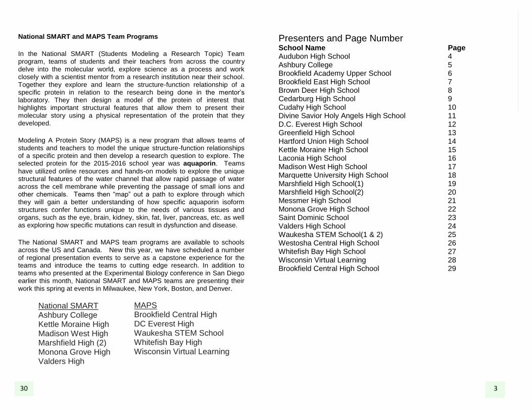
Modeling Aquaporin-4
Wisconsin Virtual Learning Poster #26
Authors: Michael Krueger, Rachel Mangiulli, Casandra Mangiulli, Emilie McNabb, Amber Wicklund, Alanna Wilson Teacher: Karen O’Donnell School: Wisconsin Virtual Learning Imagine life threatening problems which appear when you cannot regulate water properly. Problems such as, cerebral edema, tumors, or seizures arise when the protein Aquaporin- 4 (AQP4) is insufficient in the brain. AQP4 maintains the cerebral water balance, potassium uptake, movement of glial cells, and communication between astrocyte cells. These functions aid in the water transport of the brain, helping to sustain a normal amount of water. Water is important to the brain because it provides energy to produce hormones and neurotransmitters. AQP4 water channels are found in the cell membranes of astrocytes and ependymal cells, and osmosensory areas of the hypothalamus. Water transport through AQP4 depends on concentration gradients across the membrane, which is where the rate of transport is determined by astrocytes. An AQP4 deficiency would slow down the migration of astrocytes, playing a role in two types of cerebral edema called vasogenic and cytotoxic. With vasogenic brain edema, less water is transported out of the brain, while with cytotoxic brain edema, less water is transported into the brain. Someone with cytotoxic brain edema may have difficulties functioning, although the risk for brain swelling and its effects are higher with vasogenic brain edema. The Wisconsin Virtual Learning MAPS Team (Modeling a Protein Story) used 3D technology to model the structure, primarily highlighting amino acids 127, 55, 27, and 92 derived from the protein data bank file: 3GD8. More research into the function of AQP4 could possibly serve as a therapeutic source for a variety of cerebral disorders.
The Potential of Viral P19 for Use as a Suppressor Protein in
Disease States Ashbury College
Poster #2
Authors: Alexandra Akman, Flavia Dumitrascu, Kevin Hu, Tanya Nguyen, Nicholas Sullivan, Mason Xia Teacher: Susan Wall School: Ashbury College Mentors: John Pezacki, Ph.D., and Dana Danielson; University of Ottawa Throughout evolution, organisms developed differently from one another and thus defend themselves against viral attacks through the use of different systems. Animals defend themselves through complex immune systems which plants do not posses. Therefore, plants have a sophisticated silencing mechanism which detects and destroys double stranded RNA (characteristically viral) in a process called RNA interference (RNAi). Small interfering RNAs (siRNA) are cleaved from double stranded introns by an enzyme called DICER. In the plant, the role of siRNA is to target viral messenger RNA to be broken down via the RNAi pathway. P19 is a protein that is expressed by a family of viruses, tombus viruses, in order to suppress the RNA silencing pathway. P19 binds as a dimer to small RNA duplexes of a specific length and prevents them from participating in further steps of RNA silencing. The sequence-independent binding results from hydrogen-bonding and electrostatic interactions between the 8 saddle-like β- sheets of the homodimer and the sugar-phosphate backbone of the siRNA. P19 binds specifically to 21-nt siRNAs due to a higher affinity caused by two tryptophan residues located on an α helix at the N terminus of each monomer, which provide end-capping interactions to stabilize this length in the binding pocket of the dimer. In humans, small RNAs such as siRNA or microRnas (miRNAs) regulate the expression of messenger RNA (mRNA). Therefore, P19 could be used as a tool to control gene expression by inhibiting the siRNA. Developing P19 as a biotechnological tool offers unique opportunities for increasing our understanding of small RNA biology and its role in human disease. It serves as a molecular caliper, which selects siRNAs with a high degree of size selectivity (20- 22 nucleotides), but without regard for the sequence. This could be applied to detect small RNAs from biological samples such as human blood. Engineering P19 has allowed binding to human miRNA which can be used to detect human miRNAs or sequester miRNAs in human cells to allow a desired change in miRNA activity.
5 28
3GD8.pdb

Comparison of Structure and Mechanism in BoNT/A1 and BoNT/A2: Implications for Therapeutic Applications
Brookfield Academy Upper School Poster #3
Authors: Elizabeth Cinquegrani, Mark Morris, Leah Wang, Harry Kalsi,
Shalini Gundamraj, Suneri Kothari, Lena Ding, Srimayi Mylavarapu, Alex Dortzbach, Juliana Vollmer Teacher: Robbyn Tuinstra, Ph.D. School: Brookfield Academy Upper School Mentors: Madison Zuverink, Ph.D. Candidate and Joseph T. Barbieri, Ph.D.
Medical College of Wisconsin, Department of Microbiology and Molecular Genetics
Botulinum neurotoxin type A (BoNT/A) is a potent neurotoxin, causing muscle paralysis in the host by blocking the release of the neurotransmitter, acetylcholine, from motor neurons associated with skeletal muscle. Despite this toxicity, BoNT/A is used pharmaceutically as a treatment for numerous neurological diseases, including migraines, dystonias, and as an anti-wrinkle agent in cosmetic surgery. BoNT/A is one of seven serotypes of botulinum (A-G), which, along with tetanus toxin, are produced by several species of Clostridium. All clostridial neurotoxins, such as BoNT/A, are di-chain proteins, consisting of a 50-kDa light chain, the catalytic domain, connected by a disulfide bond to a 100-kDa heavy chain, containing the receptor-binding and translocation domains. BoNT/A intoxication is a multistep process. First, BoNT/A binds to presynaptic nerve endings, through interactions of the receptor-binding domain with the ganglioside, GT1b, and the protein co-receptor, SV2. SV2 is a synaptic vesicle protein that becomes exposed to the neuron cell surface as vesicles fuse with plasma membrane, releasing neurotransmitter into the synapse. BoNT/A then enters the neuron by receptor-mediated endocytosis. Following endocytosis, the synaptic vesicle acidifies, allowing for neurotransmitter uptake, which triggers the translocation domain to insert and transport the catalytic domain into the cytosol. The catalytic domain is a Zn-dependent protease that cleaves SNAP-25, one of three major proteins present in the SNARE complex. The SNARE protein complex is required for the fusion of synaptic vesicles with the neuronal membrane. Cleavage of SNAP-25 inhibits fusion of synaptic vesicles to the plasma membrane to inhibit acetylcholine release and muscle contraction, leading to flaccid paralysis. Brookfield Academy SMART (Students Modeling A Research Topic) Team students modeled BoNT using 3D printing technology, highlighting amino acid residues associated with protease activity in the catalytic domain, and GT1b and SV2 interactions within the receptor binding domain. This model is meant to help understand and communicate structure/function relationships of BoNT/A and promote potentially therapeutic uses of the toxin.
Modeling Aquaporin-1 Expression in Autosomal Polycystic Kidney
Diseased Kidneys Whitefish Bay High School
Poster #25
Authors: Jieun Heo, Ryan Davis, Mary Claire Potter, Kaitlyn Jackson, Gam K., Evan Davis, Mohammed Shaheer Teachers: Katie Brown and Paula Krukar School: Whitefish Bay High School About 500,000 people in the United States and six million people worldwide are affected by autosomal polycystic kidney disease (ADPKD). The disease drives nearly half of ADPKD patient to end-stage renal or kidney failure. The role of the human kidney is to remove toxins from the bloodstream and other body fluids through the creation of urine. In this function, the kidney is greatly aided by aquaporin-1 (AQP1). AQP1 is the membrane-inserted water-transporting protein in cell plasma membranes in kidney proximal tubules where transepithelial water transport is facilitated, playing an important role in reabsorption of water from the renal tubular fluid. AQP1 also insures against the entry of acidic substances through the cell membrane, helping regulate the internal balance of the cell and thus the organ. The Whitefish Bay High School MAPS (Modeling A Protein Story) Team used computer programing to model AQP1 and its integral role in the movement of water across membranes. In a normal human kidney, AQP1 is localized in proximal tubules and descending thin limbs of Henle’s loop. However, as the disease progresses to an end-stage disease, AQP1 can be seen on cysts that develop on the tubules. AQP1 is most abundant in normal adult kidneys and in early stage ADPKD kidneys, but is reduced in the membranes of end-stage ADPKD kidneys. Thus, after an initial increase in AQP1 expression to combat the stresses of the disease on osmotic equilibrium, there is a significant decrease in the total water expression as ADPKD progresses because cyst size increases and AQP1 expression decreases. Therefore, study of AQP1 expression can aid in finding more targeted solutions to the highly penetrating ADPKD.
3BTA.pdb
4SCK.pdb
27 6

Regulating Jeans with Genes – A Protein Driven
Pathway to Appetite Suppression
Westosha Central High School Poster #24
Authors: John Dietz, Jared Holloway, Trevor Millhouse, Connor Muff, Victoria Salerno, Zach Wermeling, Lucas Wysiatko Teacher: Jonathan Kao School: Westosha Central High School Mentor: SuJean Choi, Ph.D. Marquette University, Department of Biomedical Sciences According to the World Health Organization, over 1.9 billion people in the world are overweight. Many of these people are overweight as a direct result of overeating. Obesity could potentially be treated by inducing an anorexigenic response, or loss of appetite. Several neurotransmitters are involved in signaling for appetite suppressing or stimulating responses. BDNF, the brain-derived neurotrophin factor, works as one of many appetite regulators in the ventromedial nucleus (VMN) in the hypothalamus. Injection of BDNF into rat VMN has been shown to drastically reduce appetite, increase metabolism, and ultimately lead to weight loss. Conversely, knockout of BDNF has been shown to lead to overeating and obesity. BDNF has a high affinity for the receptor Trk-B due to neurotrophins having variable binding domains. The Westosha Central SMART (Students Modeling A Research Topic) Team modeled BDNF as it binds to Trk-B. Once bound, transduction sends a cascading signal that can stop feeding behavior. Various signals have been shown to affect BDNF concentration leading to anorexigenic and orexigenic responses. BDNF concentration correlates with increased leptin, a hormone made by adipose tissue, physical activity, and glutamate. Concentrations of BDNF decrease as the neurotransmitter GABA increases. Understanding BDNF production and signaling provides researchers with a better opportunity to control expression and elicit a specific response in an attempt to treat obesity or eating disorders.
C-fos: Got Your Back! Brookfield East High School
Poster #4 Authors: J. Allen, W. Bowers, D. Horneffer, A. Jhaveri, E. Kreger, S. Lai, M. Lazar, M. Liu, N. Santebennur, R. Wolff Teacher: Emily Barmantje School: Brookfield East High School Mentor: Audra Kramer, M.S. Marquette University, Department of Biomedical Sciences Climbing the stairs, speed-walking to class, jumping around the room –– all are instinctually effortless. Spinal cord injuries change all of that. When a spinal column injury occurs, severed nerves prevent commands from being sent to parts of the body beyond the injury site, leaving a person with little hope of a full recovery. Our understanding of the mechanisms behind spinal injury rehabilitation is currently very limited. The transcription factor c-fos is an indicator of active neurons, making it possible to assess functional recovery in a rehabilitation program. The increased presence of c-fos within a group of cells’ nuclei can be compared in different animal rehabilitation program groups to determine ideal cells for specific spinal cord injury rehabilitation methods. C-fos works with other transcription factors such as c-jun, binding to DNA specifically on the 5’-TGAGTCA-3’ sequence. For this to occur, c-fos and c-jun dimerize. Studying c-fos can aid in developing more advanced forms of spinal rehabilitation that aim on rewiring neural connections. The Brookfield East SMART (Students Modeling a Research Topic) Team designed a model of c-fos using 3D printing technology. The c-fos model focuses on the coiled-coil at the carboxy-terminal region. Further research of c-fos improves our understanding of tissues with limited regenerative abilities and allows us to utilize c-fos as a tool to assess functional tissue regeneration otherwise thought to be limited. 7
1FOS.pdb
26
1BND.pdb
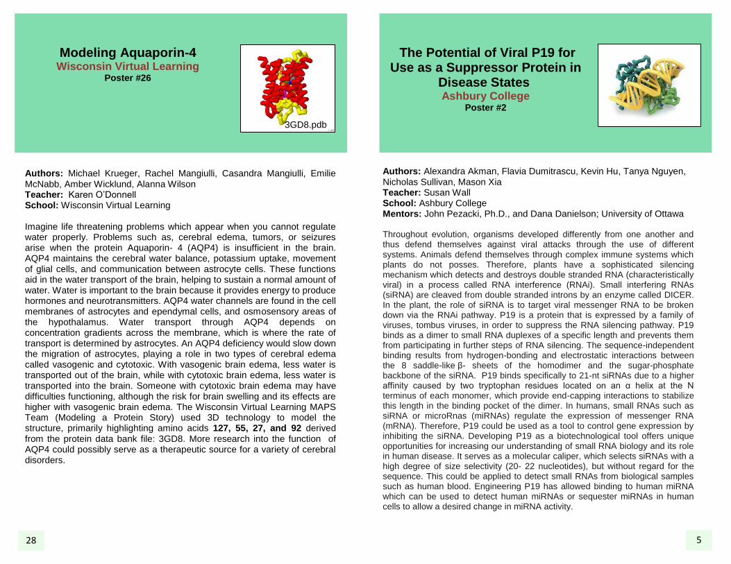
TRPA1 Makes You Feel The PA1N
Brown Deer High School Poster #5
Authors: Georgina Foran, Noah Freuler, Teylor Harris, Ashley Higgins, Justin Johnson, Isaac Ngui, Brett Poniewaz, Gloria Ramos, Luke Richmond, Noel Stoehr, Virginia Tuncel Teacher: David Sampe School: Brown Deer High School Mentor: Andy Weyer, Ph.D., DPT, and Katherine Zappia, Ph.D. Candidate Medical College of Wisconsin, Department of Cell Biology, Neurobiology and Anatomy About 3-4% of people worldwide suffer from chronic pain. The United States spends $60 billion annually treating pain. Current treatments are ineffective; opioids are addictive and lose efficacy while pregabalin, a non-opioid neuropathic pain drug, only works in 30% of patients. Pain receptors (nociceptors) are activated chemically or mechanically. The nociceptor transient receptor potential cation channel, subfamily A, member 1 (TRPA1) is an ion channel located in the membranes of free nerve endings in the skin. TRPA1 is activated by mechanical stretching, and has many active sites where ligands (e.g. tear gas, mustard gas, and wasabi) can bind to open the channel. Once opened, Ca
2+ and Na
2+ ions pass through,
depolarizing the neuron. Structurally, TRPA1 is a tetramer, with each subunit containing several domains. In the pre-S1 domain, ligands bind covalently to the sulfurs at Cys621, Cys641, Cys665, and Lys710. The TRP-like domain opens for ion passage. The ankyrin repeat domain contains 16 recurring sequences of 33 residues, and may be involved with the stretch activation of TRPA1. In domains S1- -helices hold the protein’s shape and contain binding sites for agonists to open the channel and for antagonists, which prevent functioning. One antagonist, A-967079, binds in a pocket around Phe909, forming a wedge to prevent movement and function of TRPA1. Researchers aim to develop a TRPA1 antagonist that will help treat pain sufferers. The Brown Deer SMART (Students Modeling A Research Topic) Team constructed a model of TRPA1 using 3D printing technology to assist researchers in studying its structure and function.
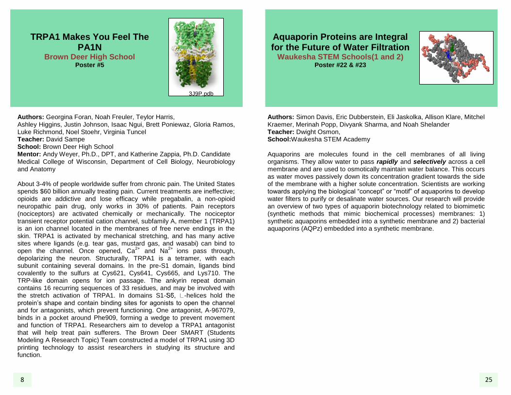
Aquaporin Proteins are Integral for the Future of Water Filtration
Waukesha STEM Schools(1 and 2) Poster #22 & #23
Authors: Simon Davis, Eric Dubberstein, Eli Jaskolka, Allison Klare, Mitchel Kraemer, Merinah Popp, Divyank Sharma, and Noah Shelander Teacher: Dwight Osmon, School:Waukesha STEM Academy Aquaporins are molecules found in the cell membranes of all living organisms. They allow water to pass rapidly and selectively across a cell membrane and are used to osmotically maintain water balance. This occurs as water moves passively down its concentration gradient towards the side of the membrane with a higher solute concentration. Scientists are working towards applying the biological “concept” or “motif” of aquaporins to develop water filters to purify or desalinate water sources. Our research will provide an overview of two types of aquaporin biotechnology related to biomimetic (synthetic methods that mimic biochemical processes) membranes: 1) synthetic aquaporins embedded into a synthetic membrane and 2) bacterial aquaporins (AQPz) embedded into a synthetic membrane.
8
3J9P.pdb
25
Modeling Protein Phosphatase-1 Complexed With the Cyanobacteria Toxin
Mycrocystin Valders High School
Poster #21 Authors: Alyssa Christianson, Sanne DeBruijn, Kassidy Dehne, Abby DeMeyer, Ireland Fenlon, Jacob Krim, Jesse Linsmeier, Madison Storm, Evelyn Tapia-Campuzano, Mariah Ulness, Trevor Wenzel Teacher: Joe Kinscher School: Valders High School Mentor: Rebecca Abler Ph.D, and Richard Hein Ph.D., University of Wisconsin-Manitowoc The cyanobacteria species Microcystis aeruginosa produces microcystin (MC) toxins that can cause liver damage in animals and have resulted in the death of dogs and livestock. MC toxins are cyclic heptapeptides, and they inhibit the enzyme protein phosphatase-1 (PP1), which plays a critical role in reversing the action of protein kinases. Kinases catalyze phosphorylation, which generally promotes cellular activity. In contrast, PP1 catalyzes dephosphorylation, which can slow cellular activity. Balanced phosphorylation and dephosphorylation is critical to liver cell homeostasis, and is disrupted when PP1 is inhibited by MC toxins. MC toxins have two unique amino acid residues: the Mdha and Adda sidechains in addition to standard amino acids. MC binding to PP1 occurs through interactions at four sites on the PP1 enzyme. Two metal atoms of PP1 can coordinate via two water molecules with the glutamic acid residue of MC. The Cys273 residue of PP1 can form a covalent linkage with the Mdha side chain of MC. The hydrophobic Adda side chain of MC fits well in the hydrophobic groove of PP1. Finally, the MeAsp residue of MC hydrogen bonds with Arg96 and Tyr134 residues of PP1. The Valders SMART Team designed a model of PP1 binding MC using 3D printing technology to understand the relationship between MC toxins and PP1.
Opsin: To See or Not to See Cedarburg High School
Poster #6
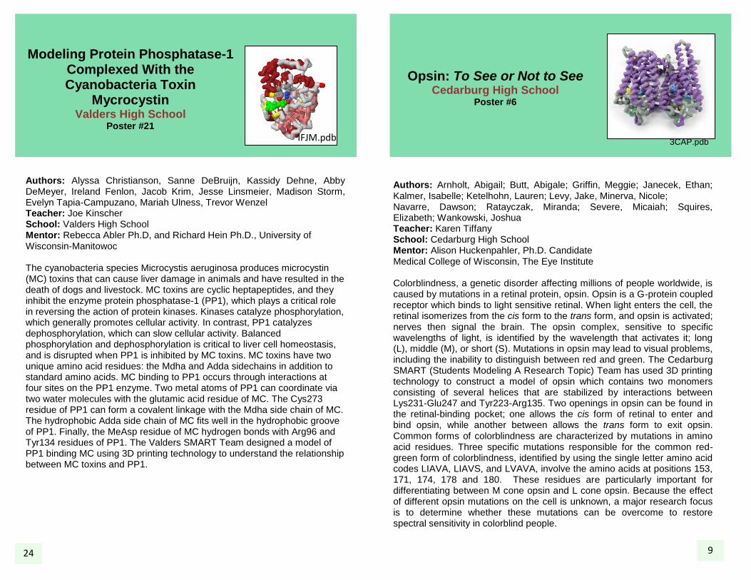
Authors: Arnholt, Abigail; Butt, Abigale; Griffin, Meggie; Janecek, Ethan; Kalmer, Isabelle; Ketelhohn, Lauren; Levy, Jake, Minerva, Nicole; Navarre, Dawson; Ratayczak, Miranda; Severe, Micaiah; Squires, Elizabeth; Wankowski, Joshua Teacher: Karen Tiffany School: Cedarburg High School Mentor: Alison Huckenpahler, Ph.D. Candidate Medical College of Wisconsin, The Eye Institute Colorblindness, a genetic disorder affecting millions of people worldwide, is caused by mutations in a retinal protein, opsin. Opsin is a G-protein coupled receptor which binds to light sensitive retinal. When light enters the cell, the retinal isomerizes from the cis form to the trans form, and opsin is activated; nerves then signal the brain. The opsin complex, sensitive to specific wavelengths of light, is identified by the wavelength that activates it; long (L), middle (M), or short (S). Mutations in opsin may lead to visual problems, including the inability to distinguish between red and green. The Cedarburg SMART (Students Modeling A Research Topic) Team has used 3D printing technology to construct a model of opsin which contains two monomers consisting of several helices that are stabilized by interactions between Lys231-Glu247 and Tyr223-Arg135. Two openings in opsin can be found in the retinal-binding pocket; one allows the cis form of retinal to enter and bind opsin, while another between allows the trans form to exit opsin. Common forms of colorblindness are characterized by mutations in amino acid residues. Three specific mutations responsible for the common red-green form of colorblindness, identified by using the single letter amino acid codes LIAVA, LIAVS, and LVAVA, involve the amino acids at positions 153, 171, 174, 178 and 180. These residues are particularly important for differentiating between M cone opsin and L cone opsin. Because the effect of different opsin mutations on the cell is unknown, a major research focus is to determine whether these mutations can be overcome to restore spectral sensitivity in colorblind people.
3CAP.pdb
24 9
IFJM.pdb
Whaddya Mean Ending Antibiotic Resistance:
S. wadayamensis May Hold Answers
Cudahy High School Poster #7
Authors: Andrew Kressin, Kaylee Day, Emily Bahling, Samantha Brzezinski, Kaycee Valine, Maddy Acherman, Lauren Ligocki, Cori Windsor, Jason Hauk, Madeline Romfoe, Cody Broeckel, Alyssa Sims, Katherine MacDonald, Ramon Rivas, Idamae Harris, Nicholas Kueker, Katelyn Pomianek Teachers: Dan Koslakiewicz and Dean Billo School: Cudahy High School Mentor: Nicholas R. Silvaggi, Ph.D. University of Milwaukee-Wisconsin, Department of Chemistry and Biochemistry Antibiotic-resistant bacteria are common and hard to treat. There is potential to create synthetic antibiotics based on natural products like enduracidin and mannopeptimycin to fight drug resistant bacteria like MRSA. MppP, an enzyme from Streptomyces wadayamensis, is required for the biosynthesis of L-enduracididine (L-end), a non-protein forming amino acid building block found in these and other antibiotics. MppP, the first step in the L-end synthesis pathway, functions to add an oxygen atom to the L-arginine substrate, and replace the α-amino group with a ketone to create 4-hydroxy-2-ketoarginine (4HKA). Hydroxylation is accomplished using a pyridoxal-5’-phosphate (PLP) cofactor, covalently bound by Lys221 to the enzyme, also held by Ser91, Asn160, Asp188, and Ser190 in the active site, which is modeled by the Cudahy SMART (Students Modeling A Research Topic) Team using 3D printing technology. During activity, PLP binds an L-arg substrate, held by Arg352, Asp27, and Asp227 as well as Thr12 and Glu15, performing a hydroxylation through which 4-hydroxy-2-ketoarginine is synthesized. The 4HKA product of the MppP-PLP dependent hydroxylation is used in subsequent reactions in the pathway leading to the creation of the amino acid L-end. Thus, MppP is a critical part of a 3-enzyme team that is responsible for making L-end. The L-end will be incorporated into the different antibiotics by other enzymes. The conversion of L-arg to L-end will help researchers in the creation of new synthetic antibiotics to fight superbugs like MRSA.
Modeling the Catalytic Domain of Activated Factor X Coagulation Protein Bound to Apixaban Saint Dominic School
Poster #20
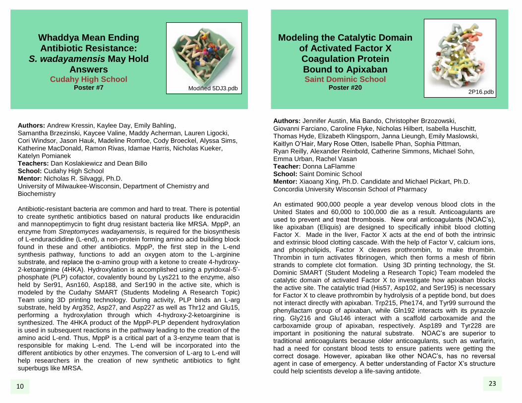
Authors: Jennifer Austin, Mia Bando, Christopher Brzozowski, Giovanni Farciano, Caroline Flyke, Nicholas Hilbert, Isabella Huschitt, Thomas Hyde, Elizabeth Klingsporn, Janna Lieungh, Emily Maslowski, Kaitlyn O’Hair, Mary Rose Otten, Isabelle Phan, Sophia Pittman, Ryan Reilly, Alexander Reinbold, Catherine Simmons, Michael Sohn, Emma Urban, Rachel Vasan Teacher: Donna LaFlamme School: Saint Dominic School Mentor: Xiaoang Xing, Ph.D. Candidate and Michael Pickart, Ph.D. Concordia University Wisconsin School of Pharmacy An estimated 900,000 people a year develop venous blood clots in the United States and 60,000 to 100,000 die as a result. Anticoagulants are used to prevent and treat thrombosis. New oral anticoagulants (NOAC’s), like apixaban (Eliquis) are designed to specifically inhibit blood clotting Factor X. Made in the liver, Factor X acts at the end of both the intrinsic and extrinsic blood clotting cascade. With the help of Factor V, calcium ions, and phospholipids, Factor X cleaves prothrombin, to make thrombin. Thrombin in turn activates fibrinogen, which then forms a mesh of fibrin strands to complete clot formation. Using 3D printing technology, the St. Dominic SMART (Student Modeling a Research Topic) Team modeled the catalytic domain of activated Factor X to investigate how apixaban blocks the active site. The catalytic triad (His57, Asp102, and Ser195) is necessary for Factor X to cleave prothrombin by hydrolysis of a peptide bond, but does not interact directly with apixaban. Trp215, Phe174, and Tyr99 surround the phenyllactam group of apixaban, while Gln192 interacts with its pyrazole ring. Gly216 and Glu146 interact with a scaffold carboxamide and the carboxamide group of apixaban, respectively. Asp189 and Tyr228 are important in positioning the natural substrate. NOAC’s are superior to traditional anticoagulants because older anticoagulants, such as warfarin, had a need for constant blood tests to ensure patients were getting the correct dosage. However, apixaban like other NOAC’s, has no reversal agent in case of emergency. A better understanding of Factor X’s structure could help scientists develop a life-saving antidote.
23 10
Modified 5DJ3.pdb 2P16.pdb

Antibiotic Mechanisms and Resistance in Bacterial
Topoisomerase II Monona Grove High School
Poster #19
Authors: Sarah Fahlberg, Brad Hanson, Katie Klauser, and Sanaa Semia School: Monona Grove High School Teachers/Mentors: Tricica Windgassen, and Katarzyna Dubiel, University of Wisconsin-Madison, Department of Biomolecular Chemistry Over the past fifteen years, a number of bacteria have displayed resistance to common antibiotics while few new antibiotics have been developed. Resistance to antibiotics is life threatening and a global issue that needs to be addressed. Because of this rise in resistance, Ciprofloxacin, a common antibiotic that binds at the active site of topoisomerase II, is becoming less effective. Topoisomerase II (TOP II) is an essential enzyme that is involved in the process of simultaneously cutting both strands of the DNA helix in order to separate replicating bacterial chromosomes and to manage DNA tangles and supercoils. When Ciprofloxacin inhibits TOP II, it does not allow the chromosomes to separate and instead the DNA breaks. Ultimately, the bacteria are not able to replicate or survive. Bacteria are prone to spread resistance to antibiotics because of their ability to replicate quickly. New research has found a drug that targets the active site of TOP II in a new way. Additional research is needed to further understand the mechanism of TOP II to help find out how drug resistance is developed. Once there is more information on drug resistance, there will be more opportunities to develop more new drugs. The Monona Grove SMART team modeled topoisomerase using 3D modeling and printing technology to use the TOP II structure to better understand the protein function. Understanding topoisomerase function can help develop new drugs and combat resistance.
Myobacterium Delirium: Inhibiting Leucine
Biosynthesis to Starve M. tuberculosis
Divine Savior Holy Angels High School
Poster #8
Authors: K Arnold, M Carrig, J David, J Diez, S Franczak, E Grogan, C Halloran, G Hilbert, S Michaeel, C Pfaff, M Pitts, T Rose, L Schauer, C Scherrer, T Siy, S Strandberg, J Strother Teachers: Stacey Strandberg and Scott Fleischmann School: Divine Savior Holy Angels High School Mentor: Martin St. Maurice, Ph.D. Marquette University, Biological Sciences
The pathogen Mycobacterium tuberculosis represents a deadly threat to the worldwide population, especially poor, developing countries, as it kills approximately 2 million people each year according to the World Health Organization. Because of overuse and increasing resistance to current antibiotics, researchers are working to develop new drugs to more effectively treat tuberculosis. M. tuberculosis alpha-isopropylmalate synthase (IPMS) is a bacterial enzyme that catalyzes the production of leucine, an essential amino acid. When cellular leucine levels are low, the biosynthetic pathway of leucine is initiated by IPMS when three ligands; alpha-ketoisovaleric acid (alpha-KIV), acetyl-CoA which is not included in our model, and Zn
2+ interact at IPMS’s
active site. IPMS has two distinct structural domains: an N-terminal alpha/beta catalytic domain which includes the active site and a C-terminal regulatory domain that includes the leucine binding site. As leucine levels increase in the organism, its biosynthetic pathway is shut down by leucine binding to an allosteric site in the C-terminal domain of IPMS which inhibits the further production of additional leucine. The protein amino acids tyr554 and ala536 play a role in the interaction with the leucine ligand. The protein amino acids arg80, asp81, his287, his285, and thr254 interact with the alpha KIV. These interactions allow for the production of leucine through the initiation of the biosynthetic pathway of leucine catalyzed by IPMS. Researchers are working to design a competitive inhibitor that would interact at the allosteric leucine binding site and shut down the pathway, thus depriving M. tuberculosis of this essential amino acid. Without the ligands leucine, Zn
2+, and KIV the tuberculosis bacteria
would die. The Divine Savior Holy Angel High School SMART (Students Modeling A Research Topic) Team modeled IPMS using 3D printing technology to investigate IPMS catalysis and feedback inhibition within the leucine biosynthetic pathway. The development of new drugs specific to M. tuberculosis offers a promising way to overcome the problem of antibiotic resistance and offers new tools to reduce life-threatening tuberculosis infections. 11
22
4OV4.pdb
2XCT.pdb

Aquaporins… Allowing (Just) Water to Go with the
Flow D.C Everest High School
Poster #9 Authors: L. Kee, E. Adams, J. Beilke, A. Bluestein, V. Bradfish, C. Ebner, M. Krueger, H. Mahmood, R. Mallum, G. Martin, P. Molling, M. Ninnemann, J. Nelson, S. Olund, V. Peak, M. Severson Teacher: Bill Heeren School: D.C Everest High School Transporting the liquid of life, aquaporins carry out the vital function of supplying water within cells in almost all living entities. Discovered only a decade ago, aquaporins are channel proteins found in cell membranes that are responsible for the transport and filtering of water and specific molecules into and out of cells. Depending on their location and function, aquaporins may transport different varieties of compounds in addition to water, such as glycerin, to assist in certain organ functions or concentration of cells. Thus, aquaporins play a major role in diseases such as diabetes insipidus due to their regulation of intake and release of water from vital organs. There are thirteen different aquaporins expressed in humans; however, aquaporins are found in all living things, ranging from animals to plants to bacteria. Though there are several types of aquaporins, their structures are similar in composition. Aquaporin monomers consist of six long segments of helices and two short segments of helices, which encompass a narrow transmembrane channel through which water and other molecules travel in single file. Only 20 Å in diameter, this channel is just wide enough for water molecules to pass through. Within cell membranes, monomers assemble as tetramers, each monomer acting independently. They are able to transport molecules at an extremely high rate--just one human AQP1 molecule has a permeation rate of three billion molecules per second, the direction of which changes depending on the prevailing osmotic gradient. Aquaporins must be highly selective in order to perform their specific functions, and they achieve this on an atomic level. The aquaporin contains two separate filters to facilitate the selection of water molecules and the exclusion of everything else: an ar/R filter (blue), and an NPA motif (magenta). Its positively charged ar/R filter allows the aquaporin to both bind to water molecules and weaken the molecule’s hydrogen bonds so it can more readily interact with the filter. Meanwhile, the NPA motif involves the local electrical fields of the atoms and amino acids along the water channel walls, which cause water molecules passing through the channel to rotate and orient themselves towards the atom’s positive charge. Because of the positive charge at the center of the channel, protons and cations are prevented from passing, thereby regulating the balance of protons inside and outside of the cell membrane.
PTP1B Inhibitors for Type 2 Diabetes
Treatment Messmer High School
Poster #18 Authors: Amya Dorsey, Hanh Nguyen, Brittany Slater, Tariq Mckinney, Melissa Anguiano, Babatunde Otukoya, April Carmicheal, Kelly Birmingham Teachers: Mark Zachar and Justin Spaeth School: Messmer High School Mentor: George Wilkinson, Ph.D. Concordia University Wisconsin School of Pharmacy
People with diabetes have difficulty regulating their blood sugar leading to the malfunctions of the heart, kidney, nerves, and brain if their blood sugar is too high. Most diabetics use insulin to control their condition. Insulin binds to its surface receptor in muscle and fat cells to trigger removal of sugar from the blood. Protein tyrosine phosphatase 1B (PTP1B) affects blood sugar regulation by dephosphorylating the insulin receptor and reducing its activity. Studying PTP1B can help us understand how its inhibitors can slow down the removal of phosphate from the insulin receptor, affecting the blood sugar regulation process. PTP1B normally functions to remove the phosphate group from the insulin receptor. Since PTP1B ordinarily reduces insulin receptor activity, blocking PTP1B could increase insulin sensitivity. The PTP1B active site has a highly positive binding pocket which binds to the highly negative phosphates on the phosphorylated insulin receptor. Many of the current inhibitors of PTP1B act by binding to this active site. However, these inhibitors have difficulty penetrating the cell membrane because of their formal charge, and are unable to inhibit PTP1B inside cells. LZP25 avoids this issue by not having a formal negative charge, but instead a polar area of similar size to phosphate. Binding to the PTP1B active site pocket (sites Ser216, Ala217, Ile219, Gln262, Gln266), its bulky side groups then prevent a key loop in the enzyme active site from closing. If PTP1B is inhibited in the insulin pathway by a potential drug based on LZP25, people who have type 2 diabetes as a result of insufficient insulin receptor activity could better regulate their blood sugar. If the insulin receptor would signal appropriately, the body’s normal control of blood sugar would improve, preventing problems with the heart, kidney, nerves, and brain. Messmer Catholic High School SMART (Students Modeling A Research Topic) Team has designed a model of PTP1B with LZP25 using 3D printing technology to investigate their structure/function relationships.
12
3EB1.pdb
21
IFQY.pdb
Phenylalanine Hydroxylase (PheOH)
Marshfield High School(2) Poster #17
Authors: Haania Khan, Aman Bassi, Roma Shah, Suhaas Bhat, Muhammad Abidi Teachers: Amy Fassler School: Marshfield High School
Nobel Prize winning author, Pearl S. Buck, gave birth to a daughter, Carol, in 1921. Unknown to her mother and the rest of the scientific community, Carol suffered from phenylketonuria, a genetic disorder that results in severe brain damage. This is caused by a mutation in the PAH gene, which codes for the enzyme phenylalanine hydroxylase (PheOH). Phenylketonuria is characterized by high levels of phenylalanine in the bloodstream, resulting from the inability of PheOH to hydrolyze phenylalanine into tyrosine. There are two leading theories to explain the severities of neurological damage resulting from PKU. One possible explanation is that tetrahydrobiopterin (BH4) cannot hydrolyze tyrosine. The other theory examines the impact of high concentrations of phenylalanine inhibiting the transfer of tyrosine across the blood-brain barrier. Recent advancements in technology, such as newborn screening, allows geneticists to determine if the child has PKU.
Not-So-Mighty Mitochondria: Neonatal
Lethality Due to Drp1 Malfunction
Greenfield High School Poster #10
Authors: Andrew Braatz, Pratyusha Emkay, Sarah Goodman, Cora Libecki, Melody Ly, Karen Nakhla, Zac Osberg, Zoe Osberg, Paige Paniagua, Lorenzo Vasallo Teacher: Julie Fangmann School: Greenfield High School Mentor: Amber Bakkum, Ph.D. Candidate and John Egner, Ph.D. Candidate Medical College of Wisconsin, Department of Biochemistry
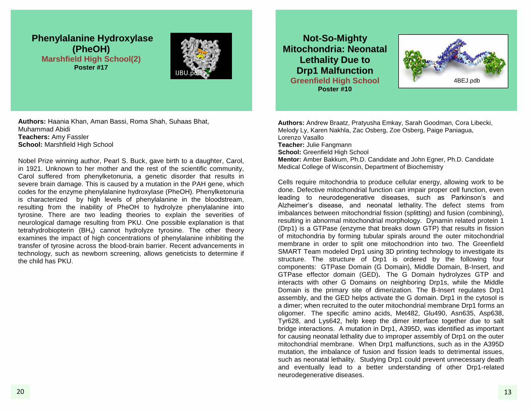
Cells require mitochondria to produce cellular energy, allowing work to be done. Defective mitochondrial function can impair proper cell function, even leading to neurodegenerative diseases, such as Parkinson’s and Alzheimer’s disease, and neonatal lethality. The defect stems from imbalances between mitochondrial fission (splitting) and fusion (combining), resulting in abnormal mitochondrial morphology. Dynamin related protein 1 (Drp1) is a GTPase (enzyme that breaks down GTP) that results in fission of mitochondria by forming tubular spirals around the outer mitochondrial membrane in order to split one mitochondrion into two. The Greenfield SMART Team modeled Drp1 using 3D printing technology to investigate its structure. The structure of Drp1 is ordered by the following four components: GTPase Domain (G Domain), Middle Domain, B-Insert, and GTPase effector domain (GED). The G Domain hydrolyzes GTP and interacts with other G Domains on neighboring Drp1s, while the Middle Domain is the primary site of dimerization. The B-Insert regulates Drp1 assembly, and the GED helps activate the G domain. Drp1 in the cytosol is a dimer; when recruited to the outer mitochondrial membrane Drp1 forms an oligomer. The specific amino acids, Met482, Glu490, Asn635, Asp638, Tyr628, and Lys642, help keep the dimer interface together due to salt bridge interactions. A mutation in Drp1, A395D, was identified as important for causing neonatal lethality due to improper assembly of Drp1 on the outer mitochondrial membrane. When Drp1 malfunctions, such as in the A395D mutation, the imbalance of fusion and fission leads to detrimental issues, such as neonatal lethality. Studying Drp1 could prevent unnecessary death and eventually lead to a better understanding of other Drp1-related neurodegenerative diseases.
13 20 13
4BEJ.pdb
IJBU.pdb
Bond … One Bond”: The Biological Blowtorch and Other
Reactive Players in CYP17
Catalyzed Production of Androgens
Hartford Union High School Poster #11
Authors: Burg, A., Diol L., Gall, M., Kastner, A., Kaul, M., Kieckhefer, T., Schmidt, N., Stoellinger, H Teacher: Mark Arnholt School: Hartford Union High School Mentors: Piotr Mak, Ph.D., and James Kincaid,Ph.D. Marquette University, Department of Chemistry Prostate cancer makes up 4.7 percent of all cancer related deaths. The exact cause of prostate cancer is unknown, but researchers have identified the leading risk factors, including excess testosterone in the body. Early treatments to reduce testosterone levels in patients involved surgical procedures. A particular heme-containing enzyme, known as CYP17, converts cholesterol-derived hormones into androgens including testosterone. Fe(III) is a key part as all reactions occur within this heme group. Pharmacological efforts, such as the drug abiraterone, decrease the efficiency of CYP17 catalysis and may present a valid treatment option for prostate cancer patients. Further efforts would be facilitated by gaining a better understanding of the mechanisms of CYP17 in the presence of its natural substrates, 17-OH progesterone and 17-OH pregnenolone. This enzyme generates an androgen precursor by catalyzing two sequential chemical conversions, the second one by an unknown process, which has been difficult to track. Using rapid-freezing, γ-ray exposure and Raman spectroscopy, the key reaction intermediates in this process are trapped and structurally defined. The Hartford Union SMART (Students Modeling A Research Topic) Team modeled the enzyme CYP17A1 with 3-D printing technology. Research efforts involving CYP17 could assist pharmaceutical companies in the development of treatments for prostate cancer.
Modeling of New Delhi Metallo-Beta-Lactamase-1, an Enzyme for
Resistance to Carbapenems Marshfield High School(1)
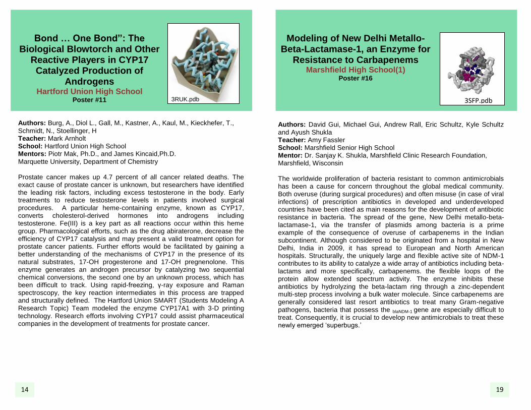
Poster #16 Authors: David Gui, Michael Gui, Andrew Rall, Eric Schultz, Kyle Schultz and Ayush Shukla Teacher: Amy Fassler School: Marshfield Senior High School Mentor: Dr. Sanjay K. Shukla, Marshfield Clinic Research Foundation, Marshfield, Wisconsin The worldwide proliferation of bacteria resistant to common antimicrobials has been a cause for concern throughout the global medical community. Both overuse (during surgical procedures) and often misuse (in case of viral infections) of prescription antibiotics in developed and underdeveloped countries have been cited as main reasons for the development of antibiotic resistance in bacteria. The spread of the gene, New Delhi metallo-beta-lactamase-1, via the transfer of plasmids among bacteria is a prime example of the consequence of overuse of carbapenems in the Indian subcontinent. Although considered to be originated from a hospital in New Delhi, India in 2009, it has spread to European and North American hospitals. Structurally, the uniquely large and flexible active site of NDM-1 contributes to its ability to catalyze a wide array of antibiotics including beta-lactams and more specifically, carbapenems. the flexible loops of the protein allow extended spectrum activity. The enzyme inhibits these antibiotics by hydrolyzing the beta-lactam ring through a zinc-dependent multi-step process involving a bulk water molecule. Since carbapenems are generally considered last resort antibiotics to treat many Gram-negative pathogens, bacteria that possess the blaNDM-1 gene are especially difficult to treat. Consequently, it is crucial to develop new antimicrobials to treat these newly emerged ‘superbugs.’ 14 19
3RUK.pdb 3SFP.pdb

Does Hope Lie Between Apoptosis and Necrosis?
Effect of Inhibiting NF-κB on Ischemia-Reperfusion-Induced Cell Apoptosis and
Necrosis in Cardiomyocytes Marquette University High School
Poster #15
Authors: P Ahn, K Arnhold, N Cowan, N Dittrich, R Foster, J Gorski, R Johnson, H Khalil, C Kim, M Malone, S Mohiuddin, L Ortega, J Otten, M Rivera, M Rose, P Rozewicz, A Sabatino, T Sargent, J Smith, D Strom, J Strom, B Tsuji, J Yamat, N Yorke Teachers: Carl Kaiser and Keith Klestinski School: Marquette University High School Mentor: Judy Maloney, Ph.D. Marquette University, Department of Biomedical Sciences
Cardiovascular disease and its complications are the leading cause of morbidity and mortality in the United States. A myocardial infarction (MI) is caused by a blood clot in a coronary artery and results in loss of blood flow to the heart (ischemia). To prevent necrosis or cell death of heart tissue, ischemia is treated with reperfusion therapy, in which blood flow is restored. Reperfusion therapy paradoxically causes damage to the heart muscle cells via the activation of Nuclear Factor Kappa Beta (NFκB) which can trigger apoptosis or programed cell death, or via the generation of reactive oxygen species and subsequently further necrosis. The extent to which the transcription factor NFκB promotes cell death or even promotes survival is unknown. However, it is known to be a key inflammatory mediator that coordinates cell homeostasis under bodily stress, as with an MI or ischemia reperfusion (IR) injury. NFκB is complexed with Inhibitor Kappa Beta (IκB) and exists as a larger IκB/NFκB complex within the cytosol of the cell until inflammatory mediators activate an IκB kinase. Phosphorylation of this larger complex separates IκB from NFκB, at which point NFκB translocates to the nucleus of the cell, binds to DNA, and initiates transcription of genes responsible for apoptosis. The Marquette University High School SMART (Students Modeling A Research Topic) Team modeled NFκB, a heterodimer, which, in the heart, is composed of the subunits, p50 and p65, complexed with DNA. To study the effect of NFκB inhibition on IR-induced apoptosis and necrosis, cultured cardiac myocytes received two hours of ischemia, and were reperfused with or without the NFκB inhibitor, N4-[2-(4-phenoxyphenyl)ethyl]-4,6-quinazolinediamine (QNZ). Caspase 3 activation and annexin V staining, measurements of apoptosis, revealed that inhibiting NF-κB caused a decrease in IR-induced apoptosis. Propidium iodide staining, a measurement of cell necrosis, revealed that inhibiting NF-κB caused an increase in IR-induced necrosis. Further studies into understanding the effect of NFκB inhibition on IR injury may help provide novel therapies.
The P2X4 Subunit and Alcohol Addiction
Kettle Moraine High School Poster #12
Authors: Mahi Gokuli, Ethan Helfenstein, Sam Kanakkanatt, Christina Nielsen, Autumn Saunders, Kaitlyn Simon, Maggie Simon, Aaron Smet, Justin Smet, and Benjamin Taylor Teacher: Melissa Kirby School: Kettle Moraine High School Mentor: Robert W. Peoples, Ph.D., Marquette University, Department of Biomedical Sciences Alcoholism is a disease. A predisposition towards alcohol is inherited: it has less to do with the type of alcohol, amount consumed, or the duration of consumption than it has to do with the compulsive need to drink that an alcoholic faces. The P2X4 receptor, part of the larger family of P2X receptors (P2XRs), has been shown to be involved in this need. These receptors, found in all mammalian tissues, are non-selective cation channels activated by the extracellular binding of synaptically-released ATP. P2XR channels can be formed by seven possible subtypes, P2X1-P2X7 receptors, which have a three-protein structure consisting of two transmembrane domains connected by a large ectodomain. For certain subtypes including the P2X4 receptor, which is the most abundant subtype in the central nervous system, ethanol at low-intoxicating concentrations inhibits ATP-gated currents; this is in contrast to subtypes such as P2X3 receptors, for which ethanol strengthens currents. There is evidence that this inhibition of P2X4 receptors by alcohol may play a role in the signalling process to cease alcohol consumption: of all the P2XRs, P2X4 receptors are the most ethanol-sensitive (this ethanol sensitivity has been observed to be at least partially regulated by the receptor’s extracellular cysteines, colored red) and have been observed to decrease innate tendencies towards ethanol consumption in mice. This has far-reaching implications-- P2X4 receptors may be involved with genetic vulnerability to alcoholism. Further investigation into the P2X4 subtype may lead to a greater understanding of its role in alcohol addiction. The Kettle Moraine High School SMART (Students Modeling A Research Topic) has modeled the P2X4 subtype using 3D printing technology to investigate structure-function
relationships.
1VKX.pdb 3I5D.pdb
15 15 18
Understanding α-Galactosidase A to Improve
Fabry Disease Treatment Laconia High School
Poster #13 Authors Allison Opheim, Alex Wood, Josie Stahmann, McKenzie Englund, Mckayla Johnson, Carissa Zibolsky, Ashley Pluim, Daniel Ihrig Teacher: Josh Demski School: Laconia High School Mentor: James Miller, M.D. and Ph.D. Candidate Medical College of Wisconsin, Department of Biochemistry Fabry disease is a debilitating lysosomal storage disease. Early in life, patients with Fabry disease experience extreme pain, especially in the extremities (acroparesthesias). Patients are eventually at risk of developing kidney disease, heart failure, and strokes, all of which may lead to a premature death. The prevalence of Fabry disease in its severest form is estimated at 1:40,000; however, the prevalence of milder, later-onset forms may be as high as 1:3,600. Fabry disease is caused by mutations in the X chromosomal GLA gene, which encodes the lysosomal enzyme, α-galactosidase A (α-Gal A). When α-Gal A activity is deficient, glycolipid substrates accumulate within lysosomes, leading to cellular, tissue, and organ pathology. To treat Fabry disease, patients are given enzyme replacement therapy (i.e., bi-weekly intravenous infusion of recombinant α-Gal A). Enzyme replacement therapy costs up to $300,000 per patient per year and commonly causes dangerous immune reactions in patients. A more cost-effective and safer treatment is desperately needed. The Laconia SMART (Students Modeling A Research Topic) Team used 3D printing
technology to generate a model of ⍺-Gal A. To be trafficked to lysosomes
and to optimally degrade glycolipids, ⍺-Gal A must be glycosylated and must dimerize. Therefore, critical residues are highlighted in the model, such as those involved in catalysis (Asp170, Asp231), N-linked glycosylation (Asn139, Asn192, Asn215), and dimerization (Phe273).
Disulfide bonds, which stabilize ⍺-Gal A tertiary structure, are also shown. Studying the structure of α-Gal A is critical in the design of a more potent therapy, which has the potential to reduce the cost and immunogenicity of Fabry disease treatment.
Investigating the Single-Stranded DNA Binding Protein Tetramer
Interface Madison West High School
Poster #14
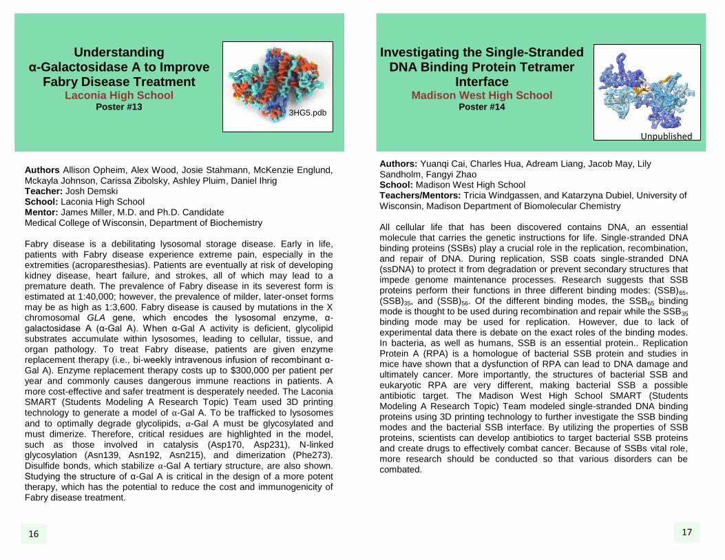
Authors: Yuanqi Cai, Charles Hua, Adream Liang, Jacob May, Lily Sandholm, Fangyi Zhao School: Madison West High School Teachers/Mentors: Tricia Windgassen, and Katarzyna Dubiel, University of Wisconsin, Madison Department of Biomolecular Chemistry All cellular life that has been discovered contains DNA, an essential molecule that carries the genetic instructions for life. Single-stranded DNA binding proteins (SSBs) play a crucial role in the replication, recombination, and repair of DNA. During replication, SSB coats single-stranded DNA (ssDNA) to protect it from degradation or prevent secondary structures that impede genome maintenance processes. Research suggests that SSB proteins perform their functions in three different binding modes: (SSB)65, (SSB)35, and (SSB)56. Of the different binding modes, the SSB65 binding mode is thought to be used during recombination and repair while the SSB35 binding mode may be used for replication. However, due to lack of experimental data there is debate on the exact roles of the binding modes. In bacteria, as well as humans, SSB is an essential protein.. Replication Protein A (RPA) is a homologue of bacterial SSB protein and studies in mice have shown that a dysfunction of RPA can lead to DNA damage and ultimately cancer. More importantly, the structures of bacterial SSB and eukaryotic RPA are very different, making bacterial SSB a possible antibiotic target. The Madison West High School SMART (Students Modeling A Research Topic) Team modeled single-stranded DNA binding proteins using 3D printing technology to further investigate the SSB binding modes and the bacterial SSB interface. By utilizing the properties of SSB proteins, scientists can develop antibiotics to target bacterial SSB proteins and create drugs to effectively combat cancer. Because of SSBs vital role, more research should be conducted so that various disorders can be combated. 17
3HG5.pdb
Unpublished
16