Cellular expression after transfectionwiththeir cDNAs … · after transfectionwiththeir...
5
Proc. Natl. Acad. Sci. USA Vol. 84, pp. 8502-8506, December 1987 Developmental Biology Cellular expression of liver and neural cell adhesion molecules after transfection with their cDNAs results in specific cell-cell binding GERALD M. EDELMAN, BEN A. MURRAY, RENE-MARC MEGE, BRUCE A. CUNNINGHAM, AND WARREN J. GALLIN The Rockefeller University, New York, NY 10021 Contributed by Gerald M. Edelman, August 26, 1987 ABSTRACT Mouse L cells, which do not express the known primary cell adhesion molecules (CAMs), were perma- nently transfected with vectors containing the simian virus 40 early promoter and cDNA sequences encoding chicken liver CAM (L-CAM) or each of the three major polypeptide forms of chicken neural CAM (N-CAM). Transfected cells in culture expressing the Ca2l-dependent L-CAM showed uniform sur- face expression of the molecule. Unlike untransfected L cells, these cells aggregated readily; the aggregation was inhibited by Fab' fragments of antibodies to L-CAM but not by fragments of anti-N-CAM. These cells spread more efficiently in culture than did their untransfected counterparts, forming small colonies of flattened cells that gradually assumed morphologies resembling closely packed L cells. Transfected L cells express- ing either the small or large intercellular domain polypeptide (sd or Id) chains of N-CAM aggregated specifically with each other or bound membrane vesicles from chick brain. Both types of binding were specifically inhibited by Fab' fragments of anti-N-CAM antibodies. These cells, in contrast to those transfected with vectors for L-CAM, showed rounded mor- phologies and spread inefficiently in culture. L cells transfected with vectors specifying the small surface domain polypeptide (ssd) chain of N-CAM showed no phenotypic changes and no evidence for linkage of ssd chains to the cell membrane by phosphatidylinositol intermediates. Instead, these cells synthe- sized the molecule and released it into the medium. These findings complete the demonstration that different CAMs have specific roles in ligating the cells that synthesize them, and they provide further evidence that L-CAM and N-CAM bind by homophilic mechanisms. The different phenotypic changes observed for each specific CAM are consistent with the hypothesis that CAM synthesis or differing associations of CAM carboxyl-terminal domains with the cell surface and cortex may lead directly or indirectly to specific alterations in the cells bound together by that CAM. The demonstration (1, 2) that cell adhesion molecules (CAMs) of different specificities play major roles in morpho- genesis, histogenesis, and regeneration has opened the pos- sibility of relating key phenotypic properties of cells with their epithelial or mesenchymal states (3). Primary CAMs of different specificities, such as the Ca2+-dependent liver CAM (L-CAM) and Ca2+-independent neural CAM (N-CAM), appear to bind by homophilic mechanisms-i.e., CAM on one cell to the same CAM on an apposing cell (4). The findings that these molecules (i) appear in defined develop- mental expression sequences (2), (ii) are specified by one or at most a few genes (5), and (iii) are under regulatory control related to boundaries of the condensed mesenchyme or epithelia that they help to ligate (6) suggest that specific CAM binding functions must be correlated with particular cell states. Recent analyses (7-9) of the structures of cDNAs speci- fying these two CAMs and of the genomic DNA for N-CAM (10) are consistent with this view. N-CAM, which appears to be closely related evolutionarily to the precursor of the entire immunoglobulin superfamily (7, 8, 11), is expressed as three major polypeptides that arise as a result of alternative splicing of RNA transcribed from a single gene (12). Each N-CAM chain has a different carboxyl-terminal domain and a different mode of association with the cell surface or cell cortical region. Of these polypeptides, the large and small intercel- lular domain (ld and sd) polypeptide chains contain intracel- lular domains of different sequence and size, whereas the small surface-domain (ssd) polypeptide chain lacks an intra- cellular domain but is linked to the cell surface by a phosphatidylinositol-containing intermediate. L-CAM is structurally unrelated to N-CAM and has another kind of intracellular domain (9). This Ca2+-dependent CAM and the different N-CAM polypeptides all have individually charac- teristic tissue distributions (2, 3, 12). The availability of appropriate cDNA clones for all of these molecules has made it possible to examine the effects of expression of CAMs on cells that ordinarily do not synthesize them. In the present experiments, vectors constructed with cDNAs specifying L-CAM and N-CAM chains were used to transfect L cells that are free of these molecules in their ordinary state. The transfected cells expressed CAMs of the correct binding specificity that ligated the cells in a homophilic fashion. Permanent lines transfected with each type of CAM showed distinct phenotypic changes in cell shape and cell spreading in culture. Transfection experiments thus open the possibility of correlating amino-terminal CAM binding specificities with the properties and cellular effects of their carboxyl-terminal surface-bound structures or their intracellular domains. Such experiments should also facili- tate the analysis of the effects of differential expression of two different CAMs in the same cell. MATERIALS AND METHODS DNA Constructs. All DNA constructs were prepared with cDNA clones for chicken N-CAM (7, 8, 13) and L-CAM (9). Constructs encompassing the coding sequence for each N-CAM polypeptide (Id, sd, and ssd) were inserted after the simian virus 40 (SV40) early promoter in a variant of the plasmid pCH110 (14). The EcoRI site downstream from the poly(A)+ addition site in pCH110 was removed by partial EcoRI digestion, and the ends were polished with the large (Klenow) fragment of DNA polymerase and rejoined by Abbreviations: CAM, cell adhesion molecule; N-CAM, neural CAM; L-CAM, liver CAM; SV40, simian virus 40; Id and sd, large and small intracellular domain polypeptides; ssd, small surface domain poly- peptide. 8502 The publication costs of this article were defrayed in part by page charge payment. This article must therefore be hereby marked "advertisement" in accordance with 18 U.S.C. §1734 solely to indicate this fact.
Transcript of Cellular expression after transfectionwiththeir cDNAs … · after transfectionwiththeir...

Proc. Natl. Acad. Sci. USAVol. 84, pp. 8502-8506, December 1987Developmental Biology
Cellular expression of liver and neural cell adhesion moleculesafter transfection with their cDNAs results in specificcell-cell bindingGERALD M. EDELMAN, BEN A. MURRAY, RENE-MARC MEGE, BRUCE A. CUNNINGHAM,AND WARREN J. GALLINThe Rockefeller University, New York, NY 10021
Contributed by Gerald M. Edelman, August 26, 1987
ABSTRACT Mouse L cells, which do not express theknown primary cell adhesion molecules (CAMs), were perma-nently transfected with vectors containing the simian virus 40early promoter and cDNA sequences encoding chicken liverCAM (L-CAM) or each of the three major polypeptide formsof chicken neural CAM (N-CAM). Transfected cells in cultureexpressing the Ca2l-dependent L-CAM showed uniform sur-face expression of the molecule. Unlike untransfected L cells,these cells aggregated readily; the aggregation was inhibited byFab' fragments of antibodies to L-CAM but not by fragmentsof anti-N-CAM. These cells spread more efficiently in culturethan did their untransfected counterparts, forming smallcolonies of flattened cells that gradually assumed morphologiesresembling closely packed L cells. Transfected L cells express-ing either the small or large intercellular domain polypeptide(sd or Id) chains of N-CAM aggregated specifically with eachother or bound membrane vesicles from chick brain. Both typesof binding were specifically inhibited by Fab' fragments ofanti-N-CAM antibodies. These cells, in contrast to thosetransfected with vectors for L-CAM, showed rounded mor-phologies and spread inefficiently in culture. L cells transfectedwith vectors specifying the small surface domain polypeptide(ssd) chain of N-CAM showed no phenotypic changes and noevidence for linkage of ssd chains to the cell membrane byphosphatidylinositol intermediates. Instead, these cells synthe-sized the molecule and released it into the medium. Thesefindings complete the demonstration that different CAMs havespecific roles in ligating the cells that synthesize them, and theyprovide further evidence that L-CAM and N-CAM bind byhomophilic mechanisms. The different phenotypic changesobserved for each specific CAM are consistent with thehypothesis that CAM synthesis or differing associations ofCAM carboxyl-terminal domains with the cell surface andcortex may lead directly or indirectly to specific alterations inthe cells bound together by that CAM.
The demonstration (1, 2) that cell adhesion molecules(CAMs) of different specificities play major roles in morpho-genesis, histogenesis, and regeneration has opened the pos-sibility of relating key phenotypic properties of cells withtheir epithelial or mesenchymal states (3). Primary CAMs ofdifferent specificities, such as the Ca2+-dependent liver CAM(L-CAM) and Ca2+-independent neural CAM (N-CAM),appear to bind by homophilic mechanisms-i.e., CAM onone cell to the same CAM on an apposing cell (4). Thefindings that these molecules (i) appear in defined develop-mental expression sequences (2), (ii) are specified by one orat most a few genes (5), and (iii) are under regulatory controlrelated to boundaries of the condensed mesenchyme orepithelia that they help to ligate (6) suggest that specific CAM
binding functions must be correlated with particular cellstates.Recent analyses (7-9) of the structures of cDNAs speci-
fying these two CAMs and of the genomic DNA for N-CAM(10) are consistent with this view. N-CAM, which appears tobe closely related evolutionarily to the precursor of the entireimmunoglobulin superfamily (7, 8, 11), is expressed as threemajor polypeptides that arise as a result of alternative splicingof RNA transcribed from a single gene (12). Each N-CAMchain has a different carboxyl-terminal domain and a differentmode of association with the cell surface or cell corticalregion. Of these polypeptides, the large and small intercel-lular domain (ld and sd) polypeptide chains contain intracel-lular domains of different sequence and size, whereas thesmall surface-domain (ssd) polypeptide chain lacks an intra-cellular domain but is linked to the cell surface by aphosphatidylinositol-containing intermediate. L-CAM isstructurally unrelated to N-CAM and has another kind ofintracellular domain (9). This Ca2+-dependent CAM and thedifferent N-CAM polypeptides all have individually charac-teristic tissue distributions (2, 3, 12).The availability ofappropriate cDNA clones for all ofthese
molecules has made it possible to examine the effects ofexpression ofCAMs on cells that ordinarily do not synthesizethem. In the present experiments, vectors constructed withcDNAs specifying L-CAM and N-CAM chains were used totransfect L cells that are free of these molecules in theirordinary state. The transfected cells expressed CAMs of thecorrect binding specificity that ligated the cells in ahomophilic fashion. Permanent lines transfected with eachtype of CAM showed distinct phenotypic changes in cellshape and cell spreading in culture. Transfection experimentsthus open the possibility of correlating amino-terminal CAMbinding specificities with the properties and cellular effects oftheir carboxyl-terminal surface-bound structures or theirintracellular domains. Such experiments should also facili-tate the analysis of the effects of differential expression oftwo different CAMs in the same cell.
MATERIALS AND METHODSDNA Constructs. All DNA constructs were prepared with
cDNA clones for chicken N-CAM (7, 8, 13) and L-CAM (9).Constructs encompassing the coding sequence for eachN-CAM polypeptide (Id, sd, and ssd) were inserted after thesimian virus 40 (SV40) early promoter in a variant of theplasmid pCH110 (14). The EcoRI site downstream from thepoly(A)+ addition site in pCH110 was removed by partialEcoRI digestion, and the ends were polished with the large(Klenow) fragment of DNA polymerase and rejoined by
Abbreviations: CAM, cell adhesion molecule; N-CAM, neural CAM;L-CAM, liverCAM; SV40, simian virus 40; Id and sd, large and smallintracellular domain polypeptides; ssd, small surface domain poly-peptide.
8502
The publication costs of this article were defrayed in part by page chargepayment. This article must therefore be hereby marked "advertisement"in accordance with 18 U.S.C. §1734 solely to indicate this fact.

Proc. Natl. Acad. Sci. USA 84 (1987) 8503
blunt-end ligation. The HindIII fragment from the N-CAMplasmid (pEC254/pEC208) pGEM construct (8) containing1800 base pairs (bp) of the 5' end of N-CAM cDNA was theninserted into the HindIII site of pCH110 found immediatelyadjacent to the SV40 early promoter. The resulting plasmidwas digested with EcoRI and ligated to EcoRI fragmentscontaining the remaining sequences for the Id (pEC208), sd(pEC281; see ref. 12), and ssd (pEC1Si) polypeptides. Thestructures of these plasmids were confirmed by restrictionmapping. We designate the clones pEC1401 (Id), pEC1402(sd), and pEC1403 (ssd).The L-CAM cDNA clones extended 5' from the region
coding for the amino terminus of the mature molecule andmore than 300 bp into the coding sequence of the precursorpolypeptide but did not extend to the 5' end of the mRNA.Therefore, an initiator methionine and signal sequence wereprovided by constructing N-CAM/L-CAM chimeras. TheL-CAM coding region, contained in the EcoRI/BamHIfragment of L-CAM clone pEC320, was inserted into theBluescript KS vector (Stratagene, La Jolla, CA) digestedwith EcoRI and BamHI to give clone B67. pEC1301 was thenprepared by ligating the 191-bp Nae I/Alu I fragment ofN-CAM clone pEC254 into the EcoRI site of the Bluescriptpolylinker of B67. pEC1301 thus contains sequences codingfor about 64 amino acids at the amino terminus of N-CAM.pEC1302 was prepared by ligating the 391-bp Nae IlEcoRIfragment of pEC254 into EcoRI/EcoRV-digested B67; thisclone codes for about 103 amino acids of the amino terminusof N-CAM. The structure and orientation of pEC1301 andpEC1302 were confirmed by DNA sequencing.pEC1301 and pEC1302 were digested with Sal I, the ends
were polished with Klenow fragment, and BamHI linkerswere added. After digestion with BamHI, these inserts werepurified on agarose gels and ligated into the Bgl II site ofpKSV-10 (Pharmacia) adjacent to the SV40 early promoter.The orientation of the insert in isolated clones was deter-mined by restriction mapping. We designate pKSV-10 havingthe pEC1301 insert as pEC1311 and the construct having thepEC1302 insert as pEC1312.
Cell Culture and Transfection. All transfections were per-formed with a calcium phosphate protocol (15) on mouseL-M(TK-) cells (American Type Culture Collection CCL1.3) grown in Dulbecco's modified Eagle's medium contain-ing 10% fetal calf serum. Cells (3-5 x 10⁶) were transfectedwith 40 ,ug of the appropriate CAM plasmid DNA and 5 ,ugof pSV2neo DNA for times ranging from 4 to 16 hr. After a2-min glycerol shock, the cells were allowed to recover for 24hr, replated in five 10-cm tissue culture dishes, and selectedwith G418 (GIBCO) at 400 ,g/ml (200 ,g/ml active). Indi-vidual colonies, which appeared 2.5-3 weeks after initiationof selection, were isolated with cloning rings and expanded.Cells from colonies that did not have uniform expression ofthe transfected genes were cloned once by limiting dilution in96-well tissue culture plates. Some cells growing on a 10-cmtissue culture dish were cultured for an additional 12-16 hrwith medium containing 10 mM sodium butyrate (16).Immunofluorescent Staining. Cells growing in 35-mm tissue
culture dishes were rinsed once with phosphate-bufferedsaline (Pi/NaCl), fixed (15 min) in P1/NaCl containing 2.5%formaldehyde and 0.02% glutaraldehyde, quenched (15 min)in Pi/NaCl containing 0.1 M glycine, and incubated (15-60min) in P1/NaCl containing 5% goat serum. Cells were thentreated with first antibody (100 jig/ml) in P,/NaCI containing5% goat serum for 1 hr at room temperature, washed fivetimes with P1/NaCl, incubated with fluorescein-conjugatedgoat anti-rabbit IgG (ICN) (1:50 to 1:100 dilution) for 0.5 hrat room temperature, washed five times with P1/NaCI, andviewed and photographed with a Zeiss Universal micro-scope.
Immunoblotting Analysis. Cells were rinsed once withPi/NaCl and then scraped off the dish in 0.5 ml ofNaDodSO4sample buffer. Extracts were heated 5 min at 950C andclarified by centrifugation. Fifty microliters of each extractwas resolved by NaDodSO4/PAGE (17). The proteins wereelectrophoretically transferred to nitrocellulose and visual-ized by immunoblotting (18).
Aggregation Assays. For the L-CAM aggregation assay,cells were released from the culture dishes by treating for 30min with P1/NaCl containing 2% fetal calf serum and 5 mMEDTA on ice. Cells were collected in Eagle's minimalessential medium modified for spinner culture and containingDNase at 10 ,ug/ml (SMEM-DNase), pelleted by centrifuga-tion, and resuspended in SMEM-DNase. Aliquots of cellsuspension were preincubated with Fab' solutions in mediumNCTC 135 on ice for 15 min and assayed for aggregation inshaking culture at 370C in Eagle's minimal essential mediumas described (19).For N-CAM aggregation assays, transfected cells on cul-
ture plates were first rinsed with P1/NaCl and then releasedfrom the dishes by trypsinization (20 ,ug/ml) at 370C for 5-10min in SMEM-DNase containing 1 mM EDTA; cell concen-trations were measured with a Coulter Counter.The binding of chick brain vesicles to transfected cells and
the inhibition ofbinding by anti-N-CAM Fab' (1 mg/ml) werecarried out as described by Grumet and Edelman (20).Cell-cell binding was assessed by measuring the disappear-ance of single cells (21).
RESULTSSynthesis and Expression of CAMs. Immunoblotting exper-
iments with specific antibodies to L-CAM and N-CAM (Fig.1) showed that the permanently transfected cell lines ex-pressed L-CAM and N-CAM polypeptides corresponding tothe appropriate cDNA in each construct. Anti-L-CAM anti-bodies revealed that the cells transfected by chimeric L-CAMconstructions made with a small 5' portion of the cDNA for
A B2 3 4 5 6 2 3
205-
- - m6- w--
66-
45-
FIG. 1. Immunoblots of proteins produced by transfected celllines. (A) Extracts of L-CAM transfectant clones NE2-5 (lanes 1 and4), NA8-4 (lanes 2 and 5), and NA8-6 (lines 3 and 6) resolved byNaDodSO4/PAGE and visualized with antibodies to chicken L-CAM (lanes 1-3) or chicken N-CAM (lanes 4-6). Lines NE2-5 wereobtained by transfection with pEC1312, whereas lines NA8-4 andNA8-6 were obtained by transfection with pEC1311. Cells wereinduced with sodium butyrate for 14 hr before extraction. The bandat Mr 65,000 seen in lanes 4-6 was also present in lanes 1-3 but is notapparent because of different exposure times; it possibly representsa proteolytic fragment or a contaminant. (B) Extracts of clones 1LB4(lane 1), 1LA4 (lane 2), and 2LA5 (lane 3) encoding Id, sd, and ssdchains of N-CAM, respectively, resolved by NaDodSO4/PAGE andvisualized with antibody to chicken N-CAM. The positions ofstandards (Mr x 10-3) are shown at the left for A and at the right forB.
Developmental Biology: Edelman et al.
...
0
j..::-
b-

8504 Developmental Biology: Edelman et al.
N-CAM linked to the cDNA for L-CAM expressed two kindsof molecules, one corresponding in size to the L-CAMpolypeptide and the other to higher molecular weight chi-meric polypeptides containing both L-CAM and segments ofN-CAM (in Fig. 1A, compare lanes 1-3 with lanes 4-6). Onlythe clone (NE2-5) with the larger N-CAM insert reacted withanti-N-CAM antibodies, however. Antibodies to N-CAMshowed that each of the lines transfected with N-CAMconstructions expressed the corresponding polypeptide ofN-CAM (Fig. 1B). Cells induced by butyrate treatment (16)expressed larger amounts of each CAM form than did thecells not so treated (data not shown).An immunohistological comparison of untransfected cells
with NE2-5 cells transfected with an L-CAM chimericconstruct is shown in Fig. 2 A-D. Individual transfected cells12 hr after butyrate induction showed no surface immuno-
FIG. 2. Matched phase-contrast (A and C) and fluorescence (Band D) photographs of NE2-5 cells induced with 10 mM butyrate for12 hr; the cells yielded no immunofluorescent staining for N-CAM(B) but gave bright uniform surface staining for L-CAM (D). (E-I)The cultured cells were released from the culture dish, incubated ina standard aggregation assay (19) for 45 min (see Table 1), and thenphotographed at low magnification under dark-field illumination toscore aggregation visually. Untransfected L cells did not aggregatein the presence of nonimmune rabbit Fab' (E), anti-L-CAM Fab' (G),or anti-N-CAM Fab' (I) after 65 min. In contrast, transfected lineNE2-5 formed visible aggregates in the presence of nonimmune Fab'after 45 min (F). This aggregation was totally inhibited by anti-L-CAM Fab' (H) and was unaffected by anti-N-CAM Fab' (J). (Bar inA = 50 ,um for A-D; bar in E = 2 mm for E-J.)
fluorescence with anti-N-CAM antibodies but exhibitedstrong uniform surface fluorescence with anti-L-CAM (com-pare B and D in Fig. 2). Correlation of these findings withthose shown in Fig. 1 indicates that some of the protein inthese cells, although made from a chimeric construct, iscleaved at the appropriate site to be expressed at the cellsurface without N-CAM determinants as mature L-CAM.After their release from the dishes by EDTA treatment, thesecells aggregated rapidly and effectively in an L-CAM-specificmanner (Fig. 2 E-J). Univalent fragments of antibodies toL-CAM strongly inhibited this aggregation, whereas corre-sponding fragments ofnonimmune Ig and ofanti-N-CAM hadno effect.
Cells separately transfected with vectors containing cDNAsequences corresponding to the sd and Id chains of N-CAMexpressed the respective chains at their cell surfaces; cellsexpressing the ld chain are shown in Fig. 3 C and D. In mostcases, it was observed that cells permanently transfected toexpress these molecules became more rounded than diduntransfected L cells and showed evidence of blebbing. Cellstransfected with vectors containing cDNA sequences for thessd chain ofN-CAM (Fig. 3 A and B) released the polypeptideinto the medium, showed no surface staining with fluoresc-ently labeled anti-N-CAM, and underwent no shape change.As indicated below, the transfected cells expressing ld and sdchains showed homophilic binding either among themselvesor to N-CAM-containing vesicles from brain membranes.
Quantitation of Binding by Transfected Cells. Aggregationand binding behavior of the transfected cells were tested byquantitative assays measuring disappearance of single cellsand its inhibition by appropriately specific antibodies. InTable 1, results are presented for various permanent linesexpressing L-CAM and for a line expressing high levels of thesd chain of N-CAM. In both cases, there was specificaggregation inhibitable only by the appropriate specificanti-CAM antibody. The two kinds of cells did not bind toeach other or to untransfected L cells (data not shown). Fab'fragments ofanti-L-CAM antibodies were more efficient thanfragments ofanti-N-CAM antibodies in inhibiting aggregationof their corresponding cells; nonetheless, the anti-N-CAM
FIG. 3. Cell surface expression of N-CAM in transfected cells:matched phase-contrast (A and C) and fluorescence micrographs (Band D) of clone 2LA5 expressing the ssd chain of N-CAM (A and B)and of clone 1LB4 expressing the Id chain of N-CAM (C and D)stained with rabbit antibodies to chicken N-CAM. Clone 2LA5 hadno detectable cell surface immunoreactive material, although itsecreted N-CAM into the medium as detected by electrophoresis andimmunoblotting. Clone 1LB4 was brightly stained at the cell surfaceand also in blebs; this clone, like others that express cell surfaceN-CAM, had a rounded morphology with cell surface membraneblebs and adhered less tightly to the culture dish than did untrans-fected cells.
Proc. Natl. Acad. Sci. USA 84 (1987)

Proc. Natl. Acad. Sci. USA 84 (1987) 8505
Table 1. Quantitation and specificity of aggregation of transfected cells
Controls
L-CAMtransfectants
N-CAM (sd chain)transfectants
Cell line*
L
NE2-5
NA8-6
Retina
8B2
8C2
Fab'NRAnti-L-CAMAnti-N-CAM
NRAnti-L-CAMAnti-N-CAMNRAnti-L-CAMAnti-N-CAM
NRAnti-N-CAMNRAnti-N-CAMNRAnti-N-CAM
% aggregationt
600
590
61340
33
804631192918
% inhibition
1000
1003
42
41
38
NR, normal rabbit Ig.*Cells were untransfected L cells or L cells transfected with (i) pEC1312 for NE2-5, (ii) pEC1311 forNA8-6, or (iii) pEC1402 for lines 8B2 and 8C2. Retinal cells were prepared from day 10 chickenembryos (26).tAggregation was determined as in refs. 19 and 20; results are averages of two separate determinations.In separate experiments under similar conditions, liver cells aggregated 48-50%, and this aggregationwas almost completely inhibited by the anti-L-CAM antibodies used in these experiments.
Fab' fragments led to considerable inhibition that was clearlyspecific.
Confirmation of this specificity was obtained by usingvesicle binding assays (Table 2). Brain plasma membranevesicles bound to a permanently transfected line expressingN-CAM Id chains, and these vesicles did not bind to cellstransfected with a control vector. Anti-N-CAM Fab' frag-ments, but not those of normal rabbit Ig, inhibited thebinding. Similar results were obtained with cells expressingthe N-CAM sd chain (data not shown).
Qualitative Observations of Phenotypic Changes FollowingTransfection. Untransfected L cells and transfected cellsexpressing N-CAM and L-CAM had identical growth rates.Cells permanently expressing L-CAM after transfectionspread more rapidly than did untransfected cells when platedinto culture dishes and tended to be found in small aggregatesthat formed colonies. Although these cells were initiallyflattened, after 36 hr in culture they began to assumemorphologies similar to that of their parent L cells (see Fig.2C).
In contrast, most permanent lines of L cells expressing sdand Id chains ofN-CAM took on a rounded morphology withblebbing (see Fig. 3). These cells plated less efficiently andwere more loosely adherent to the tissue culture substratumthan were the untransfected L cells. Cells expressing ssdchains that were released into the medium resembled
Table 2. Binding of brain vesicles to transfected cells expressingthe Id chain of N-CAMCell* Fab' % bindingt % inhibition
1LB4 NR 23 -1LB4 anti-N-CAM 2 901LA2 NR 51LA2 anti-N-CAM 5 0
NR, normal rabbit Ig.*1LB4 is an L cell line transfected with pEC1401 and expressing theId chain of N-CAM at the cell surface. 1LA2 is a control L cell linetransfected with pCH110, which encodes f3-galactosidase but con-tained no cDNA specifying a CAM.tPerformed as in ref 20; values are averages of duplicate determin-ations.
untransfected L cells in these properties, suggesting that thephenotypic changes seen with sd and Id chains resulted frominteractions with the cell surface or cortical regions ratherthan from the transfection itself.
DISCUSSIONThe present experiments indicate that permanently clonedtissue culture lines can be obtained that express CAMs ofdifferent specificities as tested by immunoblotting, surfaceimmunofluorescence, and appropriately specific quantitativeaggregation assays. Mouse L cells were successfully trans-fected with different vectors containing cDNAs encodingL-CAM or each of the three major polypeptides of N-CAM.The qualitative and quantitative findings on aggregationunequivocally demonstrate the function of CAMs in ligatingthe cells that synthesize them (2). Inasmuch as untransfectedL cells do not express genes for N- or L-CAM and aregenerally nonadhesive in the assays, the data confirm previ-ous observations (4) that N-CAM binding is homophilic andprovide evidence that the binding of the Ca2'-dependentL-CAM is also homophilic.
Transfected cells expressing L-CAM showed high levels ofuniform surface immunofluorescence, and when plated, theyaggregated and spread rapidly to form small colonies of cellslacking the spindle-shaped morphology of untransfected Lcells. After growth for 36 hr, however, these permanentlytransfected cells continued their L-CAM expression butassumed a more spindle-shaped morphology in dense culture.Such cells spread more rapidly than L cells did after platingand, when removed from culture dishes by P1/NaCl contain-ing EDTA, they aggregated specifically and rapidly. Anti-L-CAM Fab' fragments inhibited this aggregation almost com-pletely.Most cell lines expressing sd and Id polypeptides of
N-CAM after transfection showed permanent morphologicalchanges from a spindle shape to various rounded shapes withblebs. In a few cases (for example, sd lines 8B2 and 8C2;Table 1) this effect was not seen. In contrast to transfectedlines expressing L-CAM, the lines expressing sd and Idchains of N-CAM plated less efficiently and adhered onlyweakly to the substrate. Those cells transfected with cDNAs
Developmental Biology: Edelman et al.
8506 Developmental Biology: Edelman et al.
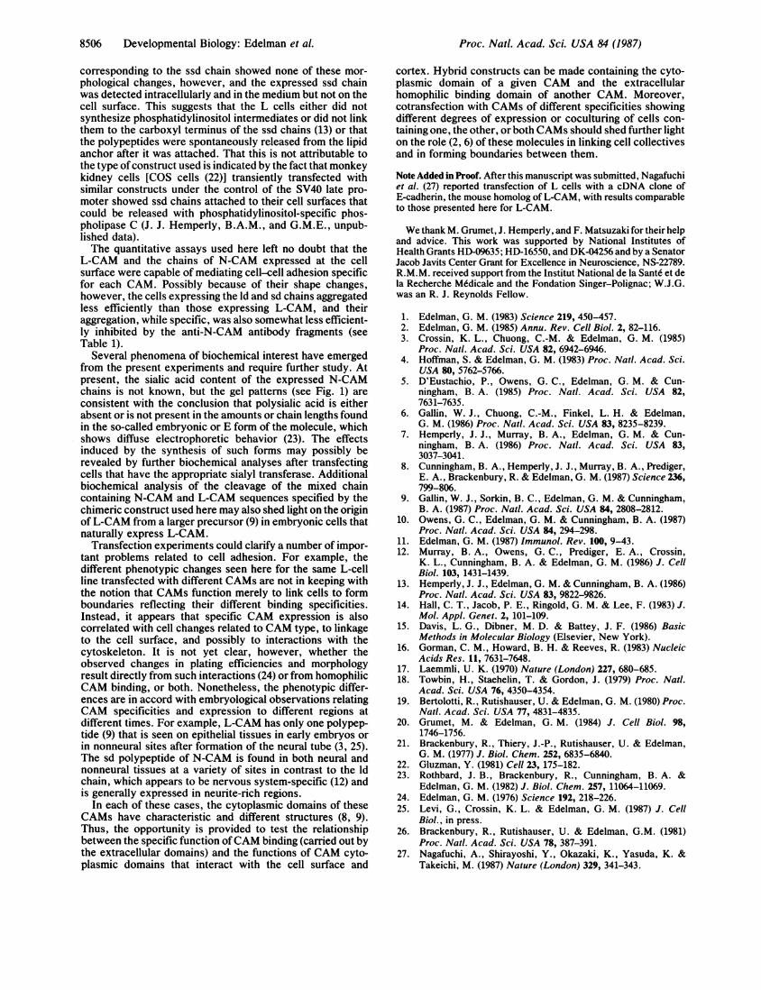
corresponding to the ssd chain showed none of these mor-phological changes, however, and the expressed ssd chainwas detected intracellularly and in the medium but not on thecell surface. This suggests that the L cells either did notsynthesize phosphatidylinositol intermediates or did not linkthem to the carboxyl terminus of the ssd chains (13) or thatthe polypeptides were spontaneously released from the lipidanchor after it was attached. That this is not attributable tothe type of construct used is indicated by the fact that monkeykidney cells [COS cells (22)] transiently transfected withsimilar constructs under the control of the SV40 late pro-moter showed ssd chains attached to their cell surfaces thatcould be released with phosphatidylinositol-specific phos-pholipase C (J. J. Hemperly, B.A.M., and G.M.E., unpub-lished data).The quantitative assays used here left no doubt that the
L-CAM and the chains of N-CAM expressed at the cellsurface were capable of mediating cell-cell adhesion specificfor each CAM. Possibly because of their shape changes,however, the cells expressing the Id and sd chains aggregatedless efficiently than those expressing L-CAM, and theiraggregation, while specific, was also somewhat less efficient-ly inhibited by the anti-N-CAM antibody fragments (seeTable 1).
Several phenomena of biochemical interest have emergedfrom the present experiments and require further study. Atpresent, the sialic acid content of the expressed N-CAMchains is not known, but the gel patterns (see Fig. 1) areconsistent with the conclusion that polysialic acid is eitherabsent or is not present in the amounts or chain lengths foundin the so-called embryonic or E form of the molecule, whichshows diffuse electrophoretic behavior (23). The effectsinduced by the synthesis of such forms may possibly berevealed by further biochemical analyses after transfectingcells that have the appropriate sialyl transferase. Additionalbiochemical analysis of the cleavage of the mixed chaincontaining N-CAM and L-CAM sequences specified by thechimeric construct used here may also shed light on the originofL-CAM from a larger precursor (9) in embryonic cells thatnaturally express L-CAM.
Transfection experiments could clarify a number of impor-tant problems related to cell adhesion. For example, thedifferent phenotypic changes seen here for the same L-cellline transfected with different CAMs are not in keeping withthe notion that CAMs function merely to link cells to formboundaries reflecting their different binding specificities.Instead, it appears that specific CAM expression is alsocorrelated with cell changes related to CAM type, to linkageto the cell surface, and possibly to interactions with thecytoskeleton. It is not yet clear, however, whether theobserved changes in plating efficiencies and morphologyresult directly from such interactions (24) or from homophilicCAM binding, or both. Nonetheless, the phenotypic differ-ences are in accord with embryological observations relatingCAM specificities and expression to different regions atdifferent times. For example, L-CAM has only one polypep-tide (9) that is seen on epithelial tissues in early embryos orin nonneural sites after formation of the neural tube (3, 25).The sd polypeptide of N-CAM is found in both neural andnonneural tissues at a variety of sites in contrast to the Idchain, which appears to be nervous system-specific (12) andis generally expressed in neurite-rich regions.
In each of these cases, the cytoplasmic domains of theseCAMs have characteristic and different structures (8, 9).Thus, the opportunity is provided to test the relationshipbetween the specific function ofCAM binding (carried out bythe extracellular domains) and the functions of CAM cyto-plasmic domains that interact with the cell surface and
cortex. Hybrid constructs can be made containing the cyto-plasmic domain of a given CAM and the extracellularhomophilic binding domain of another CAM. Moreover,cotransfection with CAMs of different specificities showingdifferent degrees of expression or coculturing of cells con-taining one, the other, or both CAMs should shed further lighton the role (2, 6) of these molecules in linking cell collectivesand in forming boundaries between them.
Note Added in Proof. After this manuscript was submitted, Nagafuchiet al. (27) reported transfection of L cells with a cDNA clone ofE-cadherin, the mouse homolog of L-CAM, with results comparableto those presented here for L-CAM.
We thank M. Grumet, J. Hemperly, and F. Matsuzaki for their helpand advice. This work was supported by National Institutes ofHealth Grants HD-09635; HD-16550, and DK-04256 and by a SenatorJacob Javits Center Grant for Excellence in Neuroscience, NS-22789.R.M.M. received support from the Institut National de la Sante et dela Recherche Mddicale and the Fondation Singer-Polignac; W.J.G.was an R. J. Reynolds Fellow.
1. Edelman, G. M. (1983) Science 219, 450-457.2. Edelman, G. M. (1985) Annu. Rev. Cell Biol. 2, 82-116.3. Crossin, K. L., Chuong, C.-M. & Edelman, G. M. (1985)
Proc. Natl. Acad. Sci. USA 82, 6942-6946.4. Hoffman, S. & Edelman, G. M. (1983) Proc. Natl. Acad. Sci.
USA 80, 5762-5766.5. D'Eustachio, P., Owens, G. C., Edelman, G. M. & Cun-
ningham, B. A. (1985) Proc. Natl. Acad. Sci. USA 82,7631-7635.
6. Gallin, W. J., Chuong, C.-M., Finkel, L. H. & Edelman,G. M. (1986) Proc. Natl. Acad. Sci. USA 83, 8235-8239.
7. Hemperly, J. J., Murray, B. A., Edelman, G. M. & Cun-ningham, B. A. (1986) Proc. Natl. Acad. Sci. USA 83,3037-3041.
8. Cunningham, B. A., Hemperly, J. J., Murray, B. A., Prediger,E. A., Brackenbury, R. & Edelman, G. M. (1987) Science 236,799-806.
9. Gallin, W. J., Sorkin, B. C., Edelman, G. M. & Cunningham,B. A. (1987) Proc. Natl. Acad. Sci. USA 84, 2808-2812.
10. Owens, G. C., Edelman, G. M. & Cunningham, B. A. (1987)Proc. Natl. Acad. Sci. USA 84, 294-298.
11. Edelman, G. M. (1987) Immunol. Rev. 100, 9-43.12. Murray, B. A., Owens, G. C., Prediger, E. A., Crossin,
K. L., Cunningham, B. A. & Edelman, G. M. (1986) J. CellBiol. 103, 1431-1439.
13. Hemperly, J. J., Edelman, G. M. & Cunningham, B. A. (1986)Proc. Natl. Acad. Sci. USA 83, 9822-9826.
14. Hall, C. T., Jacob, P. E., Ringold, G. M. & Lee, F. (1983) J.Mol. Appl. Genet. 2, 101-109.
15. Davis, L. G., Dibner, M. D. & Battey, J. F. (1986) BasicMethods in Molecular Biology (Elsevier, New York).
16. Gorman, C. M., Howard, B. H. & Reeves, R. (1983) NucleicAcids Res. 11, 7631-7648.
17. Laemmli, U. K. (1970) Nature (London) 227, 680-685.18. Towbin, H., Staehelin, T. & Gordon, J. (1979) Proc. Natl.
Acad. Sci. USA 76, 4350-4354.19. Bertolotti, R., Rutishauser, U. & Edelman, G. M. (1980) Proc.
Natl. Acad. Sci. USA 77, 4831-4835.20. Grumet, M. & Edelman, G. M. (1984) J. Cell Biol. 98,
1746-1756.21. Brackenbury, R., Thiery, J.-P., Rutishauser, U. & Edelman,
G. M. (1977) J. Biol. Chem. 252, 6835-6840.22. Gluzman, Y. (1981) Cell 23, 175-182.23. Rothbard, J. B., Brackenbury, R., Cunningham, B. A. &
Edelman, G. M. (1982) J. Biol. Chem. 257, 11064-11069.24. Edelman, G. M. (1976) Science 192, 218-226.25. Levi, G., Crossin, K. L. & Edelman, G. M. (1987) J. Cell
Biol., in press.26. Brackenbury, R., Rutishauser, U. & Edelman, G.M. (1981)
Proc. Natl. Acad. Sci. USA 78, 387-391.27. Nagafuchi, A., Shirayoshi, Y., Okazaki, K., Yasuda, K. &
Takeichi, M. (1987) Nature (London) 329, 341-343.
Proc. Natl. Acad. Sci. USA 84 (1987)