Cell Growth Differentiation
3
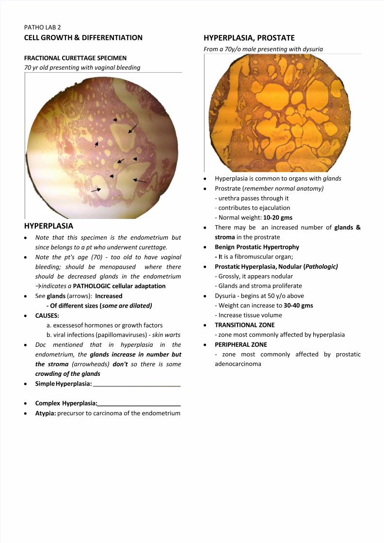
PATHO LAB 2 CELL GROWTH & DIFFERENTIATION FRACTIONAL CURETTAGE SPECIMEN 70 yr old presenting with vaginal bleeding HYPERPLASIA y Note that this specimen is the endometrium but since belongs to a pt who underwent curettage. y Note the pt's age (70) - too old to have vaginal bleeding; should be menopaused where there should be decreased glands in the endometrium indicates a PATHOLOGIC cellular adaptation y See glands (arrows): Increased - Of different sizes ( some are dilated) y CAUSES: a. excessesof hormones or growth factors b. viral inf ections (papillomaviruses) - skin warts y Doc mentioned that in hyperplasia in the endometrium, the glands increase in number but the stroma (arrowheads) don't so there is some crowding of the glands y Simple Hyperplasia: y Complex Hyperplasia: y Atypia: precursor to carcinoma of the endometrium HYPERPLASIA, PROSTATE F rom a 70y/o male presenting with dysuria y Hyperplasia is common to organs with glands y Prostrate (remember normal anatomy) - urethra passes through it - contributes to ejaculation - Normal weight: 10-20 gms y There may be an increased number of glands & stroma in the prostrate y Benign Prostatic Hypertrophy - I t is a fibromuscular organ; y Prostati c Hyperplasia, Nodular (P athologic) - Grossly, it appears nodular - Glands and stroma prolif erate y Dysuria - begins at 50 y/o above - Weight can increase to 30-40 gms - Increase tissue volume y TRANSITIONAL ZONE - zone most commonly aff ected by hyperplasia y PERIPHERAL ZONE - zone most commonly aff ected by prostatic adenocarcinoma
-
Upload
melayne-jewel-magdaong -
Category
Documents
-
view
224 -
download
0
Transcript of Cell Growth Differentiation
8/6/2019 Cell Growth Differentiation
http://slidepdf.com/reader/full/cell-growth-differentiation 1/3
PATHO LAB 2
CELL GROWTH & DIFFERENTIATION
FRACTIONAL CURETTAGE SPECIMEN
70 yr old presenting with vaginal bleeding
HYPERPLASIA
y Note that this specimen is the endometrium but
since belongs to a pt who underwent curettage.
y Note the pt's age (70) - too old to have vaginal
bleeding; should be menopaused where there
should be decreased glands in the endometrium
indicates a PATHOLOGIC cellular adaptation y See glands (arrows): Increased
- Of different sizes (some are dilated)
y CAUSES:
a. excessesof hormones or growth factors
b. viral inf ections (papillomaviruses) - skin warts
y Doc mentioned that in hyperplasia in the
endometrium, the glands increase in number but
the stroma (arrowheads) don't so there is some
crowding of the glands
y Simple Hyperplasia:
y Complex Hyperplasia:
y Atypia: precursor to carcinoma of the endometrium
HYPERPLASIA, PROSTATE
F rom a 70y/o male presenting with dysuria
y Hyperplasia is common to organs with glands
y Prostrate (remember normal anatomy) - urethra passes through it
- contributes to ejaculation
- Normal weight: 10-20 gms
y There may be an increased number of glands &
stroma in the prostrate
y Benign Prostatic Hypertrophy
- It is a fibromuscular organ;
y Prostatic Hyperplasia, Nodular (P athologic)
- Grossly, it appears nodular
- Glands and stroma prolif erate y Dysuria - begins at 50 y/o above
- Weight can increase to 30-40 gms
- Increase tissue volume
y TRANSITIONAL ZONE
- zone most commonly aff ected by hyperplasia
y PERIPHERAL ZONE
- zone most commonly aff ected by prostatic
adenocarcinoma
8/6/2019 Cell Growth Differentiation
http://slidepdf.com/reader/full/cell-growth-differentiation 2/3
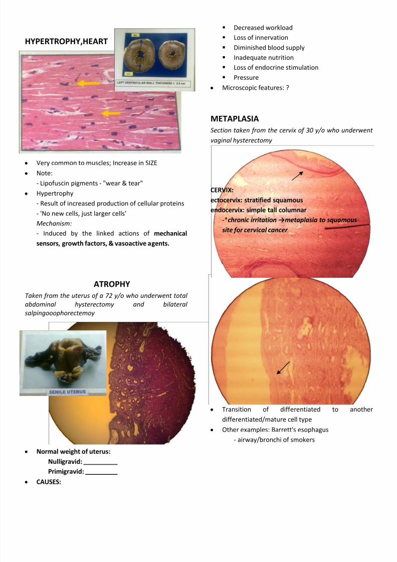
HYPERTROPHY,HEART
y Very common to muscles; Increase in SIZE
y Note:
- Lipofuscin pigments - "wear & tear"
y Hypertrophy
- Result of increased production of cellular proteins
- 'No new cells, just larger cells'
Mechanism:
- Induced by the linked actions of mechanical
sensors, growth factors, & vasoactive agents.
ATROPHY
T aken from the uterus of a 72 y/o who underwent total abdominal hysterectomy and bilateral
salpingooophorectemoy
y Normal weight of uterus:
Nulligravid:
Primigravid:
y CAUSES:
Decreased workload
Loss of innervation
Diminished blood supply
Inadequate nutrition
Loss of endocrine stimulation
Pressure
y Microscopic f eatures: ?
METAPLASIA
Section taken from the cervix of 30 y/o who underwent
vaginal hysterectomy
CERVIX:
ectocervix: stratified squamous
endocervix: simple tall columnar
-*chronic irritation metaplasia to squamous
site for cervical cancer
y Transition of diff erentiated to anothe
diff erentiated/mature cell type
y Other examples: Barrett's esophagus
- airway/bronchi of smokers
8/6/2019 Cell Growth Differentiation
http://slidepdf.com/reader/full/cell-growth-differentiation 3/3
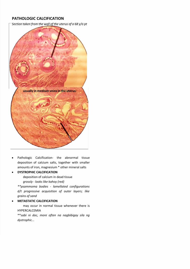
PATHOLOGIC CALCIFICATION
Section taken from the wall of the uterus of a 68 y/o pt
usually in medium veins in the uterus:
y Pathologic Calcification- the abnormal tissue
deposition of calcium salts, together with smaller
amounts of iron, magnesium * other mineral salts
y DYSTROPHIC CALCIFICATION
deposition of calcium in dead tissue
grossly - looks like kahoy (red)
**psammoma bodies - lamellated configurations
d/t progressive acquisition of outer layers; like
grains of sand
y METASTATIC CALCIFICATION
may occur in normal tissue whenever there is
HYPERCALCEMIA
**sabi ni doc, more often na nagbibigay sila ng
dystrophic...