Cell-Cycle Dependent Expression of a Translocation- Mediated
18
Cell-Cycle Dependent Expression of a Translocation- Mediated Fusion Oncogene Mediates Checkpoint Adaptation in Rhabdomyosarcoma Ken Kikuchi 1 , Simone Hettmer 2,3 , M. Imran Aslam 1 , Joel E. Michalek 4 , Wolfram Laub 5 , Breelyn A. Wilky 6 , David M. Loeb 7 , Brian P. Rubin 8 , Amy J. Wagers 2 , Charles Keller 1 * 1 Pediatric Cancer Biology Program, Pape ´ Family Pediatric Research Institute, Department of Pediatrics, Oregon Health & Science University, Portland, Oregon, United States of America, 2 The Howard Hughes Medical Institute and Department of Stem Cell and Regenerative Biology, Harvard University, Cambridge, Massachusetts, United States of America, and Joslin Diabetes Center, Boston, Massachusetts, United States of America, 3 Department of Pediatric Oncology, Dana Farber Cancer Institute and Division of Pediatric Hematology/Oncology, Children’s Hospital, Boston, Massachusetts, United States of America, 4 Department of Epidemiology and Biostatistics, University of Texas Health Science Center, San Antonio, Texas, United States of America, 5 Department of Radiation Medicine, Oregon Health & Science University, Portland, Oregon, United States of America, 6 Division of Medical Oncology, Sidney Kimmel Comprehensive Cancer Center, Johns Hopkins University, Baltimore, Maryland, United States of America, 7 Division of Pediatric Oncology, Sidney Kimmel Comprehensive Cancer Center, Johns Hopkins University, Baltimore, Maryland, United States of America, 8 Departments of Anatomic Pathology and Molecular Genetics, Taussig Cancer Center and Lerner Research Institute, Cleveland Clinic Foundation, Cleveland, Ohio, United States of America Abstract Rhabdomyosarcoma is the most commonly occurring soft-tissue sarcoma in childhood. Most rhabdomyosarcoma falls into one of two biologically distinct subgroups represented by alveolar or embryonal histology. The alveolar subtype harbors a translocation-mediated PAX3:FOXO1A fusion gene and has an extremely poor prognosis. However, tumor cells have heterogeneous expression for the fusion gene. Using a conditional genetic mouse model as well as human tumor cell lines, we show that that Pax3:Foxo1a expression is enriched in G 2 and triggers a transcriptional program conducive to checkpoint adaptation under stress conditions such as irradiation in vitro and in vivo. Pax3:Foxo1a also tolerizes tumor cells to clinically- established chemotherapy agents and emerging molecularly-targeted agents. Thus, the surprisingly dynamic regulation of the Pax3:Foxo1a locus is a paradigm that has important implications for the way in which oncogenes are modeled in cancer cells. Citation: Kikuchi K, Hettmer S, Aslam MI, Michalek JE, Laub W, et al. (2014) Cell-Cycle Dependent Expression of a Translocation-Mediated Fusion Oncogene Mediates Checkpoint Adaptation in Rhabdomyosarcoma. PLoS Genet 10(1): e1004107. doi:10.1371/journal.pgen.1004107 Editor: Marshall S. Horwitz, University of Washington, United States of America Received October 3, 2013; Accepted November 27, 2013; Published January 16, 2014 Copyright: ß 2014 Kikuchi et al. This is an open-access article distributed under the terms of the Creative Commons Attribution License, which permits unrestricted use, distribution, and reproduction in any medium, provided the original author and source are credited. Funding: This work was supported by NCI award 5R01CA133229 (to CK) as well as by grants from the Burroughs-Wellcome Fund, Harvard Stem Cell Institute, and Beckman Foundation (to AJW), and by Hope Street Kids, P.A.L.S. Bermuda/St. Baldrick’s, ALSF and Bear Necessities (to SH). The funders had no role in study design, data collection and analysis, decision to publish, or preparation of the manuscript. Competing Interests: The authors have declared that no competing interests exist. * E-mail: [email protected] Introduction Rhabdomyosarcoma (RMS) is the most common childhood soft tissue sarcoma. Historically, RMS has been thought to arise from muscle because of the expression of myogenic markers. Most childhood RMS falls into one of two biologically distinct subgroups: alveolar (aRMS) or embryonal (eRMS). aRMS is the more aggressive variant with a survival rate of less than 20% when metastatic due to chemotherapy and radiation resistance [1]. aRMS is characterized by a frequent t(2;13) chromosomal translocation, which results in the PAX3:FOXO1A fusion gene, or less frequently by a t(1;13) mediated PAX7:FOXO1A fusion oncogene [1]. Clinically, the aggressive behavior of aRMS has been attributed to PAX3:FOXO1A transcriptional reprograming because fusion negative aRMS have a more favorable outcome similar to eRMS [2,3,4]. We previously developed a mouse model of aRMS employing a conditional knock-in approach that expresses Pax3:Foxo1a from the native Pax3 locus in fetal and postnatal myoblasts [5,6,7]. In this model, Pax3:Foxo1a was necessary but not sufficient for aRMS tumor initiation. Interestingly, cells expressing high levels of Pax3:Foxo1a were more prevalent in metastatic tumors [7]. The heterogeneity of Pax3:Foxo1a expression in primary and meta- static tumors, and enrichment in the latter, suggested that Pax3:Foxo1a might be selectively expressed in a subset of aRMS cells; alternatively, Pax3:Foxo1a expression might be temporally regulated. In the current study we present striking evidence that Pax3:Foxo1a is expressed in a dynamic manner and mediates a G 2 -specific program enabling checkpoint adaptation and refrac- toriness to therapy. Results Pax3:Foxo1a expression is dynamic in mouse aRMS cells In our genetically-engineered conditional knock-in mouse model of aRMS, eYFP is expressed as a second cistron on the same mRNA as Pax3:Foxo1a (Figure 1A). We have observed heterogeneity of eYFP expression among tumor cells in situ (Figure 1B). To first examine Pax3:Foxo1a expression as a function of time, we flow sorted Pax3:Foxo1a low and Pax3:Fox- PLOS Genetics | www.plosgenetics.org 1 January 2014 | Volume 10 | Issue 1 | e1004107
Transcript of Cell-Cycle Dependent Expression of a Translocation- Mediated
Cell-Cycle Dependent Expression of a Translocation-Mediated Fusion Oncogene Mediates CheckpointAdaptation in RhabdomyosarcomaKen Kikuchi1, Simone Hettmer2,3, M. Imran Aslam1, Joel E. Michalek4, Wolfram Laub5, Breelyn A. Wilky6,
David M. Loeb7, Brian P. Rubin8, Amy J. Wagers2, Charles Keller1*
1 Pediatric Cancer Biology Program, Pape Family Pediatric Research Institute, Department of Pediatrics, Oregon Health & Science University, Portland, Oregon, United
States of America, 2 The Howard Hughes Medical Institute and Department of Stem Cell and Regenerative Biology, Harvard University, Cambridge, Massachusetts, United
States of America, and Joslin Diabetes Center, Boston, Massachusetts, United States of America, 3 Department of Pediatric Oncology, Dana Farber Cancer Institute and
Division of Pediatric Hematology/Oncology, Children’s Hospital, Boston, Massachusetts, United States of America, 4 Department of Epidemiology and Biostatistics,
University of Texas Health Science Center, San Antonio, Texas, United States of America, 5 Department of Radiation Medicine, Oregon Health & Science University,
Portland, Oregon, United States of America, 6 Division of Medical Oncology, Sidney Kimmel Comprehensive Cancer Center, Johns Hopkins University, Baltimore, Maryland,
United States of America, 7 Division of Pediatric Oncology, Sidney Kimmel Comprehensive Cancer Center, Johns Hopkins University, Baltimore, Maryland, United States of
America, 8 Departments of Anatomic Pathology and Molecular Genetics, Taussig Cancer Center and Lerner Research Institute, Cleveland Clinic Foundation, Cleveland,
Ohio, United States of America
Abstract
Rhabdomyosarcoma is the most commonly occurring soft-tissue sarcoma in childhood. Most rhabdomyosarcoma falls intoone of two biologically distinct subgroups represented by alveolar or embryonal histology. The alveolar subtype harbors atranslocation-mediated PAX3:FOXO1A fusion gene and has an extremely poor prognosis. However, tumor cells haveheterogeneous expression for the fusion gene. Using a conditional genetic mouse model as well as human tumor cell lines,we show that that Pax3:Foxo1a expression is enriched in G2 and triggers a transcriptional program conducive to checkpointadaptation under stress conditions such as irradiation in vitro and in vivo. Pax3:Foxo1a also tolerizes tumor cells to clinically-established chemotherapy agents and emerging molecularly-targeted agents. Thus, the surprisingly dynamic regulation ofthe Pax3:Foxo1a locus is a paradigm that has important implications for the way in which oncogenes are modeled in cancercells.
Citation: Kikuchi K, Hettmer S, Aslam MI, Michalek JE, Laub W, et al. (2014) Cell-Cycle Dependent Expression of a Translocation-Mediated Fusion OncogeneMediates Checkpoint Adaptation in Rhabdomyosarcoma. PLoS Genet 10(1): e1004107. doi:10.1371/journal.pgen.1004107
Editor: Marshall S. Horwitz, University of Washington, United States of America
Received October 3, 2013; Accepted November 27, 2013; Published January 16, 2014
Copyright: � 2014 Kikuchi et al. This is an open-access article distributed under the terms of the Creative Commons Attribution License, which permitsunrestricted use, distribution, and reproduction in any medium, provided the original author and source are credited.
Funding: This work was supported by NCI award 5R01CA133229 (to CK) as well as by grants from the Burroughs-Wellcome Fund, Harvard Stem Cell Institute, andBeckman Foundation (to AJW), and by Hope Street Kids, P.A.L.S. Bermuda/St. Baldrick’s, ALSF and Bear Necessities (to SH). The funders had no role in study design,data collection and analysis, decision to publish, or preparation of the manuscript.
Competing Interests: The authors have declared that no competing interests exist.
* E-mail: [email protected]
Introduction
Rhabdomyosarcoma (RMS) is the most common childhood soft
tissue sarcoma. Historically, RMS has been thought to arise from
muscle because of the expression of myogenic markers. Most
childhood RMS falls into one of two biologically distinct
subgroups: alveolar (aRMS) or embryonal (eRMS). aRMS is the
more aggressive variant with a survival rate of less than 20% when
metastatic due to chemotherapy and radiation resistance [1].
aRMS is characterized by a frequent t(2;13) chromosomal
translocation, which results in the PAX3:FOXO1A fusion gene, or
less frequently by a t(1;13) mediated PAX7:FOXO1A fusion
oncogene [1]. Clinically, the aggressive behavior of aRMS has
been attributed to PAX3:FOXO1A transcriptional reprograming
because fusion negative aRMS have a more favorable outcome
similar to eRMS [2,3,4].
We previously developed a mouse model of aRMS employing a
conditional knock-in approach that expresses Pax3:Foxo1a from the
native Pax3 locus in fetal and postnatal myoblasts [5,6,7]. In this
model, Pax3:Foxo1a was necessary but not sufficient for aRMS
tumor initiation. Interestingly, cells expressing high levels of
Pax3:Foxo1a were more prevalent in metastatic tumors [7]. The
heterogeneity of Pax3:Foxo1a expression in primary and meta-
static tumors, and enrichment in the latter, suggested that
Pax3:Foxo1a might be selectively expressed in a subset of aRMS
cells; alternatively, Pax3:Foxo1a expression might be temporally
regulated. In the current study we present striking evidence that
Pax3:Foxo1a is expressed in a dynamic manner and mediates a
G2-specific program enabling checkpoint adaptation and refrac-
toriness to therapy.
Results
Pax3:Foxo1a expression is dynamic in mouse aRMS cellsIn our genetically-engineered conditional knock-in mouse
model of aRMS, eYFP is expressed as a second cistron on the
same mRNA as Pax3:Foxo1a (Figure 1A). We have observed
heterogeneity of eYFP expression among tumor cells in situ
(Figure 1B). To first examine Pax3:Foxo1a expression as a
function of time, we flow sorted Pax3:Foxo1alow and Pax3:Fox-
PLOS Genetics | www.plosgenetics.org 1 January 2014 | Volume 10 | Issue 1 | e1004107
o1ahigh cells using eYFP signal in two independent murine aRMS
primary cultures (Figure 1C and 1D; Figure S1A and S1B).
Comparison of Pax3:Foxo1a protein levels for sorted populations
showed Pax3:Foxo1alow cells possessed much reduced levels of
Pax3:Foxo1a protein (Figure 1E and Figure S1C). However,
FACS analysis over time revealed that the eYFP signal of
Pax3:Foxo1alow and Pax3:Foxo1ahigh tended towards the mean
eYFP fluorescence intensity of unsorted tumor cells with time and/
or cell divisions (Figure 1C and 1D; Figure S1A and S1B). Thus,
Pax3:Foxo1ahigh cell could dynamically reduce expression of eYFP
from the Pax3:Foxo1a locus, and Pax3:Foxo1alow cells could
dynamically increase expression of eYFP from the Pax3:Foxo1a
locus. We further confirmed that eYFP expression was indeed
reflective of Pax3:Foxo1a expression in terms of protein half-life.
Figure S1E and S1F shows levels of eYFP signal and Pax3:Foxo1a
protein stability after translation inhibition by cycloheximide
(CHX). Akin to the strong correlation between eYFP and
Pax3:Foxo1a expression at the protein level (Figure 1 and Figure
S1C), the protein half-lives of Pax3:Foxo1a and eYFP were
roughly similar at 31.6 and 44.7 hours (Figure S1E and S1F),
thereby affirming that eYFP is a reasonable surrogate for
transcription of Pax3:Foxo1a from the Pax3 locus (we do however
acknowledge that eYFP is a better marker of the start of
Pax3:Foxo1a transcription than the end of Pax3:Foxo1a transcrip-
tion or protein expression (i.e., since eYFP is expressed on the same
mRNA as Pax3:Foxo1a, the beginning of fluorescence should
coincide with the initial presence of the Pax3:Foxo1a transcript).
Thereafter, eYFP is susceptible to photo-bleaching and possible
proteasomal degradation sooner than the 44 hours observed under
conditions of cyclohexamide treatment (Figure S1F)).
Pax3:Foxo1a expression is dynamically regulated duringthe cell cycle
To investigate what conditions affect the dynamic alteration of
Pax3:Foxo1a expression in aRMS cells, we compared eYFP
fluorescence to cell cycle phase as determined by staining with the
DNA dye Hoechst33342. Almost all Pax3:Foxo1alow cells existed
in G0/G1 (2N) stage, while to our surprise Pax3:Foxo1ahigh cells
were G2/M or hyperdiploid/multinuclear ($4N) cells (Figure 1F
and Figure S1D). We next performed time-lapse experiments of
eYFP activity by confocal microscopy. Figure 1G shows in time-
lapse images that eYFP activity during cell division is transiently
but markedly increased, particularly in pre-mitotic cells. Interest-
ingly, the level of eYFP in some multinuclear cells remained at a
high level in cells that appeared to be unable to undergo
telophase/cytokinesis (Movie S1).
We next performed QPCR of Pax3:Foxo1a and PAX3:FOXO1A
using cell cycle specific sorted mouse and human aRMS cells,
respectively. Both mouse and human aRMS cells showed
significant differences in the mRNA expression of Pax3:Foxo1a
and PAX3:FOXO1A in the transition from 2N (G1) to 3N (S phase)
and 4N (G2/M) cells (Figure 2A and 2B) affirming cross-species
relevance of the cell cycle dependent mRNA regulation of
Pax3:Foxo1a expression.
To investigate the transcriptional basis of this Pax3:Foxo1a
dynamic expression, we performed QPCR of Pax3 and Foxo1 using
cell cycle specific sorted C2C12 mouse myoblast cells of the
genotype Pax3(wt/wt) and mouse aRMS primary tumor cells of the
genotype Pax3(wt/Pax3:Foxo1a). C2C12 myoblasts showed signif-
icant increases in Pax3 mRNA levels for 4N cells when compared
with 2N cells (Figure 2C). Pax3 was not detectable in aRMS cells at
the mRNA level (data not shown), which was also reflected in the
absence of expression of Pax3 protein in aRMS cells by western
blotting (Figure 2D). This result is consistent with our prior studies
suggesting that Pax3:Foxo1a causes decreased expression of the
wildtype Pax3 locus [5,6]. By contrast, Foxo1 mRNA expression
did not differ between 2N and 4N in either C2C12 myoblasts or
aRMS tumor cells (Figure 2E). Thus, the cell cycle dependence of
Pax3:Foxo1a may in some part be attributable to increased Pax3
promoter activity at G2/M versus G1 in C2C12 myoblasts, but
Pax3:Foxo1a transcript level is so significantly increased over Pax3
in aRMS cells that other factors related to the chromosomal fusion
are likely responsible, e.g. gain of a Foxo1a 39 cis-enhancer, or loss of
a Pax3 39 cis-repressor repressor. From the design of the
conditional knock-in allele [5], this element(s) can be inferred to
exist in the 9.3 kB of the Foxo1a 39 region containing exons 2 and 3
and untranslated region (6.5 kb), or exons 8–10 of Pax3. We also
cannot exclude that stabilization of the Pax3:Foxo1a transcript may
to some degree play a role, and this stabilization may or may not
be related to the Foxo1a cis-elements on the chimeric mRNA.
Because Pdgfra [8] and Igf1r [9] are well known direct
downstream targets of Pax3:Foxo1a, we determined whether
these targets were expressed to any degree in 4N (G2/M) cells. We
first sorted aRMS tumor cells for Pdgfra or Igf1r positivity versus
negativity, then performed DNA content analysis. For both
receptor tyrosine kinases (RTKs), the majority of cells with
positive RTK surface expression were 2N (Figure 2F). However,
nearly twice as many 4N cells are Igf1r (or Pdgfra) positive versus
Igf1r (or Pdgfra) negative, suggesting these Pax3:Foxo1a targets
may have a functional role late in the cell cycle, such as the Igf1r-
mediated radioresistance seen for other forms of cancer [10].
Pax3:Foxo1a expression is specific to G2 and acts in G2/Mcheckpoint adaptation
To determine the role of Pax3:Foxo1a in G2, M or G2/M
checkpoint, we examined markers of each cell cycle phase under
non-stress or stress conditions. Immunocytochemistry is presented
in Figure 3 is a for Pax3:Foxo1a (Pax3) with phospho-histone H3
(pHH3), a marker of mitosis, or CDC2-Y15 (pCDC2), a negative
marker of entry into mitosis that is commonly expressed in G2
(CDC2-Y15 is phosphorylated by Wee1 kinase, which then
negatively regulates Cdc2 kinase [11]; CDC2-Y15 is present
starting in late G1 then also in S, and G2 phases, but absent in M
[12]). In murine aRMS primary cultures U23674 and U42369,
pHH3 positive metaphase cells did not express Pax3:Foxo1a
protein and yet most pCDC2 positive cells expressed Pax3:Foxo1a
very highly (Figure 3). These results suggest that Pax3:Foxo1a is
expressed in the G2 cell cycle phase but not M phase. Human
Author Summary
Rare childhood cancers can be paradigms from whichimportant new principles can be discerned. The childhoodmuscle cancer rhabdomyosarcoma is no exception, havingbeen the focus of the original 1969 description by Drs.Li and Fraumeni of a syndrome now know to be com-monly caused by underlying p53 tumor suppressor loss-of-function. In our studies using a conditional geneticmouse model of alveolar rhabdomyosarcoma in conjunc-tion with human tumor cell lines, we have uncovered thatthe expression level of a translocation-mediated fusiongene, Pax3:Foxo1a, is dynamic and varies during the cellcycle. Our studies support that Pax3:Foxo1a facilitate theyeast-related process of checkpoint adaptation understresses such as irradiation. The broader implication ofour studies is that distal cis elements (promoter-influenc-ing regions of DNA) may be critical to fully understandingthe function of cancer-associated translocations.
Dynamic Pax3:Foxo1a in Alveolar Rhabdomyosarcoma
PLOS Genetics | www.plosgenetics.org 2 January 2014 | Volume 10 | Issue 1 | e1004107

Figure 1. eYFP activity and Pax3:Foxo1a expression is cell cycle specific. (A) Diagrammatic representation of the conditional Pax3:Foxo1aknock-in allele by which eYFP is expressed as a second cistron on the same mRNA as Pax3:Foxo1a at the native Pax3 promoter. (B) Heterogeneity ofeYFP expression in a murine aRMS tumor by immunofluorescence. (C) eYFP fluorescence of eYFP sorted cells overtime as measured by FACS. Grey:
Dynamic Pax3:Foxo1a in Alveolar Rhabdomyosarcoma
PLOS Genetics | www.plosgenetics.org 3 January 2014 | Volume 10 | Issue 1 | e1004107

aRMS cell lines Rh3 and Rh41 showed identical results (Figure 3).
Next, we sought to understand the function of Pax3:Foxo1a in G2.
For this purpose we performed genome-wide expression analysis
using cells sorted at specific stages of the cell cycle (2N vs. 4N) with
or without Pax3:Foxo1a siRNA knockdown (Figure 4A). Because
eYFP is expressed as a second cistron in the targeted Pax3:Foxo1a-
ires-eYFP allele, we anticipated that siRNA for eYFP would knock
down not only eYFP but also Pax3:Foxo1a. Western blotting of
Pax3:Foxo1a and native Foxo1a protein 48 hours after siRNA
transfection showed that eYFP siRNA efficiently and specifically
knocked down Pax3:Foxo1a protein (Figure 4B). Protein expres-
sion of the Pax3:Foxo1a transcriptional target Pdgfra was also
reduced (Figure 4B).
From genome-wide expression analysis of 2N vs. 4N sorted cells
with or without Pax3:Foxo1a siRNA knockdown, we found several
genes implicated in the process of G2/M checkpoint adaptation to
be down-regulated in G2/M (4N cells) when Pax3:Foxo1a was
knocked down (Figure 4C; Table S1 shows all data analyzed by
ANOVA (,0.05) using the multiple comparison correction
method of Benjamini and Hochberg). Checkpoint adaptation is
the process by which unicellular organisms or cancer cells progress
through a delayed cell cycle checkpoint (G2 or by analogy the
mitotic spindle assembly checkpoint) in lieu of programmed cell
death, but before DNA damage is completely repaired [13,14,15].
Factors implicated in checkpoint adaptation are similar to those
involved in checkpoint recovery (after complete repair of DNA
damage), but additionally require anti-apoptotic signals [14].
Select G2/M checkpoint adaptation genes implicated in this
experiment, the DNA damage sensing/checkpoint progression
factors Plk1, Cdc25b, H2afx and the cell survival factor Birc5
(Survivin), were validated for differential expression by QPCR
(Figure 4D). Whether these genes are direct transcriptional targets
of Pax3:Foxo1a was investigated by interrogating loci for reported
nearby Pax3:Foxo1a binding sites [16]. Most potential regulatory
sites were greater than 60 kB away (Table S2). While regulatory
sequences can be hundreds of kBs away from the target gene, it
remains possible that these genes may also be regulated indirectly
by other Pax3:Foxo1a target genes or miRNAs.
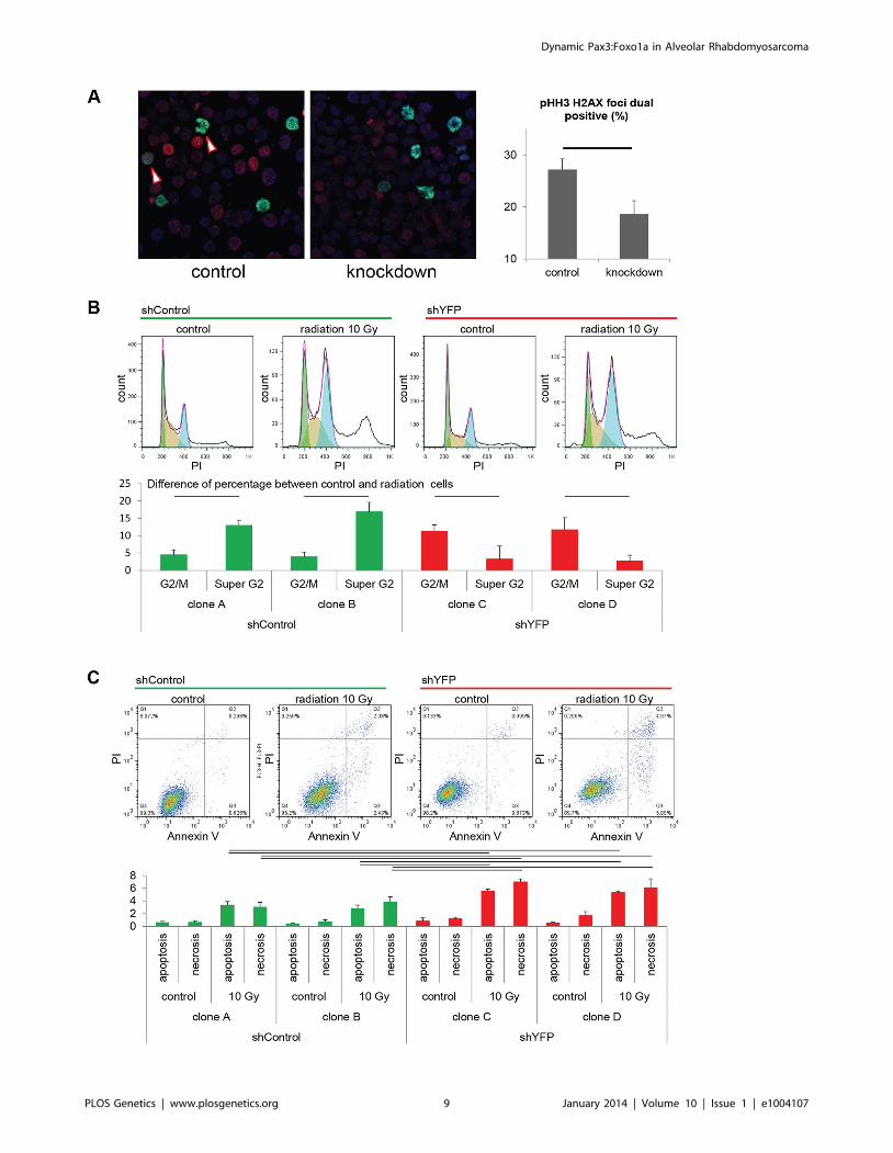
As a test of checkpoint adaptation and the permissiveness of
aRMS cells to transit from G2 to mitosis despite single- and
double-stranded DNA damage, we irradiated tumor cells with or
without Pax3:Foxo1a knockdown. Radiation resulted in a higher
fraction of DNA breaks amongst mitotic cells (as represented by
dual pHH3 positive, H2AX positive cells) under conditions of
Pax3:Foxo1a expression than its knockdown (Figure 5A and
Figure S2A), suggesting that Pax3:Foxo1a does facilitate G2 to M
transition, consistent with checkpoint adaptation. Moreover, we
performed cell cycle and Annexin V apoptosis detection assay after
treatment with 10 Gy radiation for two independent eYFP shRNA
knockdown clones compared to two other independent shRNA
controls (as stated early, eYFP knockdown also achieves
Pax3:Foxo1a knockdown) (Figure S2). Cell cycle analysis of the
shRNA clones treated with radiation revealed increasing percent-
age of cells in cells having $4N DNA content after radiation for
Pax3:Foxo1a knockdown cells compared to radiated controls
(p,0.05)(Figure 5B). This result is consistent with a role of
Pax3:Foxo1a in overcoming G2 arrest or M checkpoint arrest after
radiation. Similarly, the Annexin V apoptosis detection assay
showed a lower induction of apoptosis following radiation when
Pax3:Foxo1a expression was preserved in shControl clones than
shYFP cells (Figure 5C).
To test the acute role of Pax3:Foxo1a in tolerization to
treatment-related DNA damage in vivo, we used eYFP siRNA to
transiently knock down Pax3:Foxo1a in aRMS tumor cells treated
with radiation versus non-irradiated controls that were then
orthotopically injected into unirradiated host mice. Pax3:Foxo1a
mediated a cell survival and tumor re-establishment advantage
under the stress condition of irradiation, but not under homeo-
static conditions (p = 0.02, Figure 6A and 6B).
To assess the extent to which the fusion gene mediates
refractoriness to chemotherapy agents, we observed Pax3:Foxo1a
to facilitate 2–4 fold refractoriness to clinical agents capable of
causing double-stranded DNA breaks and mitotic arrest (vincris-
tine, actinomycin-D, topotecan) more so than agent inducing
single-strand breaks (mafosfamide, the active metabolite of
cyclophosphamide) (Figure S3A–E). That a similar role of
Pax3:Foxo1a may apply to targeted agents was previously
suggested by enriched G2 expression of Pdgfra (Figure 2F) and
then demonstrated by increased sensitivity to prototypic Pdgfr
inhibitor, imatinib, after Pax3:Foxo1a knockdown (Figure S3F).
Similarly, Pax3:Foxo1a knockdown sensitized tumor cells to siRNA
inhibition of downstream signaling mediators of acquired imatinib
resistance (Figure S3G) [17]. Thus, these in vitro and in vivo results
are consistent with a function of Pax3:Foxo1a in mediating
checkpoint adaptation and refractoriness to the established clinical
therapies of radiation and chemotherapy, or more contemporary
molecularly-targeted agents.
Discussion
A key finding of this study is that Pax3:Foxo1a expression is
dynamic and varies during the cell cycle. To our knowledge this is
first report of a translocation-mediated chimeric transcription
factor oncogene that is expressed in a cell cycle-specific manner –
much less, one that is expressed specifically during G2. The master
transcription factor MYOD is expressed strongly during G1 [18]
but is inactivated by phosphorylation during mitosis, which results
in deportation from the nucleus [19]. MYF5 is also expressed in a
cell cycle-dependent manner, but neither MYOD nor MYF5
expression is increased during G2/M as observed in our study of
Pax3:Foxo1a in aRMS. Our findings reveal that Pax3 expression
in wildtype C2C12 myoblasts is dynamic and increased during
G2/M, but that to account for the dramatic increase in
Pax3:Foxo1a expression an additional enhancer effect of Foxo1a 39
region DNA is likely to be present. This result opens the possibility
that co-factors assembled at the Pax3 promoter or fusion gene
specific cis-elements might be targeted to suppress Pax3:Foxo1a
expression.
Cell cycle progression after DNA damage is regulated by
checkpoint controls, which prevent continued transit through the
cycle until the damage has been repaired, hence protecting the
integrity of the genome. Arrest in G1 permits repair prior to
replication, whereas arrest in G2 allows repair prior to mitotic
chromosome segregation. The p53 tumor suppressor, which is
mutated in roughly half of human aRMS, has been shown to be
C2C12 (negative control), blue: no sorted cells, green: eYFP activity high cells, red: eYFP activity low cells. (D) Mean of relative eYFP activity in panel 1Cmeasured by FACS. (E) Western blot analysis using eYFP sorted cells. Plotted are relative protein levels of Pax3:Foxo1a/b-Actin. Mean 6 SE wereobtained from three independent immunoblottings. Black line shows significant difference (p,0.05). (F) eYFP activity and cell cycle analysis usingHochest33342 staining for mouse primary cell culture U23674. Green shows G0/G1 phase, brown shows S phase, and blue shows G2/M phase. (G)Time-lapse experiment of eYFP activity (select frames over 16 hours). See also corresponding Movie S1.doi:10.1371/journal.pgen.1004107.g001
Dynamic Pax3:Foxo1a in Alveolar Rhabdomyosarcoma
PLOS Genetics | www.plosgenetics.org 4 January 2014 | Volume 10 | Issue 1 | e1004107

integral to both G1 and G2 damage checkpoint machinery, but
some reports found p53 dispensable for the G2 checkpoint [13,20].
Checkpoint adaptation is defined as the ability to divide and
survive following a sustained checkpoint arrest despite the
presence of unrepairable DNA breaks [14]. Cells undergoing
checkpoint adaptation will frequently die in subsequent cell cycles
if DNA damage goes unrepaired, yet, some cells may be able to
survive and proliferate in an aneuploid state [14]. Furthermore, in
Figure 2. Pax3:Foxo1a activity is cell cycle dependent. (A) mRNA expression of Pax3:Foxo1a normalized by Gapdh in U23674 mouse aRMSprimary cell culture sorted by DNA content. Black lines show significant differences (p,0.05). (B) mRNA expression of PAX3:FOXO1A normalized byGAPDH in Rh3 and Rh41 human aRMS cell lines sorted by DNA content. (C) mRNA expression of Pax3 normalized by Gapdh in C2C12 murinemyoblasts sorted by DNA content. (D) Western blot analysis of Pax3 and Pax3:Foxo1a in unsorted murine U23674 aRMS cells (genotype Pax3(Pax3:Foxo1a activated/Pax3:Foxo1a activated)), murine U57844 aRMS cells (genotype Pax3 (wt/Pax3:Foxo1a activated)), proliferative C2C12myoblasts (pro) and differentiating C2C12 myoblasts (dif). (E) mRNA expression of Foxo1 and Pax3:Foxo1a normalized to Gapdh in C2C12 myoblastsand the U57844 mouse aRMS primary cell culture. (F) Cell cycle analysis after sorting for Pax3:Foxo1a targets Igf1r or Pdgfra in mouse aRMS tumorcells. Nearly twice as many 4N cells are Igf1r (or Pdgfra) positive versus Igf1r (or Pdgfra negative), suggesting these Pax3:Foxo1a targets may have afunctional role late in the cell cycle (* P,0.05). pos, positive. Neg, negative.doi:10.1371/journal.pgen.1004107.g002
Dynamic Pax3:Foxo1a in Alveolar Rhabdomyosarcoma
PLOS Genetics | www.plosgenetics.org 5 January 2014 | Volume 10 | Issue 1 | e1004107

unicellular eukaryotes and tumor cells, DNA repair can occur at
G1 [21]. Here, we reveal that the G2/M adaptation genes (H2afx,
Cdc25b and Plk1) were suppressed by Pax3:Foxo1a knockdown in
G2 and M cell cycle phases and that fewer cells transited from G2
to M without initiating apoptosis under conditions of Pax3:Foxo1a
knockdown in the context of radiation-induced stress. These
Figure 3. Pax3:Foxo1a is expressed in G2 for mouse and human aRMS. Immunocytochemistry for Pax3 (green), pHH3 (red) and DAPI (blue)or Pax3 (green), pCDC2 (red) and DAPI (blue). Numbers are relative rate of Pax3:Foxo1a high or low cells/pHH3 positive cells and Pax3:Foxo1a high orlow cells/CDC2-Y15 high cells. Black line shows significant difference (p,0.05).doi:10.1371/journal.pgen.1004107.g003
Dynamic Pax3:Foxo1a in Alveolar Rhabdomyosarcoma
PLOS Genetics | www.plosgenetics.org 6 January 2014 | Volume 10 | Issue 1 | e1004107
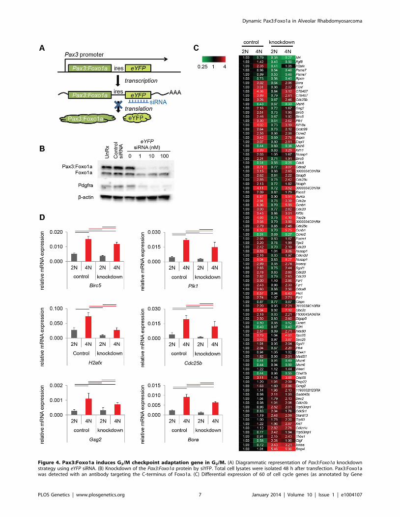
Figure 4. Pax3:Foxo1a induces G2/M checkpoint adaptation gene in G2/M. (A) Diagrammatic representation of Pax3:Foxo1a knockdownstrategy using eYFP siRNA. (B) Knockdown of the Pax3:Foxo1a protein by siYFP. Total cell lysates were isolated 48 h after transfection. Pax3:Foxo1awas detected with an antibody targeting the C-terminus of Foxo1a. (C) Differential expression of 60 of cell cycle genes (as annotated by Gene
Dynamic Pax3:Foxo1a in Alveolar Rhabdomyosarcoma
PLOS Genetics | www.plosgenetics.org 7 January 2014 | Volume 10 | Issue 1 | e1004107

results suggested that not only cell cycle dependent expression but
also a clinically-relevant biology underlying Pax3:Foxo1a expres-
sion at the G2-M checkpoint, a critical cell cycle checkpoint
following radiation or DNA double strand break inducing-
chemotherapy.
That a myogenic cancer might utilize genomic instability,
aneuploidy or multinucleation as a mechanism of cell survival or
tumor cell evolution/progression may not be so unexpected, in
retrospect. Normal myofibers are typically multi-nuclear by
definition, and genetic conditions predisposing to mitotic disjunc-
tion such as Mosaic Variegated Aneuploidy (MVA) are strongly
associated with the development of RMS [22]. Both aRMS and
eRMS have also been documented to be hyperdiploid, tetraploid,
polyploid or to even have mixed aneuploid populations [23,24,25].
At a cellular level, the heterogeneity of cells in rhabdomyosarcoma
is notable for the subpopulation of multi-nucleated rhabdomyo-
blasts which appears with giant nuclei or as multi-nucleated giant
cells, often with cross-striations – yet highly mitotic [26]. These
rhabdomyoblasts might be compared to the multinucleated
stemloid cells in fibrosarcoma, which have a tumor-repopulating
ability [27]. Our recent study of aRMS and the PKC iota inhibitor,
aurothiomalate, reveals that aRMS cells have a remarkable
tolerance to polyploidy, which induces neither apoptosis or
senescence [28]. This intrinsic capacity to tolerate aneuploidy as
well as this report’s observed Pax3:Foxo1a-mediated increase in
checkpoint adaptation gene expression may be directly relevant to
clinical care, given that decreased expression of these same factors
(i.e., PLK1, CCCNB1, BIRC5, AURKB) have been reported to
improve sensitivity to mitotic inhibitors [29]. Therefore, the interest
generated from chemical screens identifying PLK1 as a potential
therapeutic target in RMS [30] is likely warranted.
When considering the differences in treatment-related outcomes
in RMS subtypes, the role of Pax3:Fox01a in checkpoint
adaptation may be our most important clue yet as to how to
improve outcome for fusion positive patients: while aRMS are
certainly sensitive to standard chemotherapy and radiation, it is
the survival of resistant clones which is the cause of disease
progression and relapse – which occur to a greater extent in
Pax:Foxo1a positive aRMS than fusion negative aRMS or eRMS
[31,32], and which we believe to be a result of Pax3:Foxo1a-
mediated checkpoint adaptation. These effects on tumor cell
sensitivity to radiation, chemotherapy and targeted therapeutics
are likely to be cumulative and possibly critically important in
defining the otherwise very narrow therapeutic window for fusion
positive aRMS, for which the toxicity of chemotherapy and
radiation is now dose-limiting [33].
Perhaps the most interesting aspect of this genetically-engi-
neered conditional mouse model of a deadly but rare childhood
cancer is that a labor-intensive knock-in approach to modeling the
molecular pathophysiology of a fusion gene was beneficial.
Successful transgenic tumor models have been generated by
constitutive, ectopic expression of translocation-related fusion
oncogenes for synovial sarcoma [34] as well as other ‘‘driver’’
oncogene related tumors [35]; similarly, retroviral transfection of
oncogenes into hematopoietic cells has enabled this study of
translocation-associated leukemia for many years [36,37]. How-
ever, are these systems driven by non-native or partial-native
promoters to be the definitive preclinical platforms for interrogat-
ing molecular physiology – or are distal native cis- and trans-
regulation temporally critical? Every experimental system has its
advantages and limitations, yet for cell and animal models where
translocation-mediated fusion genes have yet to be modeled at the
native promoter, we may have an entirely new spectrum of cancer
genetics to explore.
Materials and Methods
Ethics statementAll animal procedures were conducted in accordance with the
Guidelines for the Care and Use of Laboratory Animals and were
approved by the Institutional Animal Care and Use Committee
(IACUC) at the University of Oregon Health & Science University
(OHSU) or the Joslin Diabetes Center (Boston, MA). Every effort
was made to minimize suffering.
MiceThe Myf6Cre,Pax3:Foxo1a,p53 conditional aRMS mouse model
has been described previously [5,6,7], is described as caMOD
Model 150064393, and is publically available through the NCI
MMHCC Repository (MMHCC Strain Codes 01XBL B6; 129-
Myf6,tm2(Cre)Mrc. and 01XBM B6; 129-Pax3,tm1Mrc.).
SHO-PrkdcscidHrhr mice were purchased from Charles River
Laboratories (Wilmington, MA) and bred/maintained at OHSU.
Primary tumor cell cultures and cell linesMouse primary cell cultures (U23674, U42369, U57844) were
established from tumor samples. Tumors were minced into small
pieces and digested with collagenase (10 mg/ml) overnight at
37uC. The dissociated cells were then incubated in Dulbecco’s
modified eagle’s media supplemented with 10% fetal bovine serum
(FBS) and 1% penicillin-streptomycin in 5% CO2 at 37uC. C2C12
mouse myoblast cells were purchased from ATCC (Manassas,
VA). Human aRMS cell lines were a gift from Peter Houghton
(Rh3; Nationwide Children’s Hospital, Columbus, OH) or Patrick
Reynolds (Rh41; COG Cell Culture and Xenograft Repository).
These cells lines were maintained in the same culture conditions as
primary tumor cell cultures: DMEM supplemented with 10%
Fetal Bovine Serum (FBS) and 1% Penicillin-Streptomycin. All
primary cell cultures experiments using cells were carried out at
passage 3–7.
Confocal imagingFor immunofluorescence staining of frozen sections, the
polyclonal antibody for green fluorescent protein (1:1000,
AB16901, Chemicon) was used with DAPI counterstain.
RNA interference studiessiRNA transfections were carried out using Lipofectamine2000
(Invitrogen, Grand Island, NY) according to manufacturer’s
recommended protocol. siRNA’s were diluted between 0.1 and
10 nM, and the final concentration of Lipofectamine2000 was
0.2%. siYFP Stealth RNAi siRNA Reporter Controls (cat. 12935-
145; Invitrogen) were used as the eYFP siRNA to knockdown the
Pax3:Foxo1a-ires-eYFP bi-cistronic mRNA, whereas Stealth RNAi
siRNA Negative Control Med GC #3 (cat. 12935-113; Invitro-
gen) was used as the siRNA control (siCont).
Ontology) for DNA content with or without Pax3:Foxo1a knockdown. (D) mRNA expression by QPCR of Plk1, Cdc25b, H2afx and Birc5 normalized toGapdh in DNA content-sorted U23674 mouse aRMS primary tumor cells with or without Pax3:Foxo1a knockdown. Black and red line shows significantdifference (p,0.05).doi:10.1371/journal.pgen.1004107.g004
Dynamic Pax3:Foxo1a in Alveolar Rhabdomyosarcoma
PLOS Genetics | www.plosgenetics.org 8 January 2014 | Volume 10 | Issue 1 | e1004107
Dynamic Pax3:Foxo1a in Alveolar Rhabdomyosarcoma
PLOS Genetics | www.plosgenetics.org 9 January 2014 | Volume 10 | Issue 1 | e1004107

Generation of shRNA tumor cell culture clonesTo establish shRNA knockdown clones of primary tumor cell
cultures, we used MISSION pLKO.1-puro eGFP shRNA Control
Transduction Particles (cat. SHC005V; Sigma Aldrich) for
Pax3:Foxo1a knockdown and MISSION pLKO.1-puro Non-
Mammalian shRNA Control Transduction Particles (cat.
SHC002V; Sigma Aldrich) as the control, respectively. shRNA
transfections and clonal selection were carried out according to
manufacturer’s recommended procedures. Mouse RMS primary
cell cultures were plated at 1.86106 cells per 150 mm dish. After
24 h, hexadimethrine bromide was added (8 mg/ml, cat. H9268;
Sigma Aldrich), followed by each particle solution (MOI 0.5). After
another 24 h, media were removed and fresh media were added.
The following day, puromycin was added (5 mg/ml, cat. P8833;
Sigma Aldrich). Puromycin-resistant clones were selected cloning
rings at day 14 (shControl) and day 17 (shYFP), with continuous
puromycin selection at all times.
RadiationCells were irradiated on a Trilogy linear accelerator (Varian,
Palo Alto, CA) with a 10610 cm AP field. Two centimeter of
bolus material was placed on top of the 2 chamber slide or 6 cm
dish and the target surface distance to the bolus was at 97 cm.
Monitor units on the linear accelerator were then set to deliver 6
Gy or 10 Gy of dose to the cells.
ImmunoblottingTumors were lysed in radioimmunoprecipitation assay (RIPA)
buffer or NP40 buffer containing both protease and phosphatase
inhibitor (Sigma). The lysates were homogenized and centrifuged
at 8000 g for 10 minutes. The resulting supernatants were used for
immunoblot analysis. Goat anti-FOXO1A antibody (cat. Sc-9808;
Santa Cruz, Santa Cruz, CA), goat anti-GFP antibody (cat. 600-
101-215, Rockland; Gilbertsville, PA) or rabbit anti-PDGFRa
antibody (cat. #3164; Cell signaling Technology, Danvers, MA).
ImmunocytochemistryCells were plated on 8-well CultureSlides (cat. 354118; BD
Falcon, Franklin Lakes, NJ), fixed with 4% paraformaldehyde,
permeabilized with 0.1% or 0.25% TritonX100, washed and
incubated with mouse monoclonal anti-skeletal myosin (FAST)
(cat. M4276; Sigma), rabbit anti-Ki67 (cat. RM-9106-F; Thermo
Scientific, Waltham, MA), mouse anti-Pax3 (cat. MAB2457; R&D
Systems), mouse anti-phospho Histone H3 (cat. #9706; Cell
Signaling Technology), rabbit anti-phospho Histone H3 (cat.
#3377; Cell Signaling Technology), mouse anti-phospho Histone
H3 (cat. #9706; Cell Signaling Technology), rabbit anti-CDC2-
Y15 (cat. #4539; Cell Signaling Technology) or rabbit anti-
phospho H2AX antibody (cat. #9718; Cell Signaling Technolo-
gy), overnight, rinsed with PBS, incubated with fluorescein
isothiocyanate-conjugated anti-mouse and rabbit IgG (1:200) for
Figure 6. Treatment-related implications for dynamic oncogene expression in rhabdomyosarcoma in vivo. (A) Kaplan-Meier survivalanalysis for disease-free survival of mice implanted with pre-irradiated (10Gy) primary murine aRMS tumor cells treated with Pax3:Foxo1a siRNA (siY)or control siRNA (siC), n = 5 animals per cohort. The p value for the difference between siY and siC groups receiving radiation was 0.02. (B)Diagrammatic representation of results in (A).doi:10.1371/journal.pgen.1004107.g006
Figure 5. Pax3:Foxo1a facilitates G2/M checkpoint adaptation. (A) Immunocytochemistry for pHH3 (green), pH2AX (red) and DAPI (Blue)using U23674 mouse aRMS primary cell culture with or without Pax3:Foxo1a knockdown treated with 6 Gy irradiation. Black line shows significantdifference (p,0.05). See Figure S2A for representative single-channel ICC images corresponding to Figure 5A. Arrowheads indicate pHH3 and pH2AXdouble positive cells. (B) Representative cell cycle analysis for U23674 transfected by shControl (Clone A) or shYFP (Clone C) with or without 10 Gyirradiation. The graph shows the differences of percentage between control and radiated cells in shControl and shYFP clones in 3 independentexperiments. Black line shows significant difference (p,0.05). (C) Annexin V apoptosis detection assay for U23674 transfected by shControl or shYFPclones with or without 10 Gy irradiation. Black line shows significant difference (p,0.05).doi:10.1371/journal.pgen.1004107.g005
Dynamic Pax3:Foxo1a in Alveolar Rhabdomyosarcoma
PLOS Genetics | www.plosgenetics.org 10 January 2014 | Volume 10 | Issue 1 | e1004107

1 h, and examined by confocal microscopy with a Zeiss LSM700
instrument. For immunocytochemistry experiments, at least 100
positive cells were scored per specimen.
FACS sortingCells were suspended in Hank’s balanced salt solution (HBSS)
with 2% FBS and 2 mM EDTA. Antibody staining was performed
for 20 minutes on ice. Prior to FACS sorting, cells were suspended
in 1 mg/ml propidium iodide (Pi) and 10 mM calcein blue
(Invitrogen) to identify viable cells (Pi2Ca+). Purity checks were
performed to confirm that the sorted eYFP+ and eYFP- cell
subsets had a purity of .98% using a eYFP expression threshold
determined by the background fluorescence of eYFP- C2C12 cells.
The following antibodies were used to evaluate receptor tyrosine
kinase surface expression: APC-conjugated Pdgfra antibody (#17-
1401-81, eBiosciences) or anti-IGF1 Receptor antibody (cat.
Ab32823; Abcam, Cambridge, MA; 1 in 25).
Cell cycle analysisMouse RMS primary cell cultures were trypsinized and
incubated with Hoechst33342 (final concentration 15 mg/ml)
and Reserpine (final concentration 5 mM). Cells were incubated
in the dark for 30 min at 37uC, and analyzed and sorted by flow
cytometry using an Influx FACS instrument (Becton Dickinson,
Franklin Lakes, NJ). Cell cycle was determined with the FlowJo
software (Tree Star, Inc., Ashland, OR).
Annexin V apoptosis detection assayMouse primary cell cultures were stained with Annexin V and
Propidium iodide using Annexin-V-FLUOS Staining Kit (cat. 11
858 777 001; Roche) following the protocol provided by the
manufacturer. Briefly, 48 hour after irradiation, 106 mouse
primary cell cultures were trypsinized, washed by PBS and
resuspended in 100 ml of Annexin-VFLUOS labeling solution,
incubated 10–15 min at 15–25uC, and analyzed by FACS
Calibur.
Quantitative RT PCR (QPCR)U23674 cells were subfractionated by FACS sorting as
described above. mRNA was isolated using RNeasy spin columns
(Qiagen, Valencia, CA) and reverse transcribed using Superscript
III First-Strand Synthesis System for RT-PCR (Invitrogen).
QPCR was performed using an AV7900 PCR system (Applied
Biosystem) with SYBR-green PCR reagents. Pax3:Foxo1a was
detected using the following primer sequences: 59-AGA-
CAGCTTTGTGCCTCCAT-39 and 59-CTCTTGCCTCCCTCTGGA-
TT-39. Other primers are Taqman Gene Expression assay, H2afx
(Mm00515990_s1), Cdc25b (Mm00499136_m1), Birc5 (Mm00599-
749_m1), Plk1 (Mm00440924_g1) and Gapdh (Mm99999915_g1)
by Invitrogen. RT-PCR data were quantified using the standard
curve method, and relative expression of Pax3:Foxo1a per sample
was determined by normalization against the quantity of 18 s
rRNA and Gapdh within each sample. For each sample, QPCR
was performed in technical duplicates and results were averaged.
In vitro growth inhibition assaysMouse RMS primary cell cultures were plated at 16103 cells of
each cohort per well in a 96-well plate. After cell incubations,
cytotoxic effects were assayed using CellTiter 96 AQueous One
Solution Cell Proliferation Assay system (Promega, Madison, WI)
and SpectraMax M5 luminometer (Molecular Devices, Sunnyvale,
CA). IC50 and C.I. were determined with CalcuSyn software
(BIOSOFT, United Kingdom).Drugs: Vincristine sulfate salt (cat.
V8879; Sigma), Actinomycin-D (cat. A9415; Sigma), Mafosfamide
(cat. sc-211761; Santa Cruz), Topotecan hydrochloride (cat.
S1231; Selleck), Eribulin mesylate (NDC 62856-389-01; Eisai) or
Imatinib Mesylate (cat. S1026; Selleck).
RNAi-assisted protein target identification (RAPID) screenFor these studies, individual siRNA were obtained from
Dharmacon (Lafayette, CO), including the mouse siRNA library
targeting the tyrosine kinome (siGENOME). These experiments
are performed at 100 nM concentration and include non-specific
pooled siRNA as a control purchased from Dharmacon.
Transfection of siRNA was carried out using Lipofectamine
2000 in Opti-MEM Reduced Serum Media (Invitrogen). After
cells were plated in 96-well plates in the presence of inhibitor or
siRNA, and incubated for 96 hours, respectively, 20 mL CellTiter
96 AQueous One solution (MTS) was added to each well and
absorbance values assessed by the BioTek Synergy 2 plate reader
(BioTek, Winooski, VT).
Genome-wide expression analysisLabeled target cRNA was prepared from 12 mouse total RNA
samples (3 independent experiments64 samples). Samples were
amplified and labeled using the Ambion MessageAmp Premier
RNA Amplification Kit following the manufacturer’s protocol.
Sample order was randomized. Each sample target was hybridized
to an Illumina MouseRef 8 v 2 Expression BeadChip Array.
Image processing and expression analysis were performed using
Illumina BeadArray Reader and GenomeStudio (v. 2010.1) Gene
Expression module (v. 1.6.0) software. Microarray data have been
accessioned with the Gene Expression Omnibus (GEO) under
series GSE41675. The following link has been created to allow
review of record GSE41675 while it remains in in review/under
private status: http://www.ncbi.nlm.nih.gov/geo/query/acc.
cgi?token = xdajbqeisomcyhq&acc = GSE41675.
In vivo studies with Pax3:Foxo1a knockdown andradiation
aRMS primary cultures (passage 5) were plated in 6 cm dishes.
The next day cells were transfected with siYFP Stealth RNAi
siRNA Reporter Controls or Stealth RNAi siRNA Negative
Control Med GC #3. Two days later cells were irradiated on a
Trilogy linear accelerator with a 10610 cm AP field with two
centimeter of bolus material was placed on top of the 6 cm dish.
The target surface distance to the bolus was at 97 cm and monitor
units on the linear accelerator were then set to deliver 10 Gy of
dose to the cells. Subsequently, cells were trypsinized and 500,000
cells were injected into the gastrocnemius muscle of SHO mice
that had been pre-injured 24 hours prior with 0.85 mg/mouse
cardiotoxin intramuscularly. Tumor volumes (cm3) were measured
3-dimensionally with electronic calipers and calculated from
formula (p/6)6length6width6height, assuming tumors to be
spheroid. For statistical analysis of disease-free survival, a tumor
volume threshold of 0.25 cc was applied. The log-rank test was
used to contrast treatments. All analyses were performed using R
3.0.0 (The R Foundation for Statistical Computing, Vienna,
Austria).
Supporting Information
Figure S1 This supplemental figure relates to Figure 1. eYFP activity
and Pax3:Foxo1a expression is dynamic. (A) eYFP fluorescence of
eYFP sorted U42369 mouse aRMS primary cell culture overtime
as measured by FACS. Grey: C2C12 (negative control), blue: no
sorted cells, green: eYFP activity high cells, red: eYFP activity low
Dynamic Pax3:Foxo1a in Alveolar Rhabdomyosarcoma
PLOS Genetics | www.plosgenetics.org 11 January 2014 | Volume 10 | Issue 1 | e1004107

cells. (B) Mean of relative eYFP activity measured by FACS. (C)
Western blot analysis using eYFP sorted cells. Plotted are relative
protein levels of Pax3:Foxo1a/b-actin. Mean 6 SE were obtained
from three independent immunoblottings. Black line shows
significant difference (p,0.05). (D) eYFP activity and cell cycle
analysis using Hochest33342 staining for mouse primary cell
culture U42369. Green shows G0/G1 phase, brown shows S
phase, and blue shows G2/M phase. (E–F) Proliferating mouse
aRMS tumor cells were treated with 10 mg/ml CHX for the
indicated incubations, and eYFP, Pax3:Foxo1a and Pdgfra protein
levels were followed by western blot analysis (E). Protein
expression quantified as relative flux normalized by b-actin for
calculation of protein half-lives (F).
(TIF)
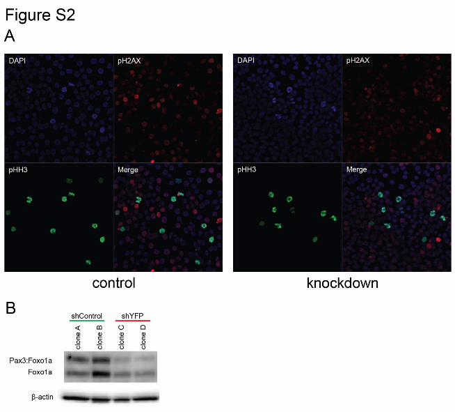
Figure S2 This supplemental figure relates to Figure 5. Pax3:Foxo1a
mediates checkpoint adaptation. (A) Individual and merged
channels for immunocytochemistry of pHH3 (green), pH2AX
(red) and DAPI (blue) using U23674 mouse aRMS primary cell
culture with or without Pax3:Foxo1a knockdown treated by 6 Gy
irradiation. (B) Western blot analysis of Pax3:Foxo1a and Foxo1a
in U23674 shControl and shYFP clones.
(TIF)
Figure S3 This supplemental figure relates to Figure 6. Pax3:Foxo1a
modifies the aRMS therapeutic response and remains an essential
target. (A–E) Pax3:Foxo1a knockdown increases select chemother-
apy sensitivities. MTS assay was performed for Pax3:Foxo1a
knockdown mouse aRMS tumor cells treated with DNA damaging
agents and microtubule inhibitors. Pax3:Foxo1a knockdown
reduced the concentration at which viability was impaired by
50% (IC50) of vincristine, actinomycin-D, topotecan and eribulin
by 2.9, 3.4, 4.8 and1.8 fold, respectively, yet did not affect the
IC50 of mafosfamide. (F–G) Imatinib IC50 determination using
mouse aRMS tumor cells transfected with siCont or siYFP,
respectively. Pax3:Foxo1a knockdown sensitized aRMS cells 5-fold
to this prototypic Pdgfra inhibitor. Given the role of Pax3:Foxo1a
in growth factor receptor transcription, we next explored the role
of Pax3:Foxo1a in driving aberrant tyrosine kinase signaling by
means of an RNAi-assisted protein target identification (RAPID)
screen after first knocking down Pax3:Foxo1a in mouse aRMS
tumor cells [38]. Cell viability was significantly decreased not only
for targets of imatinib but also for mediators of imatinib resistance
[17] in Pax3:Foxo1a knockdown cells compared with control cells
(G). * p,0.01.
(TIF)
Movie S1 This supplemental movie relates to Figure 1. Time lapse of
eYFP expression in murine aRMS cells.
(MOV)
Table S1 Genome-wide expression analysis using cell cycle
specific sorted cells (2N vs 4N) with or without Pax3:Foxo1a
knockdown.
(XLSX)
Table S2 Putative Pax3:Foxo1a binding sites of Checkpoint
Adaptation related genes.
(XLSX)
Acknowledgments
We are grateful to Dr. Charles R. Thomas for technical contribution to
radiation studies (Department of Radiation Medicine, Oregon Health &
Science University). We thank Tom Rando, Bill Forrester and Fred Barr
for critical evaluation of this study during its development.
Author Contributions
Conceived and designed the experiments: CK KK. Performed the
experiments: KK MIA SH BAW DML BPR AJW WL. Analyzed the
data: CK KK JEM SH BAW DML BPR AJW. Wrote the paper: CK KK.
References
1. Breneman JC, Lyden E, Pappo AS, Link MP, Anderson JR, et al. (2003)
Prognostic factors and clinical outcomes in children and adolescents with
metastatic rhabdomyosarcoma–a report from the Intergroup Rhabdomyosar-coma Study IV. J Clin Oncol 21: 78–84.
2. Anderson JR, Barr FG, Hawkins DS, Parham DM, Skapek SX, et al. (2010)
Fusion-negative alveolar rhabdomyosarcoma: modification of risk stratification is
premature. J Clin Oncol 28: e587–588; author reply e589–590.
3. Williamson D, Missiaglia E, de Reynies A, Pierron G, Thuille B, et al. (2010)
Fusion gene-negative alveolar rhabdomyosarcoma is clinically and molecularly
indistinguishable from embryonal rhabdomyosarcoma. J Clin Oncol 28: 2151–
2158.
4. Wexler LH, Ladanyi M (2010) Diagnosing alveolar rhabdomyosarcoma:morphology must be coupled with fusion confirmation. J Clin Oncol 28:
2126–2128.
5. Keller C, Arenkiel BR, Coffin CM, El-Bardeesy N, DePinho RA, et al.
(2004) Alveolar rhabdomyosarcomas in conditional Pax3:Fkhr mice:
cooperativity of Ink4a/ARF and Trp53 loss of function. Genes Dev 18: 2614–2626.
6. Keller C, Hansen MS, Coffin CM, Capecchi MR (2004) Pax3:Fkhr interferes
with embryonic Pax3 and Pax7 function: implications for alveolar rhabdomyo-
sarcoma cell of origin. Genes Dev 18: 2608–2613.
7. Nishijo K, Chen QR, Zhang L, McCleish AT, Rodriguez A, et al. (2009)
Credentialing a preclinical mouse model of alveolar rhabdomyosarcoma. Cancer
Res 69: 2902–2911.
8. Taniguchi E, Nishijo K, McCleish AT, Michalek JE, Grayson MH, et al. (2008)
PDGFR-A is a therapeutic target in alveolar rhabdomyosarcoma. Oncogene 27:
6550–6560.
9. Cao L, Yu Y, Bilke S, Walker RL, Mayeenuddin LH, et al. (2010) Genome-
Wide Identification of PAX3-FKHR Binding Sites in Rhabdomyosarcoma
Reveals Candidate Target Genes Important for Development and Cancer.
Cancer Res 70: 6497–508.
10. Osuka S, Sampetrean O, Shimizu T, Saga I, Onishi N, et al. (2013) IGF1
receptor signaling regulates adaptive radioprotection in glioma stem cells. Stem
cells 31: 627–640.
11. Nurse P (1994) Ordering S phase and M phase in the cell cycle. Cell 79: 547–
550.
12. Berry LD, Gould KL (1996) Regulation of Cdc2 activity by phosphorylation atT14/Y15. Progress in cell cycle research 2: 99–105.
13. Syljuasen RG, Jensen S, Bartek J, Lukas J (2006) Adaptation to the ionizing
radiation-induced G2 checkpoint occurs in human cells and depends oncheckpoint kinase 1 and Polo-like kinase 1 kinases. Cancer Res 66: 10253–
10257.
14. Syljuasen RG (2007) Checkpoint adaptation in human cells. Oncogene 26:
5833–5839.
15. Yoo HY, Kumagai A, Shevchenko A, Shevchenko A, Dunphy WG (2004)Adaptation of a DNA replication checkpoint response depends upon inactivation
of Claspin by the Polo-like kinase. Cell 117: 575–588.
16. Cao L, Yu Y, Bilke S, Walker RL, Mayeenuddin LH, et al. (2010) Genome-wide
identification of PAX3-FKHR binding sites in rhabdomyosarcoma revealscandidate target genes important for development and cancer. Cancer Res 70:
6497–6508.
17. Abraham J, Chua YX, Glover JM, Tyner JW, Loriaux MM, et al. (2012) Anadaptive Src-PDGFRA-Raf axis in rhabdomyosarcoma. Biochem Biophys Res
Commun 426: 363–368.
18. Kitzmann M, Carnac G, Vandromme M, Primig M, Lamb NJ, et al. (1998) The
muscle regulatory factors MyoD and myf-5 undergo distinct cell cycle-specific
expression in muscle cells. J Cell Biol 142: 1447–1459.
19. Batonnet-Pichon S, Tintignac LJ, Castro A, Sirri V, Leibovitch MP, et al. (2006)
MyoD undergoes a distinct G2/M-specific regulation in muscle cells. Exp CellRes 312: 3999–4010.
20. Koniaras K, Cuddihy AR, Christopoulos H, Hogg A, O’Connell MJ (2001)
Inhibition of Chk1-dependent G2 DNA damage checkpoint radiosensitizes p53mutant human cells. Oncogene 20: 7453–7463.
21. Clemenson C, Marsolier-Kergoat MC (2009) DNA damage checkpointinactivation: adaptation and recovery. DNA Repair (Amst) 8: 1101–11
09.
22. Hanks S, Coleman K, Reid S, Plaja A, Firth H, et al. (2004) Constitutionalaneuploidy and cancer predisposition caused by biallelic mutations in BUB1B.
Nat Genet 36: 1159–1161.
23. Kowal-Vern A, Gonzalez-Crussi F, Turner J, Trujillo YP, Chou P, et al. (1990)
Flow and image cytometric DNA analysis in rhabdomyosarcoma. Cancer Res50: 6023–6027.
Dynamic Pax3:Foxo1a in Alveolar Rhabdomyosarcoma
PLOS Genetics | www.plosgenetics.org 12 January 2014 | Volume 10 | Issue 1 | e1004107
24. San Miguel-Fraile P, Carrillo-Gijon R, Rodriguez-Peralto JL, Badiola IA (2004)
Prognostic significance of DNA ploidy and proliferative index (MIB-1 index) in
childhood rhabdomyosarcoma. Am J Clin Pathol 121: 358–365.
25. Shapiro DN, Parham DM, Douglass EC, Ashmun R, Webber BL, et al. (1991)
Relationship of tumor-cell ploidy to histologic subtype and treatment outcome in
children and adolescents with unresectable rhabdomyosarcoma. J Clin Oncol 9:
159–166.
26. Li G, Kikuchi K, Radka M, Abraham J, Rubin BP, et al. (2013) IL-4 receptor
blockade abrogates satellite cell - rhabdomyosarcoma fusion and prevents tumor
establishment. Stem cells 31: 2304–12.
27. Weihua Z, Lin Q, Ramoth AJ, Fan D, Fidler IJ (2011) Formation of solid tumors
by a single multinucleated cancer cell. Cancer 117: 4092–4099.
28. Kikuchi K, Soundararajan A, Zarzabal LA, Weems CR, Nelon LD, et al. (2012)
Protein kinase C iota as a therapeutic target in alveolar rhabdomyosarcoma.
Oncogene 32: 286–95.
29. Yamada HY, Rao CV (2010) Genes that modulate the sensitivity for anti-
microtubule drug-mediated chemotherapy. Curr Cancer Drug Targets 10: 623–
633.
30. Hu K, Lee C, Qiu D, Fotovati A, Davies A, et al. (2009) Small interfering RNA
library screen of human kinases and phosphatases identifies polo-like kinase 1 as
a promising new target for the treatment of pediatric rhabdomyosarcomas. Mol
Cancer Ther 8: 3024–3035.
31. Skapek SX, Anderson J, Barr FG, Bridge JA, Gastier-Foster JM, et al. (2013)
PAX-FOXO1 Fusion Status Drives Unfavorable Outcome for Children With
Rhabdomyosarcoma: A Children’s Oncology Group Report. Pediatr Blood
Cancer 60: 1411–7.32. Missiaglia E, Williamson D, Chisholm J, Wirapati P, Pierron G, et al. (2012)
PAX3/FOXO1 fusion gene status is the key prognostic molecular marker in
rhabdomyosarcoma and significantly improves current risk stratification. J ClinOncol 30: 1670–1677.
33. Gupta AA, Anderson JR, Pappo AS, Spunt SL, Dasgupta R, et al. (2012)Patterns of chemotherapy-induced toxicities in younger children and adolescents
with rhabdomyosarcoma: a report from the Children’s Oncology Group Soft
Tissue Sarcoma Committee. Cancer 118: 1130–1137.34. Haldar M, Hancock JD, Coffin CM, Lessnick SL, Capecchi MR (2007) A
conditional mouse model of synovial sarcoma: insights into a myogenic origin.Cancer Cell 11: 375–388.
35. Zhu H, Acquaviva J, Ramachandran P, Boskovitz A, Woolfenden S, et al. (2009)Oncogenic EGFR signaling cooperates with loss of tumor suppressor gene
functions in gliomagenesis. Proc Natl Acad Sci U S A 106: 2712–2716.
36. Bernt KM, Zhu N, Sinha AU, Vempati S, Faber J, et al. (2011) MLL-rearrangedleukemia is dependent on aberrant H3K79 methylation by DOT1L. Cancer
Cell 20: 66–78.37. DiMartino JF, Miller T, Ayton PM, Landewe T, Hess JL, et al. (2000) A
carboxy-terminal domain of ELL is required and sufficient for immortalization
of myeloid progenitors by MLL-ELL. Blood 96: 3887–3893.38. Tyner JW, Walters DK, Willis SG, Luttropp M, Oost J, et al. (2008) RNAi
screening of the tyrosine kinome identifies therapeutic targets in acute myeloidleukemia. Blood 111: 2238–2245.
Dynamic Pax3:Foxo1a in Alveolar Rhabdomyosarcoma
PLOS Genetics | www.plosgenetics.org 13 January 2014 | Volume 10 | Issue 1 | e1004107

SUPPORTING INFORMATION: FIGURE LEGENDS
Cell-Cycle Dependent Expression of a Translocation-mediated Fusion Oncogene mediates
Checkpoint Adaptation in Rhabdomyosarcoma
Ken Kikuchi, Simone Hettmer, M. Imran Aslam, Joel E. Michalek, Wolfram Laub, Breelyn A. Wilky, David M.
Loeb, Brian P. Rubin, Amy J. Wagers, Charles Keller
Figure S1. This supplemental figure relates to Figure 1. eYFP activity and Pax3:Foxo1a expression is
dynamic. (A) eYFP fluorescence of eYFP sorted U42369 mouse aRMS primary cell culture overtime as
measured by FACS. Grey: C2C12 (negative control), blue: no sorted cells, green: eYFP activity high cells, red:
eYFP activity low cells. (B) Mean of relative eYFP activity measured by FACS. (C) Western blot analysis
using eYFP sorted cells. Plotted are relative protein levels of Pax3:Foxo1a/�-actin. Mean ± SE were obtained
from three independent immunoblottings. Black line shows significant difference (p<0.05). (D) eYFP activity
and cell cycle analysis using Hochest33342 staining for mouse primary cell culture U42369. Green shows
G0/G1 phase, brown shows S phase, and blue shows G2/M phase. (E-F) Proliferating mouse aRMS tumor cells
were treated with 10 µg/ml CHX for the indicated incubations, and eYFP, Pax3:Foxo1a and Pdgfra protein
levels were followed by western blot analysis (E). Protein expression quantified as relative flux normalized by
�-actin for calculation of protein half-lives (F).
Figure S2. This supplemental figure relates to Figure 5. Pax3:Foxo1a mediates checkpoint adaptation. (A)
Individual and merged channels for immunocytochemistry of pHH3 (green), pH2AX (red) and DAPI (blue)
using U23674 mouse aRMS primary cell culture with or without Pax3:Foxo1a knockdown treated by 6 Gy
irradiation. (B) Western blot analysis of Pax3:Foxo1a and Foxo1a in U23674 shControl and shYFP clones.
Figure S3. This supplemental figure relates to Figure 6. Pax3:Foxo1a modifies the aRMS therapeutic
response and remains an essential target. (A-E) Pax3:Foxo1a knockdown increases select chemotherapy
sensitivities. MTS assay was performed for Pax3:Foxo1a knockdown mouse aRMS tumor cells treated with
DNA damaging agents and microtubule inhibitors. Pax3:Foxo1a knockdown reduced the concentration at
which viability was impaired by 50% (IC50) of vincristine, actinomycin-D, topotecan and eribulin by 2.9, 3.4,
4.8 and1.8 fold, respectively, yet did not affect the IC50 of mafosfamide. (F-G) Imatinib IC50 determination
using mouse aRMS tumor cells transfected with siCont or siYFP, respectively. Pax3:Foxo1a knockdown
sensitized aRMS cells 5-fold to this prototypic Pdgfra inhibitor. Given the role of Pax3:Foxo1a in growth factor
2
receptor transcription, we next explored the role of Pax3:Foxo1a in driving aberrant tyrosine kinase signaling by
means of an RNAi-assisted protein target identification (RAPID) screen after first knocking down Pax3:Foxo1a
in mouse aRMS tumor cells [1]. Cell viability was significantly decreased not only for targets of imatinib but
also for mediators of imatinib resistance [2] in Pax3:Foxo1a knockdown cells compared with control cells (G).
* p<0.01.
Supporting Information References
1. Tyner JW, Walters DK, Willis SG, Luttropp M, Oost J, et al. (2008) RNAi screening of the tyrosine kinome
identifies therapeutic targets in acute myeloid leukemia. Blood 111: 2238-2245.
2. Abraham J, Chua YX, Glover JM, Tyner JW, Loriaux MM, et al. (2012) An adaptive Src-PDGFRA-Raf axis
in rhabdomyosarcoma. Biochem Biophys Res Commun 426: 363-368.