NuclearLocalizationof deNovo ThymidylateBiosynthesis ... · this pathway is present in the...
9
Nuclear Localization of de Novo Thymidylate Biosynthesis Pathway Is Required to Prevent Uracil Accumulation in DNA * Received for publication, September 26, 2011, and in revised form, October 14, 2011 Published, JBC Papers in Press, November 4, 2011, DOI 10.1074/jbc.M111.307629 Amanda J. MacFarlane ‡1 , Donald D. Anderson § , Per Flodby ‡2 , Cheryll A. Perry ‡ , Robert H. Allen ¶ , Sally P. Stabler ¶ , and Patrick J. Stover ‡§3 From the ‡ Division of Nutritional Sciences, Cornell University, Ithaca, New York 14853, the § Graduate Field of Biochemistry, Molecular and Cell Biology, Cornell University, Ithaca, New York 14853, and the ¶ Department of Medicine and Division of Hematology, University of Colorado School of Medicine, Aurora, Colorado 80045 Background: S phase nuclei contain the thymidylate synthesis pathway. Results: Mice overexpressing a Shmt1 transgene exhibit elevated expression of SHMT1 and TYMS, impaired nuclear localiza- tion of the thymidylate biosynthesis pathway, and elevated uracil in DNA. Conclusion: SHMT1 and TYMS localization to the nucleus is essential to prevent uracil accumulation in DNA. Significance: SHMT1-mediated nuclear de novo thymidylate synthesis is critical for maintaining DNA integrity. Uracil accumulates in DNA as a result of impaired folate-de- pendent de novo thymidylate biosynthesis, a pathway composed of the enzymes serine hydroxymethyltransferase (SHMT), thy- midylate synthase (TYMS), and dihydrofolate reductase. In G 1 , this pathway is present in the cytoplasm and at S phase under- goes small ubiquitin-like modifier-dependent translocation to the nucleus. It is not known whether this pathway functions in the cytoplasm, nucleus, or both in vivo. SHMT1 generates 5,10- methylenetetrahydrofolate for de novo thymidylate biosynthe- sis, a limiting step in the pathway, but also tightly binds 5-meth- yltetrahydrofolate in the cytoplasm, a required cofactor for homocysteine remethylation. Overexpression of SHMT1 in cell cultures inhibits folate-dependent homocysteine remethylation and enhances thymidylate biosynthesis. In this study, the impact of increased Shmt1 expression on folate-mediated one-carbon metabolism was determined in mice that overexpress the Shmt1 cDNA (Shmt1 tg mice). Compared with wild type mice, Shmt1 tg mice exhibited elevated SHMT1 and TYMS protein levels in tissues and evidence for impaired homocysteine rem- ethylation but surprisingly exhibited depressed levels of nuclear SHMT1 and TYMS, lower rates of nuclear de novo thymidylate biosynthesis, and a nearly 10-fold increase in uracil content in hepatic nuclear DNA when fed a folate- and choline-deficient diet. These results demonstrate that SHMT1 and TYMS local- ization to the nucleus is essential to prevent uracil accumulation in nuclear DNA and indicate that SHMT1-mediated nuclear de novo thymidylate synthesis is critical for maintaining DNA integrity. Depletion of de novo dTMP synthesis caused by folate defi- ciency, antifolate inhibitors, or genetic disruption of the path- way results in deoxyuridine misincorporation into nuclear DNA leading to genome instability (1–3). De novo thymidylate biosynthesis is distinct from the other nucleotide synthesis pathways in that it is compartmentalized at the sites of DNA replication, namely the mitochondria (4) and nucleus (5, 6). The de novo thymidylate biosynthesis pathway requires three enzymatic activities: serine hydroxymethyltransferase (SHMT), 4 which catalyzes the conversion of serine and tetra- hydrofolate (THF) to form glycine and methyleneTHF; thymi- dylate synthase (TYMS), which catalyzes the conversion of methyleneTHF and dUMP to dTMP; and dihydrofolate (DHF). The cycle is completed by the conversion of DHF to THF in a NADPH-requiring reaction catalyzed by dihydrofolate reduc- tase (DHFR) (Fig. 1). In mitochondria, the pathway is encoded by SHMT2, TYMS, and DHFR1 (4). Disruption of the pathway in Chinese hamster ovary cell mitochondria results in a glycine auxotrophy and elevated levels of uracil in mitochondrial DNA (4). In the nucleus, the de novo thymidylate biosynthesis path- way is encoded by SHMT1 and, to a lesser extent SHMT2, which are functionally redundant in supplying methyleneTHF, TYMS, and DHFR. TYMS is the only gene that is common to and essential for mitochondrial and nuclear thymidylate bio- synthesis and is essential for early embryonic development (7). Nuclear thymidylate biosynthesis is cell cycle-regulated through the small ubiquitin-like modifier (SUMO)-dependent nuclear translocation of SHMT1, TYMS, and DHFR during S phase (5, 6, 8). Isolated intact nuclei exhibit de novo thymidylate synthesis activity, but this activity is lost in sonicated nuclei, suggesting that this pathway functions through a multienzyme complex and requires compartmentalization in an intact nucleus (8). It is not known whether thymidylate biosynthesis occurs in the cytoplasm. Shmt1 / mice are viable and fertile * This work was supported by Public Health Service Grant DK58144. 1 Present address: Nutrition Research Division, Food Directorate, Health Products and Food Branch, Health Canada, Ottawa, Ontario K1A 0K9, Canada 2 Present address: Division of Pulmonary & Critical Care Medicine, Keck School of Medicine, University of Southern California, 2011 Zonal Ave., HMR 911, Los Angeles, CA 90033. 3 To whom correspondence should be addressed: 315 Savage Hall, Division of Nutritional Sciences, Cornell University, Ithaca NY 14853. Tel.: 607-255- 9751; Fax 607-255-1033; E-mail: [email protected]. 4 The abbreviations used are: SHMT, serine hydroxymethyltransferase; Ado- Hcy, S-adenosylhomocysteine; AdoMet, S-adenosylmethionine; DHF, dihydrofolate; DHFR, DHF reductase; SUMO, small ubiquitin-like modifier; THF, tetrahydrofolate; TYMS, thymidylate synthase; ANOVA, analysis of variance. THE JOURNAL OF BIOLOGICAL CHEMISTRY VOL. 286, NO. 51, pp. 44015–44022, December 23, 2011 © 2011 by The American Society for Biochemistry and Molecular Biology, Inc. Printed in the U.S.A. DECEMBER 23, 2011 • VOLUME 286 • NUMBER 51 JOURNAL OF BIOLOGICAL CHEMISTRY 44015 by guest on April 1, 2019 http://www.jbc.org/ Downloaded from
Transcript of NuclearLocalizationof deNovo ThymidylateBiosynthesis ... · this pathway is present in the...

Nuclear Localization of de Novo Thymidylate BiosynthesisPathway Is Required to Prevent Uracil Accumulation in DNA*
Received for publication, September 26, 2011, and in revised form, October 14, 2011 Published, JBC Papers in Press, November 4, 2011, DOI 10.1074/jbc.M111.307629
Amanda J. MacFarlane‡1, Donald D. Anderson§, Per Flodby‡2, Cheryll A. Perry‡, Robert H. Allen¶, Sally P. Stabler¶,and Patrick J. Stover‡§3
From the ‡Division of Nutritional Sciences, Cornell University, Ithaca, New York 14853, the §Graduate Field of Biochemistry,Molecular and Cell Biology, Cornell University, Ithaca, New York 14853, and the ¶Department of Medicine and Division ofHematology, University of Colorado School of Medicine, Aurora, Colorado 80045
Background: S phase nuclei contain the thymidylate synthesis pathway.Results:Mice overexpressing a Shmt1 transgene exhibit elevated expression of SHMT1 and TYMS, impaired nuclear localiza-tion of the thymidylate biosynthesis pathway, and elevated uracil in DNA.Conclusion: SHMT1 and TYMS localization to the nucleus is essential to prevent uracil accumulation in DNA.Significance: SHMT1-mediated nuclear de novo thymidylate synthesis is critical for maintaining DNA integrity.
Uracil accumulates in DNA as a result of impaired folate-de-pendent de novo thymidylate biosynthesis, a pathway composedof the enzymes serine hydroxymethyltransferase (SHMT), thy-midylate synthase (TYMS), and dihydrofolate reductase. In G1,this pathway is present in the cytoplasm and at S phase under-goes small ubiquitin-like modifier-dependent translocation tothe nucleus. It is not known whether this pathway functions inthe cytoplasm, nucleus, or both in vivo. SHMT1 generates 5,10-methylenetetrahydrofolate for de novo thymidylate biosynthe-sis, a limiting step in the pathway, but also tightly binds 5-meth-yltetrahydrofolate in the cytoplasm, a required cofactor forhomocysteine remethylation. Overexpression of SHMT1 in cellcultures inhibits folate-dependent homocysteine remethylationandenhances thymidylate biosynthesis. In this study, the impactof increased Shmt1 expression on folate-mediated one-carbonmetabolismwas determined inmice that overexpress the Shmt1cDNA (Shmt1tg� mice). Compared with wild type mice,Shmt1tg� mice exhibited elevated SHMT1 and TYMS proteinlevels in tissues and evidence for impaired homocysteine rem-ethylation but surprisingly exhibited depressed levels of nuclearSHMT1 and TYMS, lower rates of nuclear de novo thymidylatebiosynthesis, and a nearly 10-fold increase in uracil content inhepatic nuclear DNA when fed a folate- and choline-deficientdiet. These results demonstrate that SHMT1 and TYMS local-ization to the nucleus is essential to prevent uracil accumulationin nuclear DNA and indicate that SHMT1-mediated nuclear denovo thymidylate synthesis is critical for maintaining DNAintegrity.
Depletion of de novo dTMP synthesis caused by folate defi-ciency, antifolate inhibitors, or genetic disruption of the path-way results in deoxyuridine misincorporation into nuclearDNA leading to genome instability (1–3). De novo thymidylatebiosynthesis is distinct from the other nucleotide synthesispathways in that it is compartmentalized at the sites of DNAreplication, namely the mitochondria (4) and nucleus (5, 6).The de novo thymidylate biosynthesis pathway requiresthree enzymatic activities: serine hydroxymethyltransferase(SHMT),4 which catalyzes the conversion of serine and tetra-hydrofolate (THF) to form glycine and methyleneTHF; thymi-dylate synthase (TYMS), which catalyzes the conversion ofmethyleneTHF and dUMP to dTMP; and dihydrofolate (DHF).The cycle is completed by the conversion of DHF to THF in aNADPH-requiring reaction catalyzed by dihydrofolate reduc-tase (DHFR) (Fig. 1). In mitochondria, the pathway is encodedby SHMT2, TYMS, and DHFR1 (4). Disruption of the pathwayin Chinese hamster ovary cell mitochondria results in a glycineauxotrophy and elevated levels of uracil in mitochondrial DNA(4). In the nucleus, the de novo thymidylate biosynthesis path-way is encoded by SHMT1 and, to a lesser extent SHMT2,which are functionally redundant in supplying methyleneTHF,TYMS, and DHFR. TYMS is the only gene that is common toand essential for mitochondrial and nuclear thymidylate bio-synthesis and is essential for early embryonic development (7).Nuclear thymidylate biosynthesis is cell cycle-regulated
through the small ubiquitin-like modifier (SUMO)-dependentnuclear translocation of SHMT1, TYMS, and DHFR during Sphase (5, 6, 8). Isolated intact nuclei exhibit de novo thymidylatesynthesis activity, but this activity is lost in sonicated nuclei,suggesting that this pathway functions through a multienzymecomplex and requires compartmentalization in an intactnucleus (8). It is not known whether thymidylate biosynthesisoccurs in the cytoplasm. Shmt1�/� mice are viable and fertile
* This work was supported by Public Health Service Grant DK58144.1 Present address: Nutrition Research Division, Food Directorate, Health
Products and Food Branch, Health Canada, Ottawa, Ontario K1A 0K9,Canada
2 Present address: Division of Pulmonary & Critical Care Medicine, Keck Schoolof Medicine, University of Southern California, 2011 Zonal Ave., HMR 911,Los Angeles, CA 90033.
3 To whom correspondence should be addressed: 315 Savage Hall, Division ofNutritional Sciences, Cornell University, Ithaca NY 14853. Tel.: 607-255-9751; Fax 607-255-1033; E-mail: [email protected].
4 The abbreviations used are: SHMT, serine hydroxymethyltransferase; Ado-Hcy, S-adenosylhomocysteine; AdoMet, S-adenosylmethionine; DHF,dihydrofolate; DHFR, DHF reductase; SUMO, small ubiquitin-like modifier;THF, tetrahydrofolate; TYMS, thymidylate synthase; ANOVA, analysis ofvariance.
THE JOURNAL OF BIOLOGICAL CHEMISTRY VOL. 286, NO. 51, pp. 44015–44022, December 23, 2011© 2011 by The American Society for Biochemistry and Molecular Biology, Inc. Printed in the U.S.A.
DECEMBER 23, 2011 • VOLUME 286 • NUMBER 51 JOURNAL OF BIOLOGICAL CHEMISTRY 44015
by guest on April 1, 2019
http://ww
w.jbc.org/
Dow
nloaded from

(3) and retain about 25% de novo thymidylate biosynthesiscapacity in isolated hepatic nuclei because of redundant func-tion of the enzyme SHMT2�, encoded by Shmt2 through alter-native promoter usage (8). Despite this functional redundancybetween SHMT1 and SHMT2� in the cytoplasm and nucleus,SHMT1 is the major provider of methyleneTHF; mice are sen-sitive to reductions in SHMT1 expression, as indicated by ele-vated levels of uracil in nuclear DNA in Shmt1�/� mice (3, 9),and their increased susceptibility to neural tube defects (10) andApcmin/�-mediated intestinal tumors (9). Interestingly, a com-mon SHMT1 human variant, L474F, is not an effective sub-strate for UBC-9-catalyzed sumoylation in vitro and exhibitsimpaired nuclear translocation at S phase (8), suggesting that itsassociationwith cardiovascular disease (11, 12) and lung cancer(13) risk may be due to impaired nuclear de novo thymidylatesynthesis.SHMT1 activity can be limiting for nuclear de novo thymidy-
late biosynthesis and is also involved in homocysteine remethy-lation in the cytoplasm. Expression of the SHMT1 cDNA in cellcultures increases rates of de novo thymidylate biosynthesis,while impairing homocysteine remethylation (14). SHMT1impairs folate-dependent homocysteine remethylation in thecytoplasm by binding and sequestering 5-methylTHF, the sub-strate for methionine synthase, thereby making it unavailablefor the homocysteine remethylation cycle (Fig. 1) (3, 14).SHMT1 expression is regulated over a wide dynamic range incell culturemodels by zinc chelators (15), ferritin (16), UV radi-ation (17), and vitamin A (18), but the effects of increasedSHMT1 expression on one-carbon metabolism have not beenexamined in an animalmodel. Here, we have generated a trans-genic mouse model that overexpresses SHMT1, Shmt1tg�mice, to determine the impact of elevated SHMT1 expressionon homocysteine remethylation and de novo thymidylate syn-thesis in vivo. In this mouse model, increased SHMT1 expres-sion impaired nuclear localization of the de novo dTMP synthe-sis pathway and markedly increased uracil accumulation innuclear DNA, indicating an essential role for nuclear thymidy-late biosynthesis in the maintenance of DNA integrity and sug-gests that de novo thymidylate biosynthesis does not occur in
the cytoplasm at rates sufficient to prevent uracil misincorpo-ration into DNA.
EXPERIMENTAL PROCEDURES
Shmt1 Expression Vector—The Shmt1 Z/EG vector (Fig. 2A)was designed to express the �geo reporter cassette under the
FIGURE 1. Folate-dependent de novo thymidylate biosynthesis. The denovo thymidylate biosynthesis pathway is comprised of SHMT1, SHMT2�,TYMS, and DHFR. During the S phase, these enzymes are sumoylated by Ubc9,which serves as a signal for nuclear import.
FIGURE 2. Generation of SHMT1 overexpressing mice. A, an inducible�geo/Shmt1 expression vector was created to allow for Cre/lox activation ofthe Shmt1 transgene. The �geo/Shmt1 expression cassette allows for detec-tion and localization of the transgene promoter activity via the lacZ reportergene and for conditional overexpression of functional SHMT1. Transgeneactivation is achieved when Shmt1tg mice are mated to Cre-expressing mice,in this case, Cre expression was controlled by the CMV promoter. Cre expres-sion results in the deletion of the �geo reporter gene while simultaneouslyrepositioning the Shmt1 transgene 3� of the chicken �-actin promoter; werefer to mice expressing Cre and the transgene as Shmt1tg�. B, Shmt1tg�
genotyping in Cre� Shmt1 wild type mice and Cre� Shmt1tg� SHMT1 overex-pressing mice. The Shmt1 transgene is detected as a 215-bp PCR product,whereas the wild type (Wt) allele is detected as a 542-bp PCR product. Thepresence of the Cre transgene was detected as a 119-bp PCR product, and theIl2 internal control gene was detected as a 324-bp PCR product. C, Southernblot of genomic DNA demonstrating a single insertion of the Shmt1 trans-gene. Total nuclear DNA from tail snips was isolated from PCR verifiedShmt1wt and Shmt1tg mice. The DNA was digested with BglII (lane 1, does notcut within the Z/EG vector), EcoR5 (lane 2, does not cut the Z/EG vector butcuts at position 418nt of the Shmt1 cDNA), or XhoI (lane 3, the site used forcloning that cuts 5� and 3� of the Shmt1 cDNA insert within the vector).Shmt1wt�, Cre� Shmt1 wild type mice; Shmt1tg�, Cre� Shmt1tg mice.
Nuclear Thymidylate Biosynthesis
44016 JOURNAL OF BIOLOGICAL CHEMISTRY VOLUME 286 • NUMBER 51 • DECEMBER 23, 2011
by guest on April 1, 2019
http://ww
w.jbc.org/
Dow
nloaded from

control of the chicken �-actin promoter. The Z/EG vector con-tains the chicken �-actin promoter with an upstream cytomeg-alovirus enhancer that drives the expression of a loxP-flanked�geo cassette (lacZ-neomycin resistance fusion) and an inter-nal ribosome entry sequence that enables the translation of anenhanced GFP (19). The murine Shmt1 cDNA (20) was cloned3� of the second loxP site and 5� of the internal ribosome entrysequence-enhanced GFP into the XhoI site (Fig. 2A). FollowingCre-mediated excision, the murine Shmt1 cDNA is locateddirectly 3� of the �-actin promoter.Generation of Shmt1tg Mice—Mice were maintained under
specific pathogen-free conditions in accordance with standarduse protocols and animal welfare regulations. All of the studyprotocols were approved by the Institutional Animal Care andUse Committee of Cornell University and conform to theNational Institutes of Health Guide for the Care and Use ofLaboratory Animals. The Z/EG vector containing the Shmt1cDNA was linearized with SfiI, which cuts 3� of the enhancedGFP cDNA. Shmt1tg mice were generated by pronuclear injec-tion of the linearized construct into FVB/N embryos at theCor-nell University Transgenic Mouse Facility. Single site integra-tion of the vector was confirmed in Shmt1tg founder mice bySouthern blot analysis of purified tail DNA (Qiagen) (Fig. 2C).Total nuclear DNAwas digested with: BglII, which does not cutwithin the Shmt1 cDNA or the vector; EcoR5, which cuts atnucleotide 418 of the Shmt1 cDNA but not in the vector; orXhoI, which was the cloning site for the Shmt1 cDNA insertioninto the vector. The digests were run on a 1.5% agarose gel,transferred to nitrocellulose, and hybridized with a 32P-labeledprobe generated from nucleotides 443–1233 of the Shmt1cDNA using protocols described elsewhere (21). DNA isolatedfrom the FVB/N-Tg(ACTB-LacZ/Shmt1)B1Stov (Shmt1tg)founder exhibited a high molecular band when digested withBglII or EcoR5. Digestion with XhoI resulted in a prominentband of 1.6 kb and a weaker smaller band, which were notobserved in DNA isolated from wild type mice (Fig. 2C). Themajor 1.6-kb band approximates the size of the Shmt1 cDNAinsert, whereas the smaller minor band likely reflects XhoI staractivity. Southern blots were repeated over three generationswithout changes in the Southern banding pattern, indicating asingle integration site. A single male founder was used to gen-erate the colony. Thereafter, Shmt1tg mice were genotyped byPCR using purified tail nuclear DNA (Qiagen) using the for-ward primer 5�-gatcccaagactggcaaagagactt-3� and the reverseprimer 5�-gatgcactcacagagctaggctacaaa-3�, which correspondto exon 9 and 10, respectively, of the Shmt1 gene, which amplify215- and 542-bp products representing the Shmt1 transgeneand wild type alleles (Fig. 2B).To achieve cDNA activation, loxP sites were placed both 5�
and 3� of the �geo cassette. The Shmt1 transgene was activatedby crossing male FVB/N-Tg(ACTB-LacZ/Shmt1)B1Stov(Shmt1tg) mice with female BALB/c-Tg(CMV-Cre)1Cgn/J(Cre; The Jackson Laboratory, Bar Harbor, ME) mice that werehomozygous for the Cre transgene. Presence of the Cre allelewas determined by PCR using the forward primer 5�-ACCAG-CCAGCTATCAACTCG-3� and the reverse primer 5�-TTAC-ATTGGTCCAGCCACC-3�, as described (The JacksonLaboratory). The forward and reverse primers 5�-CTAGGCC-
ACAGAATTGAAAGATCT-3� and 5�-GTAGGTGGAAATT-CTAGCATCATCC-3�, respectively, were used for detection ofthe Il2 internal control gene, as described (The Jackson Labo-ratory) (Fig. 2B).Diets—Mice used for characterization of protein overexpres-
sion and the nuclear dTMP synthesis assays were fed a standardrodent chow (Harlan Teklad LM-485) from weaning. Mice onthe controlled diet study were randomly weaned at 3 weeks ofage to a control diet (AIN93G containing 2 mg of folic acid/kgand 2.5 g of choline bitartrate/kg;Dyets, Inc., Bethlehem, PA) ora folate/choline-deficient diet (AIN-93G diet lacking folic acidand choline; Dyets, Inc.). The mice were maintained on thecontrolled diet for 5 or 32 weeks (until 8 or 35 weeks of age), asindicated in the results.Determination of AdoMet andAdoHcy Concentrations—The
animal feeding cycle was synchronized prior to tissue harvest toensure AdoMet concentrations reflected homocysteine rem-ethylation capacity with minimal contributions from dietarymethionine. Foodwas removed 24 hprior to killing the animals.After 12 h, each animal was given one food pellet, and the ani-mals were killed 12 h later by cervical dislocation. Tissues wereharvested and immediately flash-frozen and stored at �80 °Cuntil analysis. Frozen tissues were sonicated in 500 �l of 0.1 M
NaAcO buffer (pH 6), and protein was precipitated by adding312 �l of 10% perchloric acid to each sample. After vortexing,samples were centrifuged at 2000 � g for 10 min at 4 °C.AdoMet and AdoHcy were determined as described previously(14). AdoMet andAdoHcy values were normalized to total pro-tein (22).Immunoblotting—For tissues, total protein was extracted
and quantified by the Lowry-Bensadoun assay (22). Tissue lysiswas achieved by sonication in lysis buffer (2% SDS, 100 mM,dithiothreitol, 60 mM Tris, pH 6.8). For analysis of liver nuclei,pelleted nuclei, purified as described below, were disrupted byboiling in SDS-PAGE loading buffer for 10 min, and proteinconcentrations were quantified.Proteins (40 �g/well for tissues, 20 �g/well for nuclei) were
separated on a 10% (nuclei) or 12% (tissue) SDS-PAGE gel. Pro-teins were transferred at 4 °C to an Immobilon-P polyvi-nylidene difluoridemembrane (Millipore Corp.) using a Trans-blot apparatus (Bio-Rad). Following transfer, the membraneswere blocked in 5% (w/v) nonfat skim milk in PBS for 1 h fol-lowed by overnight incubation in primary antibody at 4 °C. Themembranes were washed with PBS containing 0.1% Tween 20and then incubated overnight with the appropriate horseradishperoxidase-conjugated secondary antibody (below). Themem-branes were visualized using the SuperSignal� West Picochemiluminescent substrate system (Pierce).For SHMT1 detection, sheep anti-mouse SHMT1 antibody
(20) was diluted 1:10,000, and rabbit anti-sheep IgG secondaryantibody (Pierce) was diluted 1:20,000. For TYMS detection,affinity-purified sheep anti-human TS antibody (Abcam) wasdiluted 1:5000, and rabbit anti-sheep IgG secondary antibody(Pierce) was diluted 1:10,000. For DHFR detection, affinity-pu-rified goat anti-human DHFR antibody (Santa Cruz Biotech-nology) was diluted 1:1000, andmouse anti-goat IgG secondaryantibody (Pierce) was diluted 1:5000. For lamin A detection,rabbit anti-lamin A (Santa Cruz Biotechnology) was diluted
Nuclear Thymidylate Biosynthesis
DECEMBER 23, 2011 • VOLUME 286 • NUMBER 51 JOURNAL OF BIOLOGICAL CHEMISTRY 44017
by guest on April 1, 2019
http://ww
w.jbc.org/
Dow
nloaded from
1:500, and goat anti-rabbit IgG secondary antibody (Pierce) wasdiluted 1:20000. For detection of the loading control GAPDH,mouse anti-human GAPDH antibody (Novus Biologicals) wasdiluted 1:100,000 dilution, and goat anti-mouse IgG secondaryantibody (Pierce) was diluted 1:10000.Plasma and Tissue Folate Concentration—Folate concentra-
tion in plasma and liver was quantified using the Lactobacilluscaseimicrobiological assay as described (14).For quantification of nuclear folate levels, four livers were
isolated from Shmt1wt� and Shmt1tg� mice and placed imme-diately in cold PBS at 5 °C containing 200 mM �-mercaptoeth-anol (Sigma) and 2% (w/v) sodium ascorbate (Sigma). Livernuclei from four age-matched males for each genotype werecombined and prepared as described under “Nuclear de NovoThymidylate Synthesis.” Nuclear folate content was deter-mined in two independent experiments.Uracil Content in Nuclear DNA—Nuclear DNA was
extracted from 25–50 mg of tissue using DNeasy Tissue andBlood Kit (Qiagen), including an incubation with RNase A(Sigma) and RNase T1 (Ambion) for 30 min at 37 °C. 10 �g ofDNA was treated with 1 unit of uracil DNA glycosylase (Epi-center) for 1 h at 37 °C. Immediately following incubation, 10 pgof [15N2]uracil (Cambridge Isotopes) was added to each sampleas an internal standard, and the sample was dried completely ina speed vacuum. 50 �l of acetonitrile, 10 �l of triethylamine,and 1�l of 3,5-bis(trifluoromethyl) benzyl bromide were addedto each sample and incubated for 25 min at 30 °C with shakingat 500 rpm. 50 �l of water followed by 100 �l of isooctane wereadded to each sample. The samples were vortexed and centri-fuged. Organic extraction of derived uracil was completed bythe removal of the aqueous phase and analysis of the organicphase.Analysis of uracil-3,5-bis(trifluoromethyl) benzyl bromide
was carried out on a ShimadzuQP2010. 1�l of sample or stand-ard was analyzed in the splitless mode with a purge activationtime of 1min and split vent flow of 50ml/min with an injectionport temperature of 280 °C. Ultrapurity helium gas was used ascarrier gas with a linear velocity of 55 ml/min. Separation ofderived uracil was obtained by using an XTI-5, 30 m, 0.25-mminner diameter, 0.25-�m column (Restek), using the followingtemperature cycles for the oven: 100 °C for 1 min, ramping to280 °C at 25 °C/min, holding for 5 min, ramping to 300 °C at5 °C/min, and holding for 5min. The interface temperaturewasheld at 300 °C with an ion source temperature of 260 °C. Ioni-zation was achieved using the NCI mode using methane as thereagent gas and monitoring for ions 337m/z for uracil and 339m/z for [15N2]uracil.Nuclear de Novo Thymidylate Synthesis—Livers from age-
matchedmale Shmt1wt and Shmt1tgmice fed a standard rodentchow diet (4–6 months of age, n � 12 per genotype) weredissected and placed immediately in cold PBS at 5 °C. Liverextracts for each genotype group were combined for nucleipurification. The nuclei were prepared using an iodixanol gra-dient as previously described (8).De novo dTMP reactions werecompleted as previously described (8). In short, purified nucleiwere suspended in 500 �l of nuclear assay buffer containing 5mM NADPH (Sigma), 100 mM �-mercaptoethanol, 25 mM
HEPES, pH 7.5, 50 mM sucrose, 5 mMMgCl2, 25 mMKCl, and 1
mM dUMP (Sigma) and quantified using a hemocytometer. 125�l of assay buffer containing equal numbers of suspendednuclei were aliquoted into four 1.5-ml plastic tubes, and 8 �Ciof [2,3-3H]-L-serine (Moravek Biochemicals) was added to eachsample. The assay was conducted under three different exper-imental conditions: lysed nuclei, nuclei were lysed with sonica-tion (Branson Sonifier 150) at 5 °C using two 10-s pulses at 10watts separated by a 10-s resting interval; intact nuclei; or intactnuclei with SHMT1 inhibitor, intact nuclei incubatedwith ami-nomethylphosphonate (Sigma) added to a final concentrationof 100 mM. The reactions were incubated for 12 h at 37 °C withshaking at 300 rpm. The nuclei were pelleted by centrifugationat 2000 rpm for 5 min, and the supernatant was collected andanalyzed for radiolabeled thymidylate by HPLC. Sample prep-aration and HPLC was performed as previously described (23,24). Fractions were collected, and tritium was quantified with ascintillation counter. The retention times of [2,3-3H]-L-serine(9 min) and [3H]thymidine (17 min) (Moravek Biochemicals)were verified prior to separation of the reaction mixtures. Thedata were normalized to the number of nuclei. All of the exper-iments were performed in duplicate.Metabolite Profile from Plasma—Total homocysteine, cysta-
thionine, total cysteine, methionine, glycine, serine, �-ami-nobutyric acid, N,N-dimethylglycine, and N-methylglycinewere assayed in mouse plasma by stable isotope dilution capil-lary gas chromatography-mass spectrometry as described pre-viously (25, 26).Statistical Analyses—Differences in genotype distribution
were analyzed by the �2 test. Differences between two groupswere determined by Student’s t test analysis. Differences amongmore than two genotypes were analyzed by two-way ANOVAand Dunnett’s post hoc test using the wild type genotype as thecontrol group. Diet � genotype effects were analyzed by two-way ANOVA and Tukey’s Honestly Significant Difference posthoc test. The groups were considered significantly differentwhen the p value � 0.05. The data are presented as the mean �S.E. All of the statistics were performed using JMP IN software,release 5.1.2.
RESULTS
Shmt1tg� Mice Are Viable—Shmt1 transgene expression wasactivated by crossing Shmt1tg mice to Cre-expressing mice,which results in deletion of the �geo cassette and relocation ofthe Shmt1 intronless transgene 3� to the�-actin promoter, gen-erating Shmt1tg� mice (Fig. 2A; � denotes Cre positive). PCR-based genotyping results in the generation of a 542-bp PCRproduct for the wild type Shmt1 allele and a 215-bp PCR prod-uct for the transgene (Fig. 2B).To determine whether Shmt1tg mice were viable upon acti-
vation of the transgene, the Shmt1tg� genotype distributionwasdetermined from crosses between BALB/c CMV-Cre homozy-gous male mice and FVB/N Shmt1tg female mice carrying onecopy of the transgenic allele (Fig. 2C). A total of 226 F1 pupsfrom 39 litters were examined (Table 1). The mean litter sizewas 5.8 pups, which approximates observed litter sizes forinbred FVB/N and BALB/c mice. The Shmt1 transgene wasdistributed as expected for Mendelian inheritance with a ratioof Shmt1wt� to Shmt1tg� mice of 97:129, and both sexes were
Nuclear Thymidylate Biosynthesis
44018 JOURNAL OF BIOLOGICAL CHEMISTRY VOLUME 286 • NUMBER 51 • DECEMBER 23, 2011
by guest on April 1, 2019
http://ww
w.jbc.org/
Dow
nloaded from
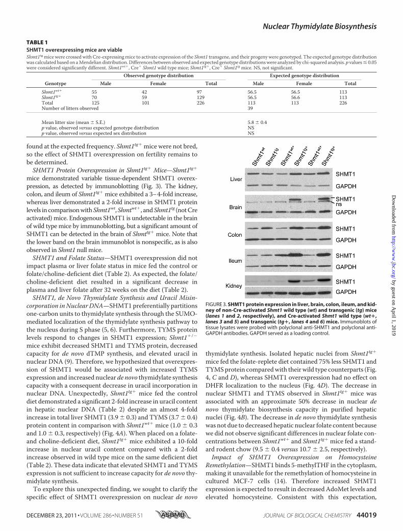
found at the expected frequency. Shmt1tg� mice were not bred,so the effect of SHMT1 overexpression on fertility remains tobe determined.SHMT1 Protein Overexpression in Shmt1tg� Mice—Shmt1tg�
mice demonstrated variable tissue-dependent SHMT1 overex-pression, as detected by immunoblotting (Fig. 3). The kidney,colon, and ileum of Shmt1tg� mice exhibited a 3–4-fold increase,whereas liver demonstrated a 2-fold increase in SHMT1 proteinlevels in comparisonwithShmt1wt,Shmtwt�, andShmt1tg (notCreactivated) mice. Endogenous SHMT1 is undetectable in the brainof wild type mice by immunoblotting, but a significant amount ofSHMT1 can be detected in the brain of Shmttg� mice. Note thatthe lower band on the brain immunoblot is nonspecific, as is alsoobserved in Shmt1 null mice.SHMT1 and Folate Status—SHMT1 overexpression did not
impact plasma or liver folate status in mice fed the control orfolate/choline-deficient diet (Table 2). As expected, the folate/choline-deficient diet resulted in a significant decrease inplasma and liver folate after 32 weeks on the diet (Table 2).SHMT1, de Novo Thymidylate Synthesis and Uracil Misin-
corporation inNuclearDNA—SHMT1preferentially partitionsone-carbon units to thymidylate synthesis through the SUMO-mediated localization of the thymidylate synthesis pathway tothe nucleus during S phase (5, 6). Furthermore, TYMS proteinlevels respond to changes in SHMT1 expression; Shmt1�/�
mice exhibit decreased SHMT1 and TYMS protein, decreasedcapacity for de novo dTMP synthesis, and elevated uracil innuclear DNA (9). Therefore, we hypothesized that overexpres-sion of SHMT1 would be associated with increased TYMSexpression and increased nuclear de novo thymidylate synthesiscapacity with a consequent decrease in uracil incorporation innuclear DNA. Unexpectedly, Shmt1tg� mice fed the controldiet demonstrated a significant 2-fold increase in uracil contentin hepatic nuclear DNA (Table 2) despite an almost 4-foldincrease in total liver SHMT1 (3.9� 0.3) and TYMS (3.7� 0.4)protein content in comparison with Shmt1wt� mice (1.0 � 0.3and 1.0 � 0.3, respectively) (Fig. 4A). When placed on a folate-and choline-deficient diet, Shmt1tg� mice exhibited a 10-foldincrease in nuclear uracil content compared with a 2-foldincrease observed in wild type mice on the same deficient diet(Table 2). These data indicate that elevated SHMT1 and TYMSexpression is not sufficient to increase capacity for de novo thy-midylate synthesis.To explore this unexpected finding, we sought to clarify the
specific effect of SHMT1 overexpression on nuclear de novo
thymidylate synthesis. Isolated hepatic nuclei from Shmt1tg�mice fed the folate-replete diet contained 75% less SHMT1 andTYMSprotein comparedwith theirwild type counterparts (Fig.4, C and D), whereas SHMT1 overexpression had no effect onDHFR localization to the nucleus (Fig. 4D). The decrease innuclear SHMT1 and TYMS observed in Shmt1tg� mice wasassociated with an approximate 50% decrease in nuclear denovo thymidylate biosynthesis capacity in purified hepaticnuclei (Fig. 4B). The decrease in de novo thymidylate synthesiswas not due to decreased hepatic nuclear folate content becausewe did not observe significant differences in nuclear folate con-centrations between Shmt1wt� and Shmt1tg� mice fed a stand-ard rodent chow (9.5 � 0.4 versus 10.7 � 2.5, respectively).Impact of SHMT1 Overexpression on Homocysteine
Remethylation—SHMT1binds 5-methylTHF in the cytoplasm,making it unavailable for the remethylation of homocysteine incultured MCF-7 cells (14). Therefore increased SHMT1expression is expected to result in decreasedAdoMet levels andelevated homocysteine. Consistent with this expectation,
TABLE 1SHMT1 overexpressing mice are viableShmt1tg mice were crossed with Cre-expressingmice to activate expression of the Shmt1 transgene, and their progeny were genotyped. The expected genotype distributionwas calculated based on aMendelian distribution. Differences between observed and expected genotype distributionswere analyzed by chi-squared analysis. p values� 0.05were considered significantly different. Shmt1wt�, Cre� Shmt1 wild type mice; Shmt1tg�, Cre� Shmt1tg mice. NS, not significant.
GenotypeObserved genotype distribution Expected genotype distribution
Male Female Total Male Female Total
Shmt1wt� 55 42 97 56.5 56.5 113Shmt1tg� 70 59 129 56.5 56.6 113Total 125 101 226 113 113 226Number of litters observed 39
Mean litter size (mean � S.E.) 5.8 � 0.4p value, observed versus expected genotype distribution NSp value, observed versus expected sex distribution NS
FIGURE 3. SHMT1 protein expression in liver, brain, colon, ileum, and kid-ney of non-Cre-activated Shmt1 wild type (wt) and transgenic (tg) mice(lanes 1 and 2, respectively), and Cre-activated Shmt1 wild type (wt�,lanes 3 and 5) and transgenic (tg�, lanes 4 and 6) mice. Immunoblots oftissue lysates were probed with polyclonal anti-SHMT1 and polyclonal anti-GAPDH antibodies. GAPDH served as a loading control.
Nuclear Thymidylate Biosynthesis
DECEMBER 23, 2011 • VOLUME 286 • NUMBER 51 JOURNAL OF BIOLOGICAL CHEMISTRY 44019
by guest on April 1, 2019
http://ww
w.jbc.org/
Dow
nloaded from

hepaticAdoMet andAdoMet:AdoHcy ratio, which is indicativeof the cellular methylation capacity, were significantly lower inShmt1tg� mice at 32 weeks post-weaning (Table 2). AdoHcywas unaffected by SHMT1 overexpression.We did not observeany genotype � diet effects on AdoMet, AdoHcy, or the Ado-Met:AdoHcy ratio.The impact of SHMT1 overexpression on homocysteine-
and folate-relatedmetabolites in the plasma ofmale and female
mice was determined at 5 weeks post-weaning. We did notobserve a significant effect of SHMT1 overexpression on any ofthe metabolites queried because of insufficient power to detectsignificant differences. A significant sex effect on plasma hom-ocysteine, methionine, cysteine, and cystathionine wasobserved. Female mice exhibited increased plasma homocys-teine and cysteine and decreased cystathionine andmethioninerelative to male mice. Plasma serine tended to be decreased in
TABLE 2Plasma and liver folate, liver S-adenosyl-methionine, S-adenosyl-homocysteine, S-adenosyl-methionine:S-adenosyl-homocysteine ratio, anduracil content in SHMT1-overexpressing mice at 32 weeks post-weaningDifferences between genotypes and diets were analyzed by Student’s t test. nuclear DNAgenotype� diet effects were analyzed by two-wayANOVAusing Tukey’s HonestlySignificantDifference post-hoc analysis. The data represent themeans� S.E. values. p values� 0.05were considered significantly different. n� 4–7/group. Folate/choline-deficient Shmt1tg�mice are significantly different than control and folate/choline-deficient Shmt1wt� and control Shmt1tg�mice, p� 0.05, as analyzed by two-wayANOVAand Tukey’s Honestly Significant Difference post-hoc test Shmt1wt�, Cre� Shmt1 wild type mice; Shmt1tg�, Cre� Shmt1tg mice. NS, not significant.
Diet Shmt1 genotype Plasma folate Liver folate AdoMet AdoHcy AdoMet:AdoHcy Liver uracil
ng/ml fmol/�gof protein
pmol/�gof protein
pmol/�gof protein
pg of uracil/�gof DNA
AIN-93G Shmt1wt� 36.3 � 7.7 43.1 � 2.3 0.7 � 0.2 0.4 � 0.1 2.1 � 0.4 0.1 � 0.0Shmt1tg� 46.8 � 5.8 51.3 � 3.4 0.3 � 0.2 0.2 � 0.2 1.2 � 0.0 0.2 � 0.0
AIN-93G minus folate & choline Shmt1wt� 7.4 � 0.2 36.1 � 2.6 0.7 � 0.1 0.6 � 0.1 1.2 � 0.2 0.3 � 0.0Shmt1tg� 5.5 � 1.5 34.0 � 5.8 0.4 � 0.0 0.6 � 0.1 0.9 � 0.3 2.3 � 0.7
p value, diet effect �0.0001 �0.0001 NS 0.006 0.04 0.01p value, genotype effect NS NS 0.02 NS 0.04 0.02p value, Diet � genotype effect NS NS NS NS NS 0.031
FIGURE 4. Hepatic nuclear SHMT1, TYMS, and DHFR protein content and de novo dTMP biosynthesis in SHMT1 overexpressing mice. A, immunoblotting forSHMT1, TYMS, and actin (loading control) from whole liver extracts from Cre-activated Shmt1 wild type (Shmt1wt�) and Shmt1tg� mice. B, nuclei were isolated fromShmt1wt� and Shmt1tg� mouse liver, and the capacity to convert dUMP and [2,3-3H]-L-serine to [3H]dTMP was determined in reactions that contained sonicated nuclei,intact nuclei, or intact nuclei incubated with 100 mM aminomethyl phosphonate, an SHMT1 inhibitor (8). De novo thymidylate biosynthesis activity was normalized tothat of Shmt1wt� intact nuclei, which was assigned an arbitrary value of 1.0. The reactions were performed in duplicate, and the experiment was repeated twice. Wedid not observe a run-dependent difference; therefore data from the two replicate experiments were combined. The data are presented as the means � S.E. C andD, immunoblotting for SHMT1, TYMS, and DHFR was performed on hepatic nuclei isolated from Shmt1 null (C only) (3) and Shmt1wt� and Shmt1tg� mice. Lamin A wasused as a nucleus-specific loading control. E, immunoblotting of cytoplasm-restricted GAPDH confirmed the purity of hepatic nuclei.
Nuclear Thymidylate Biosynthesis
44020 JOURNAL OF BIOLOGICAL CHEMISTRY VOLUME 286 • NUMBER 51 • DECEMBER 23, 2011
by guest on April 1, 2019
http://ww
w.jbc.org/
Dow
nloaded from

Shmt1tg� mice, an effect driven by samples from male mice(Table 3). The folate/choline-deficient diet was associated withincreased plasma homocysteine and cysteine and decreasedmethylglycine (Table 3). We did not observe any significantgenotype � diet effects on any of the queried metabolites.
DISCUSSION
Our study demonstrates that nuclear localization of the denovo dTMP synthesis pathway is essential to prevent uracilaccumulation in nuclear DNA and thereby maintain genomestability and confirms previous studies demonstrating thatSHMT1 expression is an important determinant of uracil accu-mulation in DNA. The results demonstrate unequivocally thatrestricting the de novo dTMP synthesis pathway to the cyto-plasm results in elevated uracil accumulation into nuclearDNA.Shmt1tg� mice demonstrated a 2–4-fold increase in SHMT1
and TYMS protein in tissues that normally express SHMT1,namely the liver, kidney, and gastrointestinal tract (Fig. 3). Nosignificant differences in genotype distribution among Cre-ac-tivated F1wild type or transgenic pupswere observed (Table 1),indicating that the achieved level of SHMT1 overexpressionhad no impact on embryo survival in dams fed a standardrodent chow diet. It will be of interest to determine the effect ofa folate deficient diet on Shmt1tg� embryo development,because we have demonstrated folate-responsive neural tubedefects in Shmt1 heterozygous null mice, which was attributedto reduced de novo thymidylate synthesis (10).Shmt1�/� mice have been reported to exhibit increased ura-
cil content in hepatic nuclear DNA, reduced levels of nuclearSHMT1 and TYMS, and consequent decreased capacity for denovo thymidylate synthesis (3, 9). In this study, contrary to
expectations, uracil content in hepatic nuclear DNA wasincreased in Shmt1tg� mice compared with wild type mice,which was exacerbated markedly by a folate- and choline-defi-cient diet. Indeed, hepatic nuclear DNA uracil content inShmt1tg� mice fed the control diet was comparable with thatobserved in Shmt1�/� mice (3, 9). Compared with wild typemice, Shmt1tg� mice exhibited a 4-fold increase in total hepaticSHMT1 and TYMS protein expression, whereas nuclear local-ization of SHMT1 and TYMS was reduced by �75%, resultingin a 50% reduction in dTMP synthesis in isolated nuclei fromfolate-repletemice (Fig. 4). Although Shmt1tg�have severalfoldincreased expression of SHMT1 and TYMS compared withShmt�/� mice, they both exhibit decreased levels of nuclearSHMT1 and TYMS protein, reduced nuclear thymidylate syn-thesis, and elevated uracil in DNA compared with wild typemice. Collectively, these results indicate that nuclear localiza-tion of the de novo thymidylate biosynthesis pathway is essen-tial for maintaining sufficient thymidine nucleotide pools forDNA replication.The mechanism by which SHMT1 and TYMS accumulation
in the nucleus is inhibited in Shmt1tg� mice is not known. Ele-vated expression of SHMT1 in the cytoplasm may impairsumoylation of SHMT1 and/or TYMS or prevent their translo-cation into the nucleus. Interestingly, the accumulation ofSHMT1 and TYMS, but not DHFR, in the nucleus is coordi-nated and dependent on SHMT1 expression.Similar to our previous studies, in which SHMT1-dependent
expression of TYMS was observed in colon and embryonic tis-sue (9, 10), the present study provides additional evidence forthe coregulation of these two proteins. Shmt1�/� mice wereshown to have �50% SHMT1 protein content, which was con-
TABLE 3Plasma metabolic profile of SHMT1 overexpressing mice at 5 weeks post-weaningDifferences between sexes, diets, and genotypes were analyzed by Student’s t test. Genotype � diet effects were analyzed by two-way ANOVA using Tukey’s HonestlySignificant Difference post-hoc analysis. The data are presented as the means � S.E. values. p values � 0.05 were considered significantly different. n � 3 males and n � 3females/diet/genotype group. Shmt1wt�, Cre� Shmt1 wild type mice; Shmt1tg�, Cre� Shmt1tg mice. NS, not significant.
Genotype Shmt1wt� Shmt1tg� p value of model effectMetabolite Sex C FCD C FCD Sex Diet Genotype Diet � genotype
Homocysteine (�M) Both 5.6 � 0.7 7.0 � 0.7 7.8 � 1.3 9.9 � 1.0 0.009 0.007 0.06 NSMale 4.8 � 0.7 5.5 � 0.2 6.6 � 2.3 8.4 � 0.6 0.10 NS NSFemale 6.4 � 1.0 8.4 � 0.3 9.0 � 1.1 11.4 � 1.6 0.03 0.08 NS
Cystathionine (nM) Both 1697 � 237 1687 � 334 1552 � 189 1555 � 70 0.002 NS NS NSMale 1979 � 399 2235 � 483 1931 � 181 1655 � 83 NS NS NSFemale 1414 � 202 1140 � 159 1173 � 54 1455 � 89 NS NS 0.08
Cysteine (�M) Both 171 � 29 200 � 20 214 � 19 219 � 15 �0.0001 0.006 0.10 NSMale 109 � 21 158 � 8 178 � 15 192 � 14 0.01 0.07 NSFemale 232 � 4 242 � 12 251 � 18 246 � 12 NS NS NS
Methionine (�M) Both 39.6 � 5.0 29.3 � 3.9 40.9 � 8.8 31.4 � 1.3 0.03 NS 0.07 NSMale 49.9 � 3.7 34.6 � 5.8 48.1 � 18.2 32.4 � 1.6 NS NS NSFemale 29.3 � 2.2 24.1 � 3.7 33.7 � 1.1 30.4 � 2.2 0.07 NS NS
�-Aminobutyric Acid (�M) Both 6.8 � 1.7 5.0 � 0.5 4.5 � 0.6 4.2 � 0.3 NS NS NS NSMale 5.8 � 1.4 4.6 � 0.4 5.0 � 1.2 4.4 � 0.5 NS NS NSFemale 7.8 � 3.3 5.4 � 1.0 4.0 � 0.3 4.0 � 0.3 NS NS NS
Glycine (�M) Both 312 � 42 286 � 22 270 � 17 289 � 20 0.0006 NS NS NSMale 383 � 59 322 � 32 291 � 23 327 � 6 NS NS NSFemale 240 � 12 249 � 9 249 � 22 250 � 20 NS NS NS
Serine (�M) Both 161 � 21 136 � 11 146 � 18 134 � 9 �0.0001 NS 0.07 NSMale 203 � 17 158 � 8 174 � 25 151 � 6 NS 0.07 NSFemale 119 � 10 113 � 8 119 � 16 117 � 9 NS NS NS
Dimethylglycine (�M) Both 7.8 � 1.7 7.1 � 0.7 6.0 � 0.7 6.2 � 0.8 0.0004 0.10 NS NSMale 5.0 � 0.1 5.6 � 0.4 5.0 � 0.7 4.6 � 0.2 NS NS NSFemale 10.5 � 2.6 8.6 � 0.6 7.0 � 1.1 7.8 � 0.5 NS NS NS
Methylglycine (�M) Both 2.8 � 0.5 2.1 � 0.2 1.4 � 0.1 2.0 � 0.3 NS 0.04 NS 0.06Male 2.3 � 0.5 2.3 � 0.3 1.4 � 0.1 1.6 � 0.1 0.02 NS NSFemale 3.3 � 1.0 1.8 � 0.2 1.3 � 0.1 2.4 � 0.7 NS NS 0.07
Nuclear Thymidylate Biosynthesis
DECEMBER 23, 2011 • VOLUME 286 • NUMBER 51 JOURNAL OF BIOLOGICAL CHEMISTRY 44021
by guest on April 1, 2019
http://ww
w.jbc.org/
Dow
nloaded from

comitant with a reduction in TYMS (9). Interestingly, SHMT1,TYMS, and thymidine kinase 1 protein expression were alsoresponsive to folate deficiency as demonstrated by theirincreased expression in mice fed the folate/choline-deficientdiet (9). Microarray analysis did not indicate significant tran-scriptional changes to TYMS expression in Shmt1�/�, suggest-ing that their coregulation occurs post-transcriptionally (9).We also have not queried the effect of SHMT1 expression onsumoylation of itself or TYMS, a mode by which SHMT1 couldinfluence nuclear translocation. The mechanism by whichSHMT1 expression influences TYMS levels is actively beinginvestigated.This study also confirms that increased SHMT1 expression
plays a modest role in the regulation of homocysteine methyla-tion in the cytoplasm. SHMT1 is a 5-methylTHF binding pro-tein (27), and therefore overexpression of SHMT1 was antici-pated to reduce hepatic methionine and AdoMet synthesis bysequestering cytoplasmic 5-methylTHF. Consistent with thisexpectation, SHMT1 overexpression decreased hepaticAdoMet, which resulted in a reduced AdoMet:AdoHcy ratio(Table 2). SHMT1 overexpression did not have a major impacton serum one-carbon metabolites, because our study wasunderpowered to detect significant differences.We previously demonstrated that the common human
L474F variant impairs the UBC9-SHMT1 interaction and con-sequently SHMT1 sumoylation and translocation to thenucleus (5). The impact of this SHMT1 variant on TYMSnuclear localization and nuclear de novo thymidylate biosyn-thesis capacity is unknown. The reduction of nuclear de novodTMP synthesis capacity in the Shmt1tg� mouse provides aunique experimental model of the human L474F SHMT1 vari-ant and permits mechanistic studies that investigate and vali-date reported epidemiological associations of this variant withlung cancer and cardiovascular risk (11–13).
Acknowledgments—We acknowledge Martha Field, Anna Beaudin,Sylvia Allen, and Rachel Slater for technical assistance.
REFERENCES1. Goulian,M., Bleile, B., and Tseng, B. Y. (1980) Proc. Natl. Acad. Sci. U.S.A.
77, 1956–19602. Blount, B. C., Mack, M. M., Wehr, C. M., MacGregor, J. T., Hiatt, R. A.,
Wang, G., Wickramasinghe, S. N., Everson, R. B., and Ames, B. N. (1997)Proc. Natl. Acad. Sci. U.S.A. 94, 3290–3295
3. MacFarlane, A. J., Liu, X., Perry, C. A., Flodby, P., Allen, R. H., Stabler, S. P.,
and Stover, P. J. (2008) J. Biol. Chem. 283, 25846–258534. Anderson, D. D., Quintero, C.M., and Stover, P. J. (2011) Proc. Natl. Acad.
Sci. U.S.A. 108, 15163–151685. Woeller, C. F., Anderson, D. D., Szebenyi, D. M., and Stover, P. J. (2007)
J. Biol. Chem. 282, 17623–176316. Anderson, D. D., Woeller, C. F., and Stover, P. J. (2007) Clin. Chem. Lab.
Med. 45, 1760–17637. Ching, Y. H.,Munroe, R. J., Moran, J. L., Barker, A. K.,Mauceli, E., Fennell,
T., Dipalma, F., Lindblad-Toh, K., Abcunas, L. M., Gilmour, J. F., Harris,T. P., Kloet, S. L., Luo, Y., McElwee, J. L., Mu,W., Park, H. K., Rogal, D. L.,Schimenti, K. J., Shen, L., Shindo, M., Shou, J. Y., Stenson, E. K., Stover,P. J., and Schimenti, J. C. (2010) BMC Genet. 11, 106–116
8. Anderson, D. D., and Stover, P. J. (2009) PLoS One 4, e58399. MacFarlane, A. J., Perry, C. A., McEntee, M. F., Lin, D.M., and Stover, P. J.
(2011) Cancer Res. 71, 2098–210710. Beaudin, A. E., Abarinov, E. V., Noden, D.M., Perry, C. A., Chu, S., Stabler,
S. P., Allen, R. H., and Stover, P. J. (2011) Am. J. Clin. Nutr 93, 789–79811. Lim, U., Peng, K., Shane, B., Stover, P. J., Litonjua, A. A., Weiss, S. T.,
Gaziano, J. M., Strawderman, R. L., Raiszadeh, F., Selhub, J., Tucker, K. L.,and Cassano, P. A. (2005) J. Nutr. 135, 1989–1994
12. Wernimont, S. M., Raiszadeh, F., Stover, P. J., Rimm, E. B., Hunter, D. J.,Tang, W., and Cassano, P. A. (2011) J. Nutr. 141, 255–260
13. Piskac-Collier, A. L., Monroy, C., Lopez, M. S., Cortes, A., Etzel, C. J.,Greisinger, A. J., Spitz, M. R., and El-Zein, R. A. (2011) Genes Chromo-somes Cancer 50, 1–12
14. Herbig, K., Chiang, E. P., Lee, L. R., Hills, J., Shane, B., and Stover, P. J.(2002) J. Biol. Chem. 277, 38381–38389
15. Perry, C., Sastry, R., Nasrallah, I. M., and Stover, P. J. (2005) J. Biol. Chem.280, 396–400
16. Woeller, C. F., Fox, J. T., Perry, C., and Stover, P. J. (2007) J. Biol. Chem.282, 29927–29935
17. Fox, J. T., Shin,W. K., Caudill, M. A., and Stover, P. J. (2009) J. Biol. Chem.284, 31097–31108
18. Nakshatri, H., Bouillet, P., Bhat-Nakshatri, P., and Chambon, P. (1996)Gene 174, 79–84
19. Novak, A., Guo, C., Yang,W., Nagy, A., and Lobe, C. G. (2000)Genesis 28,147–155
20. Liu, X., Szebenyi, D.M., Anguera,M.C., Thiel, D. J., and Stover, P. J. (2001)Biochemistry 40, 4932–4939
21. Stover, P. J., Chen, L.H., Suh, J. R., Stover, D.M., Keyomarsi, K., and Shane,B. (1997) J. Biol. Chem. 272, 1842–1848
22. Bensadoun, A., and Weinstein, D. (1976) Anal. Biochem. 70, 241–25023. Field, M. S., Szebenyi, D. M., and Stover, P. J. (2006) J. Biol. Chem. 281,
4215–422124. Friso, S., Choi, S.W., Dolnikowski, G. G., and Selhub, J. (2002)Anal Chem.
74, 4526–453125. Stabler, S. P., Lindenbaum, J., Savage, D. G., and Allen, R. H. (1993) Blood
81, 3404–341326. Allen, R. H., Stabler, S. P., and Lindenbaum, J. (1993) Metabolism 42,
1448–146027. Stover, P., and Schirch, V. (1991) J. Biol. Chem. 266, 1543–1550
Nuclear Thymidylate Biosynthesis
44022 JOURNAL OF BIOLOGICAL CHEMISTRY VOLUME 286 • NUMBER 51 • DECEMBER 23, 2011
by guest on April 1, 2019
http://ww
w.jbc.org/
Dow
nloaded from

Allen, Sally P. Stabler and Patrick J. StoverAmanda J. MacFarlane, Donald D. Anderson, Per Flodby, Cheryll A. Perry, Robert H.
Prevent Uracil Accumulation in DNA Thymidylate Biosynthesis Pathway Is Required tode NovoNuclear Localization of
doi: 10.1074/jbc.M111.307629 originally published online November 4, 20112011, 286:44015-44022.J. Biol. Chem.
10.1074/jbc.M111.307629Access the most updated version of this article at doi:
Alerts:
When a correction for this article is posted•
When this article is cited•
to choose from all of JBC's e-mail alertsClick here
http://www.jbc.org/content/286/51/44015.full.html#ref-list-1
This article cites 27 references, 17 of which can be accessed free at
by guest on April 1, 2019
http://ww
w.jbc.org/
Dow
nloaded from

![Ubiquitin and Ubiquitin-like Modifications in Viral ...1].pdf · Ubiquitin and Ubiquitin-like Modifications in Viral Infection and Immunity Abstracts of papers presented at the AUGUST](https://static.fdocuments.us/doc/165x107/5e2d68ba2a69b505b71e58fa/ubiquitin-and-ubiquitin-like-modifications-in-viral-1pdf-ubiquitin-and-ubiquitin-like.jpg)