Catheter-Directed Thrombolysis for Pulmonary...
14
EDITORIAL COMMENT Catheter-Directed Thrombolysis for Pulmonary Embolism Where Do We Stand?* Akhilesh K. Sista, MD,y Clive Kearon, MB, PHDz T here have been 2 main treatments for acute pulmonary embolism (PE)—anticoagulant therapy alone or systemic thrombolytic ther- apy. Although systemic thrombolytic therapy is eff- ective at preventing deaths from PE, it markedly increases bleeding, including intracranial and fatal bleeding (1). The recent PEITHO (Pulmonary Embo- lism Thrombolysis Study) (2), which compared tenec- teplase with placebo in 1,000 PE patients without hypotension but with right ventricular dysfunction, found no clear net benefit from systemic thrombo- lytic therapy; the reduction in cardiovascular collapse (odds ratio: 0.30) was offset by the increase in major bleeding (odds ratio: 5.2). Consequently, systemic thrombolytic therapy is usually reserved for PE pa- tients with hypotension (3). The ability to actively remove thrombus in patients with acute PE without increasing bleeding would be an important advance. Catheter-based therapy has that potential. Catheter-directed thrombolysis (CDT) was initially developed for treatment of arterial, dialysis graft, and deep vein thromboses (leg or arm). When used to treat acute PE, a wire is usually passed through the embolus, followed by placement of a multiside hole infusion catheter through which a thrombolytic drug is infused over 12 to 24 h (4). The delivery of the drug directly into the thrombus is expected to be as effective as systemic therapy but to cause less bleeding because a much lower dose of the drug is used. If more rapid thrombus removal is required, such as in a decompensating patient, fragmentation, balloon maceration, and aspiration may be used as adjunct to CDT or instead of it (i.e., in patients with a high risk of bleeding). These mechanical techniques, however, are avoided in stable patients because they may cause pulmonary artery injury. The addition of an ultrasound-emitting wire to a multiside hole infusion catheter is thought to accelerate thrombol- ysis by ultrasonically disrupting thrombus (5). Although this approach has been used to treat arterial and deep venous thromboses for about 10 years, there is uncertainty that the addition of ultrasound emis- sion increases the efficacy of CDT (6). Based partly on the findings of the SEATTLE II (A Prospective, Single- Arm Multi-Center Trial of EkoSonic Endovascular System and Activase for Treatment of Acute Pulmo- nary Embolism) study, which is reported in this issue of JACC: Cardiovascular Interventions, ultrasound- assisted CDT is now approved by the U.S. Food and Drug Administration for treatment of acute PE (7). SEATTLE II is a single-arm prospective cohort study in which 150 patients with lobar artery or more central PE (31 with and 119 without hypotension) were treated with ultrasound-assisted CDT using a standardized protocol (7). Tissue plasminogen activator was infused into each treated lung at a rate of 1 mg/h, to a total dose of 24 mg (over 12 h for bilateral lung infusions), and no additional mechanical maneuvers were used to disrupt or aspirate thrombus. When computed to- mography pulmonary angiography was repeated after 48 h, the right ventricular to left ventricular ratio was decreased by 27% and thrombus burden was reduced SEE PAGE 1382 *Editorials published in JACC: Cardiovascular Interventions reflect the views of the authors and do not necessarily represent the views of JACC: Cardiovascular Interventions or the American College of Cardiology. From the yDivision of Interventional Radiology, Department of Radi- ology, Weill Cornell Medical College, New York, New York; and the zDepartment of Medicine, Thrombosis and Atherosclerosis Research Institute, McMaster University, Hamilton, Ontario, Canada. Drs. Sista and Kearon have reported that they have no relationships relevant to the contents of this paper to disclose. JACC: CARDIOVASCULAR INTERVENTIONS VOL. 8, NO. 10, 2015 ª 2015 BY THE AMERICAN COLLEGE OF CARDIOLOGY FOUNDATION ISSN 1936-8798/$36.00 PUBLISHED BY ELSEVIER INC. http://dx.doi.org/10.1016/j.jcin.2015.06.009
Transcript of Catheter-Directed Thrombolysis for Pulmonary...

J A C C : C A R D I O V A S C U L A R I N T E R V E N T I O N S V O L . 8 , N O . 1 0 , 2 0 1 5
ª 2 0 1 5 B Y T H E AM E R I C A N C O L L E G E O F C A R D I O L O G Y F O U N DA T I O N I S S N 1 9 3 6 - 8 7 9 8 / $ 3 6 . 0 0
P U B L I S H E D B Y E L S E V I E R I N C . h t t p : / / d x . d o i . o r g / 1 0 . 1 0 1 6 / j . j c i n . 2 0 1 5 . 0 6 . 0 0 9
EDITORIAL COMMENT
Catheter-Directed Thrombolysisfor Pulmonary EmbolismWhere Do We Stand?*
Akhilesh K. Sista, MD,y Clive Kearon, MB, PHDz
SEE PAGE 1382
T here have been 2 main treatments for acutepulmonary embolism (PE)—anticoagulanttherapy alone or systemic thrombolytic ther-
apy. Although systemic thrombolytic therapy is eff-ective at preventing deaths from PE, it markedlyincreases bleeding, including intracranial and fatalbleeding (1). The recent PEITHO (Pulmonary Embo-lism Thrombolysis Study) (2), which compared tenec-teplase with placebo in 1,000 PE patients withouthypotension but with right ventricular dysfunction,found no clear net benefit from systemic thrombo-lytic therapy; the reduction in cardiovascular collapse(odds ratio: 0.30) was offset by the increase in majorbleeding (odds ratio: 5.2). Consequently, systemicthrombolytic therapy is usually reserved for PE pa-tients with hypotension (3). The ability to activelyremove thrombus in patients with acute PE withoutincreasing bleeding would be an important advance.Catheter-based therapy has that potential.
Catheter-directed thrombolysis (CDT) was initiallydeveloped for treatment of arterial, dialysis graft, anddeep vein thromboses (leg or arm). When used totreat acute PE, a wire is usually passed through theembolus, followed by placement of a multiside holeinfusion catheter through which a thrombolyticdrug is infused over 12 to 24 h (4). The delivery of thedrug directly into the thrombus is expected to be as
*Editorials published in JACC: Cardiovascular Interventions reflect the
views of the authors and do not necessarily represent the views of JACC:
Cardiovascular Interventions or the American College of Cardiology.
From the yDivision of Interventional Radiology, Department of Radi-
ology, Weill Cornell Medical College, New York, New York; and the
zDepartment of Medicine, Thrombosis and Atherosclerosis Research
Institute, McMaster University, Hamilton, Ontario, Canada. Drs. Sista and
Kearon have reported that they have no relationships relevant to the
contents of this paper to disclose.
effective as systemic therapy but to cause lessbleeding because a much lower dose of the drug isused. If more rapid thrombus removal is required,such as in a decompensating patient, fragmentation,balloon maceration, and aspiration may be used asadjunct to CDT or instead of it (i.e., in patients with ahigh risk of bleeding). These mechanical techniques,however, are avoided in stable patients becausethey may cause pulmonary artery injury. The additionof an ultrasound-emitting wire to a multiside holeinfusion catheter is thought to accelerate thrombol-ysis by ultrasonically disrupting thrombus (5).Although this approach has been used to treat arterialand deep venous thromboses for about 10 years, thereis uncertainty that the addition of ultrasound emis-sion increases the efficacy of CDT (6). Based partly onthe findings of the SEATTLE II (A Prospective, Single-Arm Multi-Center Trial of EkoSonic EndovascularSystem and Activase for Treatment of Acute Pulmo-nary Embolism) study, which is reported in this issueof JACC: Cardiovascular Interventions, ultrasound-assisted CDT is now approved by the U.S. Food andDrug Administration for treatment of acute PE (7).
SEATTLE II is a single-arm prospective cohort studyin which 150 patients with lobar artery or more centralPE (31 with and 119 without hypotension) were treatedwith ultrasound-assisted CDT using a standardizedprotocol (7). Tissue plasminogen activator was infusedinto each treated lung at a rate of 1 mg/h, to a totaldose of 24 mg (over 12 h for bilateral lung infusions),and no additional mechanical maneuvers were used todisrupt or aspirate thrombus. When computed to-mography pulmonary angiography was repeated after48 h, the right ventricular to left ventricular ratio wasdecreased by 27% and thrombus burden was reduced

Sista and Kearon J A C C : C A R D I O V A S C U L A R I N T E R V E N T I O N S V O L . 8 , N O . 1 0 , 2 0 1 5
Catheter-Directed Thrombolysis for Pulmonary Embolism A U G U S T 2 4 , 2 0 1 5 : 1 3 9 3 – 5
1394
by 30%. Pulmonary artery pressure also decreased by27% between the start to the end of CDT. These 3 im-provements were each highly statistically significant.There were 17 episodes of major bleeding in 15 patients(10%): one was associated with hypotension; all re-quired transfusion; none was intracranial; and nonewas fatal. Strengths of the SEATTLE II study include itsprospective design, inclusion of 150 patients, highpatient retention, involvement of many clinical cen-ters, standardized treatment protocol, and rigorousreporting. Limitations include that there was nocomparison group (neither anticoagulation alone norsystemic thrombolytic therapy), short-term surrogateoutcomes were used to assess efficacy, and that long-term outcomes such as quality of life or exercise ca-pacity were not assessed. SEATTLE II also did notassess whether ultrasound-assisted CDT was moreeffective than standard CDT.
So, how effective and safe is CDT? The short-term improvements in right ventricular dimensions,thrombus burden, and pulmonary hypertension inSEATTLE II are consistent with the improvementin right ventricular dimension with ultrasound-facilitated CDT in 35 patients in the ULTIMA (Ultra-sound Accelerated Thrombolysis of PulmonaryEmbolism) study (8). In ULTIMA, there was almost noimprovement in this outcome at 24 h in the 35 pa-tients who were randomized to anticoagulant therapyalone, and the difference between the CDT and anti-coagulant therapy alone groups was highly statisti-cally significant. A recently published prospectiveregistry of 101 patients with acute PE (PERFECT[Pulmonary Embolism Response to Fragmentation,Embolectomy, and Catheter Thrombolysis]) (9) re-ported similar efficacy of CDT, with or withoutultrasound-assisted thrombolysis, to ULTIMA andSEATTLE. There was no major bleeding in this re-gistry, although follow-up may not have been asstandardized as the follow-up for the SEATTLE IIand ULTIMA studies. The improvements in imagingand pulmonary artery pressure with ultrasound-assisted CDT in SEATTLE II and ULTIMA also appearto be at least as marked as the short-term improve-ments in these outcomes with systemic thrombolytictherapy (10). Therefore, based mostly on indirect
comparisons of short-term surrogate outcomes, CDTappears to be effective compared with anticoagulanttherapy alone and probably is as effective as systemicthrombolytic therapy, which uses much higher doseof the thrombolytic drug. The frequency of majorbleeding in SEATTLE II, however, suggests that CDTmay be associated with substantially more bleedingthan anticoagulation alone. Although it is encour-aging that there were no intracranial bleeds in the 150patients in SEATTLE II, the upper boundary of the95% confidence interval on this estimate is 2.4%.Although it seems likely that there is a lower risk ofnonprocedural bleeding with CDT than with systemicthrombolytic therapy, this remains uncertain.
What then is the role of CDT in patients with PE?We think that current evidence suggests that CDT ispreferred to systemic thrombolytic therapy in pa-tients with acute PE who require active thrombusremoval and have risk factors for bleeding. We sug-gest that venous puncture for CDT should always beultrasound-guided and that the total dose of throm-bolytic drug should be kept to a minimum in patientswith a high risk of bleeding (3). If there is need foractive thrombus removal in patients with a veryhigh risk of bleeding, it may be necessary to usecatheter-based therapy without thrombolytic drugor to use surgical embolectomy. We are not readyto encourage use of CDT in preference to anti-coagulation alone in stable patients with acute PEand right ventricular dysfunction. We suggest thatthere is a need for evidence that the short- and long-term benefits of CDT outweigh the associated risk ofbleeding before CDT can be recommended for suchpatients. We encourage randomized trials thatcompare CDT with systemic thrombolytic therapy inunstable patients with PE and compare CDT withanticoagulation alone in stable patients who havelarge PE and right ventricular dysfunction. Evidencefrom such studies would place the role of CDT for PEon a firmer footing.
REPRINT REQUESTS AND CORRESPONDENCE: Dr.Clive Kearon, Juravinski Hospital, Room A3-73, 711Concession Street, Hamilton, Ontario L8V 1C3,Canada. E-mail: [email protected].
RE F E RENCE S
1. Chatterjee S, Chakraborty A, Weinberg I, et al.Thrombolysis for pulmonary embolism and risk of all-cause mortality, major bleeding, and intracranial he-morrhage: a meta-analysis. JAMA 2014;311:2414–21.
2. Meyer G, Vicaut E, Danays T, et al., for thePEITHO Investigators. Fibrinolysis for patients
with intermediate-risk pulmonary embolism.N Engl J Med 2014;370:1402–11.
3. Konstantinides SV, Torbicki A, Agnelli G, et al.,for the Task Force for the Diagnosis and Manage-ment of Acute Pulmonary Embolism of the Euro-pean Society of Cardiology. 2014 ESC guidelines
on the diagnosis and management of acute pul-monary embolism. Eur Heart J 2014;35:3033–69,3069a–k.
4. Kuo WT. Endovascular therapy for acute pul-monary embolism. J Vasc Interv Radiol 2012;23:167–79.e164, quiz 179.

J A C C : C A R D I O V A S C U L A R I N T E R V E N T I O N S V O L . 8 , N O . 1 0 , 2 0 1 5 Sista and KearonA U G U S T 2 4 , 2 0 1 5 : 1 3 9 3 – 5 Catheter-Directed Thrombolysis for Pulmonary Embolism
1395
5. Braaten JV, Goss RA, Francis CW. Ultrasoundreversibly disaggregates fibrin fibers. ThrombHaemost 1997;78:1063–8.
6. Engelberger RP, Spirk D, Willenberg T, et al.Ultrasound-assisted versus conventional catheter-directed thrombolysis for acute iliofemoral deepvein thrombosis. Circ Cardiovasc Interv 2015;8:piie002027.
7. Piazza G, Hohlfelder B, Jaff MR, et al.A prospective, single-arm, multicenter trial ofultrasound-facilitated, catheter-directed, low-dose
fibrinolysis for acute massive and submassive pul-monary embolism: The SEATTLE II study. J Am CollCardiol Intv 2015;8:1382–92.
8. Kucher N, Boekstegers P, Muller OJ, et al.Randomized, controlled trial of ultrasound-assisted catheter-directed thrombolysis for acuteintermediate-risk pulmonary embolism. Circula-tion 2014;129:479–86.
9. Kuo WT, Banerjee A, Kim PS, et al. PulmonaryEmbolism Response to Fragmentation, Embolec-tomy, and Catheter Thrombolysis (PERFECT):
initial results from a prospective multicenter reg-istry. Chest 2015 Apr 9 [E-pub ahead of print].
10. Dong B, Jirong Y, Wang Q, Wu T.Thrombolytic treatment for pulmonary embo-lism. Cochrane Database Syst Rev 2006;(2):CD004437.
KEY WORDS catheter-directed, endovsaculartherapy, pulmonary embolism, thrombolysis,treatment

J A C C : C A R D I O V A S C U L A R I N T E R V E N T I O N S V O L . 8 , N O . 1 0 , 2 0 1 5
ª 2 0 1 5 B Y T H E A M E R I C A N C O L L E G E O F C A R D I O L O G Y F O U N D A T I O N I S S N 1 9 3 6 - 8 7 9 8 / $ 3 6 . 0 0
P U B L I S H E D B Y E L S E V I E R I N C . h t t p : / / d x . d o i . o r g / 1 0 . 1 0 1 6 / j . j c i n . 2 0 1 5 . 0 4 . 0 2 0
A Prospective, Single-Arm, MulticenterTrial of Ultrasound-Facilitated,Catheter-Directed, Low-Dose Fibrinolysisfor Acute Massive and SubmassivePulmonary EmbolismThe SEATTLE II Study
Gregory Piazza, MD, MS,1 Benjamin Hohlfelder, PHARMD,1 Michael R. Jaff, DO,2 Kenneth Ouriel, MD,3
Tod C. Engelhardt, MD,4 Keith M. Sterling, MD,5 Noah J. Jones, MD,6 John C. Gurley, MD,7 Rohit Bhatheja, MD,8
Robert J. Kennedy, MD,9 Nilesh Goswami, MD,10 Kannan Natarajan, MD,11 John Rundback, MD,12
Immad R. Sadiq, MD,13 Stephen K. Liu, MD,14 Narinder Bhalla, MD,15 M. Laiq Raja, MD,16 Barry S. Weinstock, MD,17
Jacob Cynamon, MD,18 Fakhir F. Elmasri, MD,19 Mark J. Garcia, MD,20 Mark Kumar, MD,21 Juan Ayerdi, MD,22
Peter Soukas, MD,23 William Kuo, MD,24 Ping-Yu Liu, PHD,25 Samuel Z. Goldhaber, MD,2
for the SEATTLE II Investigators
ABSTRACT
Fro
Ma
Bo
OBJECTIVES This study conducted a prospective, single-arm, multicenter trial to evaluate the safety and efficacy of
ultrasound-facilitated, catheter-directed, low-dose fibrinolysis, using the EkoSonic Endovascular System (EKOS, Bothell,
Washington).
BACKGROUND Systemic fibrinolysis for acute pulmonary embolism (PE) reduces cardiovascular collapse but causes
hemorrhagic stroke at a rate exceeding 2%.
METHODS Eligible patients had a proximal PE and a right ventricular (RV)-to-left ventricular (LV) diameter
ratio $0.9 on chest computed tomography (CT). We included 150 patients with acute massive (n ¼ 31) or submassive
(n ¼ 119) PE. We used 24 mg of tissue-plasminogen activator (t-PA) administered either as 1 mg/h for 24 h with a
unilateral catheter or 1 mg/h/catheter for 12 h with bilateral catheters. The primary safety outcome was major bleeding
within 72 h of procedure initiation. The primary efficacy outcome was the change in the chest CT–measured RV/LV
diameter ratio within 48 h of procedure initiation.
RESULTS Mean RV/LV diameter ratio decreased from baseline to 48 h post-procedure (1.55 vs. 1.13; mean
difference, �0.42; p < 0.0001). Mean pulmonary artery systolic pressure (51.4 mm Hg vs. 36.9 mm Hg; p < 0.0001) and
modified Miller Index score (22.5 vs. 15.8; p < 0.0001) also decreased post-procedure. One GUSTO (Global Utilization of
Streptokinase and Tissue Plasminogen Activator for Occluded Coronary Arteries)–defined severe bleed (groin hematoma
with transient hypotension) and 16 GUSTO-defined moderate bleeding events occurred in 15 patients (10%). No patient
experienced intracranial hemorrhage.
CONCLUSIONS Ultrasound-facilitated, catheter-directed, low-dose fibrinolysis decreased RV dilation, reduced pul-
monary hypertension, decreased anatomic thrombus burden, and minimized intracranial hemorrhage in patients with
acute massive and submassive PE. (A Prospective, Single-arm, Multi-center Trial of EkoSonic� Endovascular System and
Activase for Treatment of Acute Pulmonary Embolism (PE) [SEATTLE II]; NCT01513759) (J Am Coll Cardiol Intv
2015;8:1382–92) © 2015 by the American College of Cardiology Foundation.
m the 1Cardiovascular Division, Department of Medicine, Brigham and Women’s Hospital, Harvard Medical School, Boston,
ssachusetts; 2Cardiovascular Division, Department of Medicine, Massachusetts General Hospital, Harvard Medical School,
ston, Massachusetts; 3Syntactx, New York, New York; 4East Jefferson General Hospital, Metairie, Louisiana; 5Cardiovascular

AB BR E V I A T I O N S
AND ACRONYM S
CT = computed tomography
DVT = deep vein thrombosis
PE = pulmonary embolism
RV = right ventricular
RV/LV = right ventricular-to-
left ventricular
t-PA = tissue-plasminogen
activator
J A C C : C A R D I O V A S C U L A R I N T E R V E N T I O N S V O L . 8 , N O . 1 0 , 2 0 1 5 Piazza et al.A U G U S T 2 4 , 2 0 1 5 : 1 3 8 2 – 9 2 Ultrasound-Facilitated, Low-Dose Fibrinolysis for Acute PE
1383
T he Surgeon General estimates that 100,000to 180,000 deaths occur annually from pul-monary embolism (PE) in the United States
(1). Advanced therapies, such as systemic fibrinolysisand embolectomy, have the potential to reduce rightventricular (RV) pressure overload in massive (2)and submassive PE (3,4) and lower mortality byreversing pulmonary arterial obstruction and RVfailure.
In the largest study of full-dose systemic fibrino-lysis, tenecteplase reduced the risk of death or car-diovascular collapse by 56% in 1,006 submassive PEpatients (5). However, this benefit was offset by anearly 5-fold increased risk of major bleeding and a10-fold increased risk of hemorrhagic stroke. Meta-analyses of trials of systemic fibrinolysis for acutePE have demonstrated similar findings (6,7).
SEE PAGE 1393
Concern over the risk of intracranial hemorrhage,which approaches 3% to 5% outside of clinical trials(8,9), has dampened clinician enthusiasm for full-dose systemic fibrinolysis and has sparked devel-opment of alternative advanced therapies withlower bleeding risk. Pharmacomechanical catheter-directed therapy combines fibrinolytic therapy withmechanical disruption of the thrombus (10–12). Thehigh-frequency, low-power ultrasound componentof ultrasound-facilitated, catheter-directed, low-dose fibrinolysis is hypothesized to disaggregatefibrin fibers, potentially allowing greater penetra-tion of the fibrinolytic agent (13). This therapeutic
and Interventional Associates, INOVA Alexandria Hospital, Alexandra, Virgin
Heart Institute, University of Kentucky, Lexington, Kentucky; 8Florida Hear
Regional Medical Center, Melbourne, Florida; 10Prairie Heart Institute, St.
Medical Group, Indianapolis, Indiana; 12Holy Name Medical Center, Teaneck
Hospital, Hartford, Connecticut; 14Lifelink Interventional Center, Memorial
Cardiology Associates, Baptist Medical Center, Montgomery, Alabama; 16El P
Hospital and Sierra Medical Hospital, El Paso, Texas; 17Leesburg Regional Med
InterventionRadiology,DepartmentofRadiology,MontefioreMedicalCenter,
of Lakeland, Lakeland Regional Medical Center, Lakeland, Florida; 20Christian
Delaware; 21The Cardiovascular Care Group, Overlook Medical Center, Sum
Medical Center of Georgia, Macon, Georgia; 23Miriam Cardiology Inc., The M
University, Stanford, California; and the 25FredHutchinsonCancer Center, Sea
grant fromEKOS, aBTG International group company. Dr. Piazza receives resea
Sankyo, and Janssen. Dr. Jaff is a noncompensatedmember of theData Safety a
VIVA Physicians, a 501(c)(3) not-for-profit education and research organizati
Syntactx,which receives fees for core laboratory activities fromEKOS.Dr. Jone
of Medtronic and EKOS; and receives consulting fees from Cordis. Drs. Engelh
Natarajan,Rundback, Sadiq, S.K.Liu,Bhalla,Raja,Weinstock, Cynamon, Elmas
has received research support from EKOS, a BTG International Group compa
Dr. Bhatheja is a speaker for St. Jude Medical and Cardiovascular Systems In
Dr. Gurley is a speaker for EKOS. Dr.Weinstock is a consultant forW.L. Gore. D
relevant to the contents of this paper to disclose. The results of this prospectiv
stract form on March 30, 2014 at the American College of Cardiology Annual S
Manuscript received April 17, 2015; accepted April 23, 2015.
strategy requires only a fraction of the sys-temic fibrinolytic dose. This dose reductionmight improve the safety of fibrinolysis forPE. In a randomized, controlled trial of59 patients with submassive PE in Europe,there were no major bleeding complica-tions with ultrasound-facilitated, catheter-directed, low-dose fibrinolysis, which rapidlyimproved RV function compared with anti-coagulation alone (14).
To expand our understanding of the
efficacy and safety of ultrasound-facilitated, catheter-directed, low-dose fibrinolysis to reverse RV dysfunc-tion in acute massive or submassive PE, we conducteda prospective, single-arm, multicenter trial (CentralIllustration) (NCT01513759).METHODS
STUDY DESIGN. From June 2012 to February 2013, wescreened 159 hospitalized patients with acute massiveor submassive PE and enrolled 150 in this prospective,single-arm trial of ultrasound-facilitated, catheter-directed, low-dose fibrinolysis (Figure 1). Study pa-tients were recruited at 22 sites across the UnitedStates, including urban, nonurban, teaching, andnonteaching hospitals. Institutional review boardapproval was obtained at all sites, and writteninformed consent was obtained for every patient. Theresults of this prospective, single-arm, multicentertrial were presented in abstract form on March 30,2014, at the American College of Cardiology Annual
ia; 6Mt. Carmel East Hospital, Columbus, Ohio; 7Gill
t Group, Florida Hospital, Orlando, Florida; 9Holmes
John’s Hospital, Springfield, Illinois; 11St. Vincent
, New Jersey; 13Vascular Medicine Division, Hartford
Medical Center, Modesto, California; 15River Region
aso Cardiology Associates, PA, Providence Memorial
ical Center, Leesburg, Florida; 18Division of Vascular
Bronx,NewYork; 19Radiologyand ImagingSpecialists
a Care Center for Heart and Vascular Health, Newark,
mit, New Jersey; 22Macon Cardiovascular Institute,
iriam Hospital, Providence, Rhode Island; 24Stanford
ttle,Washington. This studywas fundedby a research
rch support fromEKOS,Bristol-Myers Squibb, Daiichi-
ndMonitoring Board of EKOS and a Boardmember of
on. Dr. Ouriel holds equity in and is an employee of
s receives honoraria for serving on the Speaker Bureau
ardt, Sterling, Gurley, Bhatheja, Kennedy, Goswami,
ri,Garcia,Kumar,Ayerdi, Soukas,Kuo, andGoldhaber
ny. Dr. P.-Y. Liu receives consulting fees from EKOS.
c. Dr. Goswami is a consultant for Boston Scientific.
r. Hohfelder has reported that he has no relationships
e, single-arm, multicenter trial were presented in ab-
cientific Sessions in Washington, DC.

CENTRAL ILLUSTRATION Ultrasound-Facilitated, Catheter-Directed, Low-Dose Fibrinolysis for Acute Massiveand Submassive Pulmonary Embolism
A total of 150 patients with computed tomography (CT)-confirmed pulmonary embolism (PE), symptoms for 14 days or less, and a right
ventricular (RV)/left ventricular (LV) diameter ratio of at least 0.9 underwent ultrasound-facilitated, catheter-directed, low-dose fibrinolysis
according to a standardized protocol. Ultrasound-facilitated, catheter-directed, low-dose fibrinolysis for acute massive and submassive
pulmonary embolism (PE) improved RV function, decreased pulmonary hypertension, and minimized intracranial hemorrhage. PA ¼ pulmonary
artery; tPA ¼ tissue plasminogen.
Piazza et al. J A C C : C A R D I O V A S C U L A R I N T E R V E N T I O N S V O L . 8 , N O . 1 0 , 2 0 1 5
Ultrasound-Facilitated, Low-Dose Fibrinolysis for Acute PE A U G U S T 2 4 , 2 0 1 5 : 1 3 8 2 – 9 2
1384
Scientific Sessions in Washington, DC. The trial wasdesigned and led by an Executive Committee (G.P.and S.Z.G.) from the Study Coordinating Center. TheExecutive Committee established the data analysisplan and participated in the statistical analysis.
STUDY POPULATION. Patients were potentiallyeligible to participate if they had a proximal PE (fillingdefect in at least 1 main or lobar pulmonary artery),age 18 years or older, PE symptom duration #14 days,and RV/LV diameter ratio $0.9 on contrast-enhancedchest computed tomography (CT). We includedpatients with massive (defined as syncope, systemicarterial hypotension, cardiogenic shock, or resusci-tated cardiac arrest) or submassive (defined as anormotensive patient with PE and evidence of RVdysfunction) PE. Major exclusion criteria were strokeor transient ischemic attack, head trauma, or other
active intracranial or intraspinal disease within12 months; major surgery within 7 days; recent activebleeding from a major organ; hematocrit <30%;platelets <100,000/ml; International NormalizedRatio >3; serum creatinine >2 mg/dl; and systolicblood pressure <80 mm Hg despite vasopressor orinotropic support (Online Appendix). Obesity wasdefined as a clinical diagnosis of obesity in the med-ical record.
ANTICOAGULATION. Anticoagulation was initiatedwith full-dose intravenous unfractionated heparinwith a target activated partial thromboplastin time of60 to 80 s. For patients who had already receivedlow-molecular weight heparin or fondaparinux, theinitiation of intravenous unfractionated heparin wasdelayed by 12 h. After completion of the procedure, allsubjects received full anticoagulation. The drug,

FIGURE 1 Flow of Patients Through Each Stage of the Trial
One hundred fifty nine patients with acute pulmonary embolism (PE) were screened for eligibility across 22 study sites. One hundred fifty
patients were eligible for enrollment. One patient died before the ultrasound-facilitated, catheter-directed, low-dose fibrinolytic procedure
could be completed. One hundred fifteen patients had a within-window contrast-enhanced chest computed tomogram (CT) for measurement of
change in right ventricular-to-left ventricular (RV/LV) diameter ratio. Ninety eight patients had a within-window echocardiogram to estimate
48-h pulmonary artery (PA) systolic pressure. t-PA ¼ tissue-plasminogen activator.
J A C C : C A R D I O V A S C U L A R I N T E R V E N T I O N S V O L . 8 , N O . 1 0 , 2 0 1 5 Piazza et al.A U G U S T 2 4 , 2 0 1 5 : 1 3 8 2 – 9 2 Ultrasound-Facilitated, Low-Dose Fibrinolysis for Acute PE
1385
dose, frequency, and duration of anticoagulationwere selected by the attending physician.
ULTRASOUND-FACILITATED, CATHETER-DIRECTED, LOW-
DOSE FIBRINOLYSIS. The EkoSonic EndovascularSystem (EKOS, Bothell, Washington) comprises3 components: the Intelligent Drug Delivery Catheter,a removable MicroSonic device, and a reusable Eko-Sonic control unit. The procedure was performed by anexperienced operator from Interventional Cardiology,Interventional Radiology, Vascular Surgery, or Car-diothoracic Surgery. Venous access was obtained,most often with ultrasound guidance, via the commonfemoral or internal jugular vein. After catheter place-ment but before activation of ultrasound and infusionof fibrinolytic therapy, baseline right heart pressureswere transduced through the catheters. Because thecatheters are placed in or adjacent to the PE, thefibrinolytic agent is delivered directly to the thrombus.
The fixed-dose regimen of tissue-plasminogenactivator (t-PA) (Genentech, South San Francisco,California) was 24 mg at 1 mg/h with saline coolant at35 ml/h for both unilateral and bilateral PEs. Thefibrinolytic agent is not infused under pressure, butrather drips out of microscopic side holes. For pa-tients with predominantly unilateral PE, a singlecontinuous catheter-directed pulmonary artery infu-sion of t-PA was used for 24 h. For patients withbilateral PE, 2 drug delivery devices were placed, and
a continuous infusion of t-PA was administered ineach catheter for 12 h. No adjunctive interventionaltechniques to assist thrombus removal or dissolutionwere permitted. During the procedure, intravenousunfractionated heparin was continued at intermedi-ate intensity with a target aPTT of 40 to 60 s. Afterremoval of the drug delivery device(s), the access sitewas manually compressed for at least 5 min. Fifteenminutes after achieving hemostasis, full therapeuticanticoagulation was restarted.
FOLLOW-UP. After completion of the procedure butbefore catheter removal, right heart pressures weremeasured. Right heart pressure measurements weretransduced once before activation of ultrasound andinfusion of fibrinolytic therapy and once aftercompletion of the procedure. The post-procedureinvasive hemodynamic assessment was performedat either 12 or 24 h after the initiation of fibrinolysis.In the 86% of patients with bilateral PEs whounderwent ultrasound-facilitated, catheter-directed,low-dose fibrinolysis via bilateral catheters, the post-procedure hemodynamic assessment was performedat 12 h after initiation of the procedure, before cath-eter removal. For the remaining patients who hadunilateral ultrasound-facilitated, catheter-directed,low-dose fibrinolysis via a single catheter, the post-procedure hemodynamic assessment was performedat 24 h after initiation of the procedure, before

TABLE 1 Baseline Demographics and Clinical Characteristics
(N ¼ 150)
Patient
Age, yrs 59 � 16.1
Body mass index, kg/m2 35.6 � 9.1
Female 77 (51.3)
Ethnicity/race
Caucasian 119 (79.3)
African American 22 (14.7)
Hispanic or Latino 9 (6)
Comorbid conditions
Obesity 82 (54.7)
Concomitant use of antiplatelet medications 51 (34)
Immobility within 30 days of PE 45 (30)
Diabetes mellitus 42 (28)
Family history of venous thromboembolism 31 (20.7)
Previous DVT 30 (20)
Active tobacco use 27 (18)
Active infectious illness within 30 days of PE 20 (13.3)
Atherosclerotic cardiovascular disease 18 (12)
Previous PE 15 (10)
Values are mean � SD or n (%).
DVT ¼ deep vein thrombosis; PE ¼ pulmonary embolism.
TABLE 2 Characteristics of PE and Initial Anticoagulation
(N ¼ 150)
Any symptoms of PE 150 (100)
Duration of symptoms, days
#14 149 (99.3)
>14 1 (0.7)
PE subtype
Submassive 119 (79.3)
Massive 31 (20.7)
Pre-procedure anticoagulation*
Intravenous unfractionated heparin 76 (50.7)
Enoxaparin 54 (36)
Warfarin 16 (10.7)
Other 7 (4.7)
None 24 (16)
Values are n (%). *Patients could have received more than 1 anticoagulant.
PE ¼ pulmonary embolism.
Piazza et al. J A C C : C A R D I O V A S C U L A R I N T E R V E N T I O N S V O L . 8 , N O . 1 0 , 2 0 1 5
Ultrasound-Facilitated, Low-Dose Fibrinolysis for Acute PE A U G U S T 2 4 , 2 0 1 5 : 1 3 8 2 – 9 2
1386
catheter removal. Multiple right heart pressure mea-surements or “averaging” values were not allowedfor assessment of baseline and post-procedure pres-sures. Follow-up contrast-enhanced chest CT andtransthoracic echocardiography were performedwithin 48 � 6 h after initiation of the procedure. Thechanges in the RV/LV diameter ratio and modifiedMiller angiographic obstruction index score (15) wereassessed by the contrast-enhanced chest CT per-formed at baseline and at 48 h. The maximal modifiedMiller Index score is 40, corresponding with occlu-sion of all pulmonary artery segments. Echocardiog-raphy was performed to estimate pulmonary arterysystolic pressure at 48 � 6 h. All CT scans and echo-cardiograms were analyzed at a dedicated, blindedimaging core laboratory (Syntactx, New York, NewYork).
Bleeding complications were assessed for 72 h afterthe procedure. Patients were assessed clinically forrecurrent, symptomatic PE at 30 days after the pro-cedure. Recurrent PE was defined as symptomaticand objectively confirmed with contrast-enhancedchest CT, ventilation-perfusion lung scanning, orinvasive contrast pulmonary angiography. Overall,149 of 150 patients (99.3%) completed the requiredclinical follow-up.OUTCOMES. The primary efficacy outcome was thecore laboratory–measured change in the RV/LVdiameter ratio from baseline, as assessed by contrast-enhanced chest CT at baseline and at 48 � 6 h afterinitiation of procedure (16). A secondary efficacyoutcome was the change in pulmonary artery systolicpressure, as assessed by baseline right heart cathe-terization compared with measurements obtained atthe conclusion of the procedure and as estimated bytransthoracic echocardiography at 48 h. An additionalsecondary outcome was the change in the modifiedMiller angiographic obstruction index score, asassessed by the baseline contrast-enhanced chest CTand the follow-up scan performed at 48 h.
The primary safety outcome was major bleedingwithin 72 h of initiation of the procedure. Bleedingevents were classified by the GUSTO (Global Utiliza-tion of Streptokinase and Tissue Plasminogen Acti-vator for Occluded Coronary Arteries) bleeding criteria(Online Appendix) (17). Severe or life-threateningbleeding was defined as either intracranial hemor-rhage or bleeding that caused hemodynamic compro-mise and required intervention. Moderate bleedingrequired blood transfusion but did not result inhemodynamic compromise. Mild bleeding did notmeet criteria for either severe/life-threatening ormoderate bleeding. We defined major bleeding aseither a GUSTO moderate or a GUSTO severe/life-
threatening bleeding event. All monitoring for majorbleeding within 72 h was performed during thehospitalization.
The secondary safety outcomes included symp-tomatic recurrent PE up to 30 days after the initiationof the procedure, all-cause mortality at hospitaldischarge and through 30 days, and technical proce-dural complications. Mortality was further classifiedas related to cancer, myocardial infarction, PE, orother causes. Death was attributed to PE if it waseither sudden or unsuspected or if there was evidenceto support an association with PE. All safety out-comes, including bleeding complications, were

TABLE 3 Procedural Characteristics
Total dose of t-PA, mg* 23.7 � 2.9
Successful device placement† 278 (97.5)
Access sites‡
Right femoral vein 177 (63.7)
Left femoral vein 61 (21.9)
Right internal jugular vein 31 (11.2)
Other 9 (3.2)
No. of devices per patient*
0 1 (0.7)
1 20 (13.3)
2 129 (86)
Completed infusion of t-PA‡ 272 (97.8)
Values are mean � SD or n (%). *N ¼ 150 patients (1 patient died before devicescould be placed). †Devices attempted ¼ 285. ‡Devices placed ¼ 278.
t-PA ¼ tissue-plasminogen activator.
TABLE 4 Efficacy Outcomes*
Outcome BaselineAt ProcedureCompletion
AbsoluteDifference
48 h AfterProcedure
AbsoluteDifference
PA systolic pressure,mm Hg
51.4 � 16 37.5 � 11.9† �14 � 15 36.9 � 14.9‡ �14.4 � 15.4
RV/LV diameter ratio 1.55 � 0.39 – – 1.13 � 0.2§ �0.42 � 0.36
Modified Miller Index 22.5 � 5.7 – – 15.8 � 5.9k �6.6 � 6.3
Values are mean � SD. *All p values <0.0001. †Patients with complete right heart catheterization data ¼ 147.‡Patients with transthoracic echocardiogram performed within both the baseline and the 48 � 6-h window¼ 98.§Patients with contrast-enhanced chest computed tomography performed within both the baseline and the48 � 6-h window ¼ 115. kPatients with contrast-enhanced chest computed tomography performed within boththe baseline and the 48 � 6-h window ¼ 116.
PA ¼ pulmonary artery; RV/LV ¼ right ventricular-to-left ventricular.
J A C C : C A R D I O V A S C U L A R I N T E R V E N T I O N S V O L . 8 , N O . 1 0 , 2 0 1 5 Piazza et al.A U G U S T 2 4 , 2 0 1 5 : 1 3 8 2 – 9 2 Ultrasound-Facilitated, Low-Dose Fibrinolysis for Acute PE
1387
adjudicated by a designated independent StudySafety Monitor (M.R.J.).
STATISTICAL ANALYSIS. We estimated that at 48 hafter the procedure, therewould be at least a 0.27meandecrease from baseline in the RV/LV diameter ratio,based on a study that compared RV/LV diameter ratiosat baseline and after reperfusion in patients undergo-ing systemic fibrinolysis or surgical pulmonary embo-lectomy for acute PE (18). A 20% decrease in the RV/LVdiameter ratio was observed in a previous systemicfibrinolysis study (19) and served as the basis for oursample size estimate. With recruitment of 118 evalu-able subjects in our present study, the power wasw0.89 to detect a >0.2 mean RV/LV diameter ratiodecrease, with a SD of 0.24, at a 2-sided p < 0.05 sig-nificance level by t test.
Results for continuous variables were comparedwith baseline using a paired t test or Wilcoxon signedrank test. For 2-group comparisons, a 2-sample t testor Wilcoxon rank sum test was used for continuousdata, and the Fisher exact test was used for binarydata. All reported p values were 2-sided, and ap value <0.05 was considered statistically significant.An intercept-only mixed model repeated measuresanalysis was conducted with change in RV/LV diam-eter ratio as the dependent variable and study site(n ¼ 22) fitted as a random effect to assess the vari-ance in the change in RV/LV diameter ratio across thestudy sites. All statistical analyses were performedusing SAS statistical software version 9.2 (SAS Insti-tute, Cary, North Carolina).
ROLE OF FUNDING. The sponsor had no role in datainterpretation or writing the manuscript. G.P. andS.Z.G. had full access to the data and had final re-sponsibility for the decision to submit for publication.The sponsor of the trial was in possession of thedatabase.
RESULTS
BASELINE DEMOGRAPHICS AND CLINICAL
CHARACTERISTICS. The mean age was 59 years(Table 1). The mean body mass index (35.6 kg/m2) wasconsistent with an obese patient population. Com-mon risk factors for PE included obesity (55%),immobility within 30 days of PE diagnosis (30%),family history of venous thromboembolism (21%),and personal history of deep vein thrombosis (DVT)(20%) or PE (10%). Mean serum creatinine was1.0 mg/dl.
CHARACTERISTICS OF A PE. All patients had symp-tomatic PEs (Table 2). The duration of PE symptomswas 14 days or less, with the exception of a singlepatient who was subsequently found to have symp-toms for more than 14 days. Submassive PE andmassive PE were observed in 79% and 21% of patients,respectively.
PROCEDURAL CHARACTERISTICS. The mean totaldose of t-PA was 24 mg (Table 3). Of 285 devices thatwere attempted to be placed, 98% were successfullyplaced. Right femoral venous access was mostfrequently used for device placement (64%). Ultra-sound guidance was used in 73% of vascular accessprocedures for catheter placement. Bilateral deviceswere placed in 86% of patients. Bilateral catheters totreat bilateral PE were placed through a single-accesssite in the majority of patients (57%).
EFFICACY OUTCOMES. Thirty-five patients did nothave follow-up chest CT performed within the 48 �6-h window. The mean RV/LV diameter ratio de-creased from 1.55 at baseline to 1.13 at 48 � 6 h afterinitiation of the procedure (mean difference, �0.42;p < 0.0001) (Table 4, Figure 2). Invasively measuredmean pulmonary artery systolic pressure decreasedfrom 51.4 mm Hg at baseline to 37.5 mm Hg at thecompletion of the procedure (mean difference,

FIGURE 2 Efficacy Outcomes
(A) Change in contrast-enhanced CT–determined mean RV/LV
diameter ratio from baseline to 48 � 6 h after initiation of the
ultrasound-facilitated, catheter-directed, low-dose fibrinolytic
procedure. (B) Change in invasively measured mean systolic
pulmonary artery pressure from baseline to completion of the
ultrasound-facilitated, catheter-directed, low-dose fibrinolysis
and to 48 � 6 h after the procedure as estimated by trans-
thoracic echocardiogram. (C) Change in contrast-enhanced
CT–determined mean modified Miller pulmonary angiographic
obstruction index score from baseline to 48 � 6 h after initiation
of the ultrasound-facilitated, catheter-directed, low-dose fibri-
nolytic procedure. Abbreviations as in Figure 1.
TABLE 5 Safety Outcomes (N ¼ 150)
Length of stay, SD, days 8.8 � 5
In-hospital death 3 (2)
30-day mortality* 4 (2.7)
Serious and severe adverse events potentiallyrelated to device
3 (2)
Serious and severe adverse events potentiallyrelated to t-PA
2 (1.3)
IVC filter placed 24 (16)
Major bleeding within 30 days* 15 (10)
GUSTO moderate* 14 (9.3)
GUSTO severe* 1 (0.7)
Intracranial hemorrhage 0 (0)
Values are mean � SD or n (%). *N ¼ 149 (1 patient lost to follow-up).
GUSTO ¼ Global Utilization of Streptokinase and Tissue Plasminogen Activatorfor Occluded Coronary Arteries; IVC ¼ inferior vena cava; t-PA ¼ tissue-plasminogen activator.
Piazza et al. J A C C : C A R D I O V A S C U L A R I N T E R V E N T I O N S V O L . 8 , N O . 1 0 , 2 0 1 5
Ultrasound-Facilitated, Low-Dose Fibrinolysis for Acute PE A U G U S T 2 4 , 2 0 1 5 : 1 3 8 2 – 9 2
1388
�14 mm Hg; p < 0.0001). The decrease in mean pul-monary artery systolic pressure was sustained frombaseline to 48 � 6 h, as estimated by transthoracicechocardiography (51.4 mm Hg vs. 36.9 mm Hg; meandifference, �14.4; p < 0.0001). Mean modified Millerangiographic obstruction index score decreasedfrom 22.5 at baseline to 15.8 at 48 � 6 h (meandifference, �6.6; p < 0.0001).
An analysis was conducted to determine whetherthere was any difference in the change in RV/LVdiameter ratio between patients who had a follow-up
CT scan performed with the 48 � 6-h window andthose who had a follow-up CT scan performed but itfell outside of the 48 � 6-h window. There was nodifference in the change in RV/LV diameter ratio inpatients who had a follow-up CT scan performedwithin the 48 � 6-h window and those who had afollow-up CT scan performed but it fell outside of the48 � 6-h window (mean percentage of change, �24%vs. �29%; p ¼ 0.29). Similarly, there was no differencein the change in pulmonary artery systolic pressure inpatients who had follow-up echocardiography per-formed within the 48 � 6-h window and thosewho had echocardiography performed outside of the48 � 6-h window (mean percentage of change,�14.4 mm Hg vs. �17.5 mm Hg; p ¼ 0.24).
A random-effects model analysis was performed toassess whether there was significant variance in thechange in RV/LV diameter ratio according to studysite. There was no indication of significant study sitevariance (p ¼ 0.24), and the 2-sided p value forcomparing the mean change in RV/LV diameter ratiowith the pre-specified control value of �0.2 remainedsignificant (p < 0.0001).
SAFETY OUTCOMES. Three patients died while hos-pitalized, and 1 patient died after hospital dischargewithin 30 days of the procedure (Table 5). One patientdied of massive PE before the procedure could becompleted; 1 patient changed her code status andelected to receive hospice care after multisystem or-gan failure developed during a prolonged admission;1 patient died of sepsis unrelated to the procedure;and 1 patient died of PE resulting in progressive res-piratory failure. The patient who died before theprocedure could be completed was a 61-year-oldwoman with diabetes, obesity, and recent infectiousillness who presented hemodynamically stable with
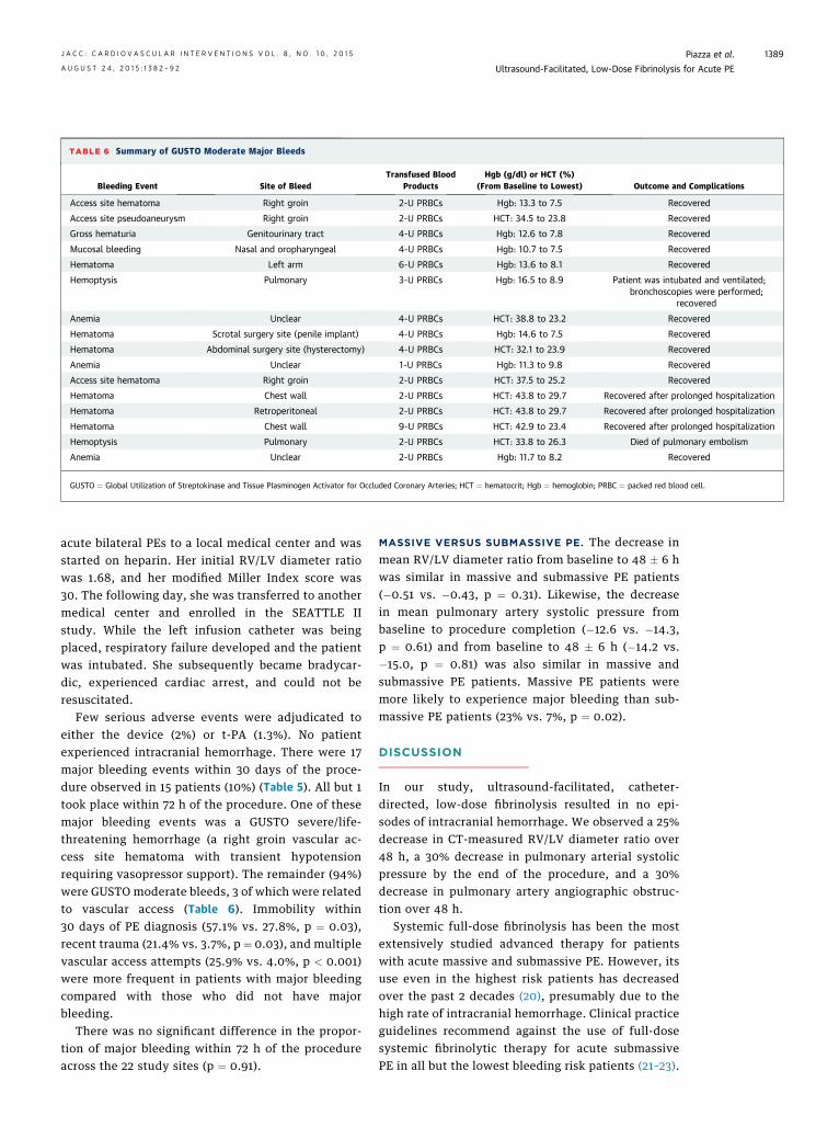
TABLE 6 Summary of GUSTO Moderate Major Bleeds
Bleeding Event Site of BleedTransfused Blood
ProductsHgb (g/dl) or HCT (%)
(From Baseline to Lowest) Outcome and Complications
Access site hematoma Right groin 2-U PRBCs Hgb: 13.3 to 7.5 Recovered
Access site pseudoaneurysm Right groin 2-U PRBCs HCT: 34.5 to 23.8 Recovered
Gross hematuria Genitourinary tract 4-U PRBCs Hgb: 12.6 to 7.8 Recovered
Mucosal bleeding Nasal and oropharyngeal 4-U PRBCs Hgb: 10.7 to 7.5 Recovered
Hematoma Left arm 6-U PRBCs Hgb: 13.6 to 8.1 Recovered
Hemoptysis Pulmonary 3-U PRBCs Hgb: 16.5 to 8.9 Patient was intubated and ventilated;bronchoscopies were performed;
recovered
Anemia Unclear 4-U PRBCs HCT: 38.8 to 23.2 Recovered
Hematoma Scrotal surgery site (penile implant) 4-U PRBCs Hgb: 14.6 to 7.5 Recovered
Hematoma Abdominal surgery site (hysterectomy) 4-U PRBCs HCT: 32.1 to 23.9 Recovered
Anemia Unclear 1-U PRBCs Hgb: 11.3 to 9.8 Recovered
Access site hematoma Right groin 2-U PRBCs HCT: 37.5 to 25.2 Recovered
Hematoma Chest wall 2-U PRBCs HCT: 43.8 to 29.7 Recovered after prolonged hospitalization
Hematoma Retroperitoneal 2-U PRBCs HCT: 43.8 to 29.7 Recovered after prolonged hospitalization
Hematoma Chest wall 9-U PRBCs HCT: 42.9 to 23.4 Recovered after prolonged hospitalization
Hemoptysis Pulmonary 2-U PRBCs HCT: 33.8 to 26.3 Died of pulmonary embolism
Anemia Unclear 2-U PRBCs Hgb: 11.7 to 8.2 Recovered
GUSTO ¼ Global Utilization of Streptokinase and Tissue Plasminogen Activator for Occluded Coronary Arteries; HCT ¼ hematocrit; Hgb ¼ hemoglobin; PRBC ¼ packed red blood cell.
J A C C : C A R D I O V A S C U L A R I N T E R V E N T I O N S V O L . 8 , N O . 1 0 , 2 0 1 5 Piazza et al.A U G U S T 2 4 , 2 0 1 5 : 1 3 8 2 – 9 2 Ultrasound-Facilitated, Low-Dose Fibrinolysis for Acute PE
1389
acute bilateral PEs to a local medical center and wasstarted on heparin. Her initial RV/LV diameter ratiowas 1.68, and her modified Miller Index score was30. The following day, she was transferred to anothermedical center and enrolled in the SEATTLE IIstudy. While the left infusion catheter was beingplaced, respiratory failure developed and the patientwas intubated. She subsequently became bradycar-dic, experienced cardiac arrest, and could not beresuscitated.
Few serious adverse events were adjudicated toeither the device (2%) or t-PA (1.3%). No patientexperienced intracranial hemorrhage. There were 17major bleeding events within 30 days of the proce-dure observed in 15 patients (10%) (Table 5). All but 1took place within 72 h of the procedure. One of thesemajor bleeding events was a GUSTO severe/life-threatening hemorrhage (a right groin vascular ac-cess site hematoma with transient hypotensionrequiring vasopressor support). The remainder (94%)were GUSTO moderate bleeds, 3 of which were relatedto vascular access (Table 6). Immobility within30 days of PE diagnosis (57.1% vs. 27.8%, p ¼ 0.03),recent trauma (21.4% vs. 3.7%, p ¼ 0.03), and multiplevascular access attempts (25.9% vs. 4.0%, p < 0.001)were more frequent in patients with major bleedingcompared with those who did not have majorbleeding.
There was no significant difference in the propor-tion of major bleeding within 72 h of the procedureacross the 22 study sites (p ¼ 0.91).
MASSIVE VERSUS SUBMASSIVE PE. The decrease inmean RV/LV diameter ratio from baseline to 48 � 6 hwas similar in massive and submassive PE patients(�0.51 vs. �0.43, p ¼ 0.31). Likewise, the decreasein mean pulmonary artery systolic pressure frombaseline to procedure completion (�12.6 vs. �14.3,p ¼ 0.61) and from baseline to 48 � 6 h (�14.2 vs.�15.0, p ¼ 0.81) was also similar in massive andsubmassive PE patients. Massive PE patients weremore likely to experience major bleeding than sub-massive PE patients (23% vs. 7%, p ¼ 0.02).
DISCUSSION
In our study, ultrasound-facilitated, catheter-directed, low-dose fibrinolysis resulted in no epi-sodes of intracranial hemorrhage. We observed a 25%decrease in CT-measured RV/LV diameter ratio over48 h, a 30% decrease in pulmonary arterial systolicpressure by the end of the procedure, and a 30%decrease in pulmonary artery angiographic obstruc-tion over 48 h.
Systemic full-dose fibrinolysis has been the mostextensively studied advanced therapy for patientswith acute massive and submassive PE. However, itsuse even in the highest risk patients has decreasedover the past 2 decades (20), presumably due to thehigh rate of intracranial hemorrhage. Clinical practiceguidelines recommend against the use of full-dosesystemic fibrinolytic therapy for acute submassivePE in all but the lowest bleeding risk patients (21–23).

Piazza et al. J A C C : C A R D I O V A S C U L A R I N T E R V E N T I O N S V O L . 8 , N O . 1 0 , 2 0 1 5
Ultrasound-Facilitated, Low-Dose Fibrinolysis for Acute PE A U G U S T 2 4 , 2 0 1 5 : 1 3 8 2 – 9 2
1390
The desire to reduce RV pressure overload and tominimize the risk of adverse outcomes, such asintracranial hemorrhage, spurred exploration ofalternative lower-dose fibrinolytic strategies, in-cluding half-dose systemic fibrinolysis (24,25) andcatheter-based pharmacomechanical therapy (26). Inthe European ULTIMA (Ultrasound AcceleratedThrombolysis of Pulmonary Embolism) trial of 59patients with submassive PE, ultrasound-facilitated,catheter-directed, low-dose fibrinolysis plus anti-coagulation improved RV function from baseline to24 h to a greater extent than anticoagulation alonewithout causing major bleeding (14). Although bothtrials used the same equipment, the ULTIMA studyused a slightly lower dose of t-PA (20 mg) than thecurrent study (24 mg). The ULTIMA study evaluatedthe procedure in 30 patients with submassive PE,whereas the current study included 150 patients withsubmassive or massive PE. The difference in thedefinitions of major bleeding between the 2 trials maypartly explain the variation in major bleeding withultrasound-facilitated, catheter-directed, low-dosefibrinolysis (0% in the ULTIMA study vs. 10% in thecurrent study). In contrast to the ULTIMA study, thecurrent study also included massive PE patientswho were more likely to experience major bleedingthan those with submassive PE. Our study expandsthe experience of ultrasound-facilitated, catheter-directed, low-dose fibrinolysis in patients withmassive and submassive PE and demonstrates thepotential of this technique. On May 21, 2014, based onthe data from our trial and previous studies, the U.S.Food and Drug Administration approved the Eko-Sonic Endovascular System for treatment of PE (27).
Increased RV/LV diameter ratio is a reproducibleand well-validated tool for identifying PE patients atrisk of adverse outcomes, in particular, increased30-day mortality (28). Although a decrease in RV/LVdiameter ratio is an important surrogate markerfor the efficacy of ultrasound-facilitated, catheter-directed, low-dose fibrinolysis, the need for trialswith clinical endpoints remains important (29). Clin-ical outcomes such as hemodynamic collapse, qualityof life (30,31), and mortality will help guide the use ofthis technology.
STUDY LIMITATIONS AND STRENGTHS. The majorlimitation of our study was the lack of a comparatorgroup. Because we did not include a comparatorgroup, we cannot comment on the efficacy or safetyof ultrasound-facilitated, catheter-directed, low-dosefibrinolysis compared with full-dose systemic fibri-nolysis, half-dose systemic fibrinolysis, or anti-coagulation alone.
Another potential comparator was catheter-directed, low-dose fibrinolysis without the ultra-sound turned “on.” A study evaluated the impact ofultrasound on catheter-directed, low-dose fibrino-lysis for acute DVT and did not demonstrate that ul-trasound bolstered efficacy (32). However, thesefindings in patients with DVT cannot be extrapolatedto acute PE because PE thrombus is more acute, lessorganized, and less fibrotic than DVT.
An important statistical limitation relates to asubset of patients who did not undergo follow-upchest CT for assessment of RV/LV diameter ratio orechocardiography for estimation of pulmonary arterysystolic pressure within the pre-specified 48 � 6-hwindow. Missing data could have biased the resultsin favor of a greater treatment benefit by removingpatients with a complicated post-procedure clinicalcourse and limited improvement in our study out-comes. Alternatively, they or their physicians mayhave canceled follow-up imaging because they hadexcellent clinical improvement. We conducted ananalysis to explore the possibility of important dif-ferences between the patients with and withoutmissing follow-up imaging data. We observed nodifference in baseline demographic, clinical charac-teristics, comorbid conditions, PE duration or sub-type, anticoagulation, procedural characteristics,length of stay, or in-hospital mortality. Furthermore,we observed no difference in the change in RV/LVdiameter ratio in patients who had a follow-up CTscan performed within the 48 � 6-h window andthose who had a follow-up CT scan performed but itfell outside of the 48 � 6-h window.
In summary, we created a precise protocol forultrasound-facilitated, catheter-directed, low-dosefibrinolysis with t-PA. Previously, there was no stan-dardized approach. Our simplified study designfacilitated expeditious enrollment of 150 patientswithin 9 months. One-half of the study populationwas female. The population was also racially andethnically diverse, with substantial representationof African-American and Hispanic/Latino patients.We emphasized safety by using a definition ofmajor bleeding (combining GUSTO severe/life-threatening and GUSTO moderate bleeds) thatwould capture a greater number of clinically relevantbleeding events.
FUTURE AREAS OF INVESTIGATION. Future con-siderations include determining which patientsamong those with hemodynamically stable PE areoptimal candidates for ultrasound-facilitated, cath-eter-directed, low-dose fibrinolysis. Clinical studieswith comparator groups of anticoagulation alone,
PERSPECTIVES
WHAT IS KNOWN? Ultrasound-facilitated, catheter-directed,
low-dose fibrinolysis for acute massive and submassive PE
improved RV function, decreased pulmonary artery angiographic
obstruction, and reduced pulmonary hypertension.
WHAT IS NEW? The discussion of advanced therapies for pa-
tients with massive or submassive PE should include the option
of ultrasound-facilitated, catheter-directed, low-dose fibrino-
lysis at medical centers with experience in the appropriate
patient selection, performance of the procedure, and post-
procedure care.
WHAT IS NEXT? Although ultrasound-facilitated, catheter-
directed, low-dose fibrinolysis appeared to improve short-term,
surrogate outcomes, subsequent studies focused on clinical and
longer term outcomes will provide a better understanding of the
optimal use of this therapy for acute PE. Subsequent studies with
comparator groups of anticoagulation alone, systemic fibrino-
lysis, or other catheter-based techniques will be critical in
defining how ultrasound-facilitated, catheter-directed, low-dose
fibrinolysis should be applied to patients with acute PE.
J A C C : C A R D I O V A S C U L A R I N T E R V E N T I O N S V O L . 8 , N O . 1 0 , 2 0 1 5 Piazza et al.A U G U S T 2 4 , 2 0 1 5 : 1 3 8 2 – 9 2 Ultrasound-Facilitated, Low-Dose Fibrinolysis for Acute PE
1391
systemic fibrinolysis, or other catheter-based tech-niques will be critical in defining how ultrasound-facilitated, catheter-directed, low-dose fibrinolysisshould be used in patients with acute PE. Strategies toreduce major bleeding related to the procedureshould also be evaluated. Health economics andoutcomes research will be critical for determiningappropriate use of this technology.
CONCLUSIONS
Ultrasound-facilitated, catheter-directed, low-dosefibrinolysis improved RV function in acute PE,decreased pulmonary artery angiographic obstruc-tion, reduced pulmonary artery systolic pressure,and did not result in intracranial hemorrhage.Ultrasound-facilitated, catheter-directed, low-dosefibrinolysis has the potential to improve outcomesand change treatment algorithms in higher risk PEpatients.
REPRINT REQUESTS AND CORRESPONDENCE: Dr.Gregory Piazza, Cardiovascular Division, Brigham andWomen’s Hospital, 75 Francis Street, Boston, Massa-chusetts 02115. E-mail: [email protected].
RE F E RENCE S
1. The surgeon general’s call to action to preventdeep vein thrombosis and pulmonary embolism. U.S.Department of Health and Human Services.2008. Available at: www.ncbi.nlm.nih.gov/books/NBK44178/. Accessed September 21, 2014.
2. Kucher N, Goldhaber SZ. Management ofmassive pulmonary embolism. Circulation 2005;112:e28–32.
3. Piazza G. Submassive pulmonary embolism.JAMA 2013;309:171–80.
4. Piazza G, Goldhaber SZ. Management of sub-massive pulmonary embolism. Circulation 2010;122:1124–9.
5. Meyer G, Vicaut E, Danays T, et al. Fibrinolysisfor patients with intermediate-risk pulmonaryembolism. N Engl J Med 2014;370:1402–11.
6. Chatterjee S, Chakraborty A, Weinberg I, et al.Thrombolysis for pulmonary embolism and risk ofall-cause mortality, major bleeding, and intracra-nial hemorrhage: a meta-analysis. JAMA 2014;311:2414–21.
7. Marti C, John G, Konstantinides S, et al. Sys-temic thrombolytic therapy for acute pulmonaryembolism: a systematic review and meta-analysis.Eur Heart J 2015;36:605–14.
8. Fiumara K, Kucher N, Fanikos J, Goldhaber SZ.Predictors of major hemorrhage following fibri-nolysis for acute pulmonary embolism. Am J Car-diol 2006;97:127–9.
9. Goldhaber SZ, Visani L, De Rosa M. Acutepulmonary embolism: clinical outcomes in the
International Cooperative Pulmonary EmbolismRegistry (ICOPER). Lancet 1999;353:1386–9.
10. Goldhaber SZ. Percutaneous mechanicalthrombectomy for acute pulmonary embolism: adouble-edged sword. Chest 2007;132:363–5.
11. Goldhaber SZ. Percutaneous mechanicalthrombectomy for massive pulmonary embolism:improve safety and efficacy by sharing informa-tion. Catheter Cardiovasc Interv 2007;70:807–8.
12. Kucher N. Catheter embolectomy for acutepulmonary embolism. Chest 2007;132:657–63.
13. Braaten JV, Goss RA, Francis CW. Ultrasoundreversibly disaggregates fibrin fibers. ThrombHaemost 1997;78:1063–8.
14. Kucher N, Boekstegers P, Muller O, et al.Randomized controlled trial of ultrasound-assisted catheter-directed thrombolysis for acuteintermediate-risk pulmonary embolism. Circula-tion 2014;129:479–86.
15. Qanadli SD, El Hajjam M, Vieillard-Baron A,et al. New CT index to quantify arterial obstructionin pulmonary embolism: comparison with angio-graphic index and echocardiography. AJR Am JRoentgenol 2001;176:1415–20.
16. Schoepf UJ, Kucher N, Kipfmueller F, Quiroz R,Costello P, Goldhaber SZ. Right ventricularenlargement on chest computed tomography: apredictor of early death in acute pulmonary em-bolism. Circulation 2004;110:3276–80.
17. An international randomized trial comparingfour thrombolytic strategies for acute myocardial
infarction. The GUSTO investigators. N Engl J Med1993;329:673–82.
18. Kipfmueller F, Quiroz R, Goldhaber SZ,Schoepf UJ, Costello P, Kucher N. Chest CTassessment following thrombolysis or surgicalembolectomy for acute pulmonary embolism. VascMed 2005;10:85–9.
19. Fasullo S, Scalzo S, Maringhini G, et al. Six-month echocardiographic study in patients withsubmassive pulmonary embolism and rightventricle dysfunction: comparison of thrombolysiswith heparin. Am J Med Sci 2011;341:33–9.
20. Stein PD, Matta F. Thrombolytic therapy inunstable patients with acute pulmonary embolism:saves lives but underused. Am J Med 2012;125:465–70.
21. Jaff MR, McMurtry MS, Archer SL, et al. Man-agement of massive and submassive pulmonaryembolism, iliofemoral deep vein thrombosis, andchronic thromboembolic pulmonary hypertension:a scientific statement from the American HeartAssociation. Circulation 2011;123:1788–830.
22. Kearon C, Akl EA, Comerota AJ, et al. Anti-thrombotic therapy for VTE disease: Antith-rombotic Therapy and Prevention of Thrombosis,9th ed: American College of Chest PhysiciansEvidence-Based Clinical Practice Guidelines. Chest2012;141:e419S–94S.
23. Konstantinides SV, Torbicki A, Agnelli G, et al.2014 ESC Guidelines on the diagnosis and man-agement of acute pulmonary embolism: the TaskForce for the Diagnosis and Management of Acute

Piazza et al. J A C C : C A R D I O V A S C U L A R I N T E R V E N T I O N S V O L . 8 , N O . 1 0 , 2 0 1 5
Ultrasound-Facilitated, Low-Dose Fibrinolysis for Acute PE A U G U S T 2 4 , 2 0 1 5 : 1 3 8 2 – 9 2
1392
Pulmonary Embolism of the European Society ofCardiology (ESC) Endorsed by the European Res-piratory Society (ERS). Eur Heart J 2014;35:3033–73.
24. Sharifi M, Bay C, Skrocki L, Rahimi F,Mehdipour M. Moderate pulmonary embolismtreated with thrombolysis (from the “MOPETT”Trial). Am J Cardiol 2013;111:273–7.
25. WangC, Zhai Z, YangY, et al. Efficacy and safetyof low dose recombinant tissue-type plasminogenactivator for the treatment of acute pulmonarythromboembolism: a randomized, multicenter,controlled trial. Chest 2010;137:254–62.
26. Kuo WT, Gould MK, Louie JD, Rosenberg JK,Sze DY, Hofmann LV. Catheter-directed therapyfor the treatment of massive pulmonary embolism:systematic review and meta-analysis of moderntechniques. J Vasc Interv Radiol 2009;20:1431–40.
27. EkoSonic� Endovascular System receives FDAClearance for the Treatment of Pulmonary
Embolism in the USA. Cath Lab Digest. Available at:http://www.cathlabdigest.com/EkoSonic%C2%AE-Endovascular-System-receives-FDA-Clearance-Treatment-Pulmonary-Embolism-USA. AccessedJune 27, 2015.
28. Meinel FG, Nance JW Jr., Schoepf UJ, et al.Predictive value of computed tomography inacute pulmonary embolism: systematic reviewand meta-analysis. Am J Med 2015;128:747–59.
29. Weinberg I, Jaff MR. Accelerated thrombolysisfor pulmonary embolism: will clinical benefitbe ULTIMAtely realized? Circulation 2014;129:420–1.
30. Kline JA, Nordenholz KE, Courtney DM, et al.Treatment of submassive pulmonary embolismwith tenecteplase or placebo: cardiopulmonaryoutcomes at 3 months: multicenter double-blind,placebo-controlled randomized trial. J ThrombHaemost 2014;12:459–68.
31. Klok FA, Cohn DM, Middeldorp S, et al. Qualityof life after pulmonary embolism: validation of thePEmb-QoL Questionnaire. J Thromb Haemost2010;8:523–32.
32. Engelberger RP, Spirk D, Willenberg T, et al.Ultrasound-assisted versus conventional catheter-directed thrombolysis for acute iliofemoral deepvein thrombosis. Circ Cardiovasc Interv 2015 Jan 8[E-pub ahead of print].
KEY WORDS catheter embolectomy,catheter thrombolysis, fibrinolysis,pulmonary embolism, right ventricularfailure, thrombolysis
APPENDIX For supplemental material,please see the online version of this article.