Prognostic factors for pulmonary metastasis in primary osteosarcoma … · 2019-06-28 ·...
48
Prognostic factors for pulmonary metastasis in primary osteosarcoma Eunah Shin Department of Medicine The Graduate School, Yonsei University
Transcript of Prognostic factors for pulmonary metastasis in primary osteosarcoma … · 2019-06-28 ·...

Prognostic factors for pulmonary metastasis in primary osteosarcoma
Eunah Shin
Department of Medicine
The Graduate School, Yonsei University

Prognostic factors for pulmonary
metastasis in primary osteosarcoma
Directed by Professor Woo Ick Yang
The Doctoral Dissertation submitted to the Department of Medicine, the Graduate School of Yonsei University
in partial fulfillment of the requirements for the degree of Doctor of Philosophy
Eunah Shin
December 2008

This certifies that the Doctoral
Dissertation of Eunah Shin is approved.
------------------------------------ Woo Ick Yang, M.D., PhD.
------------------------------------
Jin-Suck Suh, M.D., PhD.
------------------------------------ Kyeongmee Park, M.D., PhD.
------------------------------------ Sun Young Rha, M.D., PhD.
------------------------------------
Kyung-A Lee, M.D., PhD.
The Graduate School Yonsei University
December 2008

ACKNOWLEDGEMENTS
I am most grateful to Professor Woo Ick Yang and Professor
Sun Young Rha for their patience and guidance throughout my
journey to the degree. I am also greatly indebted to Professor
Jin-Suck Suh, Professor Kyeongmee Park and Professor
Kyung-A Lee for their kind and meticulous instructions.
I express my deepest gratitude to my loving husband, Dr.
Gijong Yi, and my most beloved sons Gene W. and Howon,
who are indeed the greatest joy of my everyday life. I am also
grateful to my parents-in-law who have shown me with their
lives what patience and devotion mean.
To my dearest friends Dr. Cheryn Song, Dr. Kyungwon Kim
and Dr. Jaseung Koo, I give thanks simply for just being there
for me.
I thank my brother Jaehyeok for bearing with me all the time
and lastly, I dedicate this thesis to my loving parents whom I
respect the most in every aspect of life and who have been
more than fully supportive throughout my life to this day.
December, 2008
Eunah Shin

TABLE OF CONTENTS
ABSTRACT ······························································································1
I. INTRODUCTION ···················································································· 3
II. MATERIALS AND METHODS ···························································· 5
1. Construction of tissue microarray ······················································· 5
2. Immunohistochemical stain································································· 5
3. Fluorescent in situ hybridization ························································· 7
4. cDNA microarray ················································································ 8
5. Collection of clinical data······························································· 10
6. Statistical analysis·············································································· 10
III. RESULTS····························································································· 10
1. Tissue microarray construction results·············································· 10
2. Patient demographics········································································· 11
3. Various protein expression in osteosarcoma by
immunohistochemical stain ······························································· 14
4. Fluorescent in situ hybridization results············································ 15
5. Correlation of protein expression and clinical variables··················· 15
6. Survival analyses according to ezrin and VEGF expressions··········· 17
7. Coexpressions of ezrin, VEGF, nm23, and EGFR···························· 19
8. cDNA microarray results··································································· 22

IV. DISCUSSION ······················································································ 25
V. CONCLUSION ····················································································· 31
REFERENCES··························································································· 32
ABSTRACT(IN KOREAN)·································································· 39

LIST OF FIGURES
Figure 1. Representative image of tissue microarray slides····· 11
Figure 2. Histologic subtypes of osteosarcoma························ 13
Figure 3. Immunohistochemical staining of ezrin and nm23
··················································································· 13
Figure 4. Overall and disease-free survival in ezrin-positive
and ezrin-negative osteosarcoma ······························ 18
Figure 5. Overall and disease-free survival in VEGF-positive
and VEGF-negative osteosarcoma···························· 18
Figure 6. Hierarchial clustering of cDNA microarray············· 22

LIST OF TABLES
Table 1. Antibodies used for immunohistochemical stain
······················································································ 7
Table 2. Patient demographics·················································· 12
Table 3. Immunohistochemical stain results in primary
osteosarcoma······························································· 14
Table 4. p-values for the correlation of immunohistochemical
stain results and clinical variables ······························ 15
Table 5. Correlation of protein expression and
pulmonary metastasis·················································· 17
Table 6. Correlation of pulmonary metastasis, response to
chemotherapy, overall survival, and disease-free
survival and coexpressions of ezrin, VEGF, nm23,
and EGFR···································································· 19
Table 7. List of upregulated genes in metastasis group ··········· 23
Table 8. List of downregulated genes in metastasis group ······ 24

1
ABSTRACT
Prognostic factors for pulmonary metastasis in primary osteosarcoma
Eunah Shin
Department of Medicine
The Graduate School, Yonsei University
(Directed by Professor Woo Ick Yang)
Osteosarcoma is the most common primary tumor of bone with a 5-year
survival of approximately 70%, which drops to 37% once the tumor
recurs or metastasizes. Thus, identifying prognostic factors for
pulmonary metastasis can be one of the keys to improving the overall
survival of osteosarcoma. In an endeavor to identify novel factors of
prognostic significance, a panel of immunohistochemistry and
fluorescent in situ hybridization (FISH) studies were done in tissue
microarray of 92 cases of osteosarcoma, 26 of which were tumors with
metachronous pulmonary metastasis. Additionally, to newly discover
differentially expressed genetic molecules between metastatic and
non-metastatic osteosarcoma tissues, cDNA microarray was done in 8
fresh frozen tissues of osteosarcoma, 4 of which were tumors with
metachronous pulmonary metastasis. The immunohistochemical studies
were comprised of metastasis-related molecules in osteosarcoma
previously screened by cDNA microarray studies such as ezrin,
vascular endothelial growth factor (VEGF), and nm23. Antibodies for
EGFR, c-kit, nerve growth factor receptor (NGFR), nuclear factor
kappa B (NFκB), tyrosine receptor kinase (Trk) A, B, and C, and
HER-2/neu were stained additionally. The fluorescent in situ
hybridization studies were comprised of HER-2/neu, epidermal growth
2
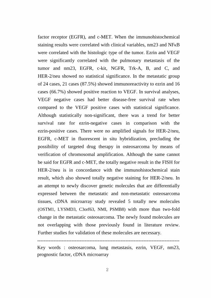
factor receptor (EGFR), and c-MET. When the immunohistochemical
staining results were correlated with clinical variables, nm23 and NFκB
were correlated with the histologic type of the tumor. Ezrin and VEGF
were significantly correlated with the pulmonary metastasis of the
tumor and nm23, EGFR, c-kit, NGFR, Trk-A, B, and C, and
HER-2/neu showed no statistical significance. In the metastatic group
of 24 cases, 21 cases (87.5%) showed immunoreactivity to ezrin and 16
cases (66.7%) showed positive reaction to VEGF. In survival analyses,
VEGF negative cases had better disease-free survival rate when
compared to the VEGF positive cases with statistical significance.
Although statistically non-significant, there was a trend for better
survival rate for ezrin-negative cases in comparison with the
ezrin-positive cases. There were no amplified signals for HER-2/neu,
EGFR, c-MET in fluorescent in situ hybridization, precluding the
possibility of targeted drug therapy in osteosarcoma by means of
verification of chromosomal amplification. Although the same cannot
be said for EGFR and c-MET, the totally negative result in the FISH for
HER-2/neu is in concordance with the immunohistochemical stain
result, which also showed totally negative staining for HER-2/neu. In
an attempt to newly discover genetic molecules that are differentially
expressed between the metastatic and non-metastatic osteosarcoma
tissues, cDNA microarray study revealed 5 totally new molecules
(OSTM1, LYSMD3, C3orf63, NMI, PSMB8) with more than two-fold
change in the metastatic osteosarcoma. The newly found molecules are
not overlapping with those previously found in literature review.
Further studies for validation of these molecules are necessary.
---------------------------------------------------------------------------------------
Key words : osteosarcoma, lung metastasis, ezrin, VEGF, nm23,
prognostic factor, cDNA microarray

3
Prognostic factors for pulmonary metastasis
in primary osteosarcoma
Eunah Shin
Department of Medicine The Graduate School, Yonsei University
(Directed by Professor Woo Ick Yang)
I. INTRODUCTION
Osteosarcoma, the most common primary tumor of bone, is the second highest
cause of cancer-related death in the pediatric age group.1 The principal treatment
modality consists of neoadjuvant chemotherapy followed by limb-salvage
operation. However, despite the introduction of adjuvant chemotherapy with
resultant tumor necrosis of considerable extent, the 5-year survival is
approximately 70% and has not changed much over the last 20 years.1,2
Moreover, this figure drops to 40%-50% if the chemotherapy induced tumor
necrosis falls short of 90% and even more dramatically to 37% once the tumor
recurs or metastasizes. Such inferior chemotherapy induced necrosis has been
correlated with higher rates of pulmonary metastasis, which is an overriding
determinant of survival.3 Approximately 20% of the patients show lung
metastasis at initial diagnosis and 30% to 40% eventually develop metastasis in
spite of the conventionally established treatment.4 There are some distinctive
features of metastasis in osteosarcoma, such as 1) long latent period between
initial diagnosis of the primary tumor and the development of metastasis, 2)
preference of lung as metastatic site, 3) relative success associated with surgical
removal of the metastasis.5 Consequently, there have been many endeavors to
identify predictive factors for pulmonary metastasis and novel markers of
therapeutic and prognostic importance for this highly debilitating and fatal entity,
but unfortunately the results have been controversial, if not futile, and have

4
resulted in the plateau of survival rate for more than 20 years.6 Recently, owing
to the fast developing biotechnology and tumor biology, identification of newly
emerging molecular markers for prognostic significance and development of
new therapeutic targets have been possible. Moreover, drugs specifically
targeted to inhibit certain genetic alteration involved in oncogenesis and tumor
progression have been developed and have led to improved outcomes for the
patients especially when combined with multiagent chemotherapy. Namely such
targeted drug therapies include trastuzumab, which is specific for HER-2/neu
amplification in breast cancers, gefitinib, which is a specific inhibitor of
epidermal growth factor receptor (EGFR), and imatinib mesylate, inhibitor of
KIT activity.7-9 Since their successful clinical utility that was more than expected,
studies exploring the possibility of their application to various other tumors have
been continued and osteosarcoma has been no exception. However, studies
involving osteosarcoma to date have been limited by a small number of index
cases or cell line studies only. For instance, Morris et al. has reported that
HER-2/neu expression is associated with significantly less tumor necrosis after
preoperative chemotherapy, and thus antibodies that target this receptor should
be considered for the treatment.10 The shortcoming is, though, that their study
has involved immunohistochemical staining only and the true gene amplification
has not been investigated and correlated.
The primary aim of this study is to identify factors which can be predictive of
higher metastatic potential of a given osteosarcoma, so that the patients with
lower chemosensitivity and higher metastatic potential can be subject to
alternative chemotherapeutic regimen earlier enough to improve overall survival.
To this end, various markers with established prognostic and therapeutic
significance in other solid tumors and molecules previously screened by cDNA
microarray studies to be associated with metastatic potential in osteosarcoma
are applied immunohistochemically to a large series of osteosarcoma in tissue
microarray and the results are analyzed with appropriate statistical methods. In
addition, the differential protein expressions of erbB family protein receptor
5
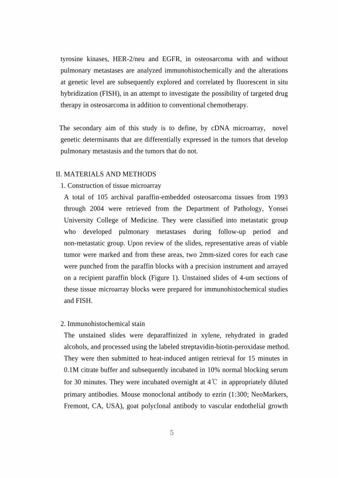
tyrosine kinases, HER-2/neu and EGFR, in osteosarcoma with and without
pulmonary metastases are analyzed immunohistochemically and the alterations
at genetic level are subsequently explored and correlated by fluorescent in situ
hybridization (FISH), in an attempt to investigate the possibility of targeted drug
therapy in osteosarcoma in addition to conventional chemotherapy.
The secondary aim of this study is to define, by cDNA microarray, novel
genetic determinants that are differentially expressed in the tumors that develop
pulmonary metastasis and the tumors that do not.
II. MATERIALS AND METHODS
1. Construction of tissue microarray
A total of 105 archival paraffin-embedded osteosarcoma tissues from 1993
through 2004 were retrieved from the Department of Pathology, Yonsei
University College of Medicine. They were classified into metastatic group
who developed pulmonary metastases during follow-up period and
non-metastatic group. Upon review of the slides, representative areas of viable
tumor were marked and from these areas, two 2mm-sized cores for each case
were punched from the paraffin blocks with a precision instrument and arrayed
on a recipient paraffin block (Figure 1). Unstained slides of 4-um sections of
these tissue microarray blocks were prepared for immunohistochemical studies
and FISH.
2. Immunohistochemical stain
The unstained slides were deparaffinized in xylene, rehydrated in graded
alcohols, and processed using the labeled streptavidin-biotin-peroxidase method.
They were then submitted to heat-induced antigen retrieval for 15 minutes in
0.1M citrate buffer and subsequently incubated in 10% normal blocking serum
for 30 minutes. They were incubated overnight at 4℃ in appropriately diluted
primary antibodies. Mouse monoclonal antibody to ezrin (1:300; NeoMarkers,
Fremont, CA, USA), goat polyclonal antibody to vascular endothelial growth

6
factor (VEGF) (1:150; R&D Systems, Minneapolis, MN, USA), rabbit
polyclonal antibody to nm23 (1:700; NeoMarkers), ready to use antibody for
EGFR using EGFR PharmDxTM, rabbit polyclonal antibody to HER-2/neu
(1:250; DAKO, Glostrup, Denmark), mouse monoclonal AE1/AE3 antibody for
CK (1:100; DAKO), rabbit polyclonal antibodies for tyrosine receptor kinase-A
(Trk-A), B, and C (1:200; Santa Cruz, Santa Cruz, CA, USA), rabbit polyclonal
antibody to nuclear factor kappa B (NFκB) (1:200; NeoMarkers), rabbit
polyclonal antibody to c-kit (1:30; DAKO), and mouse monoclonal antibody
for low affinity nerve growth factor receptor (NGFR) (1:50; DAKO) were used
for immunohistochemistry (Table 1). After washing with Tris buffer, sections
were incubated with biotin-labelled secondary antibodies and then with
streptavidin-horseradish peroxidase using the DAKO LSAB kit (DAKO) at
room temperature for 30 minutes for each step. Nova red (Vector Laboratory,
Burlingame, CA, USA) was used as the chromogen and hematoxylin as the
nuclear counterstain. This procedure was performed for all antibodies under
study except for EGFR. Ready to use antibody was employed for
immunohistochemical stain of EGFR using EGFR PharmDxTM , and the
staining procedure was done as the manufacturer’s protocol.
7
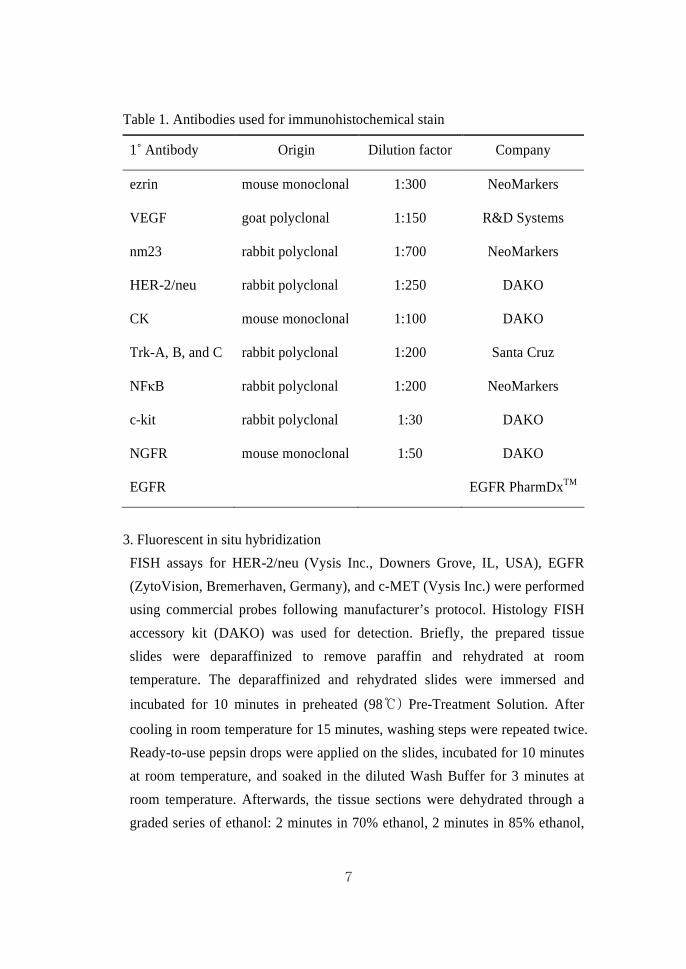
Table 1. Antibodies used for immunohistochemical stain
1˚ Antibody Origin Dilution factor Company
ezrin mouse monoclonal 1:300 NeoMarkers
VEGF goat polyclonal 1:150 R&D Systems
nm23 rabbit polyclonal 1:700 NeoMarkers
HER-2/neu rabbit polyclonal 1:250 DAKO
CK mouse monoclonal 1:100 DAKO
Trk-A, B, and C rabbit polyclonal 1:200 Santa Cruz
NFκB rabbit polyclonal 1:200 NeoMarkers
c-kit rabbit polyclonal 1:30 DAKO
NGFR mouse monoclonal 1:50 DAKO
EGFR EGFR PharmDxTM
3. Fluorescent in situ hybridization
FISH assays for HER-2/neu (Vysis Inc., Downers Grove, IL, USA), EGFR
(ZytoVision, Bremerhaven, Germany), and c-MET (Vysis Inc.) were performed
using commercial probes following manufacturer’s protocol. Histology FISH
accessory kit (DAKO) was used for detection. Briefly, the prepared tissue
slides were deparaffinized to remove paraffin and rehydrated at room
temperature. The deparaffinized and rehydrated slides were immersed and
incubated for 10 minutes in preheated (98℃) Pre-Treatment Solution. After
cooling in room temperature for 15 minutes, washing steps were repeated twice.
Ready-to-use pepsin drops were applied on the slides, incubated for 10 minutes
at room temperature, and soaked in the diluted Wash Buffer for 3 minutes at
room temperature. Afterwards, the tissue sections were dehydrated through a
graded series of ethanol: 2 minutes in 70% ethanol, 2 minutes in 85% ethanol,
8
and 2 minutes in 96% ethanol, and then air dried completely. An appropriate
amount of the probe was applied to the slides, covered with glass coverslip
overlapped by Cover slip Sealant to form a seal around the coverslip, and
placed in Dako Hybridizer. The denaturation was set to 72 °C for 10 min and
hybridization to 37 °C overnight. Washing and dehydrating steps were repeated
and the slides were counterstained with blue fluorescence counterstain
Fluorescence Mounting Medium.
The number of signals of chromosome 17 for HER-2/neu and chromosome 7
for EGFR and c-MET, signals for each probe, and the number of tumor nuclei
scored were recorded for each core. At least 30 tumor nuclei were counted per
tissue core, and the signal enumeration was performed under x1000
magnification using epifluorescence microscope with single-interference filter
sets for green (FITC), red (Texas red), and blue (DAPI) as well as triple (blue,
red, green) band pass filters. Tumors were interpreted as amplified when the
ratio of target gene signals to centromere (CEP) 17 or CEP 7 signals was ≥2.0.
The average ratio of different cores from the same tumor was used as the final
score for the determination of gene amplification status of that particular tumor.
4. cDNA microarray
A. RNA extraction and amplification
Total RNA was extracted from fresh frozen tissues, which were immediatedly
frozen at -80℃ at the time of incisional biopsy for initial diagnosis, by TRIzol
reagent (Invitrogen, Carlsbad, CA, USA) according to the manufacturer's
instructions. The Yonsei reference RNA (Cancer Metastasis Research Center,
Seoul, Korea) was prepared by pooling equivalent amounts of total RNA from
11 human cancer cell lines. The quantity and quality of RNA were confirmed
by a ND-1000 spectrophotometer (NanoDrop Technologies, Montchanin, DE,
USA) and gel electrophoresis. First-strand template cDNA was generated by
reverse transcription with an oligo(dT)15 primer coupled to a T7 RNA
polymerase recognition sequence (Applied Biosystems, Framingham, MA,

9
USA). Residual single-stranded RNA was removed by RNase H digestion and
the cDNA template was used for transcription with biotinylated CTP and UTP
nucleosides to produce a cRNA template. After purification and quantification,
the biotinylated cRNA was fragmented by hydrolysis, producing 35 to 200
nucleotide segments.
B. Hybridization
Following the institutional protocol, samples were hybridized at 45℃ to a
human cDNA chip (CMRC-GT, Seoul, Korea) containing ~17000 cDNA clone
of 300bp~3kb with a reference design. The test samples were labeled with Cy5
and individually co-hybridized with the Cy3-labeled Yonsei reference RNA
(CMRC, Seoul, Korea).
C. Preprocessing and data analysis
For further analysis, raw Cy5/Cy3 data were log2-transformed. Systemic errors
were corrected by normalization using intensity dependent, within-print, tip
normalization based on the Lowess function. After normalization, genes with
more than one missing value in all experiments were filtered, no missing
proportion (NMP) 80%. The values of repeated genes were adjusted by S-Plus
2000 software (Insightful, Seattle, WA, USA). We determined the significant
genes which could divide the tissues into metastatic and non-metastatic groups
using two-class significance analysis of microarrays (SAM)11 with selected
genes. Hierarchical clustering analysis was performed with Cluster (Eisen Lab,
http://rana.lbl.gov/EisenSoftware.htm) and the resulting dendrogram was
visualized using TreeView software (Eisen Lab). Clustering was done by
complete linkage algorithm with uncentered correlation. The distance of each
cluster represents correlation between two clusters. Annotation of the selected
genes was performed using the Database for Annotation, Visualization and
Integrated Discovery (DAVID) (http:// apps1. niaid. nih. gov/ david) and the
Stanford Online Universal Resource for Clones and Expressed Sequence Tags
(SOURCE) (http:// source. stanford. edu/ cgi-bin/ source/ source Search).

10
5. Collection of clinical data
Clinical information were collected from the medical chart review and the
clinicopathologic factors assessed for prognostic significance included age,
gender, site of involvement, histologic subtype of the tumor, percentile of
chemotherapy induced necrosis, recurrence, follow-up duration and survival.
6. Statistical analysis
Sets of statistical analysis based on the aims of the study to investigate factors
associated with increased risk of developing pulmonary metastasis were done.
Univariate analysis and multivariate analysis were performed to assess
prognostic significance and value of individual risk factors. In all statistical
analyses, a two-tailed p-value < 0.05 was considered statistically significant. All
analyses were performed using SPSS for Windows statistical software (Version
15.0)
III. RESULTS
1. Tissue microarray construction results
Of the selected 105 cases of osteosarcoma 32 cases were osteosarcomas with
metachronous lung metastasis and 73 cases were devoid of lung metastasis
neither at the time of initial diagnosis nor during the follow-up period. All slides
were reviewed and representative tumor areas were marked appropriately. Cases
with insufficient amount of tissue in paraffin blocks and those with missing
slides or blocks were dropped. The total number of evaluated cases was 92 in
toto, 66 of which comprised the non-metastatic group and 26 of which belonged
to the metastatic group. After tissue microarray construction, there were tissue
cores from each group either insufficient for evaluation or lost upon serial
sectioning. Therefore, the total number of evaluated cases differed for each
antibody or probe (Figure 1).

11
A.
B.
Figure 1. Representative image of tissue microarray slides. A) H&E stain,
B) Immunohistochemical stain
2. Patient demographics
Of the total number of 92 cases, 48 were male and 44 were female with a mean
age of 21.6 years (range 3 to 65 years). Thirty-five cases were respondent to
chemotherapy when ‘responsiveness’ is defined as chemotherapy induced tumor
necrosis more than 90% and 37 were not responsive, while chemoresponsiveness
in 20 cases were not assessed due to lack of post-chemotherapeutic specimen.
Chemotherapeutic regimen consisted of either combination of adriamycin and
intra-arterial cisplatin or combination of ifosfamide, adriamycin and intra-arterial
cisplatin. Regarding the location of the tumor, 71 cases were tumors arising in
the lower extremity, 7 cases were in the upper extremity, 8 cases were tumors
arising in sites other than upper and lower extremities. Six cases had no records
on site of involvement. As for the histologic subtype, 51 cases were osteoblastic
type, 8 were chondroblastic, 4 were fibroblastic, 12 were mixed (of which
osteoblastic mixed with chondroblastic type was the most common), 7 cases
were parosteal/ periosteal, and 10 cases were other rare subtypes including
intramedullary well differentiated, small cell, pleomorphic, and telangiectatic
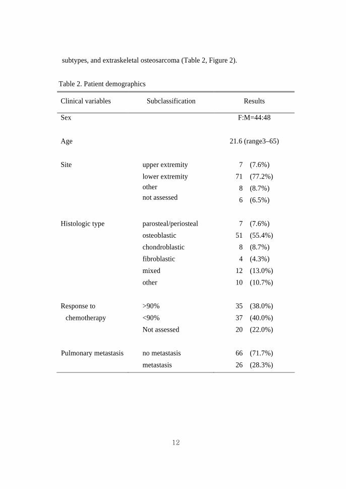
12
subtypes, and extraskeletal osteosarcoma (Table 2, Figure 2).
Table 2. Patient demographics
Clinical variables Subclassification Results
Sex
F:M=44:48
Age
21.6 (range3–65)
Site upper extremity
lower extremity
other
not assessed
7 (7.6%)
71 (77.2%)
8 (8.7%)
6 (6.5%)
Histologic type
Response to
chemotherapy
Pulmonary metastasis
parosteal/periosteal
osteoblastic
chondroblastic
fibroblastic
mixed
other
>90%
<90%
Not assessed
no metastasis
metastasis
7 (7.6%)
51 (55.4%)
8 (8.7%)
4 (4.3%)
12 (13.0%)
10 (10.7%)
35 (38.0%)
37 (40.0%)
20 (22.0%)
66 (71.7%)
26 (28.3%)

13
A
Figure 2. Histologic subtypes of osteosarcoma (H&E, x40). A) osteoblastic type,
B) chondroblastic type, C) fibroblastic type, D) intramedullary well- differentiated type.
Figure 3. Immunohistochemical stains for A) ezrin and B) nm23, showing
membranous staining for ezrin and cytoplasmic staining for nm23 (x100).
A
C D
B
A B

14
3. Various protein expression in osteosarcoma by immunohistochemical stain
As aforementioned, the total number of evalulated cases ranged from 78 to 92
due to lost cores during tissue microarray construction and serial sectioning. For
ezrin, nm23, c-kit, Trk-A, Trk-B, and NFκB, more than 50% of the cases were
immunoreactive, whereas VEGF and EGFR showed relatively low expression
rates of 42% and 25%, respectively. NGFR was predominantly non-
immunoreactive in index cases (Figure 2). None of the cases showed
immunoreactivity for Trk-C, CK, and HER-2/neu (Table 3).
Table 3. Immunohistochemical stain results in primary osteosarcoma
Protein Positive ratio
ezrin 47/83 (57%)
nm23 57/83 (69%)
c-kit 59/83 (71%)
Trk-A 64/78 (82%)
Trk-B 66/78 (85%)
NFκB 49/79 (62%)
VEGF 33/79 (42%)
EGFR 23/92 (25%)
NGFR 3/92 (3%)
Trk-C 0 /78 (0%)
CK 0 /78 (0%)
HER-2/neu 0 /78 (0%)
15
4. Fluorescent in situ hybridization results
All cases evaluated for amplification of HER-2/neu, EGFR, and c-MET showed
no amplified signal in osteosarcoma tissues.
5. Correlation of protein expression and clinical variables
When each immunohistochemical staining result was evaluated for correlation
with clinical variables other than lung metastasis, nm23 and NFκB were
positively correlated with osteoblastic subtype of the tumor (p-value 0.023 and
0.015, respectively) and no other antibody was correlated with any of the
clinical variables (Table 4).
Table 4. p-values for the correlation of immunohistochemical stain results and
clinical variables
Sex Age Site Histologic
Type
Response to
chemotherapy
ezrin 0.059 0.0586 0.395 0.093 0.166
VEGF 1.00 0.1560 0.687 0.272 0.145
nm23 0.871 0.1233 0.403 0.023 0.151
EGFR 0.603 0.8755 0.961 0.447 0.778
c-kit 0.122 0.4983 0.831 0.551 0.99
NGFR 0.621 0.3382 1.000 0.132 0.608
Trk-A 0.206 0.3133 0.450 0.518 0.489
Trk-B 0.792 0.2775 0.202 0.711 0.712
NFκB 0.531 0.4178 0.511 0.015 0.214

16
Ezrin expression was evaluated in 83 cases, of which 24 were with lung
metastasis. Among 59 non-metastatic tumors, 26 (44.1%) were positive for ezrin
and 21 out of 24 (87.5%) were positive for ezrin in lung metastasis group
(p-value 0.0003).
Nm23 was evaluated in 83 cases, and of the 60 non-metastatic tumors, 38
(63.3%) were positive for nm23 and 19 out of 23 metastatic tumors (82.6%)
were positive for nm23.
For EGFR, 13 out of 66 (19.7%) were immunoreactive in the non-metastatic
group and 10 out of 26 (38.5%) were immunoreactive in the metastatic group.
For NGFR evaluated in a total of 92 patients, only one case from the
non-metastatic group and two cases from the metastatic group showed positive
immunostaining.
Immunohistochemical staining for Trk-A and Trk-B showed similar results for
both metastatic and non-metastatic groups, with 83.3% (45 out of 54) and 85.2%
(46 out of 54) respectively in non-metastatic group and 79.2% (19 out of 24) and
83.3% (20 out of 24) respectively in metastatic group.
For VEGF and NFκB, cores of 79 cases were available. In the metastasis group,
16 out of the 24 (66.7%) were immunoreactive to VEGF and in the
non-metastasis group, only 17 out of the 55 (30.9%) were immunoreactive
(p-value 0.003). As for NFκB, 15 out of the 24 (62.5%) were immunoreactive in
the metastasis group and 34 out of the 55 (61.8%) were immunoreactive in the
non-metastasis group, which were statistically not significant.
Immunohistochemical stain results for Trk-C, CK, and HER-2/neu were totally
negative in all cases evaluated. These immunohistochemical staining results,
except for those of ezrin and VEGF, were all statistically insignificant (Table 5).

17
Table 5. Correlation of protein expression and pulmonary metastasis
Pulmonary metastasis Protein expression
Present (%) Absent (%) p-value
Positive Ezrin
Negative
21 (87.5)
3 (12.5)
26 (44.1)
33 (55.9) 0.0003
Positive VEGF
Negative
16 (66.7)
8 (33.3)
17 (30.9)
38 (69.1) 0.003
Positive nm23
Negative
19 (82.6)
4 (17.4)
38 (63.3)
22 (36.7) 0.0902
Positive EGFR
Negative
10 (38.5)
16 (61.5)
13 (19.7)
53 (80.3) 0.0613
Positive C-kit
Negative
19 (79.2)
5 (20.8)
40 (67.8)
19 (32.2) 0.3754
Positive NGFR
Negative
2 (7.7)
24 (92.3)
1 (1.5)
65 (98.5) 0.1915
Positive Trk-A
Negative
19 (79.2)
5 (20.8)
45 (83.3)
9 (16.7) 0.7516
Positive Trk-B
Negative
20 (83.3)
4 (16.7)
46 (85.2)
8 (14.8) 0.999
Positive NFκB
Negative
15 (62.5)
9 (37.5)
34 (61.8)
21 (38.2) 0.954
6. Survival analyses according to ezrin and VEGF expressions.
The overall survival and disease-free survival in relation to the ezrin
overexpression were evaluated by log-rank test. The median follow-up duration
was 90 months. The median in overall survival was 77.25 months (range 0.6 –
153.9) and the median in disease-free survival was 74.25 months (range 2.2 –

18
146.3). Although there was no statistical significance, there was a trend for
higher survival curve for those that were ezrin-negative (Figure 3). Meanwhile,
VEGF negative group showed higher disease-free survival rate in contrast to the
VEGF positive group with a statistical significance (Figure 4).
Ezrin negative
Ezrin positive
months
surv
ival
P-value=0.125
By Log-rank test
Ezrin negative
Ezrin positive
months
surv
ival
P-value=0.119
By Log-rank test
A. B.
Figure 4. A) Overall survival and B) disease-free survival in ezrin-positive and
ezrin-negative osteosarcoma
A. B.
Figure 5. A) Overall survival B) disease-free survival in VEGF-positive and
VEGF-negative osteosarcoma

19
7. Coexpressions of ezrin, VEGF, nm23, and EGFR
In addition, we evaluated the coexpressions of 4 molecules (ezrin, VEGF, nm23,
and EGFR) to see whether the coexpressions were correlated to the outcome of
osteosarcoma. There was no correlation between the coexpressions and
chemoresponsiveness or disease-free survival. However, lung metastasis was
positively correlated with 5 combinations of coexpressions and only that of ezrin
and nm23 showed statistical significance for overall survival (Table 6).
Table 6. Correlation of pulmonary metastasis, chemoresponsiveness, overall
survival, and disease-free survival with coexpressions of ezrin, VEGF,
nm23, and EGFR
Pulmonary
Metastasis
Response to
Chemotherapy
Overall
Survival
Disease-free
Survival Ezrin
and
VEGF Present Absent ≥90% <90% ≥24m <24m ≥24m <24m
+/+ 14 14 14 10 18 8 13 13
+/−, −/+ 9 15 7 9 15 4 11 8
−/− 2 26 8 16 18 5 16 7
p-value 0.0018 0.218 0.687 0.379
Pulmonary
Metastasis
Response to
Chemotherapy
Overall
Survival
Disease-free
Survival Ezrin
and
nm23 Present Absent ≥90% <90% ≥24m <24m ≥24m <24m
+/+ 7 24 18 15 24 12 20 16
+/−, −/+ 6 16 8 9 28 1 12 7
−/− 2 17 3 12 11 4 10 5
p-value 0.049 0.081 0.048 0.721

20
Pulmonary
Metastasis
Response to
Chemotherapy
Overall
Survival
Disease-free
Survival Ezrin
and
EGFR Present Absent ≥90% <90% ≥24m <24m ≥24m <24m
+/+ 10 9 8 6 15 2 10 7
+/−, −/+ 11 21 12 12 15 10 14 11
−/− 3 29 9 18 55 5 18 9
p-value 0.003 0.278 0.073 0.719
Pulmonary
Metastasis
Response to
Chemotherapy
Overall
Survival
Disease-free
Survival VEGF
and
nm23 Present Absent ≥90% <90% ≥24m <24m ≥24m <24m
+/+ 13 15 15 11 19 8 13 14
+/−, −/+ 9 25 11 14 23 5 19 9
−/− 2 15 3 10 8 4 7 5
p-value 0.0399 0.121 0.473 0.334
Pulmonary
Metastasis
Response to
Chemotherapy
Overall
Survival
Disease-free
Survival VEGF
and
EGFR Present Absent ≥90% <90% ≥24m <24m ≥24m <24m
+/+ 7 3 5 3 9 1 5 5
+/−, −/+ 12 24 15 15 22 8 17 13
−/− 6 28 9 17 20 8 18 10
p-value 0.0068 0.297 0.488 0.696

21
Pulmonary
Metastasis
Response to
Chemotherapy
Overall
Survival
Disease-free
Survival nm23
and
EGFR Present Absent ≥90% <90% ≥24m <24m ≥24m <24m
+/+ 7 11 8 8 13 2 10 5
+/−, −/+ 15 29 18 15 29 11 22 18
−/− 3 20 5 13 13 4 10 7
p-value 0.122 0.176 0.651 0.736

22
8. cDNA microarray results
Eight fresh frozen tissues acquired at the time of the incisional biopsy for initial
diagnosis were available for cDNA microarray analysis, 4 of which belonged to
the metastatic group and the remaining 4 in the non-metastatic group. Seven
hundred and eighty-three genes were differentially expressed with a p-value of
less than 0.05 (Figure 6). Five of these genes identified were upregulated greater
than two-fold in the metastasis group (Table 7) and six were downregulated with
fold change less than 0.6 (Table 8).
M M UMUMUM M M UMGR PR GR GR PR PR GR GRL S L L S L S L
No metastasisMetastasis
A. B.
Figure 6. Hierarchial clustering of cDNA microarray. A) Unsupervised clustering
of 15354 genes. B) Supervised clustering of metastatic and non-metastatic
osteosarcoma tissues with 783 genes.

23
Table 7. List of upregulated genes in metastasis group
Biologic function Gene title Symbol Fold
change
Degradation of
G-protein
Cell wall catabolic
process
Unknown
Interaction with
N-Myc and C-Myc
Multicatalytic
proteinase complex
osteopetrosis associated
transmembrane protein 1
Not given
Not given
N-Myc interactor and STAT
interactor
proteasome subunit 8,
prosome, macropain
OSTM1
LYSMD3
C3orf63
NMI
PSMB8
2.01
2.15
2.25
3.00
3.67

24
Table 8. List of downregulated genes in metastasis group
Biologic function Gene title Symbol Fold
change
GTPase activator
Extracellular matrix
structural constituent
in cartilagenous
tissue
Endocytosis, brain
development and
formation of bone
Changes in cell
shape, motility or
function
Chemoattractant
activity
Regulator of G-protein
signaling
Aggrecan/versican
proteoglycan family
Alpha2-HS glycoprotein
Wiskott-Aldrich syndrome
protein family
Macrophage inflammatory
protein-1
RGS3
ACAN
AHSG
WASF2
CCL3
RCAN2
0.26
0.27
0.46
0.51
0.54
0.56

25
IV. DISCUSSION
Putative prognostic markers for osteosarcoma, especially those that are expected
to predict pulmonary metastasis, searched to date are many.12,13 However,
several studies employing different methodologies report different results even
for the same markers. The most consistent marker of all that have been tried is
the cytoskeleton linker protein ezrin, which is involved in cell adhesion,
regulating the growth and metastatic capacity of cancer cells.14 It has been
identified as a metastasis-associated gene in cancer by cDNA microarray and
since then it has been nominated as a crucial factor for determining metastatic
potential of osteosarcoma.15-18 Recently, its mRNA expression has been reported
to be 5 times higher in a tumor sample with lung metastasis compared to the
samples without metastasis.19 However, its implication in metastatic potential of
osteosarcoma needed to be validated by a larger number of index cases.
Although this study has only evaluated the differential expression between the
osteosarcomas with lung metastasis and those without by means of
immunohistochemistry, significant difference has been demonstrated in the
expression rate of ezrin between the two groups. According to Park et al., ezrin
immunoreactivity is present in 43.7% of high-grade osteosarcoma specimens and
all low-grade osteoarcomas are negative for ezrin.20 This result has implicated
that ezrin immunoreactivity may also be increased in the osteosarcomas with
higher potential for pulmonary metastasis and our result confirmed the
implication. Nonetheless, the expectation that there must be other prognostic
markers that can predict pulmonary metastasis in osteosarcoma by significantly
different level of expression between the two groups has been somewhat
disappointed. However, in addition to ezrin, VEGF has been shown to be
differentially expressed in osteosarcomas with lung metastasis and those without.
VEGF, although well-known to be involved in angiogenesis and hence endowed
with a relatively important role in the early settlement of metastatic clones, has
not been fully investigated in human osteosarcoma. It has been reported to be
associated with tumor growth in osteosarcoma cell line and the latest reported
result on VEGF in relation to the metastatic potential of osteosarcoma was by
26
Park et al., which stated that VEGF expression in osteosarcoma is not different
between the metastatic and non-metastatic groups.21,22 However, their research
comprised of only two cases of metastatic osteosarcoma and the number is rather
small to be sufficient for validated conclusion. My result shows significantly
increased expression of VEGF in the metastatic group, in compliance with the
expectation that it must be increased in the metastatic tumors when its
angiogenetic role and the importance of angiogenesis in metastasis are taken into
account. Also it may have to do with the fact that the osteosarcoma is highly
vascularized tumor and the expression of VEGF is the booster to the metastatic
potential of the already vascular-rich tumors. According to the previous research,
it may additionally be presumed that the angiogenetic role of VEGF in
osteosarcoma is mainly for the growth or expansion of the primary tumor mass
rather than for the establishment of metastatic clones, but this of course has to be
corroborated in human tissue study in the near future. A contrasting result to the
hypothesis is the increased expression of nm23 in the metastatic group though
there was no statistical significance. nm23 is a putative metastasis-suppressor
gene originally identified in highly metastatic murine melanoma cells.23 Its
reduced expression is associated with higher rate of lymph node metastasis in
breast carcinomas and its expression is reduced at the metastatic site of gastric
and colorectal carcinomas.24 Although recent data have failed to show any
inverse relationship between nm23 expression and metastatic potential in breast
cancers, a positive correlation between nm23 and metastatic capacity has been
shown in rat osteosarcomas.25 Although the paradoxical result of more frequent
nm23 expression in metastatic tumors showed no statistical significance, Oda et
al. had reported similar result with statistical significance.26 They evaluated 25
cases of metastatic osteosarcoma with both primary tumor and metastatic tumor
tissues available, and the result was that the nm23 expression was significantly
higher in the metastatic tumor tissues than in the primary tumor tissues. Honoki
et al has shown by Northern blotting that the level of nm23 was increased in
rat-transplantable osteosarcoma cell lines that had higher metastatic potential.27
Thus by review of the literature, my result regarding nm23 is also suggesting

27
that nm23 expression in osteosarcoma is, in contrast with the epithelial tumors,
positively correlated with metastatic potential.
Some salient features of metastatic osteosarcoma, lung as the preferred site and
the success associated with surgical removal of the metastatic tumor for instance,
are somewhat reminiscent of epithelial malignancies28 and hence tried were a
few novel markers associated with epithelial and nerve growth factor receptors,
namely cytokeratin, low-affinity NGFR (p75), Trk-A, Trk-B, and Trk-C. These
markers have never been evaluated in osteosarcoma neither in cell line nor
human tissues. The Trk proto-oncogene encodes a tyrosine kinase protein of
140kDa.29 It is a member of a receptor tyrosine kinase family including related
genes Trk-B and Trk-C.30 Tyrosine kinase proteins are signaling transmembrane
receptors for neurotrophins of nerve growth factor family, which are essential for
the differentiation and development of central and peripheral nervous systems.31
Studies have demonstrated the presence of Trk proteins and nerve growth factors
(NGF) in the smooth muscles of vessels in animal models.31 Low-affinity NGFR
(p75) is a member of the tumor necrosis factor receptor family. It may modulate
binding of NGF to Trk-A.32 Since osteosarcoma is a highly vascularized tumor
and rich vasculature is a prerequisite for distant metastasis, the possibility of
differential expression of Trk family and p75 in osteosarcoma groups with and
without lung metastasis was explored and unfortunately, there was no significant
difference between the two groups. While Trk-A and B are immunoreactive in a
relatively high percentage of osteosarcomas and the expression rate is irrelevant
of the presence of lung metastasis, Trk-C is totally negative in all cases and p75
is near-totally negative. This may shed some light on the relationship between
Trk-C and p75 at molecular level and the different actions of Trk-A, B, and C.
Amplification study for HER-2/neu, EGFR, and c-MET by FISH revealed no
amplification for all three. HER-2/neu proto-oncogene is located on human
chromosome 17 and encodes a 185kDa transmembrane glycoprotein with
tyrosine kinase activity.33 Overexpression of HER-2/neu has been shown in a
variety of human tumors, especially breast carcinomas in which its expression is

28
correlated with poor prognosis but renders target therapy by trastuzumab.34
Many reports have suggested HER-2/neu overexpression as possible predictive
factor for osteosarcoma therapy, but their results are controversial.35-39 Some
studies have demonstrated significant overexpression of HER-2/neu in early
pulmonary metastases of osteosarcoma,40 while some have correlated its
overexpression with better survival of osteosarcoma patients.41 Others did not
observe any HER-2/neu expression in osteosarcoma at all.42-43 The
overexpression of EGFR is observed in non-small cell lung cancers and other
solid tumors including breast, head and neck, colon, kidney, and ovarian
cancers.44 The EGFR gene mutation is correlated with favorable outcome and
increased sensitivity to EGFR tyrosine kinase inhibitors in non-small cell lung
cancers.45-46 Although there are quite a few studies on erbB receptors in
osteosarcoma, most of them are focused on HER-2/neu and little has been
revealed on the EGFR status in osteosarcoma. The investigation of its potential
prognostic value can be rewarding if it can only render osteosarcoma patients as
candidates for anti-EGFR treatment strategies.47 In addition, MET
proto-oncogene is the receptor for cytokine hepatocyte growth factor/scatter
factor (HGF-SF), a disulfide-linked heterodimer produced by mesenchymal cells,
such as macrophages, endothelial cells, and fibroblasts.48 The functions of
HGF-SF are mediated by the c-MET tyrosine kinase receptor, which activate
various intracellular signaling pathways.49 Overexpression of HGF-SF or c-MET
has been identified in a number of epithelial tumors and various sarcoma,
including osteosarcoma.50 c-MET expression is shown to be increased in
metastatic lesions of osteosarcoma compared to primary lesions,51 whereas
another study has demonstrated negative correlation between c-MET expression
and the development of bone metastases.52 There are controversies regarding the
prognostic role of the three markers in osteosarcoma. That HER-2/neu
amplification results by FISH are known to be well-correlated with the protein
expression demonstrated by immunohistochemistry taken into account, my
results of HER-2/neu amplification study by FISH and immunohistochemistry
for protein expression are concordant and therefore, it can be safely said that
29
osteosarcoma is not associated with HER-2/neu amplification at all. On the other
hand, EGFR protein overexpression was shown by immunohistochemistry
though statistically insignificant between the metastatic and non-metastatic
groups, while no amplification was shown by FISH. This may implicate EGFR
mutation of osteosarcomas,53 not amplification as in non-small cell lung
carcinomas, and further studies at molecular level are required to investigate
possible applicability of the EGFR-targeted therapy. Even so, the total absence
of amplification in all cases is not only contradictory to all the studies reported
heretofore, but also it cannot be easily explained.
In search of more reliable and novel markers, cDNA microarray was employed
to assess differential genetic expression profiles between the metastatic and
non-metastatic osteosarcoma tissues. Five differentially upregulated novel genes
with fold change more than 2.0 were identified. Gene expression profile studies
are mostly in vitro studies employing osteosarcoma cell lines with high- and
low-metastatic potential,54 which is possibly the reason for the emergence of
totally new genes in our study, and they have yielded many differentially
upregulated novel genes associated with metastasis in osteosarcoma, among
which are ezrin, c-MET, nm23, VEGF, galectin 3, and Ki67.55 Therefore, our
study has a meaning in two aspects. One is that we have confirmed the
association of the above mentioned novel genes screened by microarray in
osteosarcoma cell lines heretofore with metastatic potential and other clinical
variables in a large number of human tissues, and the other is that we have
discovered totally novel genes differentially expressed between fresh human
tissues of osteosarcoma with and without pulmonary metastasis. In addition,
when the microarray analyses were done to search differentially expressed genes
between osteosarcoma with chemoresponsiveness (>90% preoperative
chemotherapy induced necrosis) and osteosarcoma without
chemoresponsiveness (<90% necrosis) (data not shown), there were some
overlapping genes differentially expressed both in metastatic tumors and
chemoresponsive tumors. These genes had no known function and they left us
with obligation to explore and uncover in the near future their hidden functions
30
and associations with metastatic potential and chemoresponsiveness. Among the
differentially expressed genes between the chemoresponsive and non-responsive
osteosarcoma tissues was NFκB, the receptor activator of which had been
reported to be expressed in osteosarcoma cell lines in vitro by Mori et al.56
Though it was not among the genes differentially expressed between the
metastatic and non-metastatic osteosarcoma tissues, immunohistochemical stain
of NFκB was added to the panel of immunohistochemistry. Though it was not
significantly associated with pulmonary metastasis in osteosarcoma, it was
associated with the histologic subtype of the tumor. Further research into the
molecule may reveal some insight into the understanding of the biology of
osteosarcoma.

31
V. CONCLUSION
In an endeavor to identify novel factors of prognostic and therapeutic
significance, a panel of immunohistochemistry and fluorescent in situ
hybridization studies were done in tissue microarray of 92 cases of
osteosarcoma, and cDNA microarray was done in 8 fresh frozen tissues.
When the immunohistochemical staining results were correlated with clinical
variables, nm23 and NFκB were correlated with the histologic type of the
tumor and other factors were not significantly correlated. Among the
immunohistochemically stained antibodies, ezrin and VEGF were
significantly correlated with the pulmonary metastasis of the tumor and the
remaining showed no statistical significance. Significantly higher disease-free
survival rate was shown in VEGF-negative cases in comparison with the
VEGF-positive cases. Although statistically not significant, there was a trend
for better survival rate for ezrin-negative cases in comparison with the
ezrin-positive cases. There were no amplified signals for HER-2/neu, EGFR,
and c-MET in fluorescent in situ hybridization, precluding the possibility of
current HER-2/neu and EGFR targeted drug therapies and yet developing
c-MET targeted therapy in osteosarcoma by means of verification of
chromosomal amplification.
In an attempt to newly discover differentially expressed genetic molecules in
the metastatic and non-metastatic osteosarcoma tissues, cDNA microarray
study revealed 5 totally new molecules (OSTM1, LYSMD3, C3orf63, NMI,
PSMB8) with more than two-fold change in the metastatic osteosarcoma. The
newly found molecules are not overlapping with those previously found in
literature review. Further studies for validation of these molecules will be
necessary.

32
REFERENCES
1. Wang LL. Biology of osteogenic sarcoma. Cancer J 2005;1:294–305.
2. Hellm´en E, Moller M, Blankenstein MA, Andersson L, Westermark B.
Expression of different phenotypes in cell lines from canine mammary
spindle-cell tumours and osteosarcomas indicating a pluripotent mammary stem
cell origin. Breast Cancer Res Treat 2000;6:197–210.
3. Witlox MA, van Beusechem VW, Grill J, Haisma HJ, Schaap G, Bras J.
Epidermal growth factor receptor targeting enhances adenoviral vector based
suicide gene therapy of osteosarcoma. J Gene Med 2002;4:510–6.
4. Hughes DPM, Thomas DG, Giordano TJ, McDonagh KT, Baker LH.
Essential erbB family phosphorylation in osteosarcoma as a target for CI-1033
inhibition. Pediatr Blood Cancer 2006;46:614–23.
5. Kim MS, Song WS, Cho WH, Lee SY, Jeon DG. Ezrin expression predicts
survival in stage IIB osteosarcomas. Clin Orthop Relat Res 2007;459:229–36.
6. Hasegawa S, Hirose T, Kudo E, Hizawa K, Usui M, Ishii S.
lmmunophenotypic heterogeneity in osteosarcomas. Hum Pathol
1991;22:583–90.
7. Ferrari S, Zanella L, Alberghini M, Palmerini E, Staals E, Bacchini P.
Prognostic significance of immunohistochemical expression of ezrin in
non-metastatic high-grade osteosarcoma. Pediatr Blood Cancer 2008;50:752–6.
8. Kansara M, Thomas DM. Molecular pathogenesis of osteosarcoma. DNA
Cell Biol 2007;26:1–18.
9. Somers GR, Ho M, Zielenska M, Squire JA, Thorner PS. HER2 amplification
and overexpression is not present in pediatric osteosarcoma: a tissue microarray
study. Pediatr Dev Pathol 2005;8:525–32.
10. Morris CD, Gorlick R, Huvos AG, Heller G, Meyers PA, Healey JH.

33
Human epidermal growth factor receptor 2 as a prognostic indicator in
osteogenic sarcoma. Clin Orthop Relat Res 2001;382:59–65.
11. Tusher VG, Tibshirani R, Chu G. Significance analysis of microarrays
applied to the ionizing radiation response. Proc Natl Acad Sci U S A. 2001; 98:
5116–21.
12. Rouzier R, Perou CM, Symmans WF, Ibrahim N, Cristofanilli M, Anderson
K, et al. Breast cancer molecular subtypes respond differently to preoperative
chemotherapy. Clin Cancer Res 2005;11:5678–85.
13. Khanna C, Khan J, Nguyen P, Prehn J, Caylor J, Yeung C, et al.
Metastasis-associated differences in gene expression in a murine model of
osteosarcoma. Cancer Res 2001;61:3750–9.
14. Yang HS, Hinds PW. Phosphorylation of ezrin by cyclin-dependent kinase 5
induces the release of Rho GDP dissociation inhibitor to inhibit Rac1 activity in
senescent cells. Cancer Res 2006;66:2708–15.
15. Hunter KW. Ezrin, a key component in tumor metastasis. Trends Mol Med
2004;10:201–4.
16. Curto M, Andrea I. McClatchey AI. Ezrin… a metastatic detERMinant?
Cancer Cell 2004;5:113–4.
17. Salas S, Bartoli C, Deville JL, Gaudart J, Fina F, Calisti A, et al. Ezrin and
alpha-smooth muscle actin are immunohistochemical prognostic markers in
conventional osteosarcomas. Virchows Arch 2007;451:999–1007.
18. Khanna C, Wan X, Bose S, Cassaday R, Olomu O, Mendoza A, Yeung C,
Gorlick R, Hewitt SM, Helman LJ. The membrane-cytoskeleton linker ezrin is
necessary for osteosarcoma metastasis. Nat Med 2004;10:182–6.
19. Ogino W, Takeshima Y, Mori T, Yanai T, Hayakawa A, Akisue T, et al.
High level of ezrin mRNA expression in an osteosarcoma biopsy sample with

34
lung metastasis. J Pediatr Hematol Oncol 2007;29:435–9.
20. Park HR, Jung WW, Bacchini P, Bertoni F, Kim YH, Park YK. Ezrin in
osteosarcoma: comparison between conventional high-grade and central
low-grade osteosarcoma. Pathol Res Pract 2006;202:509–15.
21. Tsai JY, Aviv H, Benevenia J, Chang VT, Patterson F, Aisner S, et al.
HER-2/neu and p53 in osteosarcoma:an immunohistochemical and fluorescence
in situ hybridization analysis. Cancer Invest 2004;22:16–24.
22. Lipinski M, Braham K, Philip I, Wiels J, Philip T, Goridis C, et al.
Neuroectoderm-associated antigens on Ewing's sarcoma cell lines. Cancer Res
1987;47:183–7.
23. Tee YT, Chen GD, Lin LY, Ko JL, Wang PH. NM23-H1: a
metastasis-associated gene. Taiwan J Obstet Gynecol 2006;45:107–13.
24. Aryee D, Strobel T, Kos K, Salzer-Kuntschik M, Zoubek A, Veron M, et al.
High nrn23-HI/NDPK-A expression in Ewing tumors: paradoxical
immunohistochemical reactivity and lack of prognostic significance. Int J
Cancer 1995;64:104–11.
25. Liao WM, Chiu KY, Li FB, Qiu JS, Han SY, Chow SP. Expression on
nm23 protein in human osteosarcoma in relationship with early metastasis.
Orthopedics 2000;2;1175–8.
26. Oda Y, Naka T, Takeshita M, Iwamoto Y, Tsuneyoshi M. Comparison of
histological changes and changes in nm23 and c-MET expression between
primary and metastatic sites in osteosarcoma: a clinicopathologic and
immunohistochemical study. Hum Pathol 2000;31:709–16.
27. Honoki K, Tsutsumi M, Miyauchi Y, Mii Y, Tsujiuchi T, Morishita T, et al.
Increased expression of nucleoside diphosphate kinase/n/w23 and c-Ha-ras
mRNA is associated with spontaneous lung metastasis in rat-transplantable
osteosarcomas. Cancer Res 1993;53:5038–42.

35
28. Okada K, Hasegawa T, Yokoyama R , Beppu Y, Itoi E. Osteosarcoma with
cytokeratin expression: a clinicopathological study of six cases with an
emphasis on differential diagnosis from metastatic cancer. J Clin Pathol
2003;56:742–6.
29. Montano X. p53 associates with trk tyrosine kinase. Oncogene 1997;15:
245–56.
30. Donovan MJ, Hempstead BL, Horvath C, Chao MV, Schofield D.
Immunohistochemical localization of trk receptor protein in pediatric small
round blue cell tumors. Am J Pathol 1993;143:1560–7.
31. Nogueira E, Navarro S, Pellin A, Llombart-Bosch A. Activation of TRK
genes in Ewing’s sarcoma Trk A receptor expression linked to neural
differentiation. Diagn Mol Pathol 1997;6:10–6.
32. Fanburg-Smith JC, Miettinen M. Low-affinity nerve growth factor receptor
(p75) in dermatofibrosarcoma protuberans and other nonneural tumors: a study
of 1,150 tumors and fetal and adult normal tissues. Hum Pathol
2001;32:976–83.
33. Willmore-Payne C, Holden JA, Zhou H, Gupta D, Hirschowitz S, Wittwer
CT, et al. Evaluation of HER-2/neu gene status in osteosarcoma by fluorescence
in situ hybridization and multiplex and monoplex polymerase chain reactions.
Arch Pathol Lab Med 2006;130:691–8.
34. Kumar VR, Nalini G, Nandita K, Sharma SC. Prognostic and predictive
value of c-erbB2 overexpression in osteogenic sarcoma. J Cancer Res Ther
2006;2:20–3.
35. Anninga JK, van de Vijver MJ, Cleton-Jansen AM, Kristel PMP, Taminiau
AHM, Nooij M, et al. Overexpression of the HER-2 oncogene does not play a
role in high-grade osteosarcomas. Eur J Cancer 2004;40:963–70.
36. Gorlick R, Huvos AG, Heller G, Aledo A, Beardsley GP, Healey JH, et al.

36
Expression of HER2/erbB-2 correlates with survival in osteosarcoma. J Clin
Oncol 1999;17:2781–8.
37. Zhou H, Randall RL, Brothman AR, Maxwell T, Coffin CM, Goldsby RE.
HER-2/neu expression in osteosarcoma increases risk of lung metastasis and
can be associated with gene amplification. J Pediatr Hematol Oncol
2003;25:27–32.
38. Maitra A, Wanzer D, Weinberg AG, Ashfaq R. Amplification of the
HER-2/neu oncogene is uncommon in pediatric osteosarcomas. Cancer
2001;92:677–83.
39. Scotlandi K , Manara MC, Hattinger CM, Benini S, Perdichizzi S, Pasello
M, et al. Prognostic and therapeutic relevance of HER2 expression in
osteosarcoma and Ewing’s sarcoma. Eur J Cancer 2005;41:1349–61.
40. Ferrari S, Bertoni F, Zanella L, Setola E, Bacchini P, Alberghini M, et al.
Evaluation of p-glycoprotein, HER-2/ErbB-2, p53, and bcl-2 in primary tumor
and metachronous lung metastases in patients with high-grade osteosarcoma.
Cancer 2004;100:1936–42.
41. Akatsuka T, Wada T, Kokai Y, Kawaguchi S, Isu K, Yamashiro K, et al.
ErbB2 expression is correlated with increased survival of patients with
osteosarcoma. Cancer 2002;94:1397–404.
42. Thomas DG, Giordano TJ, Sanders D, Biermann JS, Baker L. Absence of
HER2/neu gene expression in osteosarcoma and skeletal Ewing’s sarcoma. Clin
Cancer Res 2002;8:788–93.
43. Fellenberg J, Krauthoff A, Pollandt K, Delling G, Parsch D. Evaluation of
the predictive value of Her-2/neu gene expression on osteosarcoma therapy in
laser-microdissected paraffin-embedded tissue. Lab Invest 2004;84:113–21.
44. Hughes DPM, Thomas DG, Giordano TJ, Baker LH, McDonagh KT. Cell
surface expression of epidermal growth factor receptor and Her-2 with nuclear

37
expression of Her-4 in primary osteosarcoma. Cancer Res 2004;64:2047–53.
45. Kersting C, Agelopoulos K, Schmidt H, Korsching E, August C, Gosheger
G, et al. Biological importance of a polymorphic CA sequence within intron 1
of the epidermal growth factor receptor gene (EGFR) in high grade central
osteosarcomas. Genes Chromosomes Cancer 2008;47:657–64.
46. Kersting C, Gebert C, Agelopoulos K, Schmidt H, van Diest PJ, Juergens H,
et al. Epidermal growth factor receptor expression in high-grade osteosarcomas
is associated with a good clinical outcome. Clin Cancer Res
2007;13:2998–3005.
47. Freeman BB, Daw NC, Geyer JR, Furman WL, Stewart CF. Evaluation of
gefitinib for treatment of refractory solid tumors and central nervous system
malignancies in pediatric patients. Cancer Invest 2006;24:310–7.
48. Scotlandi K, Baldini N, Oliviero M, Di Renzo MF, Martano M, Serra M, et
al. Expression of Met/hepatocyte growth factor receptor gene and malignant
behavior of musculoskeletal tumors. Am J Pathol 1996;149:1209–19.
49. MacEwen EG, Kutzke J, Carew J, Pastor J, Schmidt JA, Tsan R, et al.
C-Met tyrosine kinase receptor expression and function in human and canine
osteosarcoma cells. Clin Exp Metastasis 2003;20:421–30.
50. Ferracini R, Angelini I, Cagliero E, Linari A, Martano M, Wunder P, et al.
MET oncogene in canine aberrant expression of osteosarcoma. J Orthop Res
2000;18:253–6.
51. Cantiani L, Manara MC, Zucchini C, De Sanctis P, Zuntini M, Valvassori L,
et al. Caveolin-1 reduces osteosarcoma metastases by inhibiting c-Src activity
and Met signaling. Cancer Res 2007;67:7675–85.
52. Coltella N, Manara MC, Cerisano V, Trusolino L, Di Renzo MF, Scotlandi
K, et al. Role of the MET/HGF receptor in proliferation and invasive behavior
of osteosarcoma. FASEB J 2003;17:1162–4.

38
53. Wen YH, Koeppen H, Garcia R, Chiriboga L, Tarlow BD, Peters BA, et al.
Epidermal growth factor receptor in osteosarcoma:expression and mutational
analysis. Hum Pathol 2007;38:1184–91.
54. Lisle JW, Choi JY, Horton JA, Allen MJ, Damron TA. Metastatic
osteosarcoma gene expression differs in vitro and in vivo. Clin Orthop Relat
Res 2008;466:2071–80.
55. Leonard P, Sharp T, Henderson S, Hewitt D, Pringle J, Sandison A, et al.
Gene expression array profile of human osteosarcoma. Br J Cancer
2003;89:2284–8.
56. Mori K, Le Goff B , Berreur M, Riet A, Moreau A, Blanchard F, et al.
Human osteosarcoma cells express functional receptor activator of nuclear
factor-kappa B. J Pathol 2007;211:555–62.

39
ABSTRACT (IN KOREAN)
원발성 골육종의 폐전이에 대한 예측인자
<지도교수 양 우 익>
연세대학교 대학원 의학과
신 은 아
골육종은 뼈에 생기는 원발성 종양 중 가장 흔한 악성
종양으로서 5년 생존율이 약 70% 정도 되며 종양이
재발하거나 특히 폐전이가 되면 이러한 생존율은 약 37%
정도로 떨어진다. 따라서 폐전이를 예측할 수 있는 인자를
찾는 것이 골육종의 생존율을 높이는 데 있어 중요한 열쇠가
될 수 있겠다. 골육종의 예후와 치료에 있어서 중요한 의미를
가질 수 있는 새로운 인자를 찾기 위하여 원발성 종양의 진단
후 폐전이가 생긴 골육종 26 증례를 포함한 92개의 골육종
조직으로 tissue microarray를 만들어 면역염색과 fluorescent in
situ hybridization (FISH) 을 시행하였다. 이 중 폐전이가 생긴
4개의 증례를 포함하여 신선 냉동 조직이 있는 총 8개의
증례에 대하여 폐전이가 있는 조직과 폐전이가 없는 조직에서
유의한 차이를 가지고 발현되는 새로운 유전인자를 찾기
위하여 cDNA microarray를 시행하였다. 면역조직화학 염색은
ezrin, VEGF, nm23 등 cDNA microarray 연구 결과 골육종에서
폐전이와 연관성이 있을 것으로 보고된 인자들에 대하여
시행하였고 fluorescent in situ hybridization은 HER-2/neu, EGFR,
c-MET에 대하여 시행하였다. 면역조직화학 염색 결과와

40
대상군의 임상적인 특징을 비교분석한 결과 nm23과 NFκB가
종양의 조직학적 분류(osteoblastic type)와 유의하게 상관성이
있었으며 nm23, EGFR, c-kit, NGFR, Trk-A,-B,-C, HER-2/neu등
다른 인자들은 대상군의 임상적인 특성과 유의한 상관성이
없었다. 또한, 폐전이에 대한 비교분석에서는 ezrin과 VEGF가
종양의 폐전이에 대하여 통계학적으로 유의한 상관성을
보였고 다른 인자들은 폐전이와 유의한 상관성이 없었다. 이들
두 인자에 대한 생존율 분석을 시행하였을 때 각각에 대하여
음성인 군이 양성인 군에 비해 높은 생존율을 보였다. FISH
연구 결과 종양은 92증례 모두 HER-2/neu, EGFR, c-MET에
대한 증폭이 없는 것으로 나타나 골육종에서 HER-2/neu나
EGFR의 증폭에 따른 표적치료의 가능성을 기대하기는 어려울
것으로 생각된다. HER-2/neu에 대한 FISH 결과는 면역조직화학
염색 결과와 상관성이 매우 높다는 사실을 감안할 때 본
연구에서 HER-2/neu에 대한 면역조직화학 염색 결과가 92증례
모두에서 음성으로 나온 것은 FISH 결과와 일치한다고 볼 수
있다. 폐전이가 있는 군과 없는 군에서 유의한 차이를 가지고
발현되는 새로운 인자를 찾기 위하여 시행한 cDNA microarray
연구 결과 5개의 새로운 인자가 (OSTM1, LYSMD3, C3orf63,
NMI, PSMB8) 폐전이가 있는 조직에서 2배 이상 높게
발현되었다. 이들 5개의 인자는 기존의 골육종 연구 결과
폐전이와 관련이 있을 것으로 밝혀진 인자들과는 다른 새로운
인자들로서 앞으로 이들 인자의 발현을 확인할 수 있는
연구가 필요할 것으로 생각된다.
---------------------------------------------------------------------------------------
핵심되는 말: 골육종, 폐전이, ezrin, VEGF, nm23, cDNA microarray