Case Study of a Critical Care Patient - WordPress.com · Septic Shock •Systemic Inflammatory...
88
CASE STUDY OF A CRITICAL CARE PATIENT The Transition into Multiple Organ Dysfunction Syndrome
Transcript of Case Study of a Critical Care Patient - WordPress.com · Septic Shock •Systemic Inflammatory...

CASE STUDY OF A CRITICAL CARE PATIENT The Transition into Multiple Organ Dysfunction Syndrome

From the Beside
• Older gentlemen, Asian descent
• Family at the bedside
• On a ventilator
• TPN, NG, ostomy, wound vac on abdominal wound
• Foley, central line
• Nonresponsive, not following commands
• Pitting edema, denuded, weeping
• Day 19
• 3 hours later: Code Blue, 300+mL bloody residuals from NG tube, evening attempt to begin wheaning fails,
• Hyperkalemia, hyperchloremia, hypocalemic

Introduction of Patient
• 88 year-old male of Chinese decent
• PMH: HTN, hyperlipidemia, and SVTs following reduction of beta blockers
• 11/7/2013: Presented to ED with abdominal pain, N & V, and small BMs. Symptoms had progressively worsened over last 3 weeks.
• Diagnosis: Adenocarcinoma in the splenic flexor ( 5.8 cm) causing a bowel obstruction. • exploratory laparotomy for resection of the mass with end-to-end
anastomosis.

Timeline
11/7/2013 Admitted w/ab pain, N&V, colectomy, exploratory laparotomy and mass removal
11/8 Transferred to PVICU (not a candidate for chemo)
11/9 SVT’s w/adenosine x2 (hx: 1st degree heart block)
11/12 Acute renal failure (intravascular volume depletion)
11/16 CT revealed abscess filled with frank, liquefied stool
11/17 Colectomy
11/17 Sepsis w/ARDS, anastomic leak & intrapelvic abscess
11/18 Exploratory midline laparotomy, terminal ileostomy & right hemicolectomy
11/26 Code blue, (3rd degree heart block) PT resolved

Overview of Patient Case Study Presents to ED
Diagnosed with colon cancer
Removal of mass with post-op complications
Abscess found and drained
Septic shock Acute kidney failure Respiratory failure
Weaned off ventilator
Discharged to ECF
Pt. is tachypneic and hypotensive at
cardiologist’s office

Septic Shock • Systemic Inflammatory Response Syndrome (SIRS) Sepsis Septic Shock Multiple Organ Dysfunction • Diagnosis Criteria:
• Proven or suspected source of infection • Fever above 101.3 F (38.5 C) or below 95 F (35 C) • Heart rate higher than 90 beats a minute • Respiratory rate higher than 20 breaths a minute • High or low WBC’s and >10% immature bands • Low PaCO2
• 10th most common cause of death in U.S. 7% increase in mortality with every 1 hr delay in antibiotic administration
• Sepsis and sepsis related deaths increasing 1.5% each year
• 16.7 billion dollars – estimated national hospital cost in U.S.

SPLANCHNIC CIRCULATION

Pathophysiology

Blood Flow

LOW ARTERIAL PRESSURE
Sympathetic activity
Splanchnic resistance
Splanchnic blood flow
10 %
100%
75%
Splanchnic resistance
Splanchnic blood flow
Autoregulatory escape
strong
mild
intense
60 Minutes

Severe Sepsis and Septic Shock
• Infection toxins SIRS damaged endothelium hypovolemic state hypermetabolic state vasoconstriction
• Severe Sepsis can lead to septic shock, continued hypotension despite adequate fluid resuscitation
• This can lead to failure of gastrointestinal tract, liver, spleen and pancreas. Which in turn results in MODS

Decreased Splanchnic Perfusion
• Ischemia leads to intestinal edema and eventually translocation of normal gut flora into systemic circulation
• Intestinal edema further compromises splanchnic circulation, pressure is increased and then exerted onto the abdominal organs
• Ischemic injury and translocation of bacteria further perpetuates inflammatory response
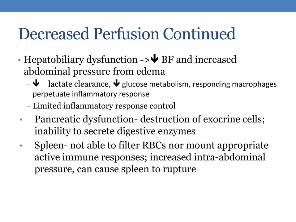
Decreased Perfusion Continued
• Hepatobiliary dysfunction -> BF and increased abdominal pressure from edema – lactate clearance, glucose metabolism, responding macrophages
perpetuate inflammatory response
– Limited inflammatory response control
• Pancreatic dysfunction- destruction of exocrine cells; inability to secrete digestive enzymes
• Spleen- not able to filter RBCs nor mount appropriate active immune responses; increased intra-abdominal pressure, can cause spleen to rupture

Relation to Rhabdomyolysis
• Sepsis can cause Rhabdomyolysis
• In preventing kidney damage; fluid resuscitation is needed.
• Fluid resuscitation can lead to increased abdominal pressure
• Poor perfusion -> Bf and pushes fluids into abdominal tissues which further compresses organs
• Broken down muscle tissue now needs to be filtered by kidneys and can potentially disrupt blood flow to other organs;

Clinical manifestations related to splanchnic circulation
• GI tract: decreased motility, malabsorption
– Weight loss, minimal bowel sounds, nausea and vomiting, paralytic ileus, GI ulcer, abdominal distention
• Pancreas: maldigestion and constipation symptoms
– Early rise in glucose, with a later decline
• Spleen: hemorrhage if ruptured; more susceptible to infection process

Clinical Manifestations Related To Splanchnic Circulation • Hepatobiliary failure-
• Liver : elevations of bilirubin, jaundice, elevated liver enzymes
• Gallblader : Cholecystis without gallstones, right upper quadrant pain and tenderness, abdomen, distention, loss of bowel sounds, fever,

Clinical Presentation
• Third spacing, pitting edema, ventilator, non-responsive
• WBC 12.8, bands >5%
• AST 64 (bile obstruction)
• Platelets 227,000 (thrombocytopenia)
• Cr 1.35, BUN 50 (renal failure)
• BNP 120 (increased fluid)
PT score: 23, high risk 28-day mortality rate: 39%

How Did This Patient Become Septic?
• PT had colon cancer which caused a small bowel obstruction
• SBO causes intestinal dilation (GI secretions, swallowed air)
• Fluid loss r/t emesis, & edema – metabolic alkalosis
• Peristalsis increases - high hydrostatic pressure (third spacing, loss of fluids & electrolytes vascularly - edema)
• Intestinal stasis – floral overgrowth – bacterial translocation across bowel wall
• SEPSIS
• Other issues: large abdominal wound from surgery, new ostomy, NG tube, Foley, ventilator, central line

Radiographic Confirmation of SBO

Treatment—Sepsis Protocol—EBP
Major Interventions
• IV antibiotics (2 or 3)
• IV fluids (for low bp)
• Therapy to support any organ dysfunction (intubation, dialysis, surgery, drainage)
Within 1st 6 Hours
• Labs/Tests (blood cultures & lactic acid w/in 15 min)
• Antibiotics Ceftriaxone, Levofloxacin, metro, Vanco)
• Fluid bolus NS 30-40ml/kg & continued fluid replacement
• Norepinephrine, Vasopressin, NPO, Foley, move to ICU

Treatment Specific to Patient
• Primaxin – bactericidal
• Peridex – ventilator induced pneumonia protocol
• TPN
• Heparin - VTE prophylaxis protocol
• Humulin R – inhibits hepatic glucose production
• Lopressor – beta blocker for high bp (PT hx)
• Fentanyl & Norco – analgesics, sedatives
• NPO, Foley, central line, HOB up, ventilator, wound vac, NG tube, q2h residual checks, q4h Foley care, q2d central line dressing change

Family Education
• I/O
• Diet
• Alcohol, drugs abstinence
• Infection prevention
• Signs and systems of infection
• Colonoscopy

CORONARY CIRCULATION

Patient Cardiology
• First Degree Heart Block
• Impulses move slowly through the heart, but each electrical impulse is till produced, lengthening the PR interval
• Second Degree Heart Block
• Affects how many impulses actually reach the ventricles, leading to an irregular heart rate
• Third Degree Heart Block (Complete Block)
• Electrical impulses that are initiated in atria never reach ventricles
• P Waves are not related to QRS complex
• SVT
• Occurs above AVE node increased heart rate
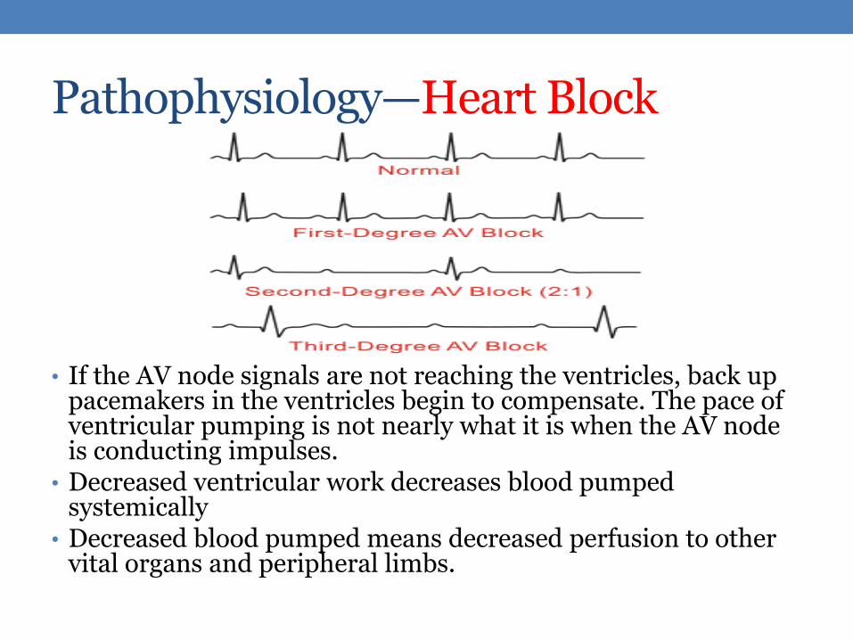
Pathophysiology—Heart Block
• If the AV node signals are not reaching the ventricles, back up pacemakers in the ventricles begin to compensate. The pace of ventricular pumping is not nearly what it is when the AV node is conducting impulses.
• Decreased ventricular work decreases blood pumped systemically
• Decreased blood pumped means decreased perfusion to other vital organs and peripheral limbs.

Pathophysiology—SVT
• Originates above Atrioventricular Node (Does not originate within ventricles), Narrow QRS complexes
• Leads to rapid heart rate
• Can deteriorate to ventricular fibrillation leading to death

Treatment—SVT & Heart Block
• Observation – 1° & 3° Heart Block
• Appears to be self limiting, resulting from Sepsis
• Amiodarone - SVT
• Antidysrhythmic: Prolongs action potential and repolarization
• Adenosine – SVT
• Antidysrhythmics: Slows conduction through AV node and interrupts AV reentry, restoring NSR

Role of the Myocardium
• Middle Layer of the Heart Muscle
• Consists of Cardiac Muscle
• Sepsis Effects on Myocardium
• Weakens Cardiac Muscle Cells Decreased CO Decreased Perfusion to Vital Organs Multiple Organ Failure

Pathophysiology—Fluid Shift
Vasodilation due to release of inflammatory chemicals
Increased capillary permeability
Edema from fluid entering interstitial tissues
Hypotension
Shock
Coagulopathy
Decreased perfusion of coronary muscle
Decreased cardiac output
Decreased perfusion of other organs
Pt. will die if left untreated

Cardiogenic Shock
• Heart is incapable of pumping enough blood to meet body requirements
• CO usually 10 to 20%
• Low Blood Pressure
• Hypoxia
• Rapid Treatment can save the patient
• Oxygen Supplement
• Fluid Replacement Therapy
• Pharmacological Interventions – Dopamine, Norepinephrine, Epinephrine

Septic Shock & Cardiac Function
• About 80% of Cardiac output goes to kidneys, GI tract, skeletal muscle, heart, and the brain.
• Cytokines released into the bloodstream begin the Inflammatory Response and release of Nitrous Oxide
• Depress cardiac contractility
• Vasodilation caused by Adenosine, Lactic Acid, and H+.
• Altered autoregulation and coronary endothelial function
• Prostanoids (Cyclooxygenase)

Synopsis of Potential Underlying Mechanisms in Septic Myocardial Infarction

Assessment Findings & Labs
Lab Result Admission Sepsis
Lactic 1.3 2.0
WBC 10.8 14.3
Hgb 14.4 8.3
Hct 43.1 24.9
Na 138 141
K 5.0 3.4
Ca 7.1 N/A
CK-MB 4.9 17
Troponin 0.024 0.084
Albumin N/A 2.0
Platelets 147 181
PT 11.3 23
PTT 28.6 32.5
INR 1.1 2.1
• Pt’s cardiac output remained close to 74%
• Overall results:
• Increased Hgb and Hct
• WBC trending high
• No ischemia present
• Troponins and CK-MB increased
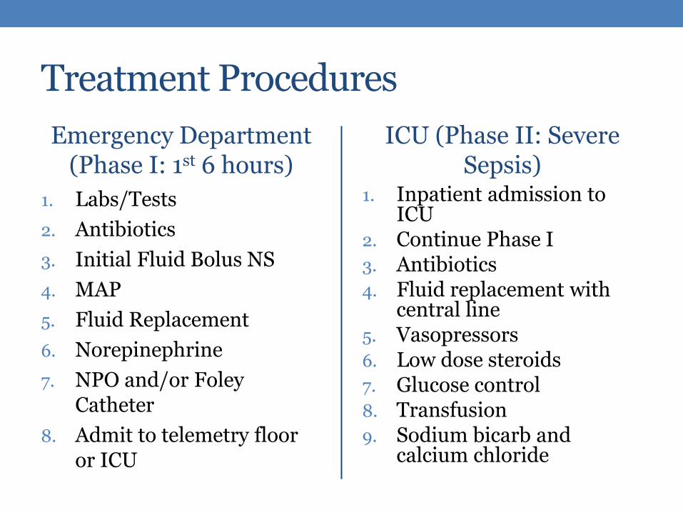
Treatment Procedures
Emergency Department (Phase I: 1st 6 hours)
1. Labs/Tests
2. Antibiotics
3. Initial Fluid Bolus NS
4. MAP
5. Fluid Replacement
6. Norepinephrine
7. NPO and/or Foley Catheter
8. Admit to telemetry floor or ICU
ICU (Phase II: Severe Sepsis)
1. Inpatient admission to ICU
2. Continue Phase I 3. Antibiotics 4. Fluid replacement with
central line 5. Vasopressors 6. Low dose steroids 7. Glucose control 8. Transfusion 9. Sodium bicarb and
calcium chloride

Medications
Medication Indication
Lotensin (Benazapril) Ace inhibitor for HTN
Metoprolol Tartrate (Lopressor) Beta blocker for HTN *Pt has history of AV block
Amlodipine Besylate (Norvasc) Calcium channel blocker for HTN
Adenosine (Adenocard) Endogenous nucleoside for treatment of SVTs
Vancomycin HCl Antibiotic to treat abdominal infections
Micafungin Sodium Antifungal antibiotic to treat Candida fungal infections
Imipenem/Cilastatin Sodium Antibiotics for severe infections

Family Education
• Watch for Signs of Sepsis Return – Racing heart feeling (Tachycardia), Respiration rate >20 breaths per minute (Tachypnea), Fever - > 100.1°
• Healthy Eating Habits
• Exercise – 30 minutes 3-5 days per week to help strengthen heart
• Drink plenty fluids to avoid dehydration

References
• Merx, M.W., Weber, C. (2007). Cardiovascular Involvement in General Medical Conditions. American Heart Association;116:793-802, doi: 10.1161/CIRCULATIONAHA.106.678359
• Perman, S.M., Goyal, M., & Gaieski, D.F. (2012). Initial
Emergency Department Diagnosis and Management of Adult Patients with Severe Sepsis and Septic Shock. Scandinavian Journal of Trauma, Resuscitation and Emergency Medicine, 20:41.
• Tanna, M.S., LeFrancois, D., Velez, C., Ali, N., Zheng, E., and
Leung, S. (2012). Abstract 15051: Left Ventricular Dilatation Improves Survival in Patients with Severe Sepsis. American Heart Association. 2012; 126: A15051. http://circ.ahajournals.org

PULMONARY CIRCULATION

Pathophysiology

Pulmonary Circulation

Pulmonary Circulation
V= Ventilation Q= Perfusion

Acute Respiratory Distress Syndrome (ARDS)
• PaO2/FiO2= <200 PaO2/FiO2= 200-300
• 69/.30=230
1. Injury/Exudative Phase (1-7 days)
2. Reparative/Proliferative Phase (1-2 weeks)
3. Fibrotic/Chronic/Latent Phase (2-3 weeks)
Acute Lung Injury (ALI)

SIRS ARDS & ALI

Assessment
ARDS & ALI
• Labs
• Diagnostics
• Physical assessment
The Patient (ALI)
• Labs: WBC, Hgb, Hct
• Diagnostics: pH, PaCO2, PaO2
• Physical assessment
• WOB
• Breath sounds
• Edema

Treatment
ARDS & ALI
• Complication prevention
• Respiratory therapy
• Supportive therapy
The Patient (ALI)
• Normal saline, hemodynamic monitoring
• HOB up
• PEEP, low FiO2
• Attempts to wean
• VTE prophylaxis, TPN, analgesics, PUD prophylaxis
Evidence-Based Practice
• 5 P’s of ARDS therapy • Perfusion • Positioning • Protective lung ventilation • Protocol weaning • Preventing complications
Powers, J. (2007). The five P’s spell positive outcomes for ARDS patients. American Nurse Today, 2(3). Retrieved from http://www.americannursetoday.com/article.aspx?id=4806

Family Education
• Explanation of necessity of ventilator
• What to expect
• Weaning

RENAL CIRCULATION

Renal System

Renal Functions
• Regulation of body fluid volume and osmolality
• Regulation of electrolyte balance
• Regulation of acid-base balance
• Excretion of waste products (urea, ammonia, drugs, toxins)
• Production and secretion of hormones (erythropoietin, renin, calcitriol)
• Regulation of blood pressure (renin)

Functional Unit of the Kidney—The Nephron
• Glomerular (filtration)
• Proximal (reabsorption)
• Loop of Henle (concentration)
• Distal (reabsorption/secretion)
• Collecting Duct (reabsorption/secretion)
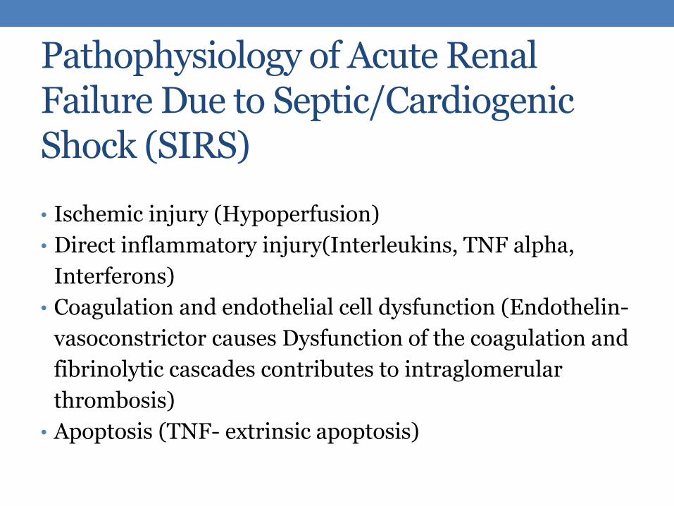
Pathophysiology of Acute Renal Failure Due to Septic/Cardiogenic Shock (SIRS)
• Ischemic injury (Hypoperfusion)
• Direct inflammatory injury(Interleukins, TNF alpha,
Interferons)
• Coagulation and endothelial cell dysfunction (Endothelin-
vasoconstrictor causes Dysfunction of the coagulation and
fibrinolytic cascades contributes to intraglomerular
thrombosis)
• Apoptosis (TNF- extrinsic apoptosis)

Types of Shock
• Cardiogenic shock:
Occurs when either
systolic or diastolic
dysfunction of the
pumping action of the
heart results in
reduced cardiac output
(CO).
• Clinical manifestations
• Increased Na and H2O retention
• Decreased renal blood flow
• Decreased urinary output
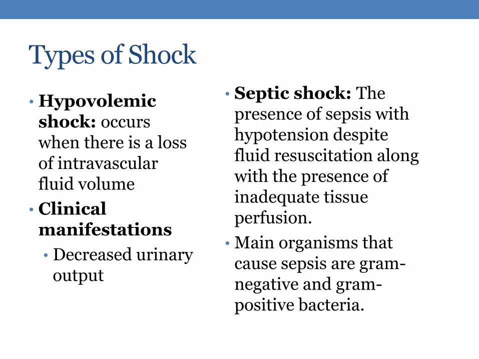
Types of Shock
• Septic shock: The presence of sepsis with hypotension despite fluid resuscitation along with the presence of inadequate tissue perfusion.
• Main organisms that cause sepsis are gram-negative and gram-positive bacteria.
• Hypovolemic shock: occurs when there is a loss of intravascular fluid volume
• Clinical manifestations
• Decreased urinary output

Case Study—Data to Support ARF Due to Dehydration
Date Creatinine BUN
11/07/13 1.36 23
11/08/13 2.54 42
11/13/15 1.36 35
11/15/13 1.54 55
11/16/13 1.66 58
11/17/13 1.64 46
• Increased Creatinine & BUN • Nausea& Vomiting • Dehydration • 11/08/13 Colectomy

Case Study—Data to Support ARF Due to Sepsis
Date Cratinine BUN
11/17/13 1.94 52
11/18/13 1.67 47
11/19/13 1.71 40
11/20/13 1.78 39
• Increased HR
• SOB
• Septic looking

Treatment
• Fluid replacement therapy
• Sympathomimetic drugs( Norepinephrine, dopamine,
phenylephrine)
• Antibiotics
• Nutritional therapy

Family Education
• Monitor daily weight
• Fluid restriction
• Diet
• Limit sodium and potassium
• Assist with position change every 2 hours
• Identify symptoms to be reported.

References
Majumdar, A. (2010). Sepsis-induced acute kidney injury .Indian Journal of Critical Care Medicine, 14, 0972-5229.
Vriese, A. (2003). Prevention and treatment of acute renal failure in sepsis. Journal of The American Society of Nephrology, 14(3), 792-805.
CEREBRAL CIRCULATION

100,000 Miles of Blood Vessels
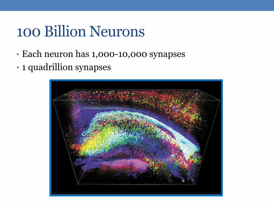
100 Billion Neurons
• Each neuron has 1,000-10,000 synapses
• 1 quadrillion synapses

Cerebral Perfusion
• 17% of cardiac output
• 750 milliliters per minute
• 20% of oxygen
• 25% of glucose

Our Patient: Cerebral Perfusion
• Unresponsive
• Recent encephalopathy
• Babinski’s reflex
• Infection
• Peripheral edema
• Heart block
• GI bleeding
• V/P mismatch
• Cardiac output
• Hypovolemia
• Renal failure
• Respiratory failure
• Anemia
• Glucose
• Inadequate nutrition
• Electrolyte imbalances

Autoregulation
Ability to maintain relatively constant blood flow despite changes in perfusion pressure
Blood flow = O2 extraction
CO2 = vasodilation = blood flow
CO2 = vasoconstriction = blood flow
Normal CPP= 70 - 90 mmHg
< 70 mmHg = ischemia

Metabolic Disturbances
Glucose, electrolytes, acid base
Circulatory Failure
cerebral blood flow
Environmental
Stressors
Medication
Toxicity
Acute Brain Dysfunction
The Brain & Sepsis
Blood-Brain Barrier
Alterations
Sepsis

Sepsis Associated Encephalopathy (SAE) • Diffuse cerebral dysfunction caused by systemic inflammatory
response to an infection
Sepsis Associated Encephalopathy
is the most frequent cause of
delirium in critical illness
Up to 70% of patients with severe systemic infection
Source: Nature Review, Neurology. 2012 Oct;8(10):557-66.
Overlooked as “just ICU delirium”
•Fluctuating mental status changes
•Inattention
•Disorganized thinking
•LOC , neuro changes Source: Annals of Intensive Care 2013, 3:15

More About Encephalopathy • Definition: Worsening of brain function
• Possible causes in this patient: Bacterial infection
Hypertension
Chronic inflammation – cancer, chemo, infx, trauma
Metabolic dysfunction- hyperkalemia, hyperchloremia, hypocalemia
Poor nutrition - TPN
Lack of blood flow to brain – low CO, heart block, ventilation
Renal failure – build up of toxins
GI Bleeding
Toxicity – build up of ammonia & other toxins, medication toxicity
• Primaxin (imipenem and cilistatin) - carbapenem antibiotic • Neurotoxicity associated with encephalopathy
• Increased risk with renal failure

Long-term Cognitive Impairment & Sepsis
• Substantial and persistent new cognitive impairment in older adults
• Functional disability
• Downturn in patients’ ability to live independently
Source: JAMA. 2010;304(16):1787-1794

Treatment
• Treat the source
• Surgery
• Antibiotics
• Electrolyte replacement
• Fluid replacement
• Glucose
• Insulin
• Control hypertension
• Monitor labs
• ABGs
• Liver function
• Kidney function
• Med toxicity—serum levels
• CBC
• Electrolytes

Evidence-Based Treatment Considerations
• Thiamine Supplements
• Thiamine depletion common in critical illness
• 50% increase in mortality
• Severe neurologic disorders such as encephalopathy
• Should be suspected in severe sepsis, lactic acidosis
• Source: Manzanares, W. , & Hardy, G. (2011). Thiamine supplementation in the critically ill. Current Opinion in Clinical Nutrition & Metabolic Care, 14(6), 610-617.
• Valproic Acid – new research
• Reverses cognitive deficits • probably via a reduction in inflammation and apoptosis in the brain
• More studies needed to refine science
• Wu, J. , Dong, L. , Zhang, M. , Jia, M. , Zhang, G. , et al. (2013).. Neurochemical Research, 38(11), 2440-2449. Class i histone deacetylase inhibitor valproic acid reverses cognitive deficits in a mouse model of septic encephalopathy

What if…?
• Patient’s cardiac output goes way up
Hemorrhagic or Ischemic Stroke
Shock vascular permeability
Heparin clotting time
Hyperlipidemia atherosclerotic plaque
Inflammation vascular vulnerability

Family Education • Educate regarding care of other organ systems
• Nutrition
• Hydration
• Exercise
• Medication
• Specific therapies and treatments
• Teach signs & symptoms of brain dysfunction • Headaches
• Seizures
• Confusion
• Memory problems
• Behavioral/mood changes
• Nausea
• LOC

END-OF-LIFE CARE

Palliative Care vs. Comfort Care
• Palliative Care
• Patient can still be receiving curative treatment
• Diagnosis does not need to be terminal
• Comfort Care
• Curative treatment is withdrawn
• Terminal diagnosis with typically 6 month life expectancy

Ethical Dilemmas
• Life Supportive Care
• Machines and Pharmaceuticals: How long is too long?
• What is occurring in Oakland - Full story, Media, Future
• Pain Medication: Passive Euthanasia?

Ethical Dilemmas Continued
• Right to Die
• Terminal Diagnosis—Should people be allowed to choose death with dignity?
• Donor Network
• Keeping people artificially alive until organs can be harvested

SUMMARY

Patient Summary—Important Events
• 11/07/13: 88-year-old male presents to ED with complaints of abd pain pain, N/V, small BM
• Diagnosed with adenocarcinoma
• Colectomy, exploratory laparotomy, removed mass
• 11/09/13: SVT x 2 treated with Adenosine and amiodarone
• 11/17/13: Anastomic leak
• Sepsis
• Respiratory Failure ventilation

Patient Summary—Important Events
• 11/22/13: Pleural effusion
• Fluid overload acute kidney failure
• 11/26/13: Myocardial infarction, code blue pt recovered
• 12/18/13: Discharged to ECF
• 01/06/14: Appointment with PCP brought back to ER
• Last H/P: R/O sepsis; remains full code

Definitions
Systemic inflammatory response syndrome (SIRS)
A systemic inflammatory response to a variety of insults (including infection, ischemia, infarct, and injury)
Sepsis A systemic inflammatory response to infection
Severe sepsis Sepsis + organ dysfunction
Multiple organ dysfunction syndrome (MODS)
Failure of more than one organ system


SIRS MODS
• Due to uncontrolled inflammatory response • Mediators released
• Endothelium damage
• Hypermetabolism
• Vasodilation
• Vascular permeability
• Coagulation cascade activated
• Decreased organ blood perfusion • hypotension + microemboli + redistributed blood flow

MODS: Respiratory System
Inflammation Endothelial
damage Increased
permeability
Alveolar
edema

MODS: Cardiovascular System
Increased capillary permeability
Third spacing Decreased venous
return
Vasodilation Hypotension + decreased SVR
Increased CO

MODS: Neurological System
Mental status
changes
Hypoxemia
Inflammatory mediators
Impaired perfusion

MODS: Renal System
Acute kidney injury
Decreased perfusion
Inflammatory mediators
Medications

MODS: Gastrointestinal System
Decreased perfusion
Decreased mucosal barrier
Increased risk for ulcer and GI bleed
Blood shunted from GI
Increased risk for ischemic injury
End-of-life Care
• Palliative care
• Comfort care
• Life support
• Example: Jahi McMath
• 13 year old girl pronounced brain-dead in Children’s Hospital Oakland
• Right to die
• Donor network


![To what extent are the terminal stages of sepsis, septic ... · 6/10/2016 · response syndrome’ (SIRS) [27-32], septic shock [3], apoptotic [33] and necrotic [34] cell death,](https://static.fdocuments.us/doc/165x107/5fc2fc4814bc76677c56d502/to-what-extent-are-the-terminal-stages-of-sepsis-septic-6102016-response.jpg)
![Septic Shock [EDocFind.com]](https://static.fdocuments.us/doc/165x107/55cf8fb1550346703b9edc7d/septic-shock-edocfindcom.jpg)