Case Report - downloads.hindawi.comdownloads.hindawi.com/archive/2011/175916.pdf · ISRN Obstetrics...
4
International Scholarly Research Network ISRN Obstetrics and Gynecology Volume 2011, Article ID 175916, 3 pages doi:10.5402/2011/175916 Case Report The “Lasso Sign”: An Early Sonographic Sign of Posterior Meningocele Alina Weissmann-Brenner, 1 Zeev Feldman, 2 and Yaron Zalel 1 1 Department of Obstetrics and Gynecology, The Chaim Sheba Medical Center, Tel HaShomer 52621, Israel 2 Pediatric Neurosurgery Unit, The Edmond and Lily Safra Children’s Hospital, The Chaim Sheba Medical Center, Tel HaShomer 52621, Israel Correspondence should be addressed to Alina Weissmann-Brenner, [email protected] Received 20 August 2010; Accepted 22 September 2010 Academic Editor: A. Herman Copyright © 2011 Alina Weissmann-Brenner et al. This is an open access article distributed under the Creative Commons Attribution License, which permits unrestricted use, distribution, and reproduction in any medium, provided the original work is properly cited. Posterior meningocele is an uncommon form of spina bifida. We present a case of unique posterior meningocele diagnosed at the early second trimester anatomical scan using 2D and 3D ultrasound. The sonographic appearance resembled “lasso”. The prenatal follow-up was uneventful, with no demonstration of tethered cord. Clinical, neurological and radiological examinations following delivery and at the age of four months were unremarkable. The differential diagnosis for a dorsal midline mass pre- senting in fetuses and newborns includes a wide range of pathological conditions, consisting of spinal dysraphisms, teratoma, hamartoma, disturbances in regression of fetal tail, and pseudotail formation [1]. Posterior meningocele is the least common form of spina bifida. It appears as a sac or cyst which contains cerebrospinal fluid. The majority of cases have no underlying bony defect or communication to the meninges. The spinal cord and nerves are not involved and their function is normal. Its incidence is 1 : 1000 live births [2, 3]. Apart from spina bifida, causes of meningocele include teratoma and other tumors of the sacrococcyx and of the presacral space and Currarino syndrome. Usually a meningocele has no negative long-term effects, although there are reports of tethered cord. We present a case of unique posterior meningocele dia- gnosed at the early second trimester anatomical scan using 2D and 3D ultrasound. A healthy 29-year-old woman presented to our ultra- sound unit at her first pregnancy for a routine anatomical scan at 15-week gestation. The anatomical scan revealed a small cystic lesion with very tiny stalk close to the caudal aspect of the spine, “floating” in the amniotic fluid, with no flow when Doppler was applied. This lesion was suspected to be meningocele, and based on its appearance we have termed it as the “Lasso sign”. No opened spina bifida was noted. 3D ultrasound using sectional planes and surface rendering was performed, and it demonstrated the same lesion and confirmed it to be meningocele alone. The anatomical scan of the brain including the posterior fossa was normal. During the present pregnancy she performed nuchal translucency of 0.8 mm. The triple test screen was normal and consisted of alpha- fetoprotein level of 0.95 MOM. The patient did not perform MRI due to claustrophobia. One of the complications of meningocele is tethered cord, caused by pathological fixation of the spinal cord, resulting in traction on the neural tissue which, in turn, leads to ischemia and progressive neurological deterioration. The prenatal diagnosis of tethered cord is based on ultrasound evaluations of the spinal cord and the conus medullaris at the level of L2-L3 at the second and third trimester [4, 5]. Repeated sonograms in our case demonstrated the same lesion at L3-L4 (see Figure 1 and supplementary movie Lasso Movie 1 in Supplementary Material available online at doi:10.5402/2011/175916) without sonographic signs of tethered cord.
Transcript of Case Report - downloads.hindawi.comdownloads.hindawi.com/archive/2011/175916.pdf · ISRN Obstetrics...

International Scholarly Research NetworkISRN Obstetrics and GynecologyVolume 2011, Article ID 175916, 3 pagesdoi:10.5402/2011/175916
Case Report
The “Lasso Sign”: An Early Sonographic Sign ofPosterior Meningocele
Alina Weissmann-Brenner,1 Zeev Feldman,2 and Yaron Zalel1
1 Department of Obstetrics and Gynecology, The Chaim Sheba Medical Center, Tel HaShomer 52621, Israel2 Pediatric Neurosurgery Unit, The Edmond and Lily Safra Children’s Hospital, The Chaim Sheba Medical Center,Tel HaShomer 52621, Israel
Correspondence should be addressed to Alina Weissmann-Brenner, [email protected]
Received 20 August 2010; Accepted 22 September 2010
Academic Editor: A. Herman
Copyright © 2011 Alina Weissmann-Brenner et al. This is an open access article distributed under the Creative CommonsAttribution License, which permits unrestricted use, distribution, and reproduction in any medium, provided the original work isproperly cited.
Posterior meningocele is an uncommon form of spina bifida. We present a case of unique posterior meningocele diagnosed at theearly second trimester anatomical scan using 2D and 3D ultrasound. The sonographic appearance resembled “lasso”. The prenatalfollow-up was uneventful, with no demonstration of tethered cord. Clinical, neurological and radiological examinations followingdelivery and at the age of four months were unremarkable.
The differential diagnosis for a dorsal midline mass pre-senting in fetuses and newborns includes a wide range ofpathological conditions, consisting of spinal dysraphisms,teratoma, hamartoma, disturbances in regression of fetal tail,and pseudotail formation [1].
Posterior meningocele is the least common form of spinabifida. It appears as a sac or cyst which contains cerebrospinalfluid. The majority of cases have no underlying bony defector communication to the meninges. The spinal cord andnerves are not involved and their function is normal. Itsincidence is 1 : 1000 live births [2, 3].
Apart from spina bifida, causes of meningocele includeteratoma and other tumors of the sacrococcyx and ofthe presacral space and Currarino syndrome. Usually ameningocele has no negative long-term effects, althoughthere are reports of tethered cord.
We present a case of unique posterior meningocele dia-gnosed at the early second trimester anatomical scan using2D and 3D ultrasound.
A healthy 29-year-old woman presented to our ultra-sound unit at her first pregnancy for a routine anatomicalscan at 15-week gestation.
The anatomical scan revealed a small cystic lesion withvery tiny stalk close to the caudal aspect of the spine,“floating” in the amniotic fluid, with no flow when Doppler
was applied. This lesion was suspected to be meningocele,and based on its appearance we have termed it as the “Lassosign”. No opened spina bifida was noted. 3D ultrasoundusing sectional planes and surface rendering was performed,and it demonstrated the same lesion and confirmed it tobe meningocele alone. The anatomical scan of the brainincluding the posterior fossa was normal.
During the present pregnancy she performed nuchaltranslucency of 0.8 mm.
The triple test screen was normal and consisted of alpha-fetoprotein level of 0.95 MOM. The patient did not performMRI due to claustrophobia.
One of the complications of meningocele is tetheredcord, caused by pathological fixation of the spinal cord,resulting in traction on the neural tissue which, in turn, leadsto ischemia and progressive neurological deterioration. Theprenatal diagnosis of tethered cord is based on ultrasoundevaluations of the spinal cord and the conus medullarisat the level of L2-L3 at the second and third trimester[4, 5]. Repeated sonograms in our case demonstrated thesame lesion at L3-L4 (see Figure 1 and supplementary movie
Lasso Movie 1 in Supplementary Material available onlineat doi:10.5402/2011/175916) without sonographic signs oftethered cord.

2 ISRN Obstetrics and Gynecology
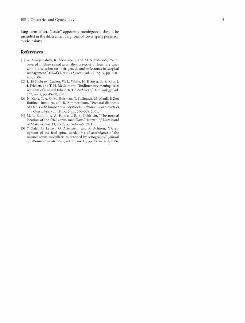
Corpus vertebra
Transverse process
Lasso sign
(a)
Lasso sign
(b)
Lasso sign
(c)
Figure 1: (a) Transverse 2D views of the lower spine revealing posterior meningocele at 15 weeks. (b) Sagittal 2D view of the posteriormeningocele at 22 + 3 weeks. (c) Sagittal 3D view of the posterior meningocele at 22 + 3 weeks.
At 40 + 2 weeks of gestation the patient underwent acesarean section because of arrest of dilatation. A 3384grfemale infant was delivered with Apgar scores of 9 and 10at 1 minute and 5 minutes, respectively.
The neonate’s physical exam revealed an atretic string ofabout 2 cm on the lower aspect of her back in the midline,confirmed by a neurosurgeon to be meningocele. Additionalevaluation included ultrasound of the brain which wasnormal, ultrasound of the sacrum which ruled out tetheredcord, and X-ray of the spine which demonstrated normalstructure of the vertebrae. Movements of four limbs andmuscle tone were normal. There was no leakage of urine, andthe tonus of the anal sphincter was good. The string driedout and fell 12 hours after birth. Pathological evaluationwas not performed; therefore, the diagnosis was based onthe prenatal ultrasound performed by gynecologists andpediatric neurosurgeons and by the clinical examinationafter birth by the pediatric neurosurgeon. Photos of the back
of the infant taken at the age of 2 days were unremarkable,apart from a tiny scar at the middle of her lower backthat disappeared after a few days. Followup examinationincluding neurological examination at the age of two monthsand four months was unremarkable. MRI performed at theage of four months was normal.
In summary, detection of the “Lasso” sign may indicatethe diagnosis of posterior meningocele. Thorough evaluationof the fetal brain and spinal cord to exclude Chiari IImalformation and spina bifida is mandatory. The use ofcolour Doppler to rule out the possibility of umbilical cordas part of the examination is necessary. Level of alpha-fetoprotein should be examined. Prenatal followup shouldinclude repeated sonographic evaluation of the brain andspinal cord with emphasis on the conus medullaris to ruleout tethered cord. 3D ultrasound may aid both in thediagnosis and followup. MRI may also be offered in order toaid in the diagnosis. In general, posterior meningocele has no
ISRN Obstetrics and Gynecology 3
long-term effect. “Lasso” appearing meningocele should beincluded in the differential diagnosis of lower spine posteriorcystic lesions.
References
[1] A. Amirjamshidi, K. Abbassioun, and M. S. Bidabadi, “Skin-covered midline spinal anomalies: a report of four rare caseswith a discussion on their genesis and milestones in surgicalmanagement,” Child’s Nervous System, vol. 22, no. 5, pp. 460–465, 2006.
[2] L. El Shabrawi-Caelen, W. L. White, H. P. Soyer, B.-S. Kim, I.J. Frieden, and T. H. McCalmont, “Rudimentary meningocele:remnant of a neural tube defect?” Archives of Dermatology, vol.137, no. 1, pp. 45–50, 2001.
[3] N. Klble, T. A. G. M. Huisman, T. Stallmach, M. Meuli, F. ZenRuffinen Imahorn, and R. Zimmermann, “Prenatal diagnosisof a fetus with lumbar myelocystocele,” Ultrasound in Obstetricsand Gynecology, vol. 18, no. 5, pp. 536–539, 2001.
[4] M. L. Robbin, R. A. Filly, and R. B. Goldstein, “The normallocation of the fetal conus medullaris,” Journal of Ultrasoundin Medicine, vol. 13, no. 7, pp. 541–546, 1994.
[5] Y. Zalel, O. Lehavi, O. Aizenstein, and R. Achiron, “Devel-opment of the fetal spinal cord: time of ascendance of thenormal conus medullaris as detected by sonography,” Journalof Ultrasound in Medicine, vol. 25, no. 11, pp. 1397–1401, 2006.

Submit your manuscripts athttp://www.hindawi.com
Stem CellsInternational
Hindawi Publishing Corporationhttp://www.hindawi.com Volume 2014
Hindawi Publishing Corporationhttp://www.hindawi.com Volume 2014
MEDIATORSINFLAMMATION
of
Hindawi Publishing Corporationhttp://www.hindawi.com Volume 2014
Behavioural Neurology
EndocrinologyInternational Journal of
Hindawi Publishing Corporationhttp://www.hindawi.com Volume 2014
Hindawi Publishing Corporationhttp://www.hindawi.com Volume 2014
Disease Markers
Hindawi Publishing Corporationhttp://www.hindawi.com Volume 2014
BioMed Research International
OncologyJournal of
Hindawi Publishing Corporationhttp://www.hindawi.com Volume 2014
Hindawi Publishing Corporationhttp://www.hindawi.com Volume 2014
Oxidative Medicine and Cellular Longevity
Hindawi Publishing Corporationhttp://www.hindawi.com Volume 2014
PPAR Research
The Scientific World JournalHindawi Publishing Corporation http://www.hindawi.com Volume 2014
Immunology ResearchHindawi Publishing Corporationhttp://www.hindawi.com Volume 2014
Journal of
ObesityJournal of
Hindawi Publishing Corporationhttp://www.hindawi.com Volume 2014
Hindawi Publishing Corporationhttp://www.hindawi.com Volume 2014
Computational and Mathematical Methods in Medicine
OphthalmologyJournal of
Hindawi Publishing Corporationhttp://www.hindawi.com Volume 2014
Diabetes ResearchJournal of
Hindawi Publishing Corporationhttp://www.hindawi.com Volume 2014
Hindawi Publishing Corporationhttp://www.hindawi.com Volume 2014
Research and TreatmentAIDS
Hindawi Publishing Corporationhttp://www.hindawi.com Volume 2014
Gastroenterology Research and Practice
Hindawi Publishing Corporationhttp://www.hindawi.com Volume 2014
Parkinson’s Disease
Evidence-Based Complementary and Alternative Medicine
Volume 2014Hindawi Publishing Corporationhttp://www.hindawi.com


![downloads.hindawi.comdownloads.hindawi.com/journals/isrn.economics/2012/703541.pdf · 2 ISRN Economics the rent to the provider (see e.g. [26–29]). This risk may be partly reduced](https://static.fdocuments.us/doc/165x107/6044a19d35376546ed3bad57/2-isrn-economics-the-rent-to-the-provider-see-eg-26a29-this-risk-may-be.jpg)